peregrine falcon
TRANSCRIPT
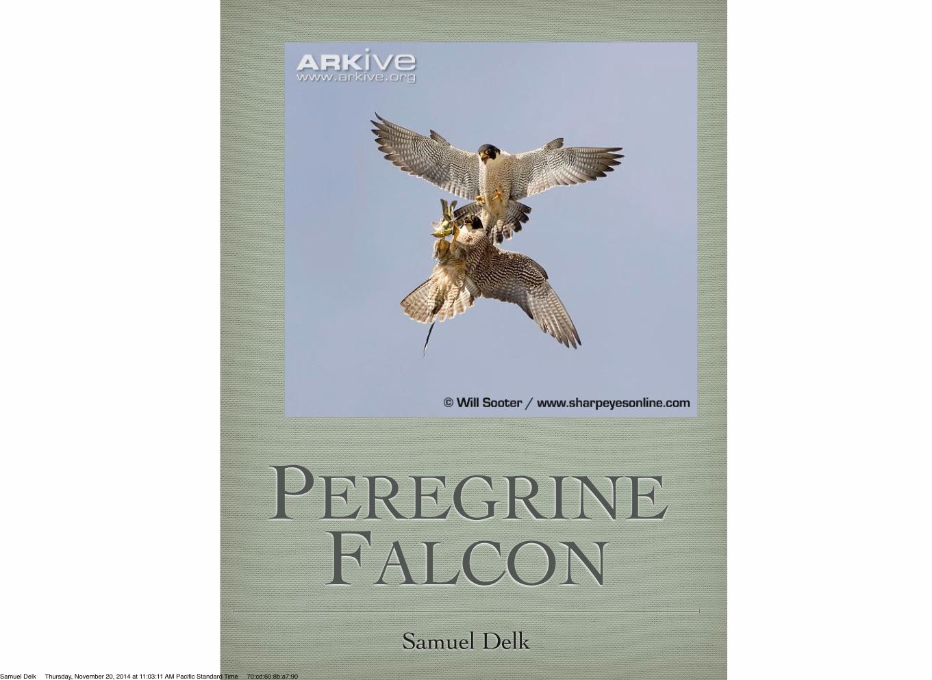
PEREGRINE FALCON
Samuel DelkSamuel Delk Thursday, November 20, 2014 at 11:03:11 AM Pacific Standard Time 70:cd:60:8b:a7:90
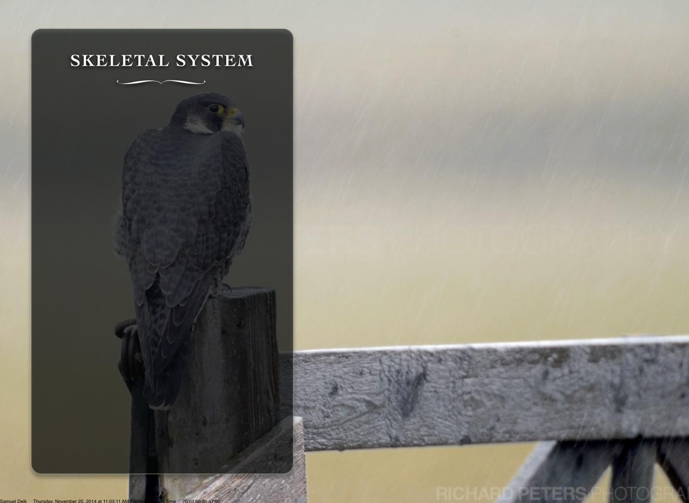
SKELETAL SYSTEM∏
Samuel Delk Thursday, November 20, 2014 at 11:03:11 AM Pacific Standard Time 70:cd:60:8b:a7:90
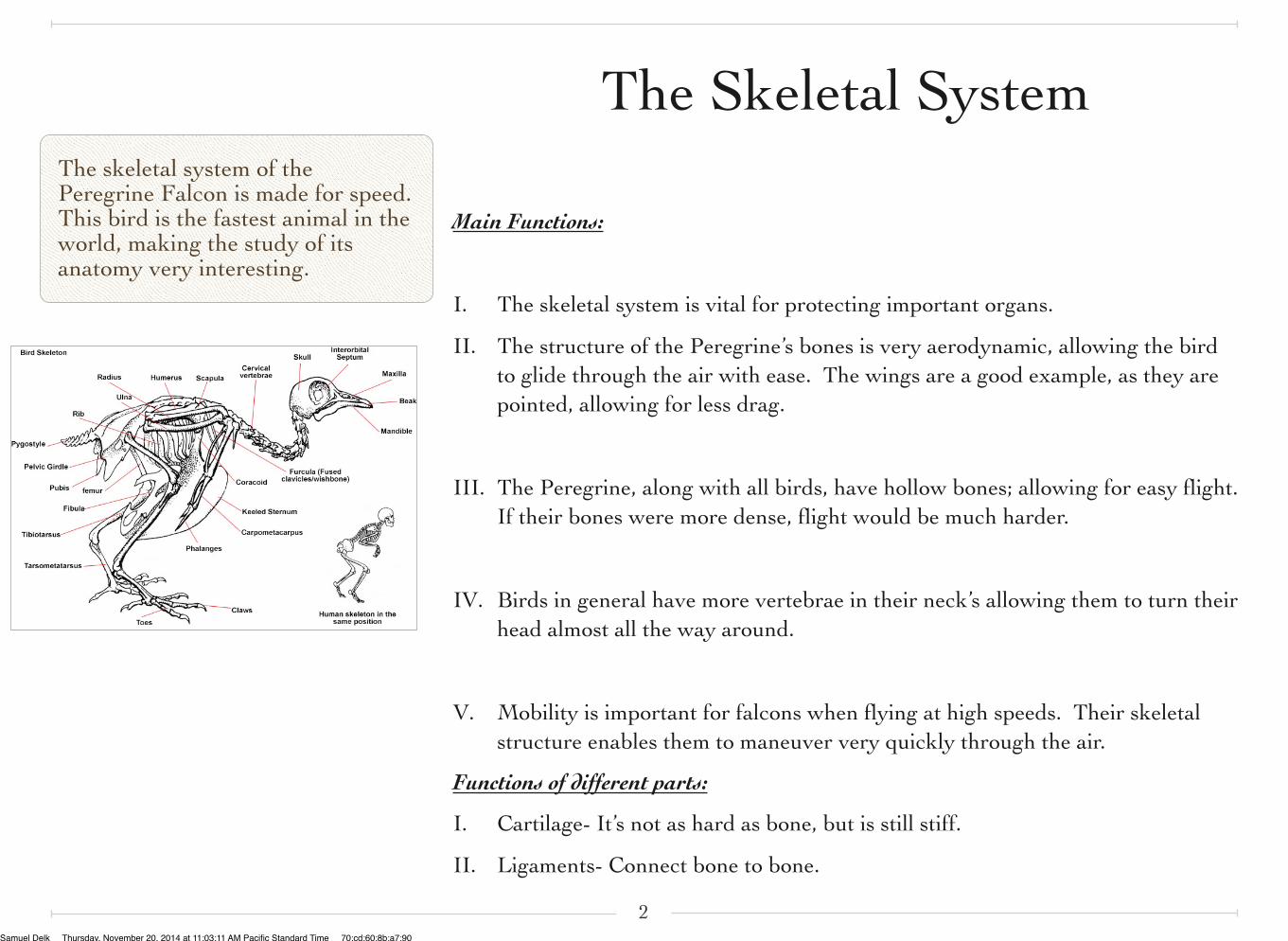
The skeletal system of the Peregrine Falcon is made for speed. This bird is the fastest animal in the world, making the study of its anatomy very interesting.
Main Functions:
I. The skeletal system is vital for protecting important organs.
II. The structure of the Peregrine’s bones is very aerodynamic, allowing the bird to glide through the air with ease. The wings are a good example, as they are pointed, allowing for less drag.
III. The Peregrine, along with all birds, have hollow bones; allowing for easy flight. If their bones were more dense, flight would be much harder.
IV. Birds in general have more vertebrae in their neck’s allowing them to turn their head almost all the way around.
V. Mobility is important for falcons when flying at high speeds. Their skeletal structure enables them to maneuver very quickly through the air.
Functions of different parts:
I. Cartilage- It’s not as hard as bone, but is still stiff.
II. Ligaments- Connect bone to bone.
2
The Skeletal System
Samuel Delk Thursday, November 20, 2014 at 11:03:11 AM Pacific Standard Time 70:cd:60:8b:a7:90
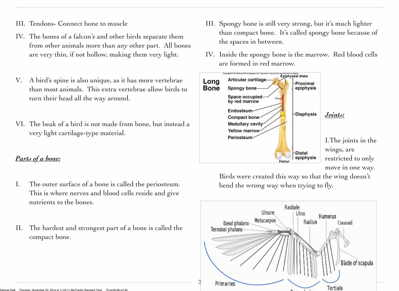
III. Tendons- Connect bone to muscle
IV. The bones of a falcon’s and other birds separate them from other animals more than any other part. All bones are very thin, if not hollow, making them very light.
V. A bird’s spine is also unique, as it has more vertebrae than most animals. This extra vertebrae allow birds to turn their head all the way around.
VI. The beak of a bird is not made from bone, but instead a very light cartilage-type material.
Parts of a bone:
I. The outer surface of a bone is called the periosteum. This is where nerves and blood cells reside and give nutrients to the bones.
II. The hardest and strongest part of a bone is called the compact bone.
III. Spongy bone is still very strong, but it’s much lighter than compact bone. It’s called spongy bone because of the spaces in between.
IV. Inside the spongy bone is the marrow. Red blood cells are formed in red marrow.
Joints:
I.The joints in the wings, are restricted to only move in one way.
Birds were created this way so that the wing doesn’t bend the wrong way when trying to fly.
3Samuel Delk Thursday, November 20, 2014 at 11:03:11 AM Pacific Standard Time 70:cd:60:8b:a7:90
II. The elbow joints of a bird are linked to the wings, so that when one moves, the other does automatically. This puts less strain on a bird when flying.
III. On the very inside of the bones, hollow spaces are filled by a jelly-like substance called bone marrow.
Parts of a Skeleton:
I. Axial- central axis bones
II. Pectoral- bones of the fore limbs
III. Pelvic- bones of the hind limbs
4Samuel Delk Thursday, November 20, 2014 at 11:03:11 AM Pacific Standard Time 70:cd:60:8b:a7:90
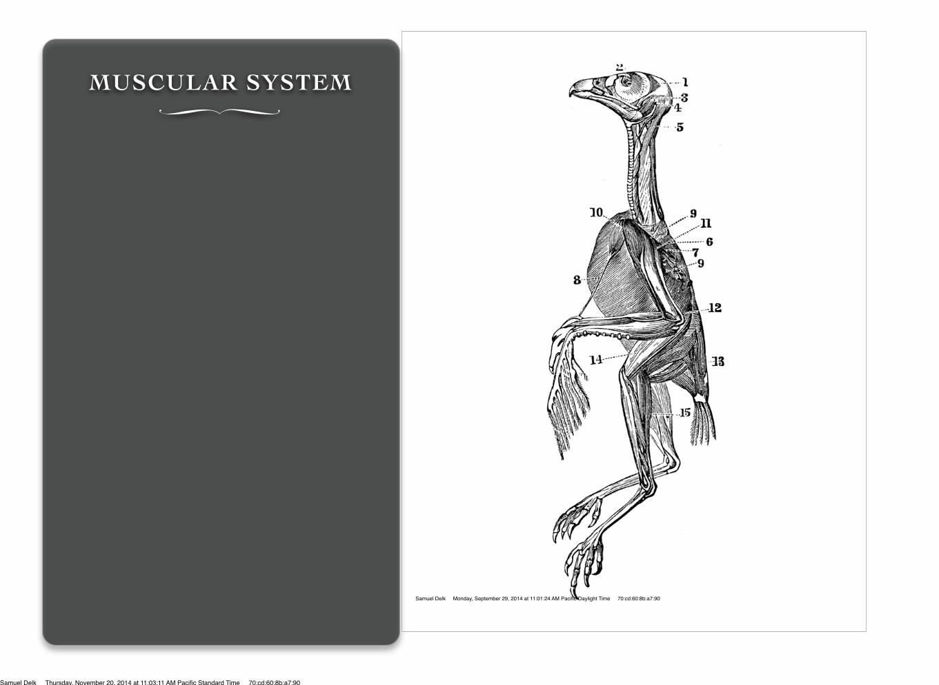
MUSCULAR SYSTEM∏
Samuel Delk Monday, September 29, 2014 at 11:01:24 AM Pacific Daylight Time 70:cd:60:8b:a7:90
Samuel Delk Thursday, November 20, 2014 at 11:03:11 AM Pacific Standard Time 70:cd:60:8b:a7:90
VOCABULARY
• Abduction- Movement away from the medial plane.
• Adduction- Movement towards the medial plane.
• Agonist- Prime mover of a joint.
• Antagonist-Opposes movement of the agonist.
• Ambulation- Moving from one place to another.
• Extension- Increasing angle between body parts.
• Flexion- Decreasing angle between body parts.
• Synergist- Muscle that indirectly aids the agonist; add extra force.
• Sarcomere- The most basic unit of a muscle (fiber).
MUSCULAR SYSTEM∏
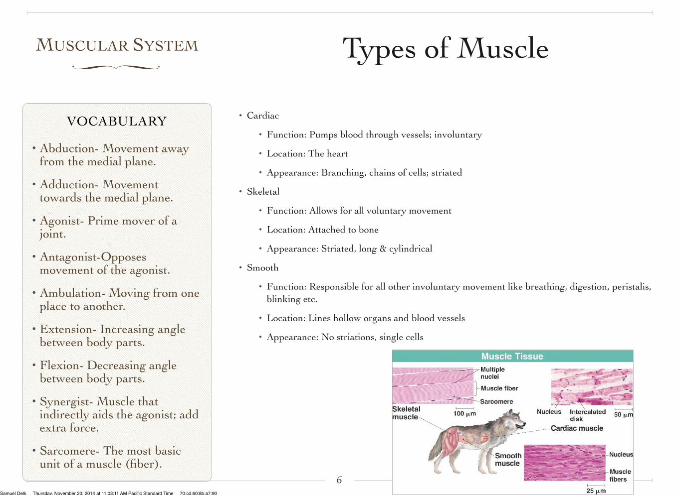
• Cardiac
• Function: Pumps blood through vessels; involuntary
• Location: The heart
• Appearance: Branching, chains of cells; striated
• Skeletal
• Function: Allows for all voluntary movement
• Location: Attached to bone
• Appearance: Striated, long & cylindrical
• Smooth
• Function: Responsible for all other involuntary movement like breathing, digestion, peristalis, blinking etc.
• Location: Lines hollow organs and blood vessels
• Appearance: No striations, single cells
6
Types of Muscle
Samuel Delk Thursday, November 20, 2014 at 11:03:11 AM Pacific Standard Time 70:cd:60:8b:a7:90
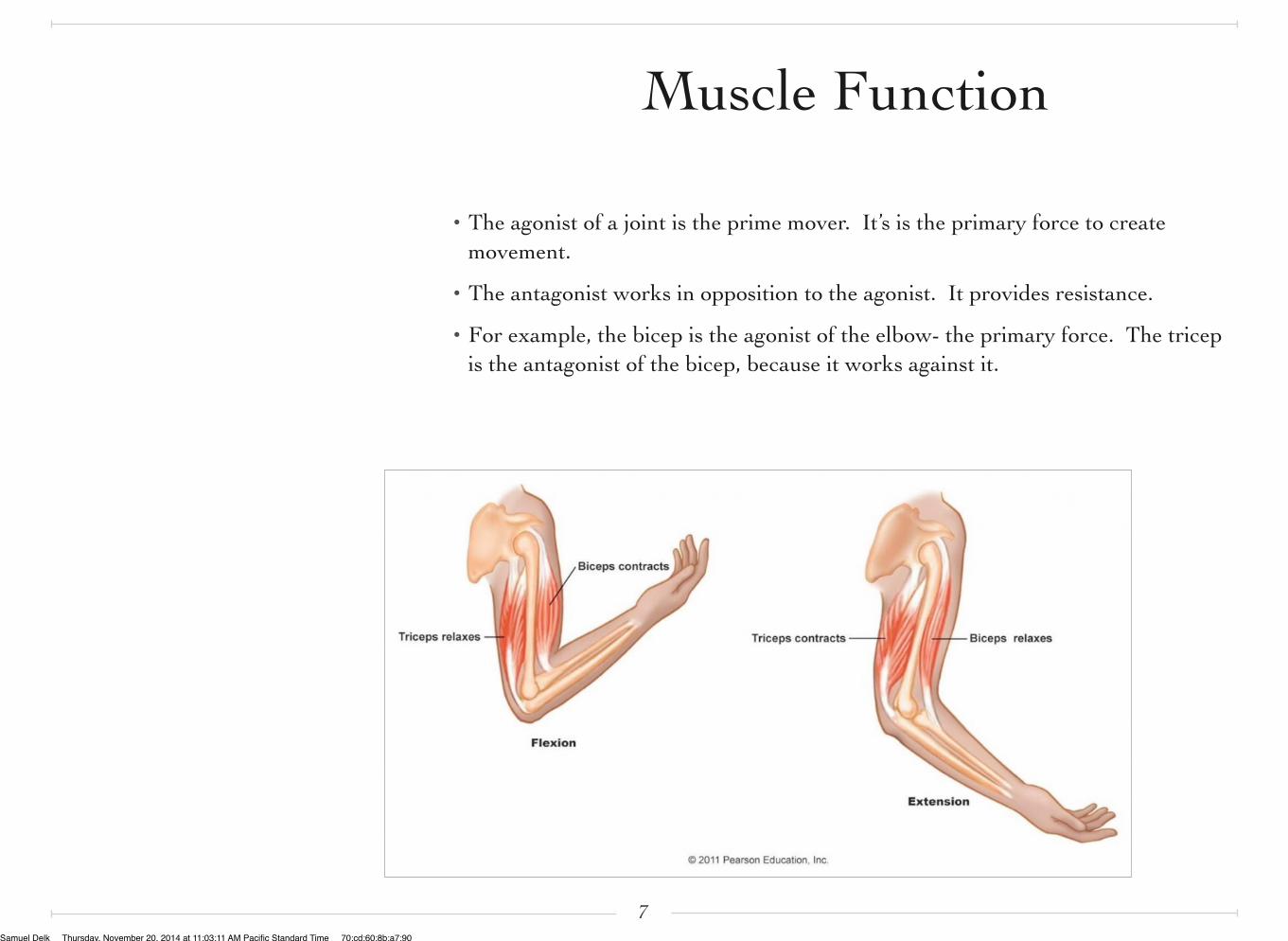
• The agonist of a joint is the prime mover. It’s is the primary force to create movement.
• The antagonist works in opposition to the agonist. It provides resistance.
• For example, the bicep is the agonist of the elbow- the primary force. The tricep is the antagonist of the bicep, because it works against it.
7
Muscle Function
Samuel Delk Thursday, November 20, 2014 at 11:03:11 AM Pacific Standard Time 70:cd:60:8b:a7:90
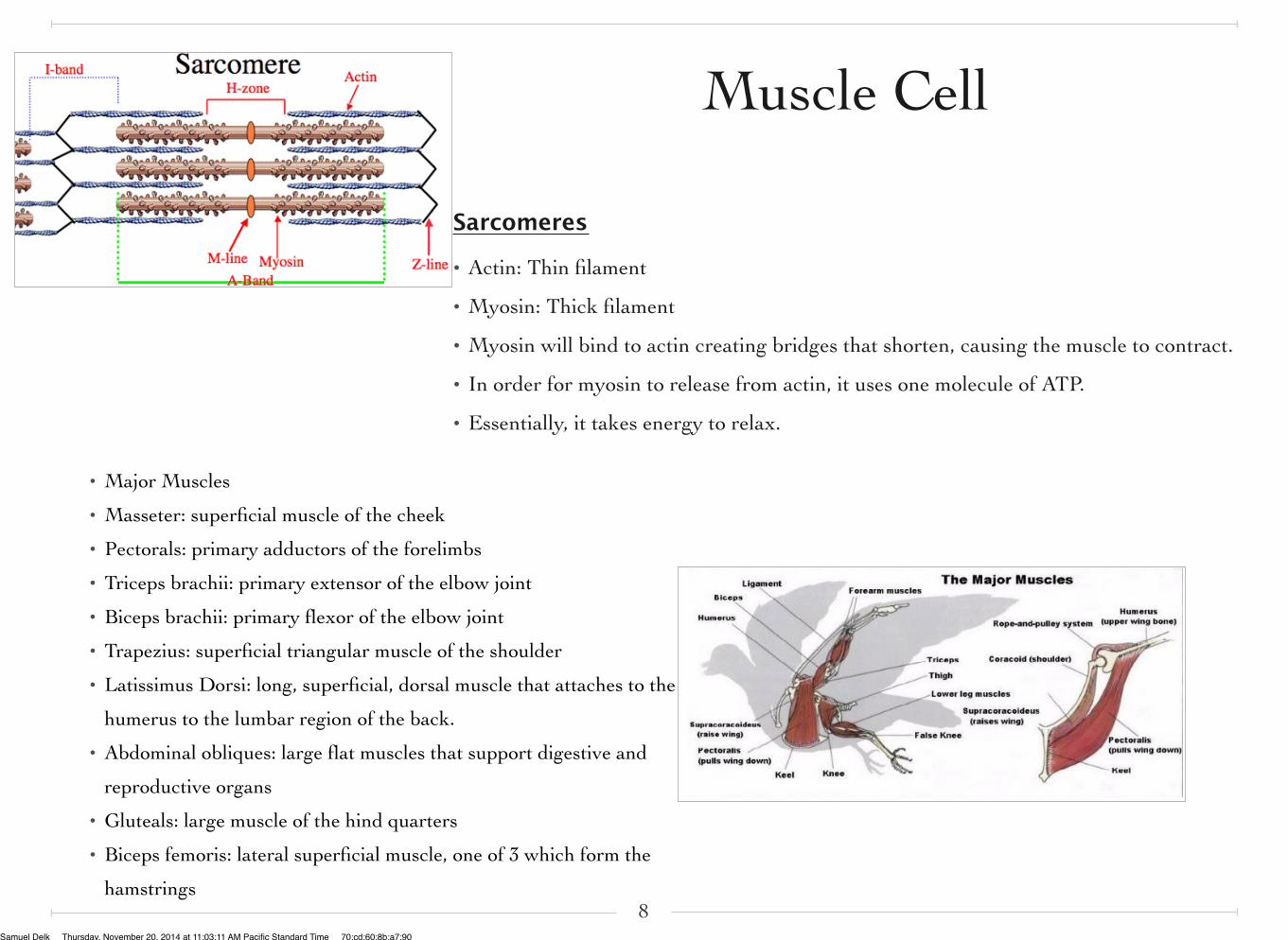
Sarcomeres
• Actin: Thin filament
• Myosin: Thick filament
• Myosin will bind to actin creating bridges that shorten, causing the muscle to contract.
• In order for myosin to release from actin, it uses one molecule of ATP.
• Essentially, it takes energy to relax.
8
Muscle Cell
• Major Muscles
• Masseter: superficial muscle of the cheek
• Pectorals: primary adductors of the forelimbs
• Triceps brachii: primary extensor of the elbow joint
• Biceps brachii: primary flexor of the elbow joint
• Trapezius: superficial triangular muscle of the shoulder
• Latissimus Dorsi: long, superficial, dorsal muscle that attaches to the
humerus to the lumbar region of the back.
• Abdominal obliques: large flat muscles that support digestive and
reproductive organs
• Gluteals: large muscle of the hind quarters
• Biceps femoris: lateral superficial muscle, one of 3 which form the
hamstrings
Samuel Delk Monday, September 29, 2014 at 11:01:42 AM Pacific Daylight Time 70:cd:60:8b:a7:90
Samuel Delk Thursday, November 20, 2014 at 11:03:11 AM Pacific Standard Time 70:cd:60:8b:a7:90
CHAPTER 3∏
NERVOUS SYSTEM
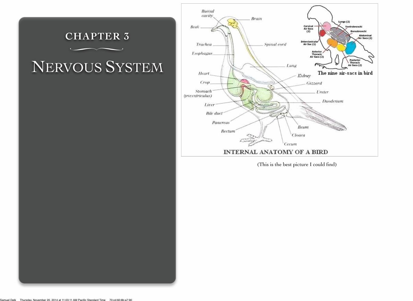
(This is the best picture I could find)
Samuel Delk Thursday, November 20, 2014 at 11:03:11 AM Pacific Standard Time 70:cd:60:8b:a7:90
• Function
• Detects and processes information
• Formulates responses
• Coordinates and control all bodily activity
• Sends and receives impulses
• Impulses: electrical signals that travel through the nervous system and provides information to the brain
• Parts of the nervous system-
• Central and Peripheral system
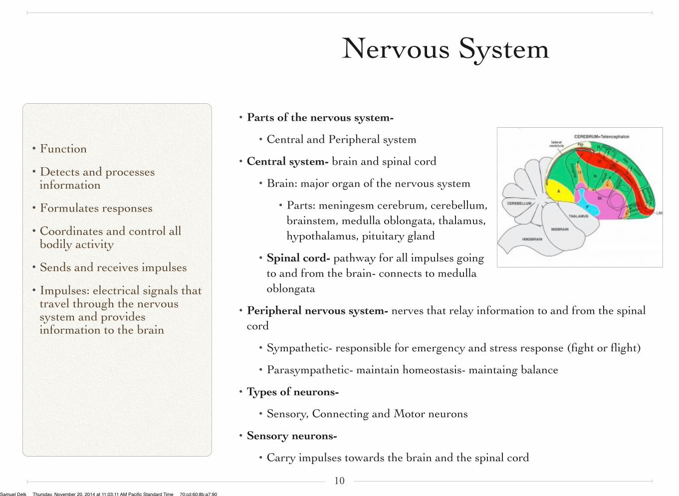
• Central system- brain and spinal cord
• Brain: major organ of the nervous system
• Parts: meningesm cerebrum, cerebellum, brainstem, medulla oblongata, thalamus, hypothalamus, pituitary gland
• Spinal cord- pathway for all impulses going to and from the brain- connects to medulla oblongata
• Peripheral nervous system- nerves that relay information to and from the spinal cord
• Sympathetic- responsible for emergency and stress response (fight or flight)
• Parasympathetic- maintain homeostasis- maintaing balance
• Types of neurons-
• Sensory, Connecting and Motor neurons
• Sensory neurons-
• Carry impulses towards the brain and the spinal cord
10
Nervous System
Samuel Delk Thursday, November 20, 2014 at 11:03:11 AM Pacific Standard Time 70:cd:60:8b:a7:90
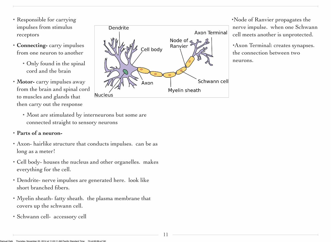
• Responsible for carrying impulses from stimulus receptors
• Connecting- carry impulses from one neuron to another
• Only found in the spinal cord and the brain
• Motor- carry impulses away from the brain and spinal cord to muscles and glands that then carry out the response
• Most are stimulated by interneurons but some are connected straight to sensory neurons
• Parts of a neuron-
• Axon- hairlike structure that conducts impulses. can be as long as a meter!
• Cell body- houses the nucleus and other organelles. makes everything for the cell.
• Dendrite- nerve impulses are generated here. look like short branched fibers.
• Myelin sheath- fatty sheath. the plasma membrane that covers up the schwann cell.
• Schwann cell- accessory cell
•Node of Ranvier propagates the nerve impulse. when one Schwann cell meets another is unprotected.
•Axon Terminal: creates synapses. the connection between two neurons.
11Samuel Delk Thursday, November 20, 2014 at 11:03:11 AM Pacific Standard Time 70:cd:60:8b:a7:90
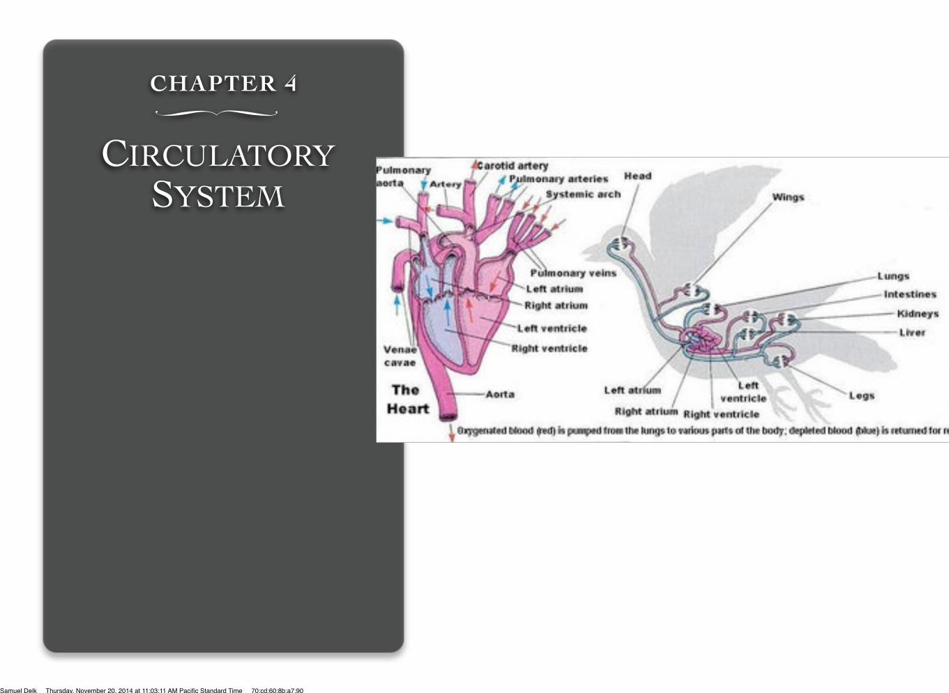
CHAPTER 4∏
CIRCULATORY SYSTEM
Samuel Delk Thursday, November 20, 2014 at 11:03:11 AM Pacific Standard Time 70:cd:60:8b:a7:90
COMPONENTS
• Heart- organ that pumps blood throughout your body.
• Veins- tubes, carrying in most cases, oxygen-depleted blood toward the heart.
• Arteries- tubes by which blood -mainly that which has been oxygenated- is conveyed from the heart to all parts of the body.
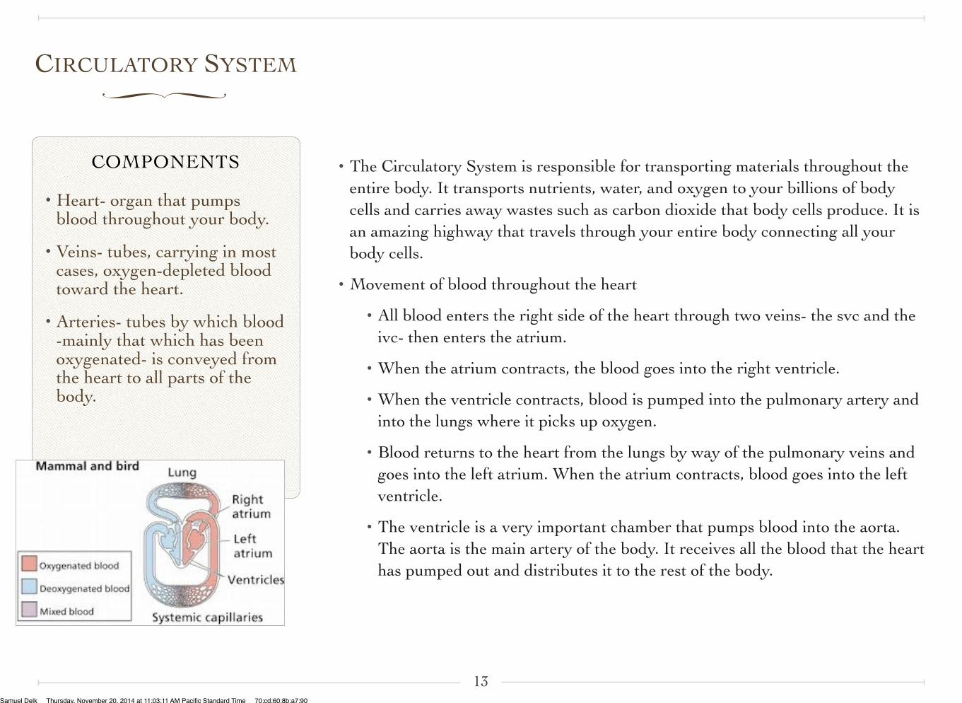
CIRCULATORY SYSTEM∏
• The Circulatory System is responsible for transporting materials throughout the entire body. It transports nutrients, water, and oxygen to your billions of body cells and carries away wastes such as carbon dioxide that body cells produce. It is an amazing highway that travels through your entire body connecting all your body cells.
• Movement of blood throughout the heart
• All blood enters the right side of the heart through two veins- the svc and the ivc- then enters the atrium.
• When the atrium contracts, the blood goes into the right ventricle.
• When the ventricle contracts, blood is pumped into the pulmonary artery and into the lungs where it picks up oxygen.
• Blood returns to the heart from the lungs by way of the pulmonary veins and goes into the left atrium. When the atrium contracts, blood goes into the left ventricle.
• The ventricle is a very important chamber that pumps blood into the aorta. The aorta is the main artery of the body. It receives all the blood that the heart has pumped out and distributes it to the rest of the body.
13Samuel Delk Thursday, November 20, 2014 at 11:03:11 AM Pacific Standard Time 70:cd:60:8b:a7:90
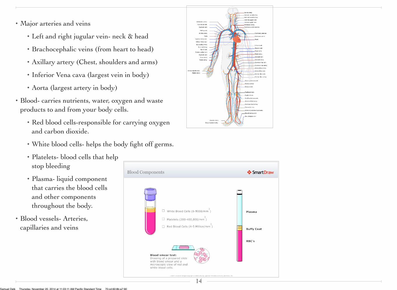
• Major arteries and veins
• Left and right jugular vein- neck & head
• Brachocephalic veins (from heart to head)
• Axillary artery (Chest, shoulders and arms)
• Inferior Vena cava (largest vein in body)
• Aorta (largest artery in body)
• Blood- carries nutrients, water, oxygen and waste products to and from your body cells.
• Red blood cells-responsible for carrying oxygen and carbon dioxide.
• White blood cells- helps the body fight off germs.
• Platelets- blood cells that help stop bleeding
• Plasma- liquid component that carries the blood cells and other components throughout the body.
• Blood vessels- Arteries, capillaries and veins
14Samuel Delk Thursday, November 20, 2014 at 11:03:11 AM Pacific Standard Time 70:cd:60:8b:a7:90
http://online.sfsu.edu/bholzman/courses/Fall99Projects/falcon.htm
http://en.wikipedia.org/wiki/Bird_anatomy
http://www2.unil.ch/biomapper/opengl/BirdFlight.html
http://takethemoment.org/?p=146
http://hes.ucfsd.org/gclaypo/circulatorysys.html#The%20Blood%20Vessels
http://www.currumbinvetservices.com.au/air_sacs.htm
15
Works Cited:
Samuel Delk Thursday, November 20, 2014 at 11:03:11 AM Pacific Standard Time 70:cd:60:8b:a7:90
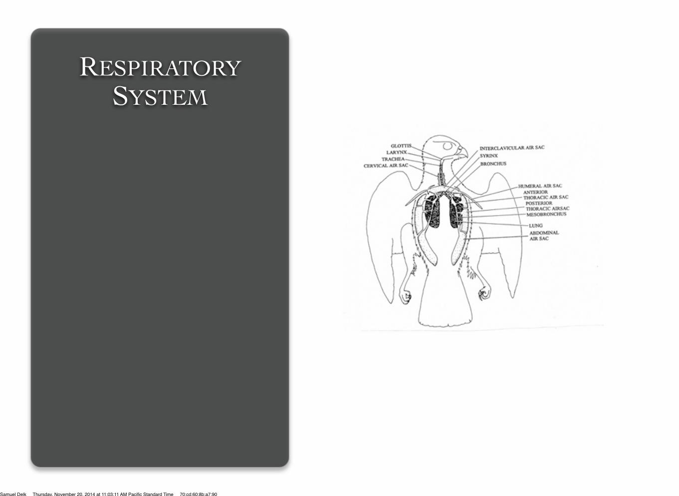
RESPIRATORY SYSTEM
Samuel Delk Thursday, November 20, 2014 at 11:03:11 AM Pacific Standard Time 70:cd:60:8b:a7:90
FUNCTION
• Bring in oxygen from the air
• Expel carbon dioxide
• Can’t be done without the circulatory system
https://answersingenesis.org/birds/peregrine-falcon-natures-top-gun/
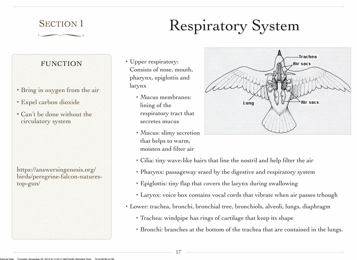
SECTION 1∏
• Upper respiratory: Consists of nose, mouth, pharynx, epiglottis and larynx
• Mucus membranes: lining of the respiratory tract that secretes mucus
• Mucus: slimy secretion that helps to warm, moisten and filter air
• Cilia: tiny wave-like hairs that line the nostril and help filter the air
• Pharynx: passageway sraed by the digestive and respiratory system
• Epiglottis: tiny flap that covers the larynx during swallowing
• Larynx: voice box contains vocal cords that vibrate when air passes trhough
• Lower: trachea, bronchi, bronchial tree, bronchiols, alveoli, lungs, diaphragm
• Trachea: windpipe has rings of cartilage that keep its shape
• Bronchi: branches at the bottom of the trachea that are contained in the lungs.
17
Respiratory System
Samuel Delk Thursday, November 20, 2014 at 11:03:11 AM Pacific Standard Time 70:cd:60:8b:a7:90
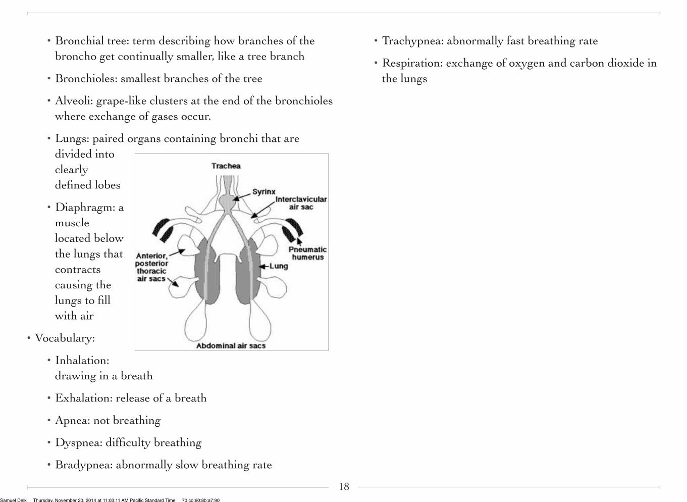
• Bronchial tree: term describing how branches of the broncho get continually smaller, like a tree branch
• Bronchioles: smallest branches of the tree
• Alveoli: grape-like clusters at the end of the bronchioles where exchange of gases occur.
• Lungs: paired organs containing bronchi that are divided into clearly defined lobes
• Diaphragm: a muscle located below the lungs that contracts causing the lungs to fill with air
• Vocabulary:
• Inhalation: drawing in a breath
• Exhalation: release of a breath
• Apnea: not breathing
• Dyspnea: difficulty breathing
• Bradypnea: abnormally slow breathing rate
• Trachypnea: abnormally fast breathing rate
• Respiration: exchange of oxygen and carbon dioxide in the lungs
18Samuel Delk Thursday, November 20, 2014 at 11:03:11 AM Pacific Standard Time 70:cd:60:8b:a7:90