photodegradation study of 3,5-diamino-6-chloro- n-(2...
TRANSCRIPT

UPTEC K 16005
Examensarbete 30 hpAugusti 2016
Photodegradation study of 3,5-diamino-6-chloro-
N-(2-(methylamino)ethyl)pyrazine-2-carboxamide
using preparative SFC and LC-MS
Sara Sillén

Teknisk- naturvetenskaplig fakultet UTH-enheten
Besöksadress: Ångströmlaboratoriet Lägerhyddsvägen 1 Hus 4, Plan 0
Postadress: Box 536 751 21 Uppsala
Telefon:018 – 471 30 03
Telefax: 018 – 471 30 00
Hemsida: http://www.teknat.uu.se/student
Abstract
Photodegradation study of 3,5-diamino-6-chloro-N-(2-(methylamino)ethyl)pyrazine-2-carboxamideusing preparative SFC and LC-MS
Sara Sillén
In this project the photodegradation of3,5-diamino-6-chloro-N-(2-(methylamino)ethyl)pyrazine-2-carboxamide was studied.A hypothetical degradation pattern for the compound was proposed and the aim ofthe project was to study the formed secondary photodegradants and to, if possible,structure elucidate some of these compounds. In order to do this, the parentcompound was photodegraded in two steps, where a primary photodegradant wasisolated using semi-preparative supercritical fluid chromatography (SFC) and thenfurther degraded into the secondary photodegradants.
The photodegradation was first carried out in aqueous solution, where the parentcompound was irradiated in UV-A light of 300-400 nm. This resulted in a primaryphotodegradant with a molecular ion of m/z = 227, where the chloride in position 6of the pyrazine group had been replaced by a hydroxyl group. During the large scalephotodegradation, prior to the preparative purification, the yield of primaryphotodegradant was very low due to the photodegradation being dependent on bothsample volume and concentration and due to the primary photodegradant also beingunstable in aqueous solution at room temperature.
Due to the above mentioned difficulties the parent compound was photodegraded inmethanol instead of water in order to avoid the freeze-drying process where a lot ofthe primary photodegradant was lost. This resulted in a primary photodegradant witha molecular ion of m/z = 241, where the chloride had been replaced by a methoxygroup instead of a hydroxyl group. This compound was more stable which allowedworkup by rotary evaporation, instead of freeze-drying, before the preparativepurification. This primary photodegradant was isolated using semi-preparative SFC on
1.2 mg material was isolated and further photodegradation tests in ordinary water
Some secondary photodegradants were observed in LC-MS analyses, and theirelement compositions were proposed by accurate mass results. Fundamentalstructures for these compounds were proposed. Further structural investigationalanalyses are needed for confirmation in the future.
ISSN: 1650-8297, UPTEC K16 005Examinator: Erik BjörkÄmnesgranskare: Richard SvenssonHandledare: Jufang Wu Ludvigsson, Per-Ola Norrby, Kristina Öhlén, Malin Härslätt
column (250 x 30 mm, 5 µm) with MeOH/NH3 100/1 v/v as organic modifier. Abouta Viridis® BEH Prep OBD TM column (250 x 30 mm, 5 µm) and a Luna HILIC
and 18O-water were conducted.

3
Contents
Populärvetenskaplig sammanfattning ........................................................................................ 5
Introduction ................................................................................................................................ 6
Experimental .............................................................................................................................. 9
Chemicals ............................................................................................................................... 9
Equipment ............................................................................................................................. 10
UV Equipment .................................................................................................................. 10
Chromatographic systems ................................................................................................. 11
Softwares ........................................................................................................................... 12
Other equipment ................................................................................................................ 12
Procedure .............................................................................................................................. 13
Photodegradation in aqueous solution .............................................................................. 13
Photodegradation of samples dissolved in methanol ........................................................ 16
Structural elucidation ........................................................................................................ 17
Results and discussion .............................................................................................................. 18
Photodegradation in aqueous solution .................................................................................. 18
Detection of m/z = 227 at all systems ............................................................................... 18
Freeze-drying .................................................................................................................... 19
Screen for a purification method ....................................................................................... 20
Purification of the primary photodegradant ...................................................................... 20
Screen for a UPLC analysis method ................................................................................. 22
Photodegradation rate of 2-pyrazinecarboxamide ............................................................ 23
Test of the UV equipment built in-house .......................................................................... 24
Photodegradation in methanol .............................................................................................. 25
Discovery of an unexpected primary photodegradant ...................................................... 25
Purification of the primary photodegradant ...................................................................... 26
Secondary photodegradants .............................................................................................. 27
Structure elucidation of the primary photodegradants ...................................................... 29
Conclusion ................................................................................................................................ 30
Acknowledgements .................................................................................................................. 31
References ................................................................................................................................ 32
Appendix .................................................................................................................................. 34
I: Optimal irradiation time for water samples ...................................................................... 34
II: Large scale freeze-drying ................................................................................................. 35

4
III: Sample stability in methanol .......................................................................................... 36
IV: Screen for a preparative separation method ................................................................... 37
V: Screen for an analysis method ......................................................................................... 40
VI: Irradiation using the UV equipment built in-house ........................................................ 43
VII: Optimal irradiation time for methanol samples ............................................................ 44
VIII: Structure elucidation .................................................................................................... 45
a. Structure elucidation of the primary photodegradant formed in water...................... 45
b. Structure elucidation of the primary photodegradant formed in methanol ............... 51
c. Structural elucidation of the secondary photodegradants .......................................... 56

5
Populärvetenskaplig sammanfattning
Aktiva ingredienser är de substanser i läkemedel som ger den farmakologiska effekten. Under
läkemedelsutvecklingen görs en mängd olika tester på den aktiva ingrediensen för att ta reda
på dess olika egenskaper, till exempel substansens distribution i kroppen, substansens
terapeutiska fönster och så vidare. Alla dessa tester ger information som pusslas ihop och
tillslut formar ett färdigt läkemedel.
Substansens farmakologiska effekt är direkt kopplad till dess molekylära struktur. Den
studerade molekylen, 3,5-diamin-6-klor-N-(2-(metylamin)etyl)pyrazin-2-karboxamid, har en
liknande molekylär struktur som amilorid, en aromatisk pyrazin. Amilorid är ett
kaliumsparande diuretikum som verkar genom att blockera kroppens vattenreabsorption.
Eftersom effekten är beroende av strukturen är det även viktigt att identifiera den aktiva
ingrediensens nedbrytningsprodukter samt deras farmakologiska effekter. Detta då dessa
skulle kunna vara mer potenta än ursprungsmolekylen, eller kanske till och med vara toxiska.
När man identifierat den aktiva substansens nedbrytningsvägar har man fått en bra grund till
att bland annat välja vilka excipienter den slutgiltiga läkemedelsformuleringen behöver
innehålla för att till så stor grad som möjligt undvika att den aktiva substansen bryts ned före
patienten intagit läkemedlet.
Nedbrytning sker ofta genom hydrolys, oxidation, termolytisk- eller fotokemisk nedbrytning.
Detta projekt har handlat om att studera hur 3,5-diamin-6-klor-N-(2-(metylamin)etyl)pyrazin-
2-karboxamid bryts ned när den utsätts för UV-ljus, s.k. fotokemisk nedbrytning. Vid
fotokemisk nedbrytning strålas molekylen med ljus av ett specifikt våglängdsintervall, vilket i
vårt fall var UV-A strålning vid 300-400 nm. Belysningen gör att molekylen absorberar
fotoner från ljuset och exciteras till ett högre energitillstånd. Detta tillstånd kan ha annan
strukturell konformation än grundtillståndet och kan ses som en helt annan molekyl, vilken
inte alls behöver reagera på samma sätt som grundtillståndet gör. Detta gör att det kan vara
svårt att förutsäga fotokemiska nedbrytningsprodukter, och gör detta till ett viktigt område att
studera. Om substansen är ljuskänslig eller inte kommer i slutändan påverka vilken sorts
förpackning samt förvaring som är passande för det färdiga läkemedlet. Ljuskänsliga
substanser bör alltså ha en förpackning som inte släpper igenom ljus samt förvaras mörkt.
För att identifiera vilka molekyler som bildats vid den fotokemiska nedbrytningen har
kromatografi använts. Kromatografi är en separationsmetod som går ut på att provet,
innehållande flera olika substanser, injiceras i en mobil fas och pumpas genom en kolonn
packad med en stationär fas. De substanser som finns i provet har olika affinitet till den
stationära fasen och retarderas därför olika mycket under färden genom kolonnen, vilket
möjliggör separation av substanserna. Separation sker med avseende på molekylernas
egenskaper, som t.ex. polaritet. Val av både stationär fas och mobil fas påverkar därför
selektiviteten.
Identifiering av vilka substanser som finns i proverna sker enklast med masspektrometri.
Masspektrometri möjliggör att detektera molekylvikten på substanserna som passerar genom
detektorn. Med hjälp av molekylvikten kan en struktur föreslås. Som ytterligare verifiering av
strukturen är isotoper av väte och syre användbara. Genom att låta reaktionen ta plats i 18
O-
vatten, dvs. vatten innehållande den tyngre syreisotopen, kan man bekräfta om en molekyl
reagerar med vatten eller med syre från luften. Detta genom att se om produkten som bildas
har motsvarande masshöjning som denna isotopreaktion bör ge.

6
Introduction
Epithelial sodium channels (ENaC) can be found for instance in the kidneys, the colon and in
the lungs. These channels actively transport sodium ions across the epithelial cell membrane
by dint of Na+/K
+-ATPase. While the sodium transportation takes place water is drawn back
into the cells alongside the ions, thereby this process passively reabsorbs water [1].
This process is important for upholding salt and water homeostasis. When the reabsorption of
water is undesired, for instance when the patient is suffering from hypertension, the active
pharmaceutical ingredient amiloride may be given as treatment in combination with thiazides
[2]. Their mechanism of action is simply blocking the ENaC and thereby decreasing water
reabsorption [3].
3,5-diamino-6-chloro-N-(2-(methylamino)ethyl)pyrazine-2-carboxamide, see Fig. 1, shares
the aromatic ring structure moiety with for example amiloride, Fig. 2. Studying the
photodegradation of 3,5-diamino-6-chloro-N-(2-(methylamino)ethyl)pyrazine-2-carboxamide
may therefore generate important information useful for other projects involving this moiety.
When analysing active pharmaceutical ingredients and their related impurities reversed phase
(RP) chromatography is commonly used [4-7]. RP- columns are non-polar and the parent
compound, 3,5-diamino-6-chloro-N-(2-(methylamino)ethyl)pyrazine-2-carboxamide, see Fig.
1, is a fairly polar compound which often results in short retention times. This compound may
therefore need a polar stationary phase in order to be retained and possibly separate from the
structure related impurities generated from photodegradation. Hydrophilic liquid
chromatography (HILIC) is a type of normal phase chromatography that utilizes polar
stationary phases and has proven to be a great alternative to reversed phase chromatography
when it comes to separation of small polar compounds [8-9]. Due to the polarity of the parent
compound both reversed phases and HILIC mode phases were screened in order to find a
suitable analysis method [10].
Fig. 1. The parent compound, 3,5-diamino-6-chloro-N-(2-(methylamino)ethyl)pyrazine-2-carboxamide.
Preparative supercritical fluid chromatography (SFC) was used to isolate the primary
photodegradant formed during the irradiation of the parent compound. SFC is a separation
technique utilizing polar stationary phases together with a mobile phase containing
supercritical carbon dioxide. Since carbon dioxide is approximately as non-polar as hexane
the mobile phase may be modified by addition of an organic modifier, typically small alcohols
such as methanol or ethanol [11]. Some parameters for controlling compound retention in
SFC are mobile phase polarity, temperature and pressure. Changes in temperature or pressure

7
affect the density of the mobile phase and an increased mobile phase density yields elution
strength and thereby decreases the retention time [12-14]. Further the peak shape might be
improved by addition of a small amount of water or ammonia to the organic modifier [15].
To confirm whether the proposed structures are formed during the photodegradation a
structural analysis using mass detection was employed. Experiments using the heavier
isotopes 2H and
18O were conducted to prove whether the degradation was due to water
hydrolysis or due to reaction with O2 from the air [16].
Previously Li et. al. [17] studied the photodegradation of amiloride hydrochloride in aqueous
solution involving both a photodegradation study at the pH range 4.5-11.0 and preparative
isolation of the major photodegradant. The pH-test was carried out using an amiloride
hydrochloride stock solution which was diluted 10-fold with buffers and then irradiated. The
used buffers for the samples were acetic acid-sodium acetate pH 2-5, potassium dihydrogen or
thiophosphate-disodium hydrogen phosphate pH 5-8, borax-hydrochloric acid pH 8-9, sodium
bicarbonate-sodium carbonate pH 10 and disodium hydrogen phosphate-sodium hydroxide
pH 11-12. The conclusion drawn from irradiating solutions of different pH was that the same
major photodegradation product was formed throughout the entire pH range, see Fig. 2, and
that the neutral form of amiloride, which was present in alkaline conditions, underwent photo
degradation approximately three times as fast as the cationic form of amiloride. The major
photodegradant, isolated using semi-preparative SFC, was analysed using mass spectrometry
and NMR spectroscopy and from the results a structure was assigned, Fig. 2. The proposed
photodegradation mechanism for the formation of this degradant was by a nucleophilic attack
of water on a radical cation of amiloride [17].
Fig. 2. Molecular structures of a) amiloride, and b) its major degradant [17].
In this project a photodegradation pattern was proposed, see Fig. 3. A corresponding primary
photodegradant as found by Li et al. [17] is likely to be formed when the parent compound is
photodegraded in aqueous solution. This compound is proposed to hydrolyse further and form
secondary degradation products. The formation of the proposed secondary degradation
product has not yet been confirmed, and this structure is only one of many possible secondary
degradation products. The aim of this project is to study the photodegradation of 3,5-diamino-
6-chloro-N-(2-(methylamino)ethyl)pyrazine-2-carboxamide and hopefully to gain
understanding of the formed secondary degradants.
a) b)

8
Fig. 3. Hypothetical photodegradation pathway of 3,5-diamino-6-chloro-N-(2-(methylamino)ethyl)pyrazine-2-
carboxamide. In aqueous solution and under UV-A irradiation the primary degradant with m/z = 227 is proposed
to be formed. If this intermediate is further degraded in aqueous solution secondary photodegradants will be
formed. A hypothetical proposal was a compound with m/z = 245, amongst many possible secondary
photodegradants.
Irradiating 3,5-diamino-6-chloro-N-(2-(methylamino)ethyl)pyrazine-2-carboxamide with UV
light of 300-400 nm will result in a molecular excitation. The excited state reacts with water
and the primary photodegradant is formed. This compound has a molecular ion of m/z = 227
and a characteristic UV spectrum similar to the parent compound, see Fig. 4. If a secondary
degradation step takes place the aromatic ring may open up, resulting in a compound without
an absorbance maximum at ~360 nm since the aromatic ring no longer exists. Studying the
UV spectra of the compounds from photodegradation therefore is a helpful tool, in
combination with mass spectrometry, when structure elucidating secondary photodegradants.
Fig. 4. UV spectra of a) the parent compound, and b) the primary photodegradant.
a) b)
9
Experimental
Chemicals
3,5-diamino-6-chloro-N-(2-(methylamino)ethyl)pyrazine-2-carboxamide (also called 2-
pyrazinecarboxamide) (95%) was bought from Chemtronica AB (Stockholm, Sweden)
HPLC-grade methanol for chromatography, isopropyl alcohol (IPA) (Reag. Ph EUR), formic
acid (Reag. Ph Eur) and acetic acid (Reag. Ph Eur) was obtained from Sigma-Aldrich (St
Louise, MO, USA). Ethanol (99.5%) for chromatography was purchased from Kemetyl AB
(Haninge, Sweden). Liquid carbon dioxide (CO2) Veriseq LIC Bleu was obtained from AGA
gas AB (Stenungsund, Sweden). Acetonitrile (Reag. Ph Eur), ammonia solution (25%, Reag.
Ph Eur) and formic acid (Reag. Ph Eur) was bought from Merck Millipore (Darmstadt,
Germany). Triethylamine (TEA) (≥99.5%) and sodium hydroxide concentrate (0.1 mol) was
obtained from Fluka (Buchs, Switzerland). Milli-Q-water was obtained from a Milli-Q
gradient A10 system. Methanol-D4 solution (99.8% atom D) was bought from Sigma Aldrich
Chemie (Steinheim, Germany) and 18
O-water (≥98 atom-%) was bought from Tayio Nippon
Sanso (Tokyo, Japan). Dry ice obtained from AGA Gas AB (Lidingö, Sweden). Potassium
phosphate dissolution media concentrate pH 6.8 obtained from Reagecon (Shannon, County
Claire, Ireland)
From the above mentioned chemicals the following mobile phases were made (composition in
volume ratios, i.e. v/v):
SFC
0.5 % ammonia in methanol (MeOH/NH3 100/0.5),
0.5 % formic acid in methanol (MeOH/FA 100/0.5),
1 % ammonia in methanol (MeOH/NH3 100/1),
0.5 % ammonia in ethanol (EtOH/NH3 100/0.5),
0.5 % ammonia in isopropyl alcohol (IPA/NH3 100/0.5),
30 mM ammonia in methanol.
UPLC
20 mM ammonia in water,
0.1 % formic acid in water,
0.1 M formic acid in water,
Na/K-phosphate buffer pH 6.9 I=0.01,
10 mM formic acid and 1 mM ammonium formeate in water, pH 3,
10 mM formic acid and 1 mM ammonium formeate in 95/5 ACN/H2O, pH 3,
0.8 % ammonia in water, pH 10,
8 mL ammonia, 42 mL water and 950 mL ACN, pH 10.

10
Equipment
UV Equipment
Irradiation was mostly carried out using an Atlas suntest xls+ with a solar ID65 filter letting
through UV-A light with wavelengths of 300-400 nm. Settings: T = 15 ºC, E = 30 W/m2.
Irradiation was also carried out using an irradiation equipment built in-house consisting of 4
black light bulbs (F4T5, 4 W) emitting UV-A light of 350-400 nm with a maximum at 365
nm, see Fig. 5. The sample was kept at room temperature using a fan.
Fig 5. The UV equipment built in-house. The sample was placed in front of four 4 W F4T5 black light bulbs and
was kept at room temperature during the irradiation using a fan (the white grid).

11
Chromatographic systems
Analytical SFC systems
SFC runs were performed on two UPC2-systems. One connected to a single quadrupole
detector (SQD) and one connected to a 3100 mass spectrometer. Both systems had photodiode
array detectors (PDA) and were obtained from Waters (Milford, MA, USA). Both systems
had three column ovens, two for 150 mm length columns and one for 250 mm columns. The
used flow rate was 3.5 mL/min. All analyses were conducted in positive mode, LC-ESI.
Chiral columns used for SFC screens: Lux Amylose-1 and Lux Cellulose (-2, -3, -4) bought
from Phenomenex, (Torrance, CA, USA). Chiralpak® (AS, IB, IC, ID) and Chiracel® OJ
obtained from Chiral Technologies (Illkirch, France). Kromasil 3-CelluCoat and Kromasil 3-
Amycoat were bought from Eka Chemicals AB (Bohus, Sweden). (S, S) Welk-O1 from Regis
(Minneapolis, MN, USA). Alcyon CHIRAL ART Amylose-SA and Alcyon CHIRAL ART
cellulose-SC obtained from YMC (Chicago, IL, USA).
The column dimension was 150 x 4.6 mm and the particle size for all the used columns was 3
µm.
Achiral columns used for SFC screens: Viridis® BEH and Viridis® BEH 2-Ethylpyridine
were obtained from Waters (Milford, MA, USA). Kromasil 60-diol, Kromasil 60-sil,
Kromasil 60-CN, Kromasil 100-NH2 were obtained from Eka Chemicals AB (Bohus,
Sweden). Luna HILIC bought from Phenomenex, (Torrance, CA, USA). Daicel DCpak® SFC
A obtained from Chiral Technologies (Illkirch, France).
The column dimension was 150 x 4.6 mm for all columns except Kromasil 60-diol and
Kromasil 60-sil which were 250 x 4.6 mm. The particle size for the used columns was 5 µm.
Preparative SFC systems
SFC purifications were performed using the UV-triggered Novasep 150 system from Novasep
(Nancy, France) and the mass-triggered TharSFCTM
SFC-MS prep-100 system from Waters
(Milford, MA, USA).
Columns for preparative SFC: Lux Cellulose-4 (Lux C4) obtained from Phenomenex,
(Torrance, CA, USA). Viridis® BEH Prep OBDTM
column obtained from Waters (Milford,
MA, USA). Luna HILIC obtained from Phenomenex, (Torrance, CA, USA).
The column dimension was 250 x 30 mm except for the Lux C4 column which was 250 x
21.2 mm. The particle size was 5 µm for all columns.
Analytical UPLC systems
UPLC runs were performed on Waters Acquity UPLC systems equipped with photodiode
array detectors (PDA) and single quadrupole detectors (SQD) or Xevo quadrupole time-of-
flight (QTof) mass spectromers. All analyses were conducted in positive mode, LC-ESI.
Columns for UPLC screen: Acquity UPLC® BEH C18 (particle size 1.7 µm), Cortecs C18
(2.7 µm), Acquity UPLC® BEH amide (1.7 µm) obtained from Waters (Milford, MA, USA).
iHILIC®-fusion (Beta test (1.8 µm) and Peek (3.5 µm)) obtained from Hilicon (Umeå,

12
Sweden). Poroshell 120 Bonus-RP (C18) (2.7 µm), Zorbax HILIC Plus (2.7 µm) and
Poroshell 120 HILIC (1.8 µm) from Agilent Technologies (Santa Clara, CA, USA). Kinetex®
(EVO C18 (1.7 µm) and F5 (1.7 µm)) obtained from Phenomenex (Torrance, CA, USA).
Halo HILIC (2.7 µm) obtained from Advanced Materials Technology (Wilmington, DE,
USA).
The column dimension was 100 x 2.1 mm for all columns except for the Cortecs C18 and
Poroshell 120 Bonus-RP (C18) which were 100 x 3.0 mm. The flow rate varied between 0.3-1
mL/min depending on particle size and system pressure.
Softwares
Chromatographic data was collected using two softwares, Empower 3 Pro and MassLynx,
from Waters (Milford, MA, USA).
Other equipment
V-10 evaporator from Biotage AB (Uppsala, Sweden). Evaporation was performed at 36 ºC
with 8 mbar pressure.
HT-4X centrifuge system from Genevac (Stone ridge, NY, USA).
Large scale freeze-drying was made on a freeze-drier built in-house at a pressure of 0.18
mbar. The sample was cooled to -78 ºC in a bath of ethanol and dry ice.
Weighing was made on a Mettler Toledo AT261 DeltaRange balance and Mettler Toledo
AX504 DeltaRange balance from Mettler Toledo (Greifensee, Switzerland).
Rotary evaporation was carried out at 20 mbar pressure using a rotary evaporator from
BÜCHI Labortechnik (Flawil, Switzerland). The water bath was set to 30 ºC and the round
bottomed flask rotating at 120 rpm.
Ultrasonification was carried out using Sonorex Super RH 100 H ultrasonic bath obtained
from Bandelin electronic (Berlin, Germany).
The pH was measured using a 780 pH meter obtained from Metrohm AG (Herisau,
Switzerland).

13
Procedure
Photodegradation in aqueous solution
Freeze-drying
20 mL 2-pyrazinecarboxamide solution with a concentration of 0.1 mg/mL was divided into 1
mL vials and irradiated in the Atlas suntest xls+. 9 mL was irradiated for 1 h, 10 mL for 2 h
and 1 mL of the solution was irradiated for 3 h. The optimal irradiation time could be
determined by UPLC analysis using a BEH C18 column with mobile phase A = 10 mM
formic acid and 1 mM ammonium formeate in water, pH 3 and mobile phase B = 10 mM
formic acid and 1 mM ammonium formeate in 95/5 ACN/H2O, pH 3 as mobile phases and a
flow rate of 0.7 mL/min. The analysis was performed using a gradient, see Table 1. Then the
vials were put in the freezer for later use.
Table 1. Gradient used for sample analysis.
Time (min) % B
0 1
1 1
6 7
6.1 1
8.1 1
One vial irradiated for 2 h was evaporated using V-10 then dissolved in 300 µl ethanol.
Another vial was freeze-dried using the HT-4X evaporation system and the dissolved in 900
µl water. Analyses were run before and after the evaporations.
10 ml 0.1 mg/mL solution of 2-pyrazinecarboxamide was prepared, irradiated for 2 h in a
round bottomed flask then freeze-dried using the large scale freeze drier built in-house. 1 mL
methanol was added to the round bottomed flask. The solution was transferred into a vial,
placed in the sample manager and repeatedly analysed for 2 days.
Detection of m/z = 227
The sample that was freeze-dried using HT-4X was analysed at two UPLC systems, one
equipped with a Xevo QTof mass spectrometer and one equipped with an SQD mass
spectrometer. The analyses were conducted using BEH C18 columns (100 x 2.1 mm, 1.7 µm)
with acidic and basic mobile phases at pH 3 and pH 10 and a gradient of 1-10 % B in 2.7 min.
Then the sample was analysed at an UPC2–SQD system using a Viridis BEH column (150 x
4.6 mm, 5 µm) isocratically with 20 % MeOH/FA 100/0.5 as organic modifier. Cone voltages
of 15, 30 and 55 V were tested using the same sample at the UPLC-SQD system. A new 2 h
sample was thawed and analysed at the UPLC-SQD system using acidic conditions. The
sample was left in the sample manager and reanalysed again the next day.
Screen for a purification method
15.3 mg of 2-pyrazinecarboxamide was dissolved in 153 mL of water and irradiated for 3 h.
The solution was divided into two round bottomed flasks then freeze-dried using the large
scale freeze-drier. Analyses were performed before and after the freeze-drying process. One
round bottomed flask containing 7 mg degraded sample was frozen for later use and the other
one, containing 13 mg of degraded material, was dissolved in methanol to a final

14
concentration of ~2 mg/mL. This solution was used to screen for a preparative separation
method.
An SFC screen on 14 chiral columns using 25% EtOH/NH3 100/0.5 as organic modifier, 40
°C and 120 bar and on 8 achiral columns using 25% MeOH/NH3 100/0.5, 25 %
EtOH/NH3 100/0.5, 25 % EtOH and 25 % IPA/NH3 100/0.5 as organic modifiers, 40 °C and
120 bar was performed. From the results a number of columns were chosen for further
optimization with respect to mobile phase composition and system pressure. In order to
increase retention, less organic modifier was used, and the other way around. All the runs
throughout the optimization were performed using MeOH/NH3 100/0.5 as organic modifier at
a pressure of 150 bar.
Photodegradation of 2-pyrazinecarboxamide
20 mL solutions of 0.1, 0.2, 0.3, 0.5 mg/mL 2-pyrazinecarboxamide in water were irradiated
separately for 2 h then analysed and the optimal irradiation concentration could be
determined.
230 mg 2-pyrazinecarboxamide was dissolved in water and diluted to 0.2 mg/mL, resulting in
a sample volume of 1150 mL. The solution was divided into 20 mL vials and irradiated for 2
h in 3 batches. The primary photodegradant yield was analysed and the vials were irradiated
for an additional 1.5 h in batches then analysed again. The solutions were freeze-dried in 8
round bottomed flasks then the content was dissolved in methanol. Drops of water and acetic
acid was added in order to improve solubility.
Preparative purification of the primary photodegradant
Preparative isolation of the primary photodegradant was performed in isocratic mode using
the Novasep 150 system with a semi preparative Lux C4 column (250 x 21.2 mm, 5 µm) at
40°C, 140 bar and with 22% MeOH/NH3 100/0.5 as organic modifier. Collected fractions,
containing the parent compound and the primary photodegradant respectively, were rotary
evaporated and analysed on a Lux C4 column using 35 % MeOH/NH3 100/0.5, 40° and 150
bar. Unsatisfying results from the fraction analysis resulted in a change of preparative column.
The remaining purification was performed in gradient mode on a Viridis® BEH column with
a flow rate of 120 g/min, 150 bar, 40 °C and MeOH/NH3 100/0.5 as organic modifier, see
Table 2.
Table 2. Gradient used for preparative isolation of the primary photodegradant with the BEH
column.
Screen for an analysis method
A UPLC screening of columns, mobile phases and gradients to get the best separation for the
parent compound and photodegradants was performed. Samples used for the screening were
irradiated for 2 h and had a concentration of 0.1 mg/mL. The analyses were carried out using
Time (s) % CO2 % Modifier
0 80 20
150 80 20
350 70 30
351 65 35
450 80 20

15
mobile phase A = 0.1 M FA in H2O and B = ACN unless otherwise stated. The flow rates
varied between 0.3-1 mL/min depending on particle size. The reversed phase analyses were
performed using a gradient of 2-20% B in 10 min and the HILIC columns were run isocraticly
using 90 % B. The Kinetex EVO C18 column was additionally analysed using 10 mM
ammonia in H2O as mobile phase A. The Kinetex F5 column was run using A = Na/K-
phosphate buffer pH 6.93 I = 0.01 and a gradient, see Table 3.
Table 3. Gradient used for the Kinetex F5 column.
Time (min) % B
0 0
2 0
10 20
12 35
12.1 0
13 0
Photodegradation rate of 2-pyrazinecarboxamide
7 ml 0.2 mg/mL of 2-pyrazinecarboxamide in aqueous solution was made and the pH of the
solution was measured. The solution was divided into 7 vials and irradiated. One sample was
taken out and analysed every 30 min for a total of 3 h. One reference sample was kept dark
and analysed together with other samples. Analyses were run using Kinetex EVO C18 column
with mobile phase A = 10 mM ammonia in H2O and mobile phase B = ACN, a gradient of 0-
20% B in 10 min, and a flow rate of 0.5 mL/min.
Another degradation rate test of 2-pyrazinecarboxamide was performed with regards to the
irradiation volume. Each time a sample was taken out of the Atlas suntest xls+ it was replaced
with a vial containing the same amount of water, keeping the irradiated volume constant
throughout the test.
Test of the UV equipment built in-house
7 ml 0.2 mg/ml of 2-pyrazinecarboxamide was irradiated for 2 h in the in-house built
irradiation equipment. Samples for analysis were taken continuously throughout the
irradiation.

16
Photodegradation of samples dissolved in methanol
Detection of an unexpected primary photodegradant
A total of 3 samples containing 0.2 mg/mL of parent compound was prepared using
MeOH/H2O 90/10 v/v as solvent. Another 3 samples of 0.2 mg/mL parent compound was
made using MeOH/H2O 90/10 v/v as solvent to which 1.5 equivalents of NaOH was added.
The samples were irradiated for 1 h and 2 h then analysed. 3 samples of 0.2 mg/mL in
methanol-D4/H2O 90/10 v/v were irradiated for 1 h and 2 h.
Purification of the primary photodegradant
1 mL of 0.2 mg/mL was irradiated for 2 h in the Atlas suntest xls+ then rotary evaporated and
dissolved in 300 µL methanol. This sample was used to screen SFC columns for a preparative
separation method. Columns used for the screen were Diacel® DCpak, Kromasil 3-CelluCoat,
Lux Amylose-1, Viridis® BEH, Kromasil-NH2 and Luna HILIC and the organic modifier
used for the screen was MeOH/NH3 100/1. The percentage of modifier used in the SFC runs
was based on previous results from screening the samples degraded in aqueous solution.
The optimal irradiation time for samples of 1 mg/mL 2-pyrazinecarboxamide in methanol was
determined. Samples of 1 mg/mL in methanol were prepared and irradiated for 2 h and 3 h.
Another 2 samples were prepared and irradiated for 1 h and 1.5 h. All samples were analysed
in gradient mode using a UPC2 system and a Luna HILIC column with 30 mM ammonia in
MeOH as organic modifier with a flow rate of 2.5 mL/min, see Table 4.
Table 4. Gradient used for analyses on the Luna HILIC column when determining optimal
irradiation time for samples photodegraded in methanol.
Time (min) % Modifier
0 5
4 50
4.1 5
110 mg 2-pyrazinecarboxamide was photodegraded in methanol in batches of 10 ml 1mg/mL
solutions divided into 10 vials. Each batch was irradiated for 1 h. 5 mg was photodegraded for
6 h using the UV equipment built in-house. All irradiated solutions were combined and rotary
evaporated. The preparative isolation of the primary photodegradant was conducted on the
Thar SFC system with 30 mM ammonia in methanol as organic modifier, 120 bar, 38 ºC and a
flow rate of 100 g/min. The first part of the purification was conducted using the Viridis®
BEH column (250 x 30 mm, 5 µm) in gradient mode, see Table 5.
Table 5. Gradient used on the Viridis® BEH column during the purification of the primary
photodegradant formed in the methanol samples.
Time (min) % Modifier
0 20
5 20
11 40
11.1 20
12 20

17
The second part of the purification was conducted using the Luna HILIC preparative column
(250 x 30 mm, 5 µm), 120 bar, 38 ºC and 30 mM ammonia in methanol as organic modifier in
gradient mode, see Table 6.
Table 6. Gradient used on the Luna HILIC column during the purification of the primary
photodegradant formed in the methanol samples.
Time (min) % Modifier
0 25
1 25
8 30
9.8 40
10 25
11 25
Structural elucidation
In order to structure elucidate the primary photodegradant formed in water, m/z = 227,
solutions of 2-pyrazinecarboxamide in water and 18
O-water respectively of 0.1 mg/ml were
irradiated for 2 h and analysed with high resolution mass spectrometry (UPLC - QTof). In
order to structure elucidate the primary photodegradant formed in methanol corresponding
experiments were done in methanol/H2O 90/10 v/v and methanol-D4/H2O 90/10 v/v solution
as well.
The isolated primary photodegradant formed in methanol, m/z = 241, was further
photodegraded in water and 18
O-water respectively of 0.1 mg/mL were irradiated for 2 h, in
order to study secondary photodegradants. Structure elucidation was performed by Jufang Wu
Ludvigsson, see Appendix VIII. Structure elucidation.

18
Results and discussion
Photodegradation in aqueous solution
Optimal irradiation time was determined by comparing the peak areas, corresponding to the
formed primary photodegradant, at 3 consecutive irradiation times of 1, 2 and 3 h, see
Appendix I: Optimal irradiation time in water samples. After 3 h of irradiation 2-
pyrazinecarboxamide was completely degraded while the formed primary photodegradant was
at an acceptable level. This irradiation time was therefore chosen. The main drive behind this
was to obtain as much of the product as possible to maximize the potential yield of the
following purification.
Detection of m/z = 227 at all systems
The photodegraded sample dissolved in water proved to be stable for less than one day at
room temperature. Early on in the project this caused some uncertainty since different LC-MS
systems, LC-SQD versus LC-Xevo QTof, showed different results. The primary
photodegradant, with m/z = 227 was detected on the Xevo QTof system but not on the SQD
system where presumably only fragments of the compound could be seen. A theory was that
different cone voltage settings on the mass spectrometers resulted in different fragmentation.
Modification of the cone voltage settings changed the degree of fragmentation, but still the
primary photodegradant could not be found. When analysing a freshly thawed vial containing
sample irradiated for 2 h the m/z = 227 peak was finally detected at all systems, see Fig. 6.
The next day when the sample was analysed again the compound, m/z =227, was gone. This
lead to the conclusion that the primary photodegradant degrades in the samples during 1 day
when left in aqueous solution at room temperature.

19
Fig.6. Analyses of the same sample analysed at both the UPLC–Xevo QTof system and the UPLC–SQD system.
The runs were performed using BEH C18 columns and with mobile phase A = 10 mM formic acid and 1 mM
ammonium formeate in water, pH 3 and mobile phase B = 10 mM formic acid and 1 mM ammonium formeate in
95/5 ACN/H2O, pH 3. A flow rate of 0.7 mL/min and a gradient of 1-7 % B in 6 min was used at both systems.
a) Chromatogram of the run at the Xevo QTof system. b) Mass spectrum of the primary photodegradant peak at
tR = 0.77 min. c) Chromatogram of the run performed at the SQD system. d) Mass spectrum of the peak at tR =
0.38 min, the retention time where the compound later was found using this system.
Freeze-drying
In order to perform SFC screen and purification the sample concentration had to be increased
in order to exceed the detection limit. Water had to be removed without causing further
degradation. Using the V-10 evaporation system the primary photodegradant peak, with m/z =
227, completely disappeared, hence this was not an alternative to be considered. However
freeze-drying using the HT-4X system was a better alternative maintaining the stability at a
satisfactory level. To be able to evaporate increased amounts of water a large scale freeze-
drier was used. From a test of the large scale freeze-drier results showed that the ratio between
the peaks of the primary photodegradant and the parent compound decreased from 50:50 to
23:77, see Appendix II: Large scale freeze-drying, proving that a large amount of the primary
photodegradant was lost during this process. Hence the primary photodegradant was less
stable than the parent compound resulting in a sample containing 8 % (peak area) of the
desired compound at this point. The analysis was performed at 220 nm where most impurities
are visible.
The stability of a freeze-dried sample dissolved in methanol, stored without being exposed to
light at room temperature, was studied during two days, see Appendix III: Sample stability in
methanol. Methanol turned out to be the preferred solvent since the sample proved to be
a) b)
c) d)

20
sufficiently stable. Still, a slight degradation could be seen, possibly due to the remaining
water after the freeze-drying.
Screen for a purification method
To be able to isolate the primary photodegradant in a sufficient amount to enable a successful
purification, a total of 150 mg of 2-pyrazinecarboxamide was degraded for 3 h with the
intension of performing a SFC screen for a suitable purification method for the primary
photodegradant. However the outcome of the irradiation did not agree with previous results,
see Fig. 7. There was still parent compound left in the sample after 3 h of irradiation. This
was not expected remembering the results from the initial small scale degradation conducted
with 10 mL sample volume divided into 10 vials. This time it was carried out using the same
concentration in 153 mL but in 1 round bottomed flask. This lead to the conclusion that the
degree of photodegradation might be volume dependent. However, this needs further
confirmation with systematical investigation.
Fig. 7. A comparison of 1 mg/mL sample degradation after 3 h of irradiation. Both samples were analysed at a
UPLC-Xevo QTof system with a BEH column, mobile phase A = 10 mM formic acid and 1 mM ammonium
formeate in water, pH 3 and mobile phase B = 10 mM formic acid and 1 mM ammonium formeate in 95/5
ACN/H2O, pH 3, a flow rate of 0.7 mL/min and a gradient of 1-7 % B in 6 min. a) 153 mL sample volume. The
parent compound is still present in the sample. b) 10 mL sample volume. The samples were analysed at 360 nm
since this is the wavelength where both the parent compound and the primary photodegradant have their
absorbance maxima.
A thorough SFC method development screen was performed comprising several different
stationary phases together with a wide range of mobile phases, exemplified in Appendix I
V: Screen for a preparative separation method. Finally the Lux C4 column using 35 %
MeOH/NH3 100/0.5 as organic modifier, a system pressure of 150 bar and a temperature of 40
°C was chosen as preparative method.
Purification of the primary photodegradant
A study to determine the optimal concentration for large scale purpose was carried out, see
Table 7. The peak areas did not increase proportionally to sample concentrations, indicating
that the degree of photodegradation was dependent on sample concentration. A theory was
that with increased concentration the molecules block each other from being exposed to light,
leaving the molecules in the middle unexcited and unable to form the primary
a) b)
21
photodegradant. From this analysis the concentration of 0.2 mg/mL was chosen for the large
scale degradation.
Table 7. Peak areas of the primary photodegradant when irradiating samples of varying
concentrations for 2 h. The study was conducted at 360 nm.
Concentration (mg/mL) Peak areaprimary photodegradant
0.1 5349
0.2 7874
0.3 7047
0.5 7998
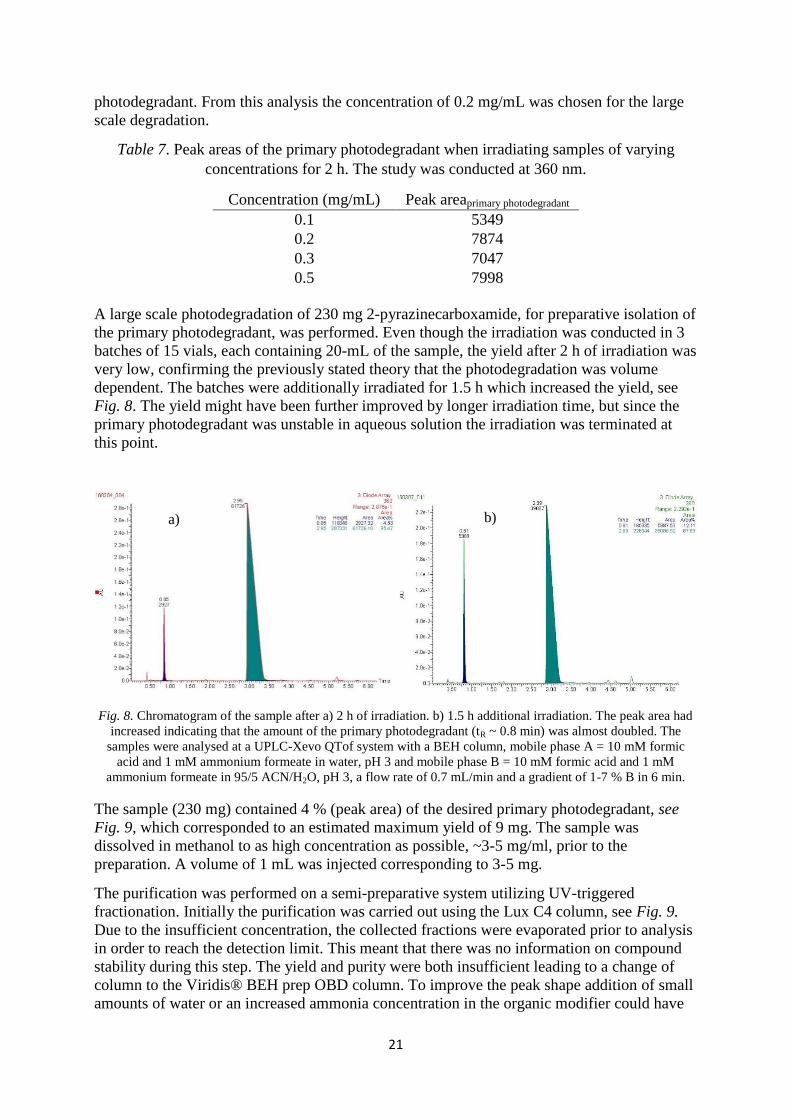
A large scale photodegradation of 230 mg 2-pyrazinecarboxamide, for preparative isolation of
the primary photodegradant, was performed. Even though the irradiation was conducted in 3
batches of 15 vials, each containing 20-mL of the sample, the yield after 2 h of irradiation was
very low, confirming the previously stated theory that the photodegradation was volume
dependent. The batches were additionally irradiated for 1.5 h which increased the yield, see
Fig. 8. The yield might have been further improved by longer irradiation time, but since the
primary photodegradant was unstable in aqueous solution the irradiation was terminated at
this point.
Fig. 8. Chromatogram of the sample after a) 2 h of irradiation. b) 1.5 h additional irradiation. The peak area had
increased indicating that the amount of the primary photodegradant (tR ~ 0.8 min) was almost doubled. The
samples were analysed at a UPLC-Xevo QTof system with a BEH column, mobile phase A = 10 mM formic
acid and 1 mM ammonium formeate in water, pH 3 and mobile phase B = 10 mM formic acid and 1 mM
ammonium formeate in 95/5 ACN/H2O, pH 3, a flow rate of 0.7 mL/min and a gradient of 1-7 % B in 6 min.
The sample (230 mg) contained 4 % (peak area) of the desired primary photodegradant, see
Fig. 9, which corresponded to an estimated maximum yield of 9 mg. The sample was
dissolved in methanol to as high concentration as possible, ~3-5 mg/ml, prior to the
preparation. A volume of 1 mL was injected corresponding to 3-5 mg.
The purification was performed on a semi-preparative system utilizing UV-triggered
fractionation. Initially the purification was carried out using the Lux C4 column, see Fig. 9.
Due to the insufficient concentration, the collected fractions were evaporated prior to analysis
in order to reach the detection limit. This meant that there was no information on compound
stability during this step. The yield and purity were both insufficient leading to a change of
column to the Viridis® BEH prep OBD column. To improve the peak shape addition of small
amounts of water or an increased ammonia concentration in the organic modifier could have
a) b)

22
been advantageous. However, due to compound instability in presence of water this was not
an option. Again the analysis of the collected primary degradant fraction showed poor purity
and yield.
Fig. 9. a) Analysis of the sample prior to purification. The analysis was performed using a Lux C4 column (150
x 4.6 mm, 3 µm) at 40ºC, 150 bar, a flow rate of 3.5 mL/min and with 35% MeOH/NH3 100/0.5 as organic
modifier. The peak at tR = 3.50 min was the wanted primary photodegradant. The peak was 4 % (peak area) at
220 nm. b) Chromatogram from the purification using the Lux C4 column (250 x 21.2 mm, 5 µm), 40 ºC, 140
bar, a flow rate of 75 g/min and with 35 % MeOH/NH3 100/0.5 as organic modifier. The wavelength used when
collecting fractions was 360 nm. Fraction 5 contained the parent compound and fraction 6 contained the
photodegradant.
The amount of desired photodegradant received after irradiation would need to be improved
before another attempt at purifying this compound may be conducted. Purification of a
compound with such a low starting purity, in this case 4 %, is a difficult task. A mass
spectrometer would have been helpful in terms of detection and collection of the target
compound and its surrounding impurities.
Screen for a UPLC analysis method
The UPLC analysis method initially used, BEH C18 analysis with mobile phases at pH 3 was
not optimal. The peaks with short retention time overlapped and did not leave any space for
more degradation products to be analysed. It would have been ideal to have an analysis
method where these peaks were more retained, leaving space between them for other
compounds. A screen for a suitable analysis method was conducted and resulted in Kinetex
EVO C18 column with mobile phase A =10 mM ammonia in H2O and B = ACN, and a
gradient of 0-20 % B being the best. This method was used for analysis of the
photodegradation rate of the parent compound. For chromatograms from this screen, see
Appendix V: Screen for an analysis method. Later on another analysis method was found.
Kinetex F5 column with mobile phase A = Na/K phosphate buffer pH 6.93, I = 0.01 and
mobile phase B = ACN. All analyses were performed using a wavelength of 254 nm since this
was where the most degradants were visible, and the primary photodegradant in a sample
photodegraded for 2 h showed up as a significant peak.
a) b)

23
Photodegradation rate of 2-pyrazinecarboxamide
The study of the photodegradation rate of 2-pyrazinecarboxamide, see Table 8 and Fig. 10,
showed that the compound had a photodegradation order between 0 and 1. In other words the
photodegradation was somewhat dependent on concentration, which strengthens the
conclusion found during the study of optimal sample concentration.
Table 8. The peak areas in the photodegradation rate study of 2-pyrazinecarboxamide.
Time
(h)
Peak area Sample concentration
(mg/mL)
0 2032732 0.19
0.5 1517779 0.14
1 1069295 0.10
1.5 810445 0.08
2 470754 0.04
2.5 337842 0.03
3 105334 0.01
Fig. 10. Plot of concentration vs time. A linear trendline indicates reaction order 0 and an exponential trendline
indicates reaction order 1. The values in this case seem to be somewhere between following reaction order 1 and
reaction order 0.
During the large scale photodegradation the theory that the photodegradation was volume
dependent was confirmed. Taking that into consideration, a second photodegradation rate test
was conducted in a different manner. This time the sample vial taken out was replaced with a
vial containing the same volume of water, making the irradiated volume constant at all times.
In the previously performed photodegradation test one vial at a time was taken out of the
irradiation apparatus resulting in less and less volume being irradiated at the same time.
Assuming that the photodegradation has reaction order 1, the rate constants in both cases were
determined to 0.8 h-1
indicating that the sample volume does not influence the
photodegradation at this scale.
C/C0= e-0,811t
R² = 0,9210
C/C0 = -0,3503t + 1
R² = 0,9357
-0,2
0
0,2
0,4
0,6
0,8
1
1,2
0 0,5 1 1,5 2 2,5 3 3,5
C/C
0
Irradiation time (h)
Photodegradation rate of 2-pyrazinecarboxamide

24
The initial pH of the solutions in the photodegradation rate tests were 8.5-8.7 in both cases.
For future studies it may be of interest to study the photodegradation rate of the parent
compound at some different starting pHs, using buffered systems, to get information of the
influence pH has on the photodegradation rate. It may also be a parameter that influences the
further degradation of the wanted primary photodegradant, and may therefore be important to
optimize in order to obtain a higher amount of the primary photodegradant in the samples.
Something else to take into consideration when studying the results is that during the
photodegradation reaction HCl is formed, changing the pH of the samples throughout the
degradation. The change in pH may affect the photodegradation rate and may also be an
explanation to why the photodegradation order is in-between 0 and 1 instead of conclusively
1st order. Confirmation of this may be conducted in a buffered solution where the pH is
controlled throughout the study.
Test of the UV equipment built in-house
In an attempt to decrease the number of photodegradants formed by absorption of the lower
wavelengths, and thereby reducing the number of impurities, irradiation was carried out using
an in-house built UV equipment. The test was successful and a profile of slightly less
impurities was formed during irradiation using this equipment, see Appendix VI: Irradiation
using the UV equipment built in-house. This test also confirmed that the wanted
photodegradant was formed by excitation of light at about 360 nm. This equipment did not
allow for larger volumes to be irradiated at the same time, which the Atlas suntest xls+ did.
Something worth looking into may be a filter to the Atlas suntest xls+ which lets through a
narrower range of wavelengths, around 360 nm, allowing for fewer impurities to be formed in
the samples.

25
Photodegradation in methanol
Discovery of an unexpected primary photodegradant
In order to avoid the freeze-drying process, where a lot of material was lost, a degradation
study where samples dissolved in MeOH/H2O 90/10 v/v instead of water was conducted. This
way the solvent could easily be evaporated prior to the preparative SFC purification step.
However, in these samples a primary photodegradant with m/z = 241 was formed instead of
the expected with m/z = 227, proving that methanol was a better nucleophile than water. This
compound also proved to be more stable than the corresponding compound formed in aqueous
solution, making it easier to work with. e.g. rotary evaporation could be used to concentrate
the sample before the preparative isolation.
The photodegradant, m/z = 241, was structure elucidated using methanol-D4 and LC-MS/MS,
see Fig.11.
Fig. 11. The confirmed structure of the primary photodegradant formed when samples dissolved in methanol
were irradiated in the Atlas suntest xls+. Methanol acted as a better nucleophile than water and by a nucleophilic
attack on the aromatic carbon in position 6 the primary photodegradant with m/z = 241 was formed.
Another photodegradation study was conducted with 1.5 equivalents of NaOH added to the
sample resulting in a different photodegradant selectivity. The expected primary
photodegradant with m/z = 227 was still not found in this sample. Instead the photodegradant
with m/z = 241 was found and the photodegradation rate seemed to be higher in this sample.
Since the formed photodegradant in these samples contained a methoxy group instead of a
hydroxyl group in position 6 of the aromatic ring, another hypothetical degradation pathway
was proposed with a mechanism corresponding to the one from the aqueous samples, see Fig.
12. It is still important to keep in mind that this is one of many possible secondary
photodegradants with the ring opening mechanism.

26
Fig. 12. Hypothetical protodegradation pathway for 2-pyrazinecarboxamide when irradiated in methanol. The
primary photodegradant in this reaction is further degraded in water to a hypothetical secondary degradation
product, assumed to be formed with the same mechanism as for the secondary photodegradant formed in water.
Purification of the primary photodegradant
SFC purification of the primary photodegradant formed in methanol was to be performed. A
new method development was done in the same manner as formerly stated, resulting in two
alternative methods on two different stationary phases, Luna HILIC and Viridis® BEH Prep
OBDTM
respectively. Prior to purification the optimal irradiation time at a concentration of 1
mg/mL in methanol was determined using SFC-MS, see Appendix VII: Optimal irradiation
time for methanol samples. From this study the optimal irradiation time was determined to 1 h
since the amount of the photodegradant did not show a significant increase after 1.5 h of
irradiation. In fact, after 1.5 h the area started to decrease again owing to further degradation
of the target molecule. Additionally this short cycle time allowed for many batches to be
irradiated in 1 day. The optimized conditions were applied to degrade 110 mg of 2-
pyrazinecarboxamide in the Atlas suntest xls+. These samples were mixed with the 5 mg that
was degraded in the in-house built UV equipment, resulting in a sample containing 8 % (peak
area) of the wanted primary photodegradant at 220 nm, yielding an estimated maximum
amount of only 9 mg. Due to shortage of time and material the purification was performed on
two different stationary phases, Luna HILIC and Viridis® BEH Prep OBDTM
columns, in
order to optimize the chances of obtaining pure compound. Due to the insufficient
concentration in the collected fractions, the fractions were evaporated prior to analysis in
order to reach the detection limit. A total amount of 1.2 mg was obtained, where 0.6 mg with
a purity of 95 % was obtained using the Luna HILIC column and 0.6 mg with a purity of 90
% was obtained using the Viridis® BEH Prep OBDTM
column. The two purification methods
gave somewhat different impurity profiles. Had there been more time and a higher amount of
the primary degradant in the samples the purification could have been performed in two steps
including both methods, Luna HILIC and Viridis® BEH Prep OBDTM
, and a sample with

27
higher purity could have been generated. As a way of improving the yield in the future, a
design of experiment (DOE) study can be conducted, in order to optimize the photodegradant
yield. Even though the starting purity with respect to the target compound in this case was low
the purity could successfully be increased to 95 % at 220 nm.
Secondary photodegradants
The isolated compound, m/z = 241, was dissolved in water and 18
O-water and further
photodegraded. The analyses of these samples can be seen in Fig. 13. A secondary
photodegradant with m/z = 259 was formed. A comparison of the mass spectra from the
sample degraded in water and the sample degraded in 18
O-water showed a mass increase of +4
Da for this compound indicating that it was partly formed by a reaction with O2 from the air,
and from the element composition a fundamental structure was proposed, see Fig.14. At this
point no conclusion of whether the compound was formed by an initial reaction with water or
with O2 from the air could be drawn. A way of sorting this out would be to identify the
intermediate peaks formed in this reaction, and by repeatedly analysing the sample with short
intervals to figure out which one of these intermediates forms first.
Two other secondary degradants, with m/z = 258 and m/z = 256, were also observed and
structures of these compounds could be proposed by studying their element composition
acquired from accurate mass and their degradation conducted in water and 18
O-water, see
Appendix VIII.c. Structural elucidation of the secondary photodegradants, structures for these
compounds could be proposed, see Fig. 14. These are only fundamental structural proposals
and could be ring opened structures as well. Since the concentration of these compounds in
the sample was very low and there was no baseline separation between peaks, the UV spectra
of these peaks were not conclusive. No conclusion of whether these compounds have peaks at
360 nm from an aromatic ring or not can be drawn at this point. It would also be good to
perform NMR analysis of each of these secondary photodegradants in order to obtain their
definitive structures.

28
Fig. 13. UV-, TIC- and extracted mass chromatograms from the sample where the primary photodegradant, m/z
= 241, were further photodegraded in water. Three secondary degradants were of interest. These compounds had
m/z = 256, m/z = 258 and m/z = 259 and their extracted chromatograms can be seen above. In H2O18
m/z = 258
increased to m/z = 260 and m/z = 256 to m/z = 258 indicating that these compounds contained only one oxygen
from reaction with water.
Fig. 14. Proposed structures for the secondary photodegradants formed in aqueous solution when degrading the
primary degradant isolated from methanol samples. Proposed structures for a) the m/z = 258 peak, b) the m/z =
259 peak, c) the m/z = 256 peak.
a) b) c)
m/z = 258
m/z = 256
m/z = 259
TIC
UV spectrum

29
Structure elucidation of the primary photodegradants
Structural conformation of the two proposed primary photodegradants formed when the
parent compound was irradiated in aqueous solution and in methanol were conducted.
Fragmentational analyses and elementary composition from LC-MS/MS confirmed the
proposed structures for the primary photodegradants. For structural elucidation of all the
mentioned secondary photodegradants, see Appendix IX: Structure elucidation.
For further strengthening of the primary- and secondary photodegradant structures NMR
analysis would be needed, even though the LC-MS/MS analyses provided solid evidence for
the structures of the primary photodegradants. In this project this could not be executed due to
low yield after the irradiation step. If an attempt to isolate the primary photodegradant formed
in methanol, m/z = 241, will be done again in the future the amount of primary
photodegradant from the photodegradation of the parent compound need to be improved to
increase the possibility of getting more material after the photodegradant isolation. A higher
amount of the wanted compound would also open up the possibility of using both purification
methods, Luna HILIC and Viridis® BEH Prep OBDTM
column, consecutively in order to
improve the purity even more.
Both primary photodegradants studied in this project were formed by nucleophilic aromatic
substitution when the parent compound was being irradiated with light of ~360 nm, but no
reaction was observed in the reference samples that were kept dark. This might not be
expected but it can be explained by the reaction involving the excited state of the molecule is
much more energetically favourable. The ground state of the pyrazine structure is aromatic
but an excitation may induce a change to anti-aromaticity. A reaction changing the
aromaticity of the ring demands much more energy than a reaction with the excited state,
which has higher energy and is less aromatic, would, see Fig. 15. Hence the need of light for
this reaction to take place.
Fig. 15. Systematic scheme over the nucleophilic aromatic substitution involving a photoexcitation of the
compound from ground state to a higher state. The amount of energy needed for a reaction with the excited state
compared to the ground state of the molecule is much lower.

30
Conclusion
The aim of the project, to study the photodegradation of 2-pyrazinecarboxamide in order to
find secondary photodegradants, was successfully conducted. The structures of the primary
photodegradants formed in both water and methanol samples could be elucidated. Also
structures for three secondary photodegradants, formed from photodegradation of the isolated
primary photodegradant formed in methanol, were proposed. Above that, a lot of knowledge
about photodegradation was achieved e.g. that the degree of photodegradation is dependent on
both sample volume and sample concentration. This is helpful information for future
photodegradation studies. Another piece of information that may be useful for further
photodegradation studies using 2-pyrazinecarboxamide was the photodegradation rate, which
was determined to 0.8 h-1
at the conditions used in this project.
Photodegradation works best at low sample concentration and compound purification works
best at high sample concentration. This caused some trouble during the project since the
primary photodegradant formed in aqueous solution was unstable during the evaporation of
water. Alternative photodegradation conditions with methanol was tried and the formed
methoxylated photodegradant turned out to have more favourable properties e.g. it was more
stable, allowing for rotary evaporation, instead of freeze-drying, to be used to concentrate
samples prior to purification.
The yield of primary photodegradant after the irradiation step was very low when
photodegrading large amount of the parent compound. When increasing sample volume the
irradiation light did not seem to be enough to cover the entire volume, and when increasing
the sample concentration the molecules close to the glass of the vial seemed to be blocking
the molecules in the middle, resulting in poor yield of the desired primary photodegradant.
We did not find a way around this issue during the project. Something that could be looked
into is a filter for the Atlas suntest xls+ that lets through a narrower range of wavelengths,
around 360 nm, where the primary photodegradant is formed. This would allow for a smaller
number of impurities to be formed in the samples and thereby improve selectivity at the
UPLC-analyses. Another way of increasing the amount of primary photodegradant formed in
the samples is to conduct a design of experiment (DOE) study in order to find other
parameters affecting the photodegradation and open up the possibility of optimizing the yield.
Parameters to be tested is e.g. irradiation temperature, sample pH, and sample volume. Even
though the starting purity with respect to the target compound in this case was low, it could
successfully be increased to a UV purity of 95 % (peak area) at 220 nm proving that the
purification method was excellent. If the amount of primary photodegradant is increased the
purification could be performed in two steps, with two different methods, in order to further
improve the purity.
Structures for the secondary photodegradants could with element composition be proposed.
These proposals are fundamental structures and there is a possibility of these being ring
opened structures instead. NMR analyses of these compounds would be useful in sorting this
out. However, solid evidence for the structures of the primary photodegradants were
presented.

31
Acknowledgements
I would like to thank all my supervisors at AstraZeneca: Jufang Wu Ludvigsson, Per-Ola
Norrby, Kristina Öhlén and Malin Härslätt. Your knowledge and guidance throughout the
project has been invaluable!
Additionally, I thank Joakim Bergman for his enthusiasm and help with the in-house built UV
equipment.
I would also like to thank my colleagues at AstraZeneca for making these past months a fun
experience and my friend My Järtelius for passing this master thesis opportunity along to me.
Thank you!

32
References
[1] J. Loffing, D. Loffing-Cueni, V. Valderrabano, L. Kläusli, S. C. Hebert, B. C. Rossier, J.
G. J. Hoenderop, R. J. M. Bindels, and B. Kaissling, “Distribution of transcellular calcium
and sodium transport pathways along mouse distal nephron,” Am. J. Physiol. - Ren.
Physiol., vol. 281, no. 6, pp. F1021–F1027, Dec. 2001.
[2] J. P. Thomas and W. H. Thomson, “Comparison of thiazides and amiloride in treatment of
moderate hypertension,” Br. Med. J. Clin. Res. Ed, vol. 286, no. 6383, pp. 2015–2018,
Jun. 1983.
[3] M. L. Chalfant, K. Peterson-Yantorno, T. G. O’Brien, and M. M. Civan, “Regulation of
epithelial Na+ channels from M-1 cortical collecting duct cells,” Am. J. Physiol., vol. 271,
no. 4 Pt 2, pp. F861–870, Oct. 1996.
[4] B. Dimitrova, I. Doytchinova, and M. Zlatkova, “Reversed-phase high-performance liquid
chromatography for evaluating the distribution of pharmaceutical substances in
suppository base-phosphate buffer system,” J. Pharm. Biomed. Anal., vol. 23, no. 6, pp.
955–964, Nov. 2000.
[5] S. W. Baertschi, “Analytical methodologies for discovering and profiling degradation-
related impurities,” TrAC Trends Anal. Chem., vol. 25, no. 8, pp. 758–767, Sep. 2006.
[6] J. Fiori, R. Gotti, C. Bertucci, and V. Cavrini, “Investigation on the photochemical
stability of lercanidipine and its determination in tablets by HPLC-UV and LC-ESI-
MS/MS,” J. Pharm. Biomed. Anal., vol. 41, no. 1, pp. 176–181, Apr. 2006.
[7] S. E. Biffar and D. J. Mazzo, “Reversed-phase determination of famotidine, potential
degradates, and preservatives in pharmaceutical formulations by high-performance liquid
chromatography using silica as a stationary phase,” J. Chromatogr. A, vol. 363, no. 2, pp.
243–249, Jan. 1986.
[8] R. Nageswara Rao and V. Nagaraju, “An overview of the recent trends in development of
HPLC methods for determination of impurities in drugs,” J. Pharm. Biomed. Anal., vol.
33, no. 3, pp. 335–377, Oct. 2003.
[9] A. Periat, B. Debrus, S. Rudaz, and D. Guillarme, “Screening of the most relevant
parameters for method development in ultra-high performance hydrophilic interaction
chromatography,” J. Chromatogr. A, vol. 1282, pp. 72–83, Mar. 2013.
[10] Y. Guo and S. Gaiki, “Retention behavior of small polar compounds on polar stationary
phases in hydrophilic interaction chromatography,” J. Chromatogr. A, vol. 1074, no. 1–2,
pp. 71–80, May 2005.
[11] L. Toribio, J. L. Bernal, M. T. Martín, J. Bernal, and M. J. del Nozal, “Effects of organic
modifier and temperature on the enantiomeric separation of several azole drugs using
supercritical fluid chromatography and the Chiralpak AD column: SFC chiral separation
of azole drugs,” Biomed. Chromatogr., vol. 28, no. 1, pp. 152–158, Jan. 2014.
[12] W. Majewski, E. Valery, and O. Ludemann‐Hombourger, “Principle and Applications of
Supercritical Fluid Chromatography,” J. Liq. Chromatogr. Relat. Technol., vol. 28, no. 7–
8, pp. 1233–1252, Apr. 2005.
[13] D. Åsberg, M. Enmark, J. Samuelsson, and T. Fornstedt, “Evaluation of co-solvent
fraction, pressure and temperature effects in analytical and preparative supercritical fluid
chromatography,” J. Chromatogr. A, vol. 1374, pp. 254–260, Dec. 2014.
[14] A. Tarafder, J. F. Hill, P. C. Iraneta, and K. J. Fountain, “Use of isopycnic plots to
understand the role of density in SFC – I. Effect of pressure variation on retention
factors,” J. Chromatogr. A, vol. 1406, pp. 316–323, Aug. 2015.

33
[15] L. Nováková, A. G.-G. Perrenoud, I. Francois, C. West, E. Lesellier, and D. Guillarme,
“Modern analytical supercritical fluid chromatography using columns packed with sub-2
μm particles: a tutorial,” Anal. Chim. Acta, vol. 824, pp. 18–35, May 2014.
[16] X. Yao, A. Freas, J. Ramirez, P. A. Demirev, and C. Fenselau, “Proteolytic 18
O Labeling
for Comparative Proteomics: Model Studies with Two Serotypes of Adenovirus,” Anal.
Chem., vol. 73, no. 13, pp. 2836–2842, Jul. 2001.
[17] Y. N. Li, D. E. Moore, and B. N. Tattam, “Photodegradation of amiloride in aqueous
solution,” Int. J. Pharm., vol. 183, no. 2, pp. 109–116, Jun. 1999.

34
Appendix
I: Optimal irradiation time for water samples
The optimal irradiation time for samples of 0.1 mg/mL in Atlas suntest xls+ was determined
to 3 h, see Fig.16, since the parent compound had completely degraded in this sample.
Fig. 16. Samples of 0.1 mg/mL 2-pyrazinecarboxamide were irradiated. a) The 1 h sample, b) the 2 h sample and
c) the 3 h sample. The peak at tR ~ 0.79 min is the primary photodegradant and the peak at tR ~ 2.90 min is the
parent compound. In the sample irradiated for 3 h the parent compound seems to have completely degraded. The
analyses were performed at a UPLC-Xevo QTof system with BEH C18 column, mobile phase A = 10 mM
formic acid and 1 mM ammonium formeate in H2O, pH 3 and mobile phase B = 10 mM formic acid and 1 mM
ammonium formeate in 95/5 ACN/H2O, a flow rate of 0.7 mL/min and a gradient of 1-7 % B in 6 min.
a)
b)
c)

35
II: Large scale freeze-drying
Comparison of the chromatograms from the analyses before and after the freeze-drying, Fig.
17, showed that a great amount of the desired photodegradant was lost in this process.
Fig. 17. a) The sample before freeze-drying in the large scale freeze-dryer. The ratio between the primary
photodegradant and the parent compound is 50:50 when analysing at a wavelength of 360 nm. b) The sample
after freeze-drying. The ratio between the primary photodegradant and the parent compound had decreased to
23:77 when analysing at a wavelength of 360 nm. The analyses were performed at a UPLC-Xevo QTof system
with BEH C18 column, mobile phase A = 10 mM formic acid and 1 mM ammonium formeate in H2O, pH 3 and
mobile phase B = 10 mM formic acid and 1 mM ammonium formeate in 95/5 ACN/H2O, a flow rate of 0.7
mL/min and a gradient of 1-7 % B in 6 min.
a)
b)

36
III: Sample stability in methanol
The sample stability in methanol was investigated. A photodegraded sample was left in the
sample manager for 2 days and repeatedly analysed during that time. A comparison of the
first and last analysis, Fig. 18, showed that the sample had slightly degraded during these
days. This can be explained by the fact that the sample was not completely dry after the
freeze-drying.
Fig. 18. a) Chromatogram of the first run of the freeze-dried sample dissolved in methanol. b) Chromatogram of
the sample run 2 days later. The the peaks at tR = 0.78 min and tR = 0.83 min respectively corresponded to the
primary photodegradant. The degradation that had occurred could have been due to the samples not being
completely dry after the freeze-drying. Therefore one can draw the conclusion that the primary photodegradant
seems stable when dissolved in methanol. The sample was at both times analysed at a UPLC-Xevo QTof system
with BEH C18 column, mobile phase A = 10 mM formic acid and 1 mM ammonium formeate in H2O, pH 3 and
mobile phase B = 10 mM formic acid and 1 mM ammonium formeate in 95/5 ACN/H2O, a flow rate of 0.7
mL/min and a gradient of 1-7 % B in 6 min.
a)
b)

37
IV: Screen for a preparative separation method
The SFC screens for a preparative separation method resulted in a number of potential
methods, see Fig. 19-24. All these runs were performed at a temperature of 40 °C and a
pressure of 150 bar using MeOH/NH3 100/0.5 as organic modifier. The method chosen to the
preparative isolation of the primary photodegradant was Lux Cellulose-4 column with 35 %
organic modifier.
Fig. 19. Kromasil 3-Cellucoat with 15 % organic modifier. The peak at tR = 2.73 min was the primary
photodegradant and the peak at tR = 3.01 min was the parent compound.
Fig. 20. Kromasil 60-CN with 10 % organic modifier. The peak at tR = 1.69 min was the primary photodegradant
and the peak at tR = 2.90 min was the parent compound.

38
Fig. 21. Daicel DCpak® SFC A with 15 % organic modifier. The peak at tR = 2.41 min was the primary
photodegradant and the peak at tR = 3.02 min was the parent compound.
Fig. 22. Lux Cellulose-3 with 25 % organic modifier. The peak at tR = 1.09 min was the primary photodegradant
and the peak at tR = 0.94 min was the parent compound.

39
Fig. 23. Viridis® BEH with 35 % organic modifier. The peak at tR = 1.43 min was the primary photodegradant
and the peak at tR = 1.89 min was the parent compound.
Fig. 24. The separation method chosen for the preparative isolation of the primary photodegradant. This SFC
separation method uses a Lux Cellulose-4 column and 35 % organic modifier. The peak at tR = 2.50 min was the
primary photodegradant and the peak at tR = 1.97 min was the parent compound.

40
V: Screen for an analysis method
The screen for a suitable UPLC analysis method was conducted using mobile phase A = 0.05
M FA in H2O and mobile phase B = ACN unless other is stated. For the C18 columns a
gradient of 2-20 % B in 10 min was used and for the HILIC columns the runs were performed
using 90 % B isocraticly. The samples used in this study were irradiated for 2 h and freshly
made/thawed from the freezer. The best analysis methods can be seen in Fig. 25-29.
Fig.25. Chromatogram of a run with the Cortecs C18 column.
Fig. 26. Chromatogram of a run with the Halo HILIC column.

41
Fig. 27. Chromatogram of a run with the Kinetex® EVO C18 column using a gradient of 0-20 % B in 10 min.
Fig. 28. Chromatogram of a run with the Kinetex® EVO C18 column and with 10 mM ammonia in H2O and
ACN as mobile phases. The sample was analysed using a gradient of 0-20 % B in 10 min.

42
Fig. 29. Kinetex® F5 column with a method of 2 min in isocratic mode using 0 % B followed by a gradient of 0-
20 % B in 8 minutes and then to 35 % B in 2 min.

43
VI: Irradiation using the UV equipment built in-house
Irradiating samples in the UV equipment built in-house instead of in the Atlas suntest xls+
resulted in samples where slightly less impurities were formed, see Fig. 30.
Fig. 30. a) TIC of the sample irradiated in the UV equipment built in-house. b) TIC of the sample irradiated in
the Atlas suntest xls+. Both samples were analysed at a UPLC-SQD system on a BEH C18 column with mobile
phase A = 0.1 M FA in H2O and mobile phase B = ACN, a flow rate of 0.4 mL/min and a gradient of 2-20 % B
in 10 min. The peak at tR = 0.85 min is the primary photodegradant and the peak at tR ~ 2.65 min is the parent
compound. Both samples were irradiated for 2 h. The sample irradiated using the in-house built UV equipment
seems to contain less impurities than the sample irradiated in the Atlas suntest xls+.
a)
b)

44
VII: Optimal irradiation time for methanol samples
This study of optimal irradiation time was conducted using samples of 1 mg/mL 2-
pyrazinecarboxamide dissolved in methanol. The samples in this study do not come from the same
stock solution and therefore only provide a guide in the choice of optimal irradiation time. Samples
were irradiated for 1, 1.5, 2 and 3 h and the optimal irradiation time was determined to 1 h, see Fig 31.
Fig. 31. Chromatograms of a) 1 h, b) 1.5 h, c) 2 h and d) 3 h of irradiation time for the methanol samples of 1
mg/mL 2-pyrazinecarboxamide. The analyses were performed at a UPC2 system using a Luna HILIC column,
40ºC, 120 bar, 30 mM ammonia in methanol as organic modifier with a gradient of 5-50 % modifier in 4 min.
The peak at tR ~ 2.31 min was the primary photodegradant and the peak at tR ~ 2.65 min was the parent
compound. Comparison of the peak areas for the primary photodegradant showed that after 1 h the sample
contained the highest amount of the primary photodegradant. This was an advantage since having a short
irradiation time would allow for more material to be degraded before the preparation.
a)
b)
c) d)

45
VIII: Structure elucidation
The structural elucidation was performed by Jufang Wu Ludvigsson at a Waters UPLC
system equipped with a Xevo (QTof) mass spectrometer. The analyses were run using a BEH
C18 column with mobile phase A = 0.1 % FA in H2O and mobile phase B = ACN, at 40 ºC
with a flow rate of 0.6 mL/min and a gradient of 2-10 % B in 8 min.
a. Structure elucidation of the primary photodegradant formed in water
The primary photodegradant had m/z = 227 and was by analysis if samples photodegraded in
water and 18
O-water, see Fig. 32-39, and by element compositions, see Table 9-10, confirmed
to be the same compound that Li et. al[17] found, which is the same molecule as the proposed
primary photodegradant.
Fig. 32. a) TIC of the reference solution kept dark. b) TIC of the solution irradiated for 1 h. c) UV chromatogram
of the irradiated sample. The peak at tR = 0.54 min has molecular ion m/z = 227.
Solution in darkness
Solution exposed to light for 1 h
a)
b)
c)

46
Fig. 33. MS spectrum for the m/z = 227 compound.
Table 9. Table of element composition for the molecular ion of m/z = 227.
Composition i-FIT Confidence
(%) Predicted m/z
m/z error
(PPM)
m/z error
(mDa) DBE
C8H14N6O2 100.0 227.125100 1.768157 0.399812 5.0

47
Fig. 34. MS/MS chromatogram of the primary photodegradant of m/z = 227 and the parent compound of m/z =
245.
From Fig. 32-34 and Table 9 a structure of the primary photodegradant could be proposed,
see Fig. 35, and confirmed by comparison of UV spectra, see Fig. 36.
Fig. 35. Structure proposal of m/z 227.

48
Fig. 36. UV spectra of the main peak and m/z = 227, confirming that these compounds are 2-
pyrazinecarboxamide and the proposed primary photodegradant.
Fig. 37. Proposed fragmentation pathways of 2-pyrazinecarboxamide, m/z = 245.

49
Fig. 38. Proposed fragmentation pathways of primary photodegradant, m/z = 227.
The proposed structure for the primary photodegradant was confirmed by H2O18
experiment.
The element composition of the m/z = 229 compound formed in H2O18
was determined by
accurate mass, see Table 10. The fragments increasing by +2 Da for this compound, see Fig.
39, correlate to the proposed fragments containing the hydroxyl group from a reaction with
water, confirming the primary photodegradants structure to be the proposed one.
Table 10. Results of photodegradation in H2O18
.
Composition Predicted m/z m/z error
(PPM)
m/z error
(mDa) DBE
C8H14N6O[18
O] 229.129346 2.647850 0.604035 5.0

50
Fig. 39. LC-MS/MS of the m/z = 229 peak in the H2O18
sample and of the m/z = 227 peak formed in the water
samples. The fragments showing a mass increase of +2 in the H2O18
sample contain the oxygen from H2O18
.
These peaks also correspond with the proposed fragments containing the hydroxyl group in position 6 of the
pyrazine ring, see Fig. 38.

51
b. Structure elucidation of the primary photodegradant formed in methanol
The primary photodegradant in methanol/H20 90/10 v/v samples had m/z = 241 and was by
analysis of samples photodegraded in methanol and methanol-D4, see Fig. 40-44, and by
elemental compositions, see Table 11-12, confirmed to be the proposed primary
photodegradant for methanol samples, see Fig. 12.
Fig. 40. TIC of methanol/H2O 90/10 v/v samples. a) TIC of the reference. b) TIC of a sample irradiated for 2 h.
The peak at ~1.33 min had both the parent molecular ion m/z = 245 and the primary degradant m/z = 241.
a)
b)
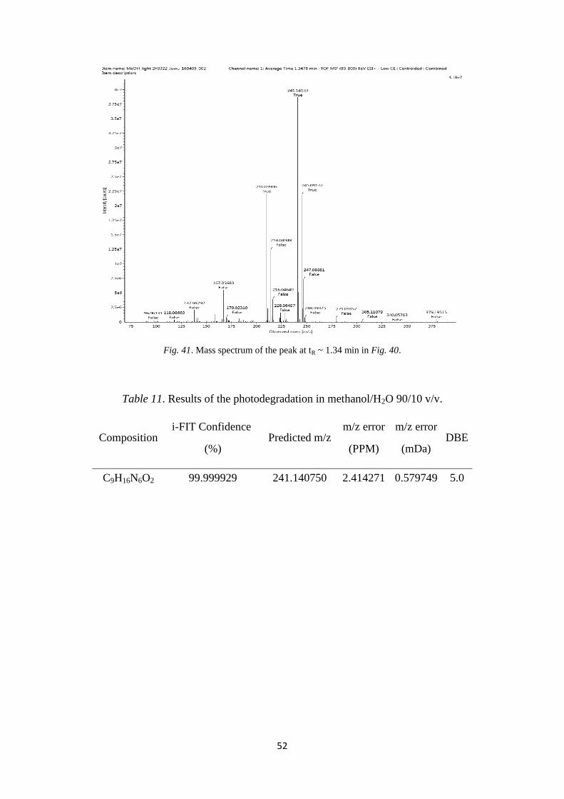
52
Fig. 41. Mass spectrum of the peak at tR ~ 1.34 min in Fig. 40.
Table 11. Results of the photodegradation in methanol/H2O 90/10 v/v.
Composition i-FIT Confidence
(%) Predicted m/z
m/z error
(PPM)
m/z error
(mDa) DBE
C9H16N6O2 99.999929 241.140750 2.414271 0.579749 5.0
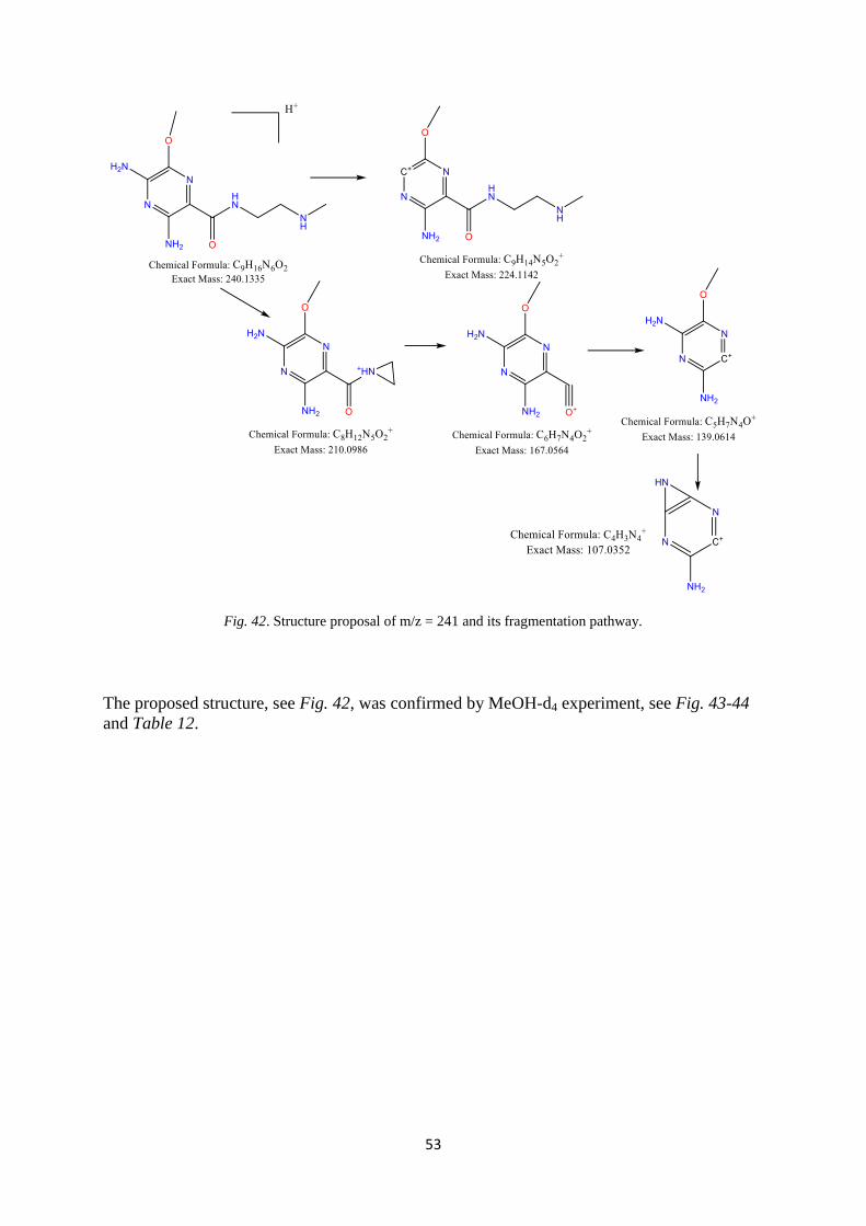
53
Fig. 42. Structure proposal of m/z = 241 and its fragmentation pathway.
The proposed structure, see Fig. 42, was confirmed by MeOH-d4 experiment, see Fig. 43-44
and Table 12.

54
Fig. 43. Results of photodegradation in MeOH-D4. LC-MS/MS analysis of the m/z = 244 peak.
Table 12. Results of photodegradation in methanol-D4/H2O 90/10 v/v.
Composition Predicted m/z m/z error
(PPM)
m/z error
(mDa) DBE
C9H13[2H]3N6O2 244.159580 1.026148 0.249511 5.0
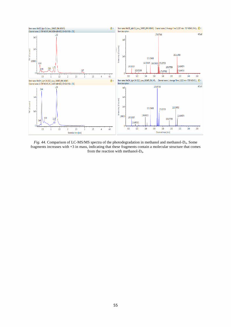
55
Fig. 44. Comparison of LC-MS/MS spectra of the photodegradation in methanol and methanol-D4. Some
fragments increases with +3 in mass, indicating that these fragments contain a molecular structure that comes
from the reaction with methanol-D4.

56
c. Structural elucidation of the secondary photodegradants
Three secondary photodegradants of interest were formed from degradation of the primary
photodegradant formed in methanol, m/z = 241. These compounds had m/z = 259, m/z = 258
and m/z = 256.
The compound with m/z = 259
By analysis of samples photodegraded in water and 18
O-water, see Fig. 45-48, and by
elemental compositions, see Table 13-14, a structure for the photodegradant with m/z = 259
could be proposed, see Fig. 49. The elemental composition of this compound indicated that
the primary photodegradant had reacted both with water and with O2 from the air.
Fig. 45. a) The TIC and c) UV spectra of the sample where the preparatively purified primary photodegradant
from the methanol photodegradation were further photodegraded in water. b) Extracted ion chromatogram of m/z
= 259.
Extracted ion chromatography of m/z = 259
a)
b)
c)
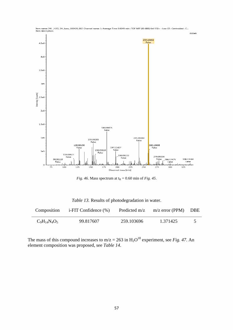
57
Fig. 46. Mass spectrum at tR = 0.60 min of Fig. 45.
Table 13. Results of photodegradation in water.
Composition i-FIT Confidence (%) Predicted m/z m/z error (PPM) DBE
C9H14N4O5 99.817607 259.103696 1.371425 5
The mass of this compound increases to m/z = 263 in H2O18
experiment, see Fig. 47. An
element composition was proposed, see Table 14.

58
Fig. 47. Mass spectrum of the m/z = 263 peak from the 18
O-sample sample.
Table 14. Result from the photodegradation in H2O18
.
Composition i-FIT Confidence
(%)
Predicted
m/z
m/z error
(PPM)
m/z error
(mDa) DBE
C9H14N4O3[18
O]2 93.406533 263.112188 0.924838 0.242405 5.0

59
MS/MS spectra for the m/z = 259 compound formed in regular water and m/z = 263 formed
in H2O18
was run, see Fig. 48. No fragmentation pathway was proposed at this stage.
Fig. 48. LC-MS/MS spectra of a) the m/z = 259 peak, and b) the m/z = 263 peak formed in H2O18
.
a)
b)

60
By comparing this element composition, C9H14N4O3[18
O]2, with the element composition of
the primary photodegradant, C9H16N6O2, a loss of two nitrogen and gain of three oxygen was
observed in this secondary degradant, of which two oxygen atoms were from a reaction with
water. The remaining oxygen must be from the air. It was assumed that an N-oxide was
formed. A fundamental structure was proposed, see Fig. 49.
Fig. 49. Proposed structure for the secondary photodegradant with m/z = 259.
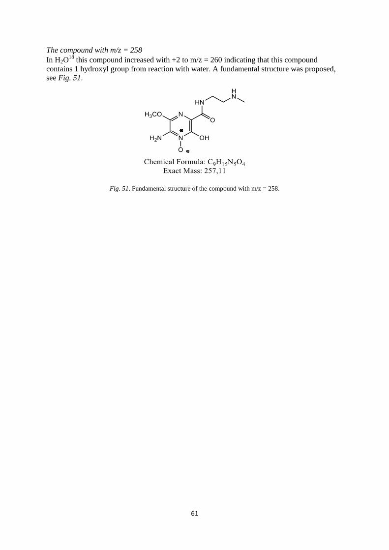
61
The compound with m/z = 258
In H2O18
this compound increased with +2 to m/z = 260 indicating that this compound
contains 1 hydroxyl group from reaction with water. A fundamental structure was proposed,
see Fig. 51.
Fig. 51. Fundamental structure of the compound with m/z = 258.

62
The compound with m/z = 256
In H2O18
this compound increased with +2 to m/z = 258 indicating that this compound
contains 1 hydroxyl group from reaction with water. A fundamental structure was proposed,
see Fig. 52.
Fig. 52. Fundamental structure of the compound with m/z = 256.