physics of ultrasound - mkon 1 - ultrasou… · 2/14/18 1 physics of ultrasound and why you should...
TRANSCRIPT

2/14/18
1
Physics of Ultrasound And why you should know about it… Misha Bhat, MD AEPC basic echo course 2018
Outline � Understanding basic concepts of sound � How ultrasound is generated, captured
and interpreted � Possibilities of ultrasound och technologies � Limitations
� Understanding how to optimize your images and image what you want
� Hopefully you wont fall asleep by the end of the talk
Physics of Sound � Sound is made by longitudinal waves of
compression and rarefaction
V=1540 m/s in soft tissue
compression
rarefaction

2/14/18
2
Definition � Ultra = “beyond” “above” � Sonus = “noise”
� Sound with frequencies higher than what is audible to human ear (20 kHz)
� Medical US is 2-15 MHz
https://sofiasounds.weebly.com/ultrasonic-sound-and-infrasonic-sound.html
bbc.co.uk http://zaprilepite.biodiversity.bg/
How does it work?
� Piezoelectric crystals � Change shape with
current
� Application of alternating current
http://people.bath.ac.uk/rjm64/Site/present%20applications.html www.makeagif.com

2/14/18
3
How does it work? � Piezoelectric crystals
� Alternating current � Cycles of deformation
and rarefaction – acoustic wave
� Acoustic wave is transmitted from probe and into the tissue @1540 m/s
http://people.bath.ac.uk/rjm64/Site/present%20applications.html www.makeagif.com
Reflection � Results in sound returning to the transducer
� At interfaces between tissues esp if different density; myocardium and blood pool
� Diaphragm and mediastinal surface are specular (mirror-like) reflectors
� Almost all is reflected off bone – no penetration beyond
� Most reflection if perpendicular. If parallel to the surface, less reflection, which may cause false “droupout”.

2/14/18
4
False drop-out in atrial septum
Scatter � As sound wave hits smaller
particles (RBC, tissues), the waves scatter.
� A smaller portion will bounce back towards the transducer (back scatter) and is used for generating image.
2/14/18
5
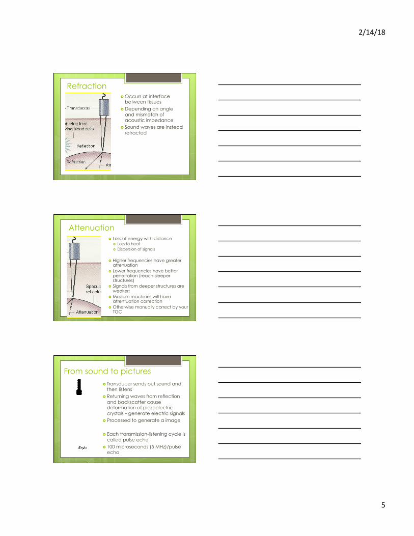
Refraction � Occurs at interface
between tissues � Depending on angle
and mismatch of acoustic impedance
� Sound waves are instead refracted
Attenuation � Loss of energy with distance
� Loss to heat � Dispersion of signals
� Higher frequencies have greater attenuation
� Lower frequencies have better penetration (reach deeper structures)
� Signals from deeper structures are weaker:
� Modern machines will have attentuation correction
� Otherwise manually correct by your TGC
From sound to pictures � Transducer sends out sound and
then listens � Returning waves from reflection
and backscatter cause deformation of piezoelectric crystals – generate electric signals
� Processed to generate a image
� Each transmission-listening cycle is called pulse echo
� 100 microseconds (5 MHz)/pulse echo
2/14/18
6
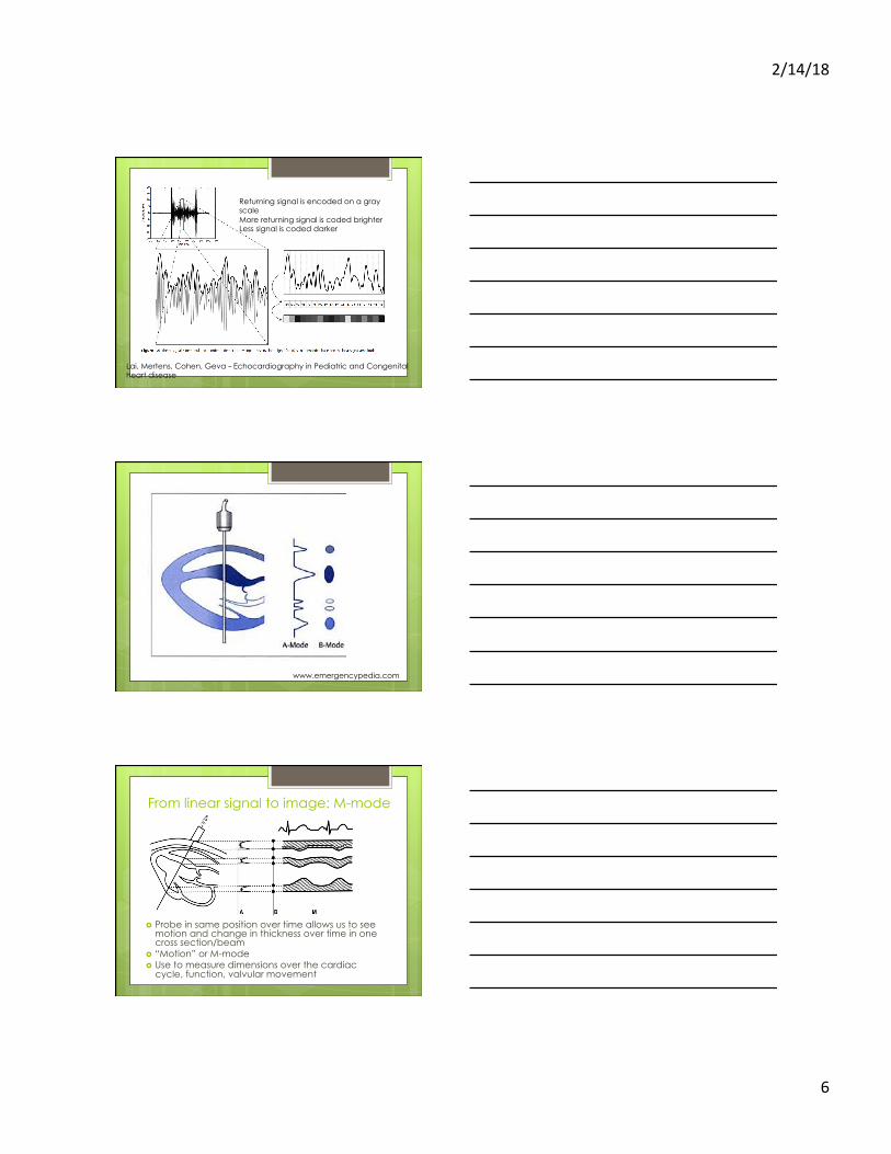
Returning signal is encoded on a gray scale More returning signal is coded brighter Less signal is coded darker
Lai, Mertens, Cohen, Geva – Echocardiography in Pediatric and Congenital heart disease
www.emergencypedia.com
From linear signal to image: M-mode
� Probe in same position over time allows us to see motion and change in thickness over time in one cross section/beam
� “Motion” or M-mode � Use to measure dimensions over the cardiac
cycle, function, valvular movement

2/14/18
7
2D image/B-mode
Phased Array Transducers � Sequentially
activating the crystal allows form form beams in different directions
� Allows wide area of imaging with smaller probe size
� Arranged as a fan with scan lines
� If arranged as a matrix – 3D imaging
Ok but what about image quality

2/14/18
8
� Resolution is the ability to see two near objects as separate
� Direct relationship between higher frequency and greater resolution
� However higher frequency has poor penetration

2/14/18
9
A quick note on trade-off
DEPTH VS RESOLUTION
Resolution types in ultrasound
� Lateral � Axial � Elevational
TEMPORAL
SPATIAL
Lateral resolution
Better with � Higher frequency � In focal zone � Narrower beam width � Decreased depth � Larger probe
(limitations of size)

2/14/18
10
Axial Resolution
� Directly related to higher frequency
� Attenuation may limit depth
Elevational resolution
� Depends on beam thickness
� Inferior to lateral resolution due to multiple reasons which cannot be modified
� Less of a problem in matrix array transducers
Temporal resolution � How many times a second
the entire image refreshes (frame rate - Hz)
� Number of scan lines/beams + distance traveled
� Movement is smoother, allows you to follow movement and structures
� Regular TV = 25Hz
� Frame increases with: � Decreased depth � Narrowing sector width � Lower line density � Single focal point � Parallel beam forming � M-mode>2D>3D

2/14/18
11
Summary � Ultrasound can help image heart and
other structures � Only part of the ultrasound is reflected
back, rest is lost � Resolution is key feature and linked to
frequency used � Higher frequency gives better lateral and
axial resolution but at tradeoff of depth � Temporal resolution important to study
movement of the heart