prenatal diagnosis of a heterozygote for mucopolysaccharidosis type vii (β-glucuronidase...
TRANSCRIPT

PRENATAL DIAGNOSIS, VOL. 2,251-256 (1982)
PRENATAL DIAGNOSIS OF A HETEROZYGOTE FOR MUCOPOLYSACCHARIDOSIS TYPE VII (0-glucuronidase
deficiency)
L. POENARU*, L. CASTELNAU~, J. MOSSMAN?, J . B O U B ~ AND J. c. DREYFUS"
*Institat de Pathologie Mol&ulaire, Chu Cochin, Inserm U129 24 rue du Fg Saint Jacques, 75674 Paris Cedex 14, France
tlnstitut of Child Health, University of London, 30 Guildford Street, London WClN IEH, England
SGroiipe de Recherches. de Biologie Prdnatale, INSERM U 73, Chriteau de Longchamp, 75016 Paris, France
SUMMARY
We had the opportunity of investigating a case (BK) of a severe form of mucopolysacchari- dosis with nearly total deficiency of 8-glucuronidase in serum, leucocytes and fibroblasts.
We here report results obtained by prenatal diagnosis of a clinically normal child (BK's sister), and point out the difficulty in interpreting a heterozygous level of 8-glucuronidase activity in cultured amniotic cells.
Four successive passages of amniotic cells were tested for 8-glucuronidase and cr-manno- sidase activity in at-risk and control cells. In different passages, enzyme activity was between 8 and 49 per cent of controls but 2 to 18 times higher than fibroblasts from the affected brother (BK).
The highest activity was observed in the first passage and the lowest in the third. The electrophoretic separation of GAGS from at-risk amniotic fluid showed a normal
pattern. We discuss the correlation between enzyme levels in different passages of cultured cells and that found in leucocytes and fibroblasts from the propositus and parents.
From a practical point of view, we conclude that the first passage gives the most reliable results for prenatal diagnosis.
KEY WORDS Prenatal diagnosis 8-Glucuronidase Cultured amniotic cells Mucopolysaccharidosis VII Amniotic fluid GAGS
INTRODUCTION
Mucopolysaccharidosis type VII, clinically characterized by short stature, mental retardation, hepatosplenomegaly, progressive skeletal deformities and excessive urinary excretion of mucopolysaccharides (Sly el al., 1973) is associated with a deficiency of ,&glucuronidase (EC.3. 2.1.31) in serum, leucocytes (GIaser and Sly, 1973) and fibroblasts (Hall et al., 1973).
To our knowledge, only eight cases, with very different phenotypic expression, have been reported (Quinton et al., 1971 ; Sly et al., 1973; Gehler, et al., 1974; Danes and Segnan, 1974; Beaudet et al., 1975; Pfeiffer el al., 1977; Guibaud et al., 1979). A prenatal diagnosis was also mentioned by Guibaud et al. in 1979.
We had the opportunity of investigating a case (BK) of a severe form of mucopoly- saccharidosis with nearly total deficiency of fLglucuronidase in serum, leucocytes and fibroblasts. Prenatal detection was proposed to the parents and the next pregnancy was monitored.
019?-3851/82/04O25 1-06 $01 .OO 0 1982 by John Wiley & Sons, Ltd.
Received 30 December 1981 Revised 3 March 1982
Accepted 8 March 1982

252 L. POENARU ET AL.
We here report results obtained by prenatal diagnosis of a clinically normal child (BK's sister), and point out the difficulty of interpreting a heterozygous level of ,B-glucuronidase activity in cultured amniotic cells.
MATERIALS AND METHODS
Serum, leucocytes and skin fibroblasts from BK and his parents were used for enzyme determination. Cultured amniotic cells were used for prenatal diagnosis ; leucocytes and serum for postnatal confirmation. Leucocytes were prepared by sedimentation followed by hemolysis in 0-17 M NH4Cl.
Skin fibroblasts were grown in RPMI medium with 20 per cent fetal calf serum. Fetal cells were cultivated from amniotic fluid taken by amniocentesis during the seventeenth week of pregnancy. Primary cultures were trypsinized at day 14-17 and then subcultured (BouC et al., 1976). The first four passages were tested. The cell density was standardized. Cells were maintained 3 days after confluence in medium with 2 per cent calf serum and then used.
All cellular extractions were carried out in 0.1 per cent Triton-X-100. Control amniotic cells were cultivated in identical conditions: 50 p1 of 1 mmol solution of 4-methylumbelliferyl-/3-~-glucuronide-trihydrate (Koch-Light) in citrate-phosphate buffer pH 5 was used as substrate for /3-glucuronidase determination. a-Mannosidase and N-acetyl /3-glucosaminidase were tested as standard enzymes using respectively 4-methylumbelliferyl-a-~-mannopyranoside and 4-methylumbelliferyl-2-acetamide-2- deoxy-/3-D-glucopyranoside as substrate (Dreyfus and Poenaru, 1975).
Glycosaminoglycans (GAGS) were isolated from amniotic fluid according to Whiteman and Henderson (1977).
RESULTS
Enzyme activity in leucocytes and serum
Table 1 shows that /3-glucuronidase activity was very low in patient BK's leucocytes and serum (less than 0-2 per cent of control mean value). The parents, obligatorily heterozygotes, were in the very low heterozygous range (10 per cent of normal in leucocytes and 20 per cent in serum).
Table 1. Enzyme activity (nmol/h/mg protein) in leucocytes and serum
/3-Glucuronidase a-Mannosidase
Leucocytes Serum Leucocytes Serum BK patient 0.1 0.2 716 27.2 Mrs B 68.8 3.8 242 29.1 Mr B 55.3 4.2 244 38.5 Controls (20) 511.2 28.4 863 38.3
(1 12-1 277) (10 5-1 14) (1 23-2072) ( 16-75)
Enzyme activity in skin jibroblasts
Table 2 confirms /3-glucuronidase results obtained for leucocytes and serum: noticeable deficiency for patient BK (1-3 per cent of control mean); heterozygous

MPS TYPE VII 253
Table 2. Enzyme activity (nmol/h/mg protein) in fibroblasts
p-Glucuronidase a-Mannosidase B-Glucosaminidase
BK 0.5-3.2 32.5 5164 Mrs B 25.2 320 3740 Mr B 24.5 286 5144 Controls (20) 52.8 252.2 4203.7
(23.5-132) (1 37-698) (1 525-6406)
levels for the parents. A low level of a-mannosidase activity was observed in BK fibroblasts but in the context of very large control variation it is probably not significant.
Enzyme activity in cultured amniotic cells
Four successive passages of amniotic cells were tested for j3-glucuronidase and a-mannosidase activity in at-risk and control cells (Table 3). The activity for all passages of at-risk amniotic cells was lower than for controls. In different passages, activity was between 8 and 49 per cent of controls but 2 to 18 times higher than fibroblasts from the affected brother (BK).
The highest activity was observed in the first passage and the lowest in the third. The ratio of j3-glucuronidase to a-mannosidase activity was also higher in the
first passage.
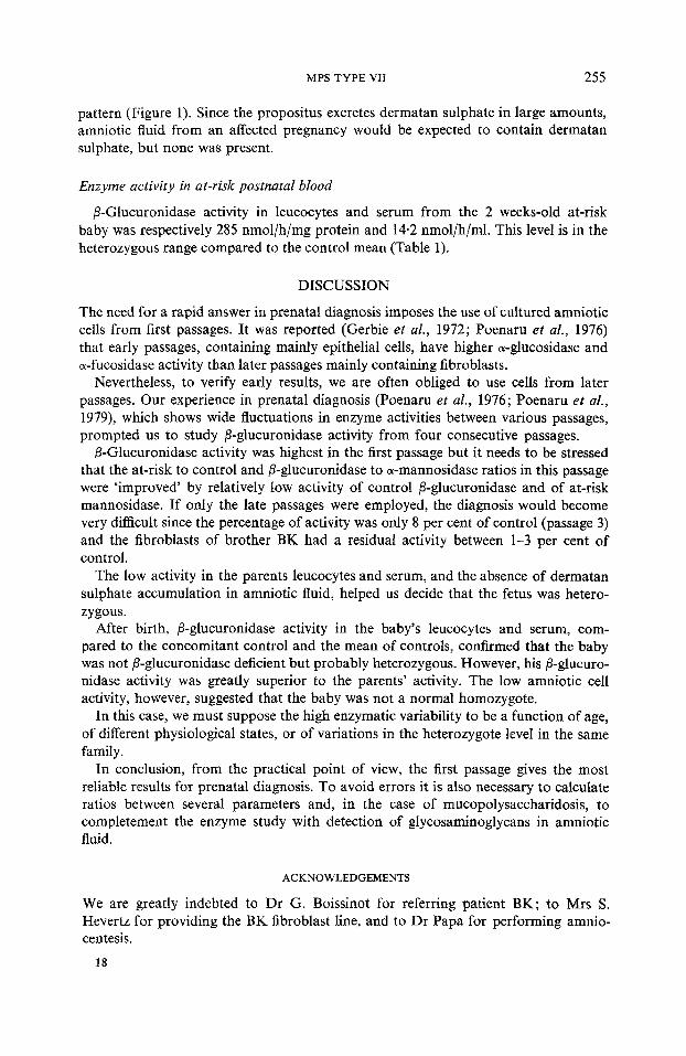
Isolation of glycosaminoglycans (GAGS) from amniotic fluid
The electrophoretic separation of GAGS from at-risk amniotic fluid showed chondroitin sulphate and hyaluronic acid only, which is a normal amniotic fluid
I =First run in pyridine: acetic acid buffer CS = Chondroiten sulphate 11 =Second run in barium acetate buffer DS =Dermatan sulphate A =At-risk amniotic fluid HS =Heparan sulphatc B =GAGS standards KS = Keratan sulphate C =Control amniotic fluid HA =Hyaluronic acid
HEP = A heparin-like component
Figure 1. Electrophoretic separation of amniotic fluid glycosaminoglycans (GAGS)

Tabl
e 3.
Enz
yme
activ
ity (n
mol
/h/m
g pr
otei
n) a
nd r
atio
s in
am
niot
ic fl
uid
cells
Pass
age
P-G
lucu
roni
dase
fi-
Glu
curo
nida
se %
of
a-M
anno
sida
se
fi-G
lucu
rcni
dase
a-M
anno
sida
se
No.
Fe
tus
(B)
Con
trol
the
sam
e da
y co
ntro
l Fe
tus
(B)
Con
trol
r
Fetu
s (B
) C
ontro
l v
i2 I
8.9
18.2
49
76
.2
139.
5 0.
11
0.13
* 3 z ;a
C
b
I1
6.1
45-2
13
10
7.6
137.
8 0.
05
0.3
I11
3.5
40.8
8
55
31.3
0.
06
1.3
IV
6.0
35.4
16
15
0 81
*6
0.
04
0.4
?
Def
icie
nt
0.5-
3.2
1-3
32.5
0.
015
fibro
blas
ts
BK
MPS TYPE VII 255
pattern (Figure 1). Since the propositus excretes dermatan sulphate in large amounts, amniotic fluid from an affected pregnancy would be expected to contain dermatan sulphate, but none was present.
Enzyme activity in at-risk postnatal blood
/3-Glucuronidase activity in leucocytes and serum from the 2 weeks-old at-risk baby was respectively 285 nmol/h/mg protein and 14.2 nmol/h/ml. This level is in the heterozygous range compared to the control mean (Table 1).
DISCUSSION
The need for a rapid answer in prenatal diagnosis imposes the use of cultured amniotic cells from first passages. It was reported (Gerbie et al., 1972; Poenaru e l al., 1976) that early passages, containing mainly epithelial cells, have higher a-glucosidase and a-fucosidase activity than later passages mainly containing fibroblasts.
Nevertheless, to verify early results, we are often obliged to use cells from later passages. Our experience in prenatal diagnosis (Poenaru et al., 1976; Poenaru et al., 1979), which shows wide fluctuations in enzyme activities between various passages, prompted us to study /?-glucuronidase activity from four consecutive passages.
B-Glucuronidase activity was highest in the first passage but it needs to be stressed that the at-risk to control and /3-glucuronidase to a-mannosidase ratios in this passage were ‘improved’ by relatively low activity of control /3-glucuronidase and of at-risk mannosidase. If only the late passages were employed, the diagnosis would become very difficult since the percentage of activity was only 8 per cent of control (passage 3) and the fibroblasts of brother BK had a residual activity between 1-3 per cent of control.
The low activity in the parents leucocytes and serum, and the absence of dermatan sulphate accumulation in amniotic fluid, helped us decide that the fetus was hetero- zygous.
After birth, p-glucuronidase activity in the baby’s leucocytes and serum, com- pared to the concomitant control and the mean of controls, confirmed that the baby was not /3-glucuronidase deficient but probably heterozygous. However, his ,tI-glucuro- nidase activity was greatly superior to the parents’ activity. The low amniotic cell activity, however, suggested that the baby was not a normal homozygote.
In this case, we must suppose the high enzymatic variability to be a function of age, of different physiological states, or of variations in the heterozygote level in the same family.
In conclusion, from the practical point of view, the first passage gives the most reliable results for prenatal diagnosis. To avoid errors it is also necessary to calculate ratios between several parameters and, in the case of mucopolysaccharidosis, to completement the enzyme study with detection of glycosaminoglycans in amniotic fluid.
ACKNOWLEDGEMENTS
We are greatly indebted to Dr G. Boissinot for referring patient BK; to Mrs S. Hevertz for providing the BK fibroblast line, and to Dr Papa for performing amnio- centesis.
18

256 L. POENARU ET AL
REFERENCES
Beaudet, A.L., Di Ferante, N.M., Ferry, C.D., Nichols, B.L. (1975). Variation in the pheno- typic expression of ,!?-glucuronidase deficiency, J. Pediat., 86, 388-394.
Boue, A., Nicolesco, H., Ravise, H., Boue, J. (1976). La culture des cellules du liquide amniotique. ExpCrience de 750 cultures. In: Boue, A. (Ed.). Prenatal Diagnosis, Paris :
Danes, S.B., Segnan, M. (1974). Different clinical and biochemical phenotypes associated with ,8-glucuronidase deficiency, Birth Defects: Orig. Avil. Ser. X, 12, 251-257.
Dreyfus, J.C., Poenaru, J. (1975). Le diagnostic enzymatique dans les maladies lysosorniales, Arch. Frac. Pkdiat., 32, 503-513.
Gerbie, A.B., Melance, S.B., Ryon, C., Nadler, H.L. (1972). Cultivated epithelial-like cells and fibrobIasts from amniotic fluid; their relationship to enzymatic and cytoIogic analysis, Amer. J. Obstet. Gynec., 114, 314-320.
Gehler, J., Cantz, M., Tolcsdorf, M., Spranger, J., Gilbert, E., Drube, H. (1974). Muco- polysaccharidosis VII, 8-glucuronidase deficiency, Humangenetik, 23, 149-158.
Glaser, J.H., Sly, W.S., (1973). ,B-glucuronidase deficiency rnucopolysaccharidosis methods for enzymatic diagnosis, J . Lab. Clin. Med., 82, 969-977.
Guibaud, P., Maire, I., Gopdon, R., Teyssier, G., Zabot, M.T., Mandon, G. (1979). Muco- polysaccharidosis type VII par deficit en ,8-glucuronidase. Etude d’une famille, J. Genet. Hum., 2 7 , 2 9 4 2 .
Hall, C.W., Cantz, M ., Neufeld, E.F. (1 973). A ,!?-glucuronidase deficiency mucopolysac- charidosis : Studies in culture fibroblasts, Arch. Biochem. Biophys., 155, 32-38.
Pfeiffer, R.A., Kresse, H., Baumer, N., Sattinger, E. (1977). 8-glucuronidase deficiency in a girl with unusual clinical features, Europ. J . Pediat., 126, 155-161.
Poenaru, L., Dreyfus, J.C., Boue, J., Nicolesco, H., Ravise, N., Bamberger, J. (1976). Prenatal diagnosis of fucosidosis, Clin. Genet. 10, 260-264.
Poenaru, L., Girard, S. , Thepot, F., Madelenat, P., Huraux-Rendu, C., Vinet, M.C., Dreyfus, J.C. (1979). Antenatal diagnosis in three pregnancies at risk for mannosidosis, Clin. Genet., 16,428432.
Quinton, B.A., Sly, W.S., McAlister, W.H., Rimoin, D.L., Hall, C.W., Neufeld, E.F. (1971). ,!?-glucuronidase deficiency: a new mucopolysaccharide storage disease, Sc. Pediat. Res. Atlantic City p. 198.
Sly, W.S., Quinton, B.A., McAlister, W.H., Rimoin, D.L. (1973). ,!?-glucuronidase deficiency: Report of clinical, radiologic and biochemical features of a new mucosaccharidosis, J. Pediat., 82, 249-257.
Whiteman, P., Henderson, H. (1977). A method for the determination of amniotic fluid glycosaminoglycan and its application to prenatal diagnosis of Hurler and Sanfilippo diseases, Clin. Chim. Acta., 79, 99-105.
INSERM, 69-79.