preparation of porous scaffolds with controlled drug ... · preparation of porous scaffolds with...
TRANSCRIPT
Preparation of Porous Scaffolds withControlled Drug Release for Tissue Engineering
著者 Himansu Sekhar Nandayear 2014その他のタイトル 組織工学のための薬物徐放機能を有する多孔質足場
材料の作製学位授与大学 筑波大学 (University of Tsukuba)学位授与年度 2014報告番号 12102甲第7098号URL http://hdl.handle.net/2241/00126613

i
Preparation of Porous Scaffolds with Controlled Drug Release for Tissue Engineering
Himansu Sekhar Nanda
Doctoral Program in Materials Science and Engineering
Submitted to the Graduate School of
Pure and Applied Sciences
in Partial Fulfillment of the Requirements
for the Degree of Doctor of Philosophy in
Engineering
at the
University of Tsukuba


i


i
Content
List of abbreviations ............................................................................................................................................... iv
Chapter 1 General introduction ............................................................................................................................. 1
1.1. Tissue engineering............................................................................................................................................. 1
1.1.1. Scaffold ................................................................................................................................................... 2
1.1.1.1. Biomaterials for scaffold preparation ........................................................................................... 3
1.1.1.2. Scaffold fabrication techniques .................................................................................................... 7
1.1.2. Cells ....................................................................................................................................................... 10 1.1.3. Bioactive factors for tissue engineering ................................................................................................ 10
1.2. Drug delivery in tissue engineering ................................................................................................................ 11
1.3. Motivation and objectives .............................................................................................................................. 12
1.4. References ........................................................................................................................................................ 13
Chapter 2 Preparation of collagen scaffolds with controlled insulin release for cartilage tissue regeneration
................................................................................................................................................................................. 24
2.1. Executive summary ........................................................................................................................................ 24
2.2. Introduction..................................................................................................................................................... 24
2.3. Materials and methods ................................................................................................................................... 25
2.3.1. Materials ................................................................................................................................................ 25 2.3.2. Methods ................................................................................................................................................. 26
2.3.2.1. Insulin microencapsulation ........................................................................................................ 26
2.3.2.2. Preparation of collagen-microbead hybrid porous scaffold ....................................................... 26
2.3.2.3. Scanning electron microscopy (SEM) ....................................................................................... 26
2.3.2.4. Microbead size analysis ............................................................................................................. 27
2.3.2.5. Insulin loading efficiency (LE) .................................................................................................. 27
2.3.2.6. Mechanical strength: Compression test ..................................................................................... 27
2.3.2.7. In vitro insulin release and microbead degradation .................................................................... 27
2.3.2.8. In vitro chondrocyte culture ....................................................................................................... 28
2.3.2.9. Statistical analysis ...................................................................................................................... 29
2.4. Results and discussion .................................................................................................................................... 29

ii
2.4.1. Morphology, size, size distribution and LE of insulin loaded PLGA microbeads ................................. 29 2.4.2. Porous scaffold microstructure and mechanical strength ...................................................................... 29 2.4.3. In vitro insulin release and degradation ................................................................................................. 30 2.4.4. Cell adhesion, viability and proliferation .............................................................................................. 32
2.5. Conclusion ....................................................................................................................................................... 35
2.6. References ........................................................................................................................................................ 35
Chapter 3 Preparation of collagen porous scaffold with sustained release of insulin for skin tissue
regeneration ........................................................................................................................................................... 38
3.1. Executive summary ........................................................................................................................................ 38
3.2. Introduction..................................................................................................................................................... 38
3.3. Materials and methods ................................................................................................................................... 39
3.3.1. Materials ................................................................................................................................................ 39 3.3.2. Methods ................................................................................................................................................. 40
3.3.2.1. Preparation of insulin loaded PLGA microbeads ....................................................................... 40
3.3.2.2. Preparation of collagen microbead porous hybrid scaffold ........................................................ 40
3.3.2.3. Scanning Electron Microscopy (SEM) ...................................................................................... 41
3.3.2.4. Microbead size analysis ............................................................................................................. 41
3.3.2.5. Insulin loading efficiency (LE) .................................................................................................. 41
3.3.2.6. In vitro insulin release ................................................................................................................ 42
3.3.2.7. In vitro cell culture and bioactivity evaluation ........................................................................... 42
3.3.2.8. Statistical analysis ...................................................................................................................... 43
3.4. Results .............................................................................................................................................................. 43
3.4.1. Preparation and characterization of microbeads .................................................................................... 43 3.4.2. Preparation and characterization of collagen-microbead hybrid scaffolds ............................................ 44 3.4.3. In vitro insulin release and microbead degradation ............................................................................... 45 3.4.4. Bioactivity of hybrid scaffolds .............................................................................................................. 47
3.5. Discussion ........................................................................................................................................................ 50
3.6. Conclusion ....................................................................................................................................................... 51
3.7. References ........................................................................................................................................................ 51
Chapter 4 Preparation of dexamethasone loaded collagen microbead functionalized PLLA-collagen hybrid
scaffold for osteogenic differerntiation of mesenchymal stem cells ................................................................... 55
4.1 Executive summary ......................................................................................................................................... 55
4.3. Materials and methods ................................................................................................................................... 56
iii
4.3.1. Preparation of ice collagen particulates ................................................................................................. 56 4.3.2. Preparation of porous scaffold ............................................................................................................... 56 4.3.3. SEM observation ................................................................................................................................... 57 4.3.4. In vitro Dex release ............................................................................................................................... 57 4.3.5. In vitro cell culture ................................................................................................................................ 58
4.3.5.1. Cell adhesion, viability and proliferation ................................................................................... 58
4.3.5.2 Osteogenic differentiation ........................................................................................................... 58
4.3.6. Statistical analysis ................................................................................................................................. 59
4.4 Results ............................................................................................................................................................... 60
4.4.1 Porous scaffold characterization ............................................................................................................. 60 4.4.2 In vitro Dex release and degradation ...................................................................................................... 60 4.4.3 In vitro cell culture ................................................................................................................................. 62
4.4.3.1 Cell adhesion, viability and proliferation .................................................................................... 62
4.4.3.2. Osteogenic differentiation: RT-PCR, ALP and Alizarin red S staining ...................................... 65
4.6. Conclusion ....................................................................................................................................................... 68
4.7. References ........................................................................................................................................................ 68
Chapter 5 Concluding remarks and future prospects ........................................................................................ 70
List of publications ................................................................................................................................................ 72
Acknowledgements ................................................................................................................................................ 73

iv
List of abbreviations ALP Alkaline phosphatase ANOVA Analysis of variance BMSC Bone marrow derived mesenchymal stem cells BAC Bovine articular chondrocytes BSA Bovine serum albumin cDNA Complementary DNA COL 1 Collagen type 1 CAD Computer aided design Dex Dexamethasone DMEM Dulbecco's modified Eagle medium DNA Deoxyribonucleic acid ECM Extracellular matrix EDC 1-Ethyl-3-[3-dimethylaminopropyl] carbodiimide EOG Ethylene oxide gas ESC Embryonic stem cells FDA Food and drug administration FBS Fetal bovine serum GAPDH Glyceraldehyde-3-phosphate dehydrogenase GAG Glycosaminoglycan HA Hyaluronic acid IBSP Integrin binding sialoprotein IGF-1 Insulin-like growth factor-1 mRNA Messenger ribonucleic acid NHS N-hydroxysuccinimide esters NHDF Neonatal human dermal fibroblast PLLA Poly(L-Lactide) PBS Phosphate buffer saline PFA Perfluoroalkoxy PLGA Poly(DL-lactic-co-glycolic acid) RGD Argine-glycine-aspartic acid RNA Ribonucleic acid RT-PCR Reverse transcripton polymerase chain reaction RUNX2 Runt-related transcription factor 2 SD Standard deviation SEM Scanning electron microscope SPP1 VEGF
Secreted phosphoprotein 1, new name of Osteopontin (OPN) Vascular endothelial growth factor
3D Three dimensional
General introduction
1
Chapter 1
General introduction
1.1. Tissue engineering A wide range of tissue defects resulting from traumatic injury, oncological resection, congenital
deformities and progressive degenerative diseases has elicited the development of tissue engineering as a promising and alternative therapeutic approach for repair and regeneration of damaged tissues and organs [1, 2]. Tissue engineering serves as an alternative therapeutic strategy to replace the conventional treatment methods such as tissue or organ transplantation (autologous and allogenic), artificial prosthesis and drug administration. The ultimate goal of the technology is to find an appropriate solution to the problems related to current medical treatment such as 1.the difficulty in regeneration of extensive damaged tissues or organs, 2.the problems of donor shortage, 3.rejection of artificial biomedical devices and 4.the problems of invasive and traumatizing surgery [1, 3]
Cells Bioactive Factors
Scaffolds
Engineered skin
Engineered bone
Engineered Cartilage(Source: the Institute for Prospective Technological Studies and Joint Research Centre of the European Commission)
(Source: Tissue Regeneration Materials Unit, National Institute for Materials Science, Japan)
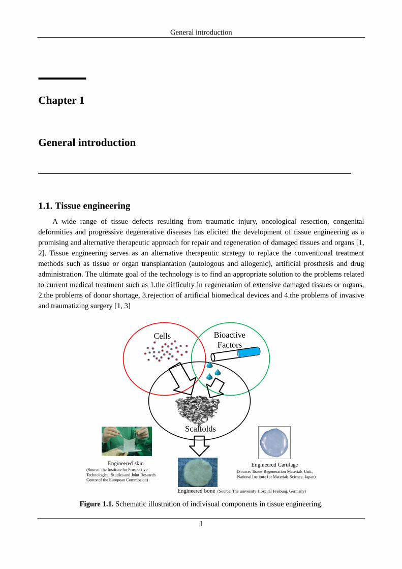
(Source: The university Hospital Freiburg, Germany) Figure 1.1. Schematic illustration of indivisual components in tissue engineering.

Chapter 1
2
The principle of tissue engineering involves the expansion of appropriate living cells over a three dimensional (3D) porous material (scaffold) in presence of bioactive factors such as growth, differentiation migration factors and therapeutics for regeneration of a functional tissue or organ of the patient [4, 5]. The cell/scaffold constructs thus generetaed, can either directly be transplanted to a damaged tissue site of the patient or can be used in in-vitro to regenerate a functional bioengineered substitute (tissues/organs). Bioengineered organ substitutes are extremely useful to meet the donor shortage problems in organ transplantation therapy. The principal components of tissue engineering are schematically illustrated in Figure 1.1. The individual components include 1.scaffolds, 2.cells and 3.bioactive factors. Successful tissue regeneration requires the complex and successful interplay among these three vital components [6-8].
1.1.1. Scaffold Scaffolds are 3D platform that mimic the microenvironments of a natural tissue or organ of a human
body. Scaffolds play a pivotal role in maintaining an appropriate niche for the cells to perform their own function as well as guide the new tissue regeneration [1, 8, 9]. Scaffolds serve as a temporary platform which functions as artificial extracellular matrices (ECM) of the tissues and eventually degrades with the progress of new tissue formation [8]. Therefore, it is necessary to mimic sufficient ECM characteristics in a scaffold in order to provide an appropriate biologic environment to the cells. Preparation of an ideal scaffold should meet several design criterions. Though, the final design of a scaffold depends on tissue type however the basic requirements remain unaltered irrespective of the type of tissue regeneration. The basic characteristics of an ideal 3D scaffold are listed as follows [1, 6, 8-23]. 1. The scaffolds should be biodegradable. Biodegradation refers to the degradation of the materials over the time of tissue regeneration. Degradation can occur either in the way of hydrolytic or enzymatic or both. The degradation of the scaffolds should produce nontoxic products or molecules which can easily be directed towards the usual metabolic pathways of the body such as glycolysis, Tri carboxylic acid (TCA) cycle and so on. This process is important for successful elimination of the degradation products. This can minimize the undesirable effect supposed to be elicited by these degradation products from scaffolds. 2. The scaffolds should be biocompatible such that it should not elicit any serious immunological response against the living system after implantation. Evaluation of biocompatibility of the material prior to design the scaffolds is prerequisite. The material should not possess any chemical or biological contaminants, which can easily elicit the immunological reaction that may cause the rejection of the implanted scaffolds. For an ideal scaffold preparation, the degradation property of the materials should be precisely be tuned such that the rate of degradation of the scaffold should be nearly same as the rate of tissue regeneration. This is necessary to regenerate the tissues fitted to exact damaged dimension. 3. Cell adhesion is a primary step for expansion of the cells over the scaffolds. Therefore the scaffolds should support cell adhesion. 4. The scaffolds should have open porous architecture for good cell seeding in order to achieve high cell seeding efficiencies. 5. In order to facilitate the effective cell migration as well as cell-cell communication, the scaffolds should have high porosity, optimal pore size and sufficient pore interconnections. Scaffolds with high porosity and good pore interconnectivity can generate the cell/scaffold construct with even cell distribution. This can further facilitate the formation of a homogeneous tissue. 6. The scaffold should possess an impressive mechanical strength in order to have high structural integration and stability. The physical parameters such as compressive Young’s modulus and tensile strength of the scaffolds should be high enough to avoid easy collapse of the scaffolds during cell culture and implantation

General introduction
3
process. The mechanical strength should be of good enough to withstand sufficient compression from the cell mediated contractile forces. 7. Bioactive factors are essential to provide suitable biochemical cues for growth, differentiation and migration of the cells. Because of low in vivo stability and unpredictable biological effect, external supply of these bioactive factors in cell culture medium may not be appropriate for subtle growth and differentiation of the cells inside scaffolds. Therefore, it is desirable to include these important cues inside the scaffolds during the fabrication procedure either via direct mixing with scaffolding materials or by incorporating via controlled drug delivery devices. This can facilitate a spatio-temporal release of these cues from the scaffolds. This method of integration can generate the scaffolds with high bioactivity. The design and preparation of bioactive scaffolds is the key driving force for development of novel biomaterial scaffolds for tissue engineering and regenerative medicine.
1.1.1.1. Biomaterials for scaffold preparation The selection of suitable biomaterials plays an important role in preparation of ideal scaffolds. The
materials should not contain any biological or chemical contaminant which may induce inflammation after the implantation of scaffolds. The bioresorbable and cell adhesive property of materials are most important characteristics for successful elimination of materials and to achieve a good cell-material interaction. Biomaterials of natural and synthetic origin have been extensively used for the preparation of scaffolds [3, 4, 6, 8]. Accoding to National Institute of Health (NIH), the materials for scaffold preparation can be broadly categorized into two different types such as natural materials (materials of biologic origin) and synthetic materials (materials of man made origin). Scaffolds can be prepared from either of these materials or from the combination of both the materials (often categorized under hybrid biomaterials) [8, 24, 25]. These two key categories of materials can be further divided into three subtypes such as biomaterials derived from polymer (polymeric biomaterials), from ceramics (Ceramic biomaterials or bioceramics) and from metals (metallic biomaterials).
A. Polymeric biomaterials Polymers are extensively studied for preparation of scaffolds due to their controlled degradation, ease of
preparation and their high bioactive properties. Polymeric materials can be available in abundant quantities form biologic origin (natural polymers) and synthesized (synthetic polymers) in large scale in chemical industries through established synthetic chemical reactions. The property of the polymers (surface chemistry, degradation and so on) can be precisely tuned based on their application (type of tissue to be regenerated). The polymers for the scaffold preparation can be categorized into two basic types such as natural and synthetic polymers [3, 4].
a. Natural polymers Natural polymers are derived from biological origin. These polymers have higher degree of bioactivity
than any other categories of materials. The high bioactivity is due to presence of inherent cell recognition molecules (often called signalling molecules) in the materials. Cell recognition molecules in the materials can facilitate cell adhesion and promotion of cell proliferation. The common disadvantage of these groups of materials includes the susceptibility towards biological contamination (contamination by animal viruses), weak mechanical strength and poor control over biodegradation. Biological contamination may elicit immunological reactions during implantation which may lead to inflammation and rejection of implanted materials. Natural polymers include protein based polymers (collagen, gelatin, fibrin and so on.) and

Chapter 1
4
polysaccharide based polymers (chitosan, hyaluronic acid (HA), glycosaminoglycan (GAG), alginate and so on.). Protein based polymers are more attractive than polysaccharide based polymers because of their high cell adhesive property [3, 4, 6, 8, 26, 27].
I. Protein based polymers
i. Collagen Collagen is a fibrous animal protein and constitutes major structural component of ECM of human body.
It constitutes 30% of all proteins in human body. Structurally, these are triple helical fibrils having role in maintaining structural and mechanical integrity of the connective tissues. The high bioactivity of the collagen plays an important role in cell adhesion, wound healing, platelet activation and angiogenesis. The high bioactivity is due to presence of best known cell recognition signalling sequence RGD peptides. Several types of collagen can be found from ECM of connective tissues in human body. Amongst, type I, II, III and IV are most common known so far. Type I collagen is commonly used for the preparation of biomimetic scaffolds due to their impressive mechanical property and higher bioactive property than any other types of collagen [28-30]. The purity of collagen plays an important role in protecting the materials from contamination. Collagen is best known for its low antigenicity. Collagen molecule is composed of a G-X-Y amino acid sequence which differs among the different animal species. The slight amount of antigenicity of collagen is thought to be due to the presence of terminal telopeptides which do not contain the G-X-Y sequence. Commercially collagen type 1 is available in different purity grades. Atelocollagen is the purest form of collagen available so far [31-34]. Atelocollagen is synthesized by protease digestion of terminal telopeptides sequence of collagen triple helices. Absence of terminal telopeptides in atelocollagen lowers the antigenic character of natural collagen.
ii. Gelatin Gelatin is obtained from collagen by partial hydrolysis and denaturation process. Two basic processes
are involved in preparation of gelatin from collagen [32]. 1. Heat treatment around 40 °C in presence of water which lead to destruction of both hydrogen bond and the electrostatic interactions. 2. Presence of water facilitates the hydrolytic degradation of covalent linkages either in acidic or alkaline conditions. The acidic degradation leads to formation of type A and alkaline degradation lead to formation of type B gelatin. In acidic process, collagen is treated with dilute acids and extracted at pH=4, where noncollagenous tissue proteins are eliminated because of their insolubility or partial solubility. In contrast, the alkaline treatment leads to dissolution of many contaminants (which are not usually soluble in acidic conditions) and can be selectively removed during the extraction process. Type B gelatin is considered as purest form of gelatin.
Gelatin has been widely used for the preparation of scaffolds for tissue engineering and drug delivery devices for controlled drug delivery applications [35-40]. However, the weak mechanical property of gelatin remains a biggest an obstacle behind its successful clinical applications [41]. Preparation of hybrid scaffolds from gelatin and synthetic degradable polymers were focused for tissue various engineering applications [24, 42-45].
iii. Fibrin Fibrin is a protein product of two important blood clotting factors such as fibrinogen and thrombin. It is
solid fibrous mesh structure and plays a role in binding the blood cells and other tissues. Fibrin has been used for preparation of scaffolds for variety of tissue regeneration studies [46-50]. Due to a rapid degradation (few
General introduction
5
weeks), it is more applicable for a short term tissue regeneration but unsuitable for long term regeneration. Fibrin gels can promote cell migration, proliferation and differentiation. Therefore, fibrin can be considered as a good candidate for scaffold preparation. Present research interest lies in preparation of durable scaffolds using a combination of fibrin gels and synthetic biomaterials. The durable scaffolds may have a great importance in long term tissue regeneration and therapy [51].
II. Polysaccharide based polymers Polysaccharide based polymers constitute the sugar unit as monomers. Cellulose, chitosan, hyaluronic
acid, glycosaminoglycan and alginate are the major polysaccharides studied for their application in preparation of scaffolds for various tissue engineering. However chitosan, hyaluronic acid and alginate are among the most popular category of polysaccharides used in the field of tissue engineering [52-57]. Chitosan is also extensively used in therapeutic delivery due to their cationic nature [58-61].
i. Chitosan Chitosan is composed of β-1, 4-linked N-acetyl-D-glucosamine (20%) and β-1, 4-linked D-glucosamine
(80%). It is prepared by the partial deacetylation of chitin in hot alkali. It has net positive charge due to its unique polycationic characteristics and thus a cationic natural polymer. It has been studied for tissue engineering because of its biocompatibility, biodegradability, antibacterial property and presence of necessary reactive groups for functionalization. It can be degraded by the enzymes such as chitosanase and lysozyme [62]. It is capable of forming scaffolds by ionic or chemical cross linking and its ability to promote cell attachment can be enhanced by combining it with other proteins [63-65]. The polycationic characteristic of the chitosan has attracted its application in drug delivery. The cationic characteristic of the chitosan encourages easy and specific functionalization with the anionic drug conjugates and makes it as an excellent material for therapeutic delivery applications [58-61, 66, 67].
ii. Alginate Alginate is a biopolymer mostly found in walls of brown algae. It is structurally similar to cellulose and
commonly well known for its high structural stability. The structural stability is due to the β-glycosidic linkages which resists the chemical break down. The structural stability of alginate makes it an excellent candidate for the scaffold preparation. However, alginate has lower biocompatibility with a mammalian host, which reduces its ability to promote cell attachment, proliferation and differentiation [68]. Nonetheless, the easy and fast gelation feature of alginate still attracts much attention for tissue engineering applications [69, 70]. Alginate is also known for promoting wound healing therefore finds its application in preparation of bandages for wrapping burn wounds. Alginate beads were also studied for preparation of drug delivery devices [71-73].
iii. Hyaluronic acid (HA) Hyaluronic acid (HA) is a hydrophilic and natural glycosaminoglycan found mostly in human
connective tissues includes skin, cartilage, intra-articular joint fluid and vitreous humor of eye. HA plays an important role in cartilage growth and burn tissue repair, thus makes it an attractive candidate for improving the rate of tissue regeneration. It also demonstrates a favourable biodegradation profile. HA based scaffolds have been widely used in the tissue engineering of skin, cartilage tissue, bone and soft tissue filler [74-76]. It has been reported that HA can regulate cell motility and mediate cell differentiation. Various chemically modified HA derivatives have been developed to improve mechanical strength and cell attachment to the

Chapter 1
6
porous scaffolds [77-79].
b. Synthetic polymers Members of linear aliphatic polyesters such as poly(glycolic acid) (PGA), Poly(lactic acid) (PLA) and
poly(lactic acid-co-glycolic acid) (PLGA) have been approved by food and drug administration (FDA) for use as engineering materials for preparation of scaffolds and clinical applications [80-82]. These synthetic polymers have demonstrated well proven mechanical strength and a tuneable degradation rate based on their monomer unit and molecular weight. Therefore, the scaffolds prepared from these materials are well suited for hard tissue regeneration such as regeneration of bone and cartilage [83-88]. The common disadvantage of these materials includes lack of cell recognition signals which causes inadequate cell adhesion and low cell seeding efficiency in prepared scaffolds [89-91]. To overcome such problems, preparation of hybrid biomaterial scaffolds by combination of natural and synthetic polymers has been reported [24, 25, 92, 93].
i. Poly(glycolic acid) (PGA) PGA is hydrophilic biodegradable polyester widely used for the preparation of biodegradable suture
implants and engineering scaffolds. Due to high hydrophilicity, the degradation of these materials are too fast which lead to weak mechanical strength among other synthetic polyesters. Preparation of nonwoven fibrous fabrics from PGA is one of the widely used scaffolds for tissue regeneration [94-97].
ii. Poly(lactic acid) (PLA) PLA is a hydrophobic biodegradable polyester. Its degradation is too slow due to its high hydrophobicity
offered by additional methyl groups present in the polymer. The disadvantage of this material is longer degradation time sometimes months to years. So it is often copolymerized with PGA to control its degradation. PLA is widely used in preparation of porous scaffolds for bone tissue engineering [24, 25, 98-104]. In order to increase the hydrophilicity and wettability of PLA, it is often hybridized with natural polymers for the preparation of hybrid scaffolds for tissue engineering [101, 103, 104]. Surface modification of PLA is also an alternative way to enhance the hydrophilic properties of the materials for tissue engineering application.
iii. Poly(lactic-co-glycolic acid) (PLGA) PLGA is one of the widely accepted polymers for tissue engineering and drug delivery applications. It is
a copolymer of PGA and PLA [105]. The degradation rate of the PLGA can be precisely tuned based on the application. It is often simple by changing the ratio of PGA to PLA during the copolymerization reaction. Industrially it is available in several copolymer compositions such as PLGA 50:50, PLGA 75:25 and PLGA 85:15 [105]. The degradation of PLGA is about a few months. Due to controlled degradation and flexibility to mould into different shapes, it has been widely used as scaffold preparation in tissue engineering [88, 92, 93, 102, 106]. It has also been widely applied for preparation of controlled release formulation for several proteins and drugs [107-113].
B. Ceramic biomaterials (Bio-ceramics) Ceramic biomaterials are widely used for their application in hard tissue regeneration such as
regeneration of a bone tissue [115,116]. The property of hardness and wear resistance makes these materials unique for hard tissue engineering. These are mostly inorganic minerals such as naturally occurring calcium phosphates which include calcium hydroxyapatite and tricalcium phosphate [116,117-119]. Both these

General introduction
7
materials are well proven for bone tissue engineering for their osteoinductive (ability to induce new bone formation by bone cell growth) and osteoconductive (ability to promote bone cell adhesion and differentiation) properties [120-122]. These inorganic minerals may either be used alone or in combination with polymeric materials forming hybrid materials [123,124]. The disadvantages of these materials include brittleness and difficult to process into highly porous structures [120].
i. Calcium hydroxyapatite It is naturally occurring calcium phosphate with molecular formula Ca10 (PO4)6OH2 found in bone and
teeth of human body [125]. It is commonly used as bone filler as well as coating of the bone implants to promote bone in-growth. Commercially available calcium hydroxyapatite usually comes from two sources such as direct chemical synthesis and heating the coral skeletons [126-128].
ii. Tricalcium phosphate (TCP) TCP otherwise called calcium orthophosphate (Ca3 (PO4)2) is commonly referred as bone ash [130]. It
is a combustion product of bone. It is often used in combination with biodegradable polyesters for preparation of scaffolding materials [131-134]. TCP acts as an antaacid and helps in balancing the unfavourable acidic environment caused due to degradation of biodegradable polyesters. In nature, it is found as mixed with sand stone and phosphorous oxides. It can also be synthesized chemically in the laboratory by simple chemical reaction of calcium with phosphoric acid.
C. Metallic biomaterials Metallic biomaterials are particularly useful in fabrication of the load bearing implants [135-137]. High
corrosion resistant property, low density and high mechanical strength have attracted metallic biomaterials for use in load bearing applications. Titanium (Ti) is an attractive candidate for metallic biomaterials applications [136]. Ti based implants have been widely used in preparation of dental implants and implants for joint replacement [138,139]. It has good osseointegration property. Other such candidates for metallic biomaterials applications include inox steel, C0-Cr alloys and Ti6Al4V etc.
1.1.1.2. Scaffold fabrication techniques Three dimensional scaffolds are designed to accomplish the function of cell adhesion, migration and
proliferation to guide new tissue regeneration. In order to generate a scaffold for optimal cell function, the fulfilment of several design criterions are necessary as discussed in section 1.1. Various methods of scaffold fabrication have been reported in literatures and each of the technique has their own advantages as well as disadvantages. Some of the commonly used techniques for scaffold preparation are described as below.
i. Solvent casting and particulate leaching It is a simplest method of preparation of porous polymeric scaffold [140-142]. The primary step
involves the preparation of a polymer solution by addition of polymer in a suitable organic solvent .The polymer solution is further mixed with required quantity of porogen materials (sugar, salt, paraffin and so on.). The mixture is casted on a teflon template of required dimension. The highly volatile solvent is allowed to evaporate under vacuum or at room temperature leaving the polymer dispersed with porogen materials. The porogen is gradually leached from the polymer by washing the template with water or hexane (based on the type of porogen used in preparation process) leaving behind the porous scaffold [141]. The scaffold porosity can be easily controlled by controlling the amount of porogen in polymer matrix as well as polymer

Chapter 1
8
concentration. The potential advantage of this method is that the method is quite simple and the pore structure can be controlled based on the diameter of porogen materials as well as concentration of polymer materials. The disadvantages of this method include 1. Complete removal of organic solvent from the porous polymer scaffold is practically impossible and may cause the toxicity to the cultured cells. 2. The incorporation of bioactive factors such as growth factors and therapeutic agents during the fabrication can cause the easy loss of bioactivity due to involvement of organic solvents in the preparation process. 3. The leaching step of the scaffold preparation increases the duration of the preparation of final scaffold and therefore a time consuming process.
ii. Freeze drying method Freeze drying is one of the attractive method for preparation of porous scaffolds from natural and
synthetic polymers [8, 24, 25, 28-30, 87, 89, 92, 93]. The preparation process involves the formation of ice crystals inside the polymer matrix during the freezing and subsequent lypholization process [141, 143]. The porous architecture of the scaffold can be modulated by changing the conditions of freeze drying such as freezing temperature, freezing time and so on. The change in temperature affects the pattern of ice crystal formation as well as crystal distribution inside the polymer matrices which modulate the pore architecture inside the scaffold. The potential advantages of this method are 1.Simple and reproducible 2.The scaffolds prepared by this method are highly porous and having sufficient pore interconnections 3. Pore structure can be precisely controlled. Incorporation of porogen leaching such as ice particulates on freeze drying process often results in formation of highly porous and well ordered pores in prepared scaffold. The disadvantages include the longer processing period.
iii. Phase separation Phase separation method has been widely adopted for the preparation of porous scaffolds with micro
and nano architecture [144-146]. The principle can be applied to preparation of porous scaffolds from both natural as well as synthetic polymer. Further more, the preparation of nanofibrous 3D scaffold and bioactive scaffold (scaffolds containing bioactive factors) through phase separation process is noteworthy [147]. Phase separation in a homogeneous multicomponent polymeric system can be induced either by thermally or by a nonsolvent. However thermal mode is widely adopted for preparation of scaffolds for tissue engineering. This is due to the induction of phase separation by a nonsolvent often results in preparation of scaffolds of non-uniform pore architecture which is not suitable for tissue engineering. Thermal induction of the polymer solution results in separation of polymer solution into two phases such as polymer rich phase and polymer lean phase (solvent). Solvent undergoes crystallization at low temperature. Subsequent sublimation of the crystals results in pore formation in the polymer matrix formed by polymer rich phase. The whole process can generate the 3D scaffolds with porous structure [141]. The temperature and the concentration of polymer can control the pore structure in the scaffold. The process is often referred as solid liquid phase separation.
iv. Gas foaming Gas foaming technique is used to eliminate the use of organic solvent as well as high temperature
treatment during the preparation of porous scaffolds [148-150]. The residual organic solvent present in the scaffolds after fabrication process can have deteriorous impact to cells. The high temperature treatment often cause the inactivation of bioactive molecules incorporated in the scaffold during fabrication process. Gas foaming process adds the advantage over such undesirable effects. In this process, CO2 gas is usually used to saturate a polymer (for example PLGA) solution. Gradual reduction in the pressure of the system creates

General introduction
9
instability of the whole system and results in clustering of the gas molecules to minimize the free energy of the total system. Eventually, the pores are generated which lead to decrease in polymer density. A 3D porous structure is generated after the foaming process [141]. Incorporation of particle leaching on gas foaming process often results in formation of open pores in prepared scaffolds.
v. Electrospinning Electrospinning is a simple and versatile technique to produce polymeric fibers ranging from nano to
microscale [151, 152]. The process involves the injection of positive or negative charge to the polymer droplet generated by a syringe pump in a high potential difference [153]. The charge injection to the polymer droplet generates electrostatic repulsive force. When the repulsive force is sufficient to overcome the surface tension force of the polymer droplet, the droplet becomes a continuous fiber and is attracted to the collector of opposite electrode at other end. The dimension of the fibers varies from micro to nanoscale depending on the processing parameters. A number of parameters decide the optimal production of fibers of specific diameter which include viscosity, molecular weight, concentration of polymer, applied voltage, flow rate of the polymer solution, distance between the capillary and collector and ambient parameters such as temperature and humidity. Electrospun nanofibers from wide variety of the natural and synthetic polymers have been studied for the tissue engineering and therapeutic encapsulation for controlled drug release applications [154]. The key advantages of the method are 1. Process is simple and versatile 2.Cost effective for production of nanofibers.
vi. Fiber bonding Nonbonded fiber meshes do not have a proper mechanical integrity for in vivo tissue regeneration. To
overcome this problem, fiber bonding method has been developed to bind the fibers together at points of intersection. In brief, PGA fibers are immersed in a PLLA solution. When the solvent evaporates, the network of PGA fibers is embedded in PLLA. The composite is then heated to above the melting temperature of both polymers. The PLLA melts first and fills all voids left by the fibers. This helps retain the spatial arrangement of fibers so that when the PGA begins to melt, the fiber structure does not collapse. Instead, in order to minimize interfacial energy, fibers at the cross-points become "welded" (melted) together, forming highly porous foam. The PLLA is then removed by dissolution with methylene chloride. This method improves the mechanical properties of fabric scaffolds. The fiber bonding scaffold fabrication technique is desirable for its simplicity, the retention of the fibers original properties, the use of only biocompatible materials and structural advantages. The disadvantages of fiber bonding are the shortage of control over porosity and pore size, the availability of suitable solvents, immiscibility of the two polymers in the melt state and the required relative melting temperature of the polymers.
vii. Rapid prototyping Rapid prototyping is an advanced technique for porous scaffold preparation [155]. In this method
scaffolds with desired property can be generated in an efficient way. The method uses computer program i.e. computer aided design (CAD) to first generate the structure of the defect in the form of a 3D model [155, 156]. This model of the defect can be sliced into layers by the computer. Corresponding to each cross section rapid prototype machine can lay down the layer of material starting from the bottom and moving up a layer at a time to create the scaffolds. This technique has several advantages such as it has ability to control matrix architecture, mechanical property, degradation kinetics and biological effect of the scaffold.

Chapter 1
10
1.1.2. Cells The cells for tissue engineering must be identified, isolated as well as amplified by transplantation over
the porous scaffold. Various cell sources have been utilized to isolate the cells for tissue engineering. Some of the major cell sources include autologous (patient’s own cell), allogenic (from a donor of the same species) and xenogenic (from the donor of an entirely different species) [157, 158]. Amongst, autologous cell source is considered as most ideal for scaffold based tissue engineering because of its low risk of pathogen transmission and least chance of immune rejection [157, 158]. Some of the autologous cells utilized for the tissue engineering include dermal fibroblasts for skin tissue engineering, chondrocytes for cartilage tissue engineering and so on. However, there are some problems associated in use of these cells for tissue engineering approach. The concern for inadequate amount of cell from aged patients as well as chronic burn patients, unavailability of these cells in patients with genetic disease and chronic pain associated with surgery during extraction of the cells from the patients are the common problems associated with use of these cell types for tissue engineering. To resolve these issues, stem cells are used as an attractive and alternative target cell for various tissue engineering [159, 160]. Stem cells have unlimited capability of self renewal and the ability to maintain its stemness. Furthermore it can differentiate to the cells of specific tissues or organs when subjected to suitable biochemical cues [159, 160]. Stem cells can be embryonic stem cells (ESCs) and adult stem cells. ESCs have the capability to differentiate into any types of cells of the tissues. In other words, it can give rise to any types of tissues of the human body. However, ethical issues on ESCs have limited its use as a major cell sources for tissue engineering. Adult stems cells are the tissue specific stem cells help in maintaining the integrity of the specific tissue. Mesenchymal stem cells (MSCs) have shown to be an attractive cell source for tissue engineering of cartilage, bone and so on [161]. MSCs are the adult pluripotent stem cells derived from bone marrow, fat adipose tissue and umbilical chord. MSCs have the ability to differentiate into cartilage (for cartilage tissue engineering), bone (bone tissue engineering), ligament (ligament tissue engineering), neural cells (neural tissue engineering) and so on.
1.1.3. Bioactive factors for tissue engineering Bioactive factors include cell growth factors, cell adhesion factors, cytokines and drugs [12, 13,
162-164]. These molecules provide molecular cues to the cells which are pre-requisite for neo tissue formation and otherwise called morphogens or tissue inductive factors. Chemically these are primarily proteins. The action of these molecules on the cells is either pleiotrophic or redundant. They act on the cell surface receptors especially membrane receptor proteins. The generated signal produced via these interations is subsequently transferred and amplified through phosphorylation of secondary messengers within the cells. This process causes phenotypic expression of target genes. Bioactive factors for tissue engineering are primarily employed for controlling cell functions such as cell proliferation, migration and gene expression for differentiation [12, 13, 20, 22, 162-164].
Commonly used bioactive factors include growth factors such as basic fibroblast growth factors(b-FGF), platelet derived growth factors (PDGF), transforming growth factor β (TGF-β), insulin like growth factors-1 (IGF-1), vascular endothelial growth factors (VEGF), bone morphogenic proteins (BMP) and so on [162]. The delivery of these molecules in appropriate concentration for a required time frame has become an important tool for directing cell proliferation, differentiation, migration and angiogenesis in neo tissues [162-164]. However, poor in vivo stability and unpredictable biological effect are the major concerns related to the application of these factors in tissue engineering [162]. Protein based bioactive factors have very short half life. For example, the biologic half-lives of PDGF, b-FGF, and VEGF are 2, 3, 50 min, respectively,

General introduction
11
upon intravenous injection [164]. Therefore, the bioactivity of the proteins can be lost very easily. It is also hard to control the concentration of these factors in optimal dose needed for the cells during different time scales of tissue regeneration process. At times, the concentration may be very high enough to kill the cells and makes it undesirable for regeneration process. Sometimes, it may be lower than minimum threshold value needed for the optimal cellular activities. Such an unhealthy condition can lead to cell death due to inability in performing the biochemical activities of the cells. It is also imperative to target these molecules to desired cell populations and keeping safe to non targeted cells and tissues. Therefore, the delivery of these chemical molecules should be carefully considered in order to harness the appropriate biological effect [162-165]. An appropriate delivery strategy should be identified in order to program the delivery of these molecules in bioactive form for a prolonged duration as well as to control over the release concentration. Integration of controlled release function into three dimensional porous scaffolds serves as a valuable tool for controlled and sustained delivery of drugs for a desired time frame [162, 165]. Until now, a wide variety of strategies have been applied for the integration of controlled release function into an engineered scaffold. Some of the key strategies are discussed in subsequent headings.
1.2. Drug delivery in tissue engineering Drugs have an important role in controlling cell function in tissue regeneration process. The most
important consideration should be the search for an appropriate mode of delivery for making these drugs available at the desired tissue sites at right time [164]. The use of appropriate delivery strategies to locally deliver these molecules in a controlled fashion for a required time frame is prerequisite [165]. Selection of a suitable controlled delivery vehicle to maintain its biological activity for relatively longer duration and to release it in required concentration for a desired time frame is pre-requisite. Furthermore, the release profile of the drugs from the delivery device should be precisely controlled in a spatio-temporal fashion. Temporal control over the concentration and spatial localization control can affect the extent of tissue formation as well as pattern of tissue formation. In order to fulfil the described design criterion, a variety of polymeric delivery systems have been designed for drug delivery in tissue engineering [162-165]. Polymeric systems are of key interest because of their easy processiablity and a good control over their degradation to control the release profile of the encapsulated drugs [164].
One of the simplest delivery techniques used in tissue engineering involve direct incorporation of the drugs into polymeric matrices during the scaffold fabrication process [166]. Drug release depends on the physicochemical properties of the polymers that are used to construct the scaffolds. The release is also dependant upon the pore structures in scaffolds. Two fundamental release mechanisms such as diffusion and degradation induce the release of an encapsulated drug. Hydrolytic or enzymatic degradation of polymeric matrices leads to cleavage of chemical bonds in matrix network and causes the detachment of drugs from the internal porous network to diffuse out from matrix.
Biodegradable polymeric drug delivery systems encapsulating the drugs such as drug loaded microspheres or microbeads can be incorporated in hydrogels or prefabricated scaffolds for spatio-temporal delivery [163-166]. In this method, either a single or a combination of drugs can be used for delivery from a single material platform [162]. Polymer matrices that incorporate uniformly distributed drugs are commonly used for drug delivery include biodegradable microbeads made from PLGA and other natural polymers such as gelatin, alginate and collagen. Drug release from biodegradable polymer matrices is not only dependent on the diffusion, but also the degradation rate of the polymers [167]. The degradation process of these materials involves the hydrolysis of polymer back bones into non-toxic monomers. The release rate can be controlled by changing the degradation rate of polymers and can easily be achieved by tailor made properties of the

Chapter 1
12
polymeric materials. Synthetic polymers such as biodegradable polyesters are more frequently used to microencapsulation of the drugs for the application in tissue engineering. Microencapsulation process via biodegradable microbeads made from natural and synthetic polymers is a powerful mean to protect the bioactive molecules from in vivo degradation as well as to obtain a sustained release profile. PLGA has already been demonstrated its great success in microencapsulation of a wide variety of drugs [168]. Drug release from the PLGA microbeads can be easily controlled by changing the copolymer composition as well as molecular weight of PLGA. PLGA microbeads can be prepared by a number of techniques which has been explained in many literatures. Techniques that have been used so far include multiple emulsion technique, spray drying, phase separation, microfluidic preparation, self-assembly of supramolecules and various polymerization techniques [168].
The most popular method for preparing drug incorporated microbeads is the emulsion based method [168]. To microencapsulate hydrophilic drugs such as protein based drugs, one step emulsion water-in-oil (w/o) or double emulsion water-in-oil-in-water (w/o/w) techniques are often used. Double emulsion technique is often considered as the best method to microencapsulate hydrophilic drugs for its ability to maintain higher bioactivity of the encapsulated drug and to yield the microbeads with high drug encapsulation efficiency as well as low initial burst release [168]. High encapsulation efficiency and low initial burst of the microbeads is important to sustain the release of an encapsulated drug for a relatively longer time frame. In this method, an aqueous phase containing desired drug (w1) is dispersed into the oil phase such as PLGA dissolved in an organic solvent (o) using a mechanical device such as vortex mixture, high speed homogenizer or a shear mixture. The generated w1/o emulsion is further re-emulsified in a surfactant containing aqueous solution such as polyvinyl alcohol (w2) to form a double (w1-o-w2) emulsion. The resulting double emulsion is then poured into a surfactant (PVA) containing aqueous continuous phase and stirred for several hours at room temperature to allow adequate solvent evaporation. The hardened microbeads can be recovered after centrifugation and washed with pure water. The microbeads are lyophilized and can be used in controlled delivery applications in tissue engineering.
The third common drug delivery method is accomplished through drug loaded hydrogels [169]. A hydrogel can be fabricated into many geometrical configurations, for example, cylinders, slabs, disks, or spheres (microbeads). Hydrogel are often referred to as macro-gels. Hydrogels made with synthetic (polyethylene glycol, polyvinyl alcohol and so on) and natural polymers (gelatin, collagen and so on) usually have a porous surface and a complex internal network. The swelling property of hydrogels in water allows free movement of drugs throughout cavities in hydrogels. Drugs can be loaded into hydrogels before or after scaffold preparation.
The last and least common used method is pre-fabricated porous polymeric matrices that are soaked with drug solutions [166]. In this method the drug release from the scaffold is governed by the process of diffusion. This method has the major drawback of higher initial burst release and poor sustained release of the drugs from the scaffolding materials.
1.3. Motivation and objectives Cell-cell as well as cell-scaffold interactions are two the fundamental processes plays an important role
in new tissue regeneration. This cross talk is facilitated by a number of signalling molecules called growth factors and drugs. Therefore the ability of scaffolds to release these powerful therapuetics in a controlled and prolonged fashion is pre-requisite for an improved cell-cell as well as cell-scaffold interactions. The objective of the present research is to design a few kinds of bioactive polymeric scaffolds with controlled drug release function for application in various tissue engineering. Technology involving spatial localization

General introduction
13
of drugs via carrier based system such as drug delivery devices made from natural and synthetic polymer is considered as most effective way to control the tissue regeneration process. Drug loaded biodegradable microbeads prepared from synthetic or natural polymers immobilized with porous scaffolds of impressive mechanical strength are promising in the areas of tissue engineering applied for wound healing (skin regeneration), neovascularisation, and pluripotent stem cell differentiation. The use of biodegradable microbeads may protect the biological activity of the drugs to avoid rapid clearance in in vivo environment. This may also facilitate a controlled and sustained release of the drug molecule for prolonged period to meet the local therapeutic demand. Furthermore the system can provide a valuable tool to deliver the drug exactly inside of the scaffold microenvironment for mantinance of a local concentration required for the cells.
In present report, our purpose was to prepare a few kinds of bioactive hybrid scaffolds with controlled pore structure and controlled drug release function. A well control over the pore structure of porous scaffolds is expected to improve the spatial cell distribution for homogeneous tissue formation and the associated controlled release function is expected to facilitate the release of required drug in a spatio-temporal fashion. The released drug at local 3D microenvironment might cause enhanced bioactivation of cells to proliferate and differentiate to neo tissues.
1.4. References 1. Hollister, Scott J. "Porous scaffold design for tissue engineering." Nature materials 4, no. 7 (2005):
518-524. 2. Burg, Karen JL, Scott Porter, and James F. Kellam. "Biomaterial developments for bone tissue
engineering." Biomaterials 21, no. 23 (2000): 2347-2359. 3. Hutmacher, D. W., J. C. Goh, and S. H. Teoh. "An introduction to biodegradable materials for tissue
engineering applications." Annals of the Academy of Medicine, Singapore 30, no. 2 (2001): 183-191. 4. Lanza, Robert, Robert Langer, and Joseph P. Vacanti, eds. Principles of tissue engineering. Academic
press, 2011. 5. Muschler, George F., Chizu Nakamoto, and Linda G. Griffith. "Engineering principles of clinical
cell-based tissue engineering." The Journal of Bone & Joint Surgery 86, no. 7 (2004): 1541-1558. 6. Freed, Lisa E., Gordana Vunjak-Novakovic, Robert J. Biron, Dana B. Eagles, Daniel C. Lesnoy,
Sandra K. Barlow, and Robert Langer. "Biodegradable polymer scaffolds for tissue engineering." Nature Biotechnology 12, no. 7 (1994): 689-693.
7. Chapekar, Mrunal S. "Tissue engineering: challenges and opportunities." Journal of biomedical materials research 53, no. 6 (2000): 617-620.
8. Chen, Guoping, Takashi Ushida, and Tetsuya Tateishi. "Scaffold design for tissue engineering." Macromolecular Bioscience 2, no. 2 (2002): 67-77.
9. Hutmacher, Dietmar W. "Scaffold design and fabrication technologies for engineering tissues—state of the art and future perspectives." Journal of Biomaterials Science, Polymer Edition 12, no. 1 (2001): 107-124.
10. Yang, Shoufeng, Kah-Fai Leong, Zhaohui Du, and Chee-Kai Chua. "The design of scaffolds for use in tissue engineering. Part I. Traditional factors." Tissue engineering 7, no. 6 (2001): 679-689.
11. Griffith, Linda G. "Emerging design principles in biomaterials and scaffolds for tissue engineering." Annals of the New York Academy of Sciences 961, no. 1 (2002): 83-95.
12. Hubbell, Jeffrey A. "Biomaterials in tissue engineering." Nature Biotechnology 13, no. 6 (1995): 565-576.
13. Eisenbarth, E. "Biomaterials for tissue engineering." Advanced Engineering Materials 9, no. 12
Chapter 1
14
(2007): 1051-1060. 14. Elisseeff, Jennifer Hartt, Yoshihiko Yamada, and Robert Langer. "Biomaterials for tissue engineering."
Tissue Engineering and Biodegradable Equivalents. New york: Marcel Dekker (2002): 1-23. 15. Shin, Heungsoo, Seongbong Jo, and Antonios G. Mikos. "Biomimetic materials for tissue
engineering." Biomaterials 24, no. 24 (2003): 4353-4364. 16. Ma, Peter X. "Biomimetic materials for tissue engineering." Advanced drug delivery reviews 60, no. 2
(2008): 184-198. 17. Chua, Chee Kai, Lay Poh Tan, and Jia An. "Advanced nanobiomaterials for tissue engineering and
regenerative medicine." Nanomedicine 8, no. 4 (2013): 501-503. 18. Bishop, Corey J., Jayoung Kim, and Jordan J. Green. "Biomolecule Delivery to Engineer the Cellular
Microenvironment for Regenerative Medicine." Annals of biomedical engineering (2013): 1-16. 19. Spiller, Kara L., and Gordana Vunjak-Novakovic. "Clinical translation of controlled protein delivery
systems for tissue engineering." Drug Delivery and Translational Research (2013): 1-15. 20. Vo, Tiffany N., F. Kurtis Kasper, and Antonios G. Mikos. "Strategies for controlled delivery of growth
factors and cells for bone regeneration." Advanced drug delivery reviews 64, no. 12 (2012): 1292-1309.
21. Rice, Jeffrey J., Mikaël M. Martino, Laura De Laporte, Federico Tortelli, Priscilla S. Briquez, and Jeffrey A. Hubbell. "Engineering the regenerative microenvironment with biomaterials." Advanced healthcare materials 2, no. 1 (2013): 57-71.
22. Koria, Piyush. "Delivery of growth factors for tissue regeneration and wound healing." BioDrugs 26, no. 3 (2012): 163-175.
23. Nair, Lakshmi S., and Cato T. Laurencin. "Polymers as biomaterials for tissue engineering and controlled drug delivery." In Tissue Engineering I, pp. 47-90. Springer Berlin Heidelberg, 2006.
24. Lu, Hongxu, Hwan Hee Oh, Naoki Kawazoe, Kozo Yamagishi, and Guoping Chen. "PLLA–collagen and PLLA–gelatin hybrid scaffolds with funnel-like porous structure for skin tissue engineering." Science and Technology of Advanced Materials 13, no. 6 (2012): 064210.
25. Chen, Guoping, Naoki Kawazoe, and Tetsuya Tateishi. "HYBRID POROUS SCAFFOLDS OF BIODEGRADABLE SYNTHETIC POLYMERS AND COLLAGEN FOR TISSUE ENGINEERING." Handbook of Intelligent Scaffold for Tissue Engineering and Regenerative Medicine (2012): 417.
26. Seliktar, Dror. "Designing cell-compatible hydrogels for biomedical applications." Science 336, no. 6085 (2012): 1124-1128.
27. Sell, Scott A., Patricia S. Wolfe, Koyal Garg, Jennifer M. McCool, Isaac A. Rodriguez, and Gary L. Bowlin. "The use of natural polymers in tissue engineering: a focus on electrospun extracellular matrix analogues." Polymers 2, no. 4 (2010): 522-553.
28. Lu, Hongxu, Young-Gwang Ko, Naoki Kawazoe, and Guoping Chen. "Cartilage tissue engineering using funnel-like collagen sponges prepared with embossing ice particulate templates." Biomaterials 31, no. 22 (2010): 5825-5835.
29. Zhang, Qin, Hongxu Lu, Naoki Kawazoe, and Guoping Chen. "Preparation of collagen scaffolds with controlled pore structures and improved mechanical property for cartilage tissue engineering." Journal of Bioactive and Compatible Polymers 28, no. 5 (2013): 426-438.
30. Zhang, Qin, Hongxu Lu, Naoki Kawazoe, and Guoping Chen. "Preparation of collagen porous scaffolds with a gradient pore size structure using ice particulates." Materials Letters 107 (2013): 280-283.

General introduction
15
31. Yamaoka, H., Y. Tanaka, S. Nishizawa, Y. Asawa, T. Takato, and K. Hoshi. "The application of atelocollagen gel in combination with porous scaffolds for cartilage tissue engineering and its suitable conditions." Journal of Biomedical Materials Research Part A 93, no. 1 (2010): 123-132.
32. Gómez-Guillén, M. C., B. Giménez, M. E. López-Caballero, and M. P. Montero. "Functional and bioactive properties of collagen and gelatin from alternative sources: A review." Food Hydrocolloids 25, no. 8 (2011): 1813-1827.
33. Park, Si-Nae, Sang-Hee Bae, and Yong Su Lee. "Method for separating atelocollagen, method for preparing modified atelocollagen, atelocollagen prepared by using the same and collagen-based matrix." U.S. Patent Application 13/698,017, filed April 21, 2011.
34. O'Brien, Fergal J. "Biomaterials & scaffolds for tissue engineering." Materials Today 14, no. 3 (2011): 88-95.
35. Wu, X., Y. Liu, X. Li, P. Wen, Y. Zhang, Y. Long, X. Wang, Y. Guo, F. Xing, and J. Gao. "Preparation of aligned porous gelatin scaffolds by unidirectional freeze-drying method." Acta biomaterialia 6, no. 3 (2010): 1167-1177.
36. He, Chuanglong, Fan Zhang, Lijun Cao, Wei Feng, Kexin Qiu, Yanzhong Zhang, Hongsheng Wang, Xiumei Mo, and Jinwu Wang. "Rapid mineralization of porous gelatin scaffolds by electrodeposition for bone tissue engineering." Journal of Materials Chemistry 22, no. 5 (2012): 2111-2119.
37. Ovsianikov, Aleksandr, Andrea Deiwick, Sandra Van Vlierberghe, Michael Pflaum, Mathias Wilhelmi, Peter Dubruel, and Boris Chichkov. "Laser fabrication of 3D gelatin scaffolds for the generation of bioartificial tissues." Materials 4, no. 1 (2011): 288-299.
38. Kumari, Avnesh, Sudesh Kumar Yadav, and Subhash C. Yadav. "Biodegradable polymeric nanoparticles based drug delivery systems." Colloids and Surfaces B: Biointerfaces 75, no. 1 (2010): 1-18.
39. Gunji, Shutaro, Kazukata Obama, Makoto Matsui, Yasuhiko Tabata, and Yoshiharu Sakai. "A novel drug delivery system of intraperitoneal chemotherapy for peritoneal carcinomatosis using gelatin microspheres incorporating cisplatin." Surgery 154, no. 5 (2013): 991-999.
40. Huang, Sha, and Xiaobing Fu. "Naturally derived materials-based cell and drug delivery systems in skin regeneration." Journal of Controlled Release 142, no. 2 (2010): 149-159.
41. Grover, Chloe N., Ruth E. Cameron, and Serena M. Best. "Investigating the morphological, mechanical and degradation properties of scaffolds comprising collagen, gelatin and elastin for use in soft tissue engineering." Journal of the mechanical behavior of biomedical materials 10 (2012): 62-74.
42. Irani, Shiva, Marziyeh Hedayati, Seyed Mohammad Atyabi, Seyed Mahdi Saeed, Najmeh Salamian, and Mojgan Zandi. "A comparative study of cell behavior on PLGA, gelatin and PLGA/gelatin scaffolds." Nano Bulletin 2, no. 2 (2013): 130213.
43. Lee, Jongman, James J. Yoo, Anthony Atala, and Sang Jin Lee. "The effect of controlled release of PDGF-BB from heparin-conjugated electrospun PCL/gelatin scaffolds on cellular bioactivity and infiltration." Biomaterials 33, no. 28 (2012): 6709-6720.
44. Meng, Z. X., Y. S. Wang, C. Ma, W. Zheng, L. Li, and Y. F. Zheng. "Electrospinning of PLGA/gelatin randomly-oriented and aligned nanofibers as potential scaffold in tissue engineering." Materials Science and Engineering: C 30, no. 8 (2010): 1204-1210.
45. Van Vlierberghe, Sandra, Peter Dubruel, and Etienne Schacht. "Biopolymer-based hydrogels as scaffolds for tissue engineering applications: a review." Biomacromolecules 12, no. 5 (2011): 1387-1408.
46. Thomson, Kassandra S., F. Steven Korte, Cecilia M. Giachelli, Buddy D. Ratner, Michael Regnier, and
Chapter 1
16
Marta Scatena. "Prevascularized microtemplated fibrin scaffolds for cardiac tissue engineering applications." Tissue Engineering Part A 19, no. 7-8 (2013): 967-977.
47. Johnson, Philip J., Stanley R. Parker, and Shelly E. Sakiyama‐Elbert. "Fibrin‐based tissue engineering scaffolds enhance neural fiber sprouting and delay the accumulation of reactive astrocytes at the lesion in a subacute model of spinal cord injury." Journal of Biomedical Materials Research Part A 92, no. 1 (2010): 152-163.
48. de la Puente, Pilar, Dolores Ludeña, Ana Fernández, Jose L. Aranda, Gonzalo Varela, and Javier Iglesias. "Autologous fibrin scaffolds cultured dermal fibroblasts and enriched with encapsulated bFGF for tissue engineering." Journal of Biomedical Materials Research Part A 99, no. 4 (2011): 648-654.
49. Carletti, Eleonora, Antonella Motta, and Claudio Migliaresi. "Scaffolds for tissue engineering and 3D cell culture." In 3D cell culture, pp. 17-39. Humana Press, 2011.
50. Barsotti, Maria Chiara, Francesca Felice, Alberto Balbarini, and Rossella Di Stefano. "Fibrin as a scaffold for cardiac tissue engineering." Biotechnology and applied biochemistry 58, no. 5 (2011): 301-310.
51. Barsotti, Maria Chiara, Francesca Felice, Alberto Balbarini, and Rossella Di Stefano. "Fibrin as a scaffold for cardiac tissue engineering." Biotechnology and applied biochemistry 58, no. 5 (2011): 301-310.
52. Correia, Clara R., Liliana S. Moreira-Teixeira, Lorenzo Moroni, Rui L. Reis, Clemens A. van Blitterswijk, Marcel Karperien, and João F. Mano. "Chitosan scaffolds containing hyaluronic acid for cartilage tissue engineering." Tissue Engineering Part C: Methods 17, no. 7 (2011): 717-730.
53. Dahlmann, Julia, Andreas Krause, Lena Möller, George Kensah, Markus Möwes, Astrid Diekmann, Ulrich Martin, Andreas Kirschning, Ina Gruh, and Gerald Dräger. "Fully defined< i> in situ</i> cross-linkable alginate and hyaluronic acid hydrogels for myocardial tissue engineering." Biomaterials 34, no. 4 (2013): 940-951.
54. Collins, Maurice N., and Colin Birkinshaw. "Hyaluronic acid based scaffolds for tissue engineering—A review." Carbohydrate polymers 92, no. 2 (2013): 1262-1279.
55. Iwasaki, Norimasa, Yasuhiko Kasahara, Shintarou Yamane, Tatsuya Igarashi, Akio Minami, and Shin-ichiro Nisimura. "Chitosan-based hyaluronic acid hybrid polymer fibers as a scaffold biomaterial for cartilage tissue engineering." Polymers 3, no. 1 (2010): 100-113.
56. Yu, Chia-Cherng, Jung-Jhih Chang, Yen-Hsien Lee, Yu-Cheng Lin, Meng-Hsiu Wu, Ming-Chien Yang, and Chiang-Ting Chien. "Electrospun scaffolds composing of alginate, chitosan, collagen and hydroxyapatite for applying in bone tissue engineering." Materials Letters 93 (2013): 133-136.
57. Ninan, Neethu, Muthunarayanan Muthiah, In-Kyu Park, Anne Elain, Sabu Thomas, and Yves Grohens. "Pectin/carboxymethyl cellulose/microfibrillated cellulose composite scaffolds for tissue engineering." Carbohydrate polymers 98, no. 1 (2013): 877-885.
58. Bernkop-Schnürch, Andreas, and Sarah Dünnhaupt. "Chitosan-based drug delivery systems." European Journal of Pharmaceutics and Biopharmaceutics 81, no. 3 (2012): 463-469.
59. Bhattarai, Narayan, Jonathan Gunn, and Miqin Zhang. "Chitosan-based hydrogels for controlled, localized drug delivery." Advanced drug delivery reviews 62, no. 1 (2010): 83-99.
60. Garcia-Fuentes, Marcos, and Maria J. Alonso. "Chitosan-based drug nanocarriers: where do we stand?." Journal of Controlled Release 161, no. 2 (2012): 496-504.
61. Hu, Liming, Yun Sun, and Yan Wu. "Advances in chitosan-based drug delivery vehicles." Nanoscale 5, no. 8 (2013): 3103-3111.

General introduction
17
62. Laffleur, Flavia, Fabian Hintzen, Deni Rahmat, Gul Shahnaz, Gioconda Millotti, and Andreas Bernkop-Schnürch. "Enzymatic degradation of thiolated chitosan." Drug development and industrial pharmacy 39, no. 10 (2013): 1531-1539.
63. Muzzarelli, Riccardo AA. "Chitosan composites with inorganics, morphogenetic proteins and stem cells, for bone regeneration." Carbohydrate Polymers 83, no. 4 (2011): 1433-1445.
64. Leipzig, Nic D., Ryan G. Wylie, Howard Kim, and Molly S. Shoichet. "Differentiation of neural stem cells in three-dimensional growth factor-immobilized chitosan hydrogel scaffolds." Biomaterials 32, no. 1 (2011): 57-64.
65. Bhardwaj, Nandana, and Subhas C. Kundu. "Silk fibroin protein and chitosan polyelectrolyte complex porous scaffolds for tissue engineering applications." Carbohydrate Polymers 85, no. 2 (2011): 325-333.
66. Sezer, Ali Demir, and Erdal Cevher. "Topical drug delivery using chitosan nano-and microparticles." Expert opinion on drug delivery 9, no. 9 (2012): 1129-1146.
67. de la Fuente, Maria, Manuela Raviña, Patrizia Paolicelli, Alejandro Sanchez, Begoña Seijo, and Maria Jose Alonso. "Chitosan-based nanostructures: a delivery platform for ocular therapeutics." Advanced drug delivery reviews 62, no. 1 (2010): 100-117.
68. Lee, Kuen Yong, and David J. Mooney. "Alginate: properties and biomedical applications." Progress in polymer science 37, no. 1 (2012): 106-126.
69. Jin, Hyeong-Ho, Dong-Hyun Kim, Tae-Wan Kim, Keun-Koo Shin, Jin Sup Jung, Hong-Chae Park, and Seog-Young Yoon. "< i> In vivo</i> evaluation of porous hydroxyapatite/chitosan–alginate composite scaffolds for bone tissue engineering." International Journal of Biological Macromolecules 51, no. 5 (2012): 1079-1085.
70. Rani, V. V., Roshni Ramachandran, K. P. Chennazhi, H. Tamura, S. V. Nair, and R. Jayakumar. "Fabrication of alginate/nanoTiO< sub> 2</sub> needle composite scaffolds for tissue engineering applications." Carbohydrate Polymers 83, no. 2 (2011): 858-864.
71. Gombotz, Wayne R., and Siow Fong Wee. "Protein release from alginate matrices." Advanced drug delivery reviews 64 (2012): 194-205.
72. Feng, Chao, Ruixi Song, Guohui Sun, Ming Kong, Zixian Bao, Yang Li, Xiaojie Cheng, Dongsu Cha, Hyun-Jin Park, and Xiguang Chen. "Immobilization of Coacervate Microcapsules in Multilayer Sodium Alginate Beads for Efficient Oral Anticancer Drug Delivery." Biomacromolecules (2014).
73. Lee, Kuen Yong, and David J. Mooney. "Alginate: properties and biomedical applications." Progress in polymer science 37, no. 1 (2012): 106-126.
74. Collins, Maurice N., and Colin Birkinshaw. "Hyaluronic acid based scaffolds for tissue engineering—A review." Carbohydrate polymers 92, no. 2 (2013): 1262-1279.
75. Kim, Iris L., Robert L. Mauck, and Jason A. Burdick. "Hydrogel design for cartilage tissue engineering: a case study with hyaluronic acid." Biomaterials 32, no. 34 (2011): 8771-8782.
76. Davidenko, Natalia, J. J. Campbell, E. S. Thian, C. J. Watson, and Ruth Elizabeth Cameron. "Collagen–hyaluronic acid scaffolds for adipose tissue engineering." Acta biomaterialia 6, no. 10 (2010): 3957-3968.
77. Prestwich, Glenn D. "Hyaluronic acid-based clinical biomaterials derived for cell and molecule delivery in regenerative medicine." Journal of Controlled Release 155, no. 2 (2011): 193-199.
78. Collins, Maurice N., and Colin Birkinshaw. "Hyaluronic acid based scaffolds for tissue engineering—A review." Carbohydrate polymers 92, no. 2 (2013): 1262-1279.
79. Nimmo, Chelsea M., Shawn C. Owen, and Molly S. Shoichet. "Diels− Alder Click Cross-Linked

Chapter 1
18
Hyaluronic Acid Hydrogels for Tissue Engineering." Biomacromolecules 12, no. 3 (2011): 824-830. 80. Boccaccini, A. R., X. Chatzistavrou, J. J. Blaker, and S. N. Nazhat. "Degradable and bioactive
synthetic composite scaffolds for bone tissue engineering." In Degradation of Implant Materials, pp. 111-137. Springer New York, 2012.
81. Tian, Huayu, Zhaohui Tang, Xiuli Zhuang, Xuesi Chen, and Xiabin Jing. "Biodegradable synthetic polymers: Preparation, functionalization and biomedical application." Progress in Polymer Science 37, no. 2 (2012): 237-280.
82. Liu, Xiaohua, Jeremy M. Holzwarth, and Peter X. Ma. "Functionalized synthetic biodegradable polymer scaffolds for tissue engineering." Macromolecular bioscience 12, no. 7 (2012): 911-919.
83. Gloria, Antonio, Roberto De Santis, and Luigi Ambrosio. "Polymer-based composite scaffolds for tissue engineering." Journal of Applied Biomaterials & Biomechanics 8, no. 2 (2010).
84. Zhang, Hualin, and Zhiqing Chen. "Fabrication and characterization of electrospun PLGA/MWNTs/hydroxyapatite biocomposite scaffolds for bone tissue engineering." Journal of Bioactive and Compatible Polymers 25, no. 3 (2010): 241-259.
85. Bose, Susmita, Mangal Roy, and Amit Bandyopadhyay. "Recent advances in bone tissue engineering scaffolds." Trends in biotechnology 30, no. 10 (2012): 546-554.
86. Chen, Jyh-Ping, and Chien-Hao Su. "Surface modification of electrospun PLLA nanofibers by plasma treatment and cationized gelatin immobilization for cartilage tissue engineering." Acta biomaterialia 7, no. 1 (2011): 234-243.
87. Kawazoe, Naoki, Chieko Inoue, Tetsuya Tateishi, and Guoping Chen. "A cell leakproof PLGA‐collagen hybrid scaffold for cartilage tissue engineering." Biotechnology progress 26, no. 3 (2010): 819-826.
88. Pan, Zhen, and Jiandong Ding. "Poly(lactide-co-glycolide) porous scaffolds for tissue engineering and regenerative medicine." Interface focus 2, no. 3 (2012): 366-377.
89. Sugiyama, Kazuyuki, Aiko Okamura, Naoki Kawazoe, Tetsuya Tateishi, Shigeo Sato, and Guoping Chen. "Coating of collagen on a poly (l-lactic acid) sponge surface for tissue engineering." Materials Science and Engineering: C 32, no. 2 (2012): 290-295.
90. Bakry, Ahmed, Andrea Martinelli, Mariano Bizzarri, Alessandra Cucina, Lucio D'Ilario, Iolanda Francolini, Antonella Piozzi, and Sara Proietti. "A new approach for the preparation of hydrophilic poly (L‐lactide) porous scaffold for tissue engineering by using lamellar single crystals." Polymer International 61, no. 7 (2012): 1177-1185.
91. Oh, Se Heang, and Jin Ho Lee. "Hydrophilization of synthetic biodegradable polymer scaffolds for improved cell/tissue compatibility." Biomedical Materials 8, no. 1 (2013): 014101.
92. Dai, Wenda, Naoki Kawazoe, Xiaoting Lin, Jian Dong, and Guoping Chen. "The influence of structural design of PLGA/collagen hybrid scaffolds in cartilage tissue engineering." Biomaterials 31, no. 8 (2010): 2141-2152.
93. Lu, Hongxu, Young-Gwang Ko, Naoki Kawazoe, and Guoping Chen. "Culture of bovine articular chondrocytes in funnel-like collagen-PLGA hybrid sponges." Biomedical Materials 6, no. 4 (2011): 045011.
94. Cao, Hong, and Noboru Kuboyama. "A biodegradable porous composite scaffold of PGA/β-TCP for bone tissue engineering." Bone 46, no. 2 (2010): 386-395.
95. Rampichová, Michala, E. Kostakova, Eva Filová, Eva Prosecká, Martin Plencner, Larisa Ocheretná, Andriy Lytvynets, D. Lukas, and Evžen Amler. "Non-woven PGA/PVA fibrous mesh as an appropriate scaffold for chondrocyte proliferation." Physiol. Res 59, no. 5 (2010): 773-781.

General introduction
19
96. Zhao, Qinghua, Shouguo Wang, Jiwei Tian, Lei Wang, Shuanghai Dong, Tian Xia, and Zhenkai Wu. "Combination of bone marrow concentrate and PGA scaffolds enhance bone marrow stimulation in rabbit articular cartilage repair." Journal of Materials Science: Materials in Medicine 24, no. 3 (2013): 793-801.
97. Hern, D., P. Vacantii, and R. Langer. "Stabilized polyglycolic acid fibre-based tubes for tissue engineering." The Biomaterials: Silver Jubilee Compendium: Silver Jubilee Compendium 17, no. 2 (2011): 129.
98. Li, Jiashen, Yun Chen, Arthur FT Mak, Rocky S. Tuan, Lin Li, and Yi Li. "A one-step method to fabricate PLLA scaffolds with deposition of bioactive hydroxyapatite and collagen using ice-based microporogens." Acta biomaterialia 6, no. 6 (2010): 2013-2019.
99. Izal, Iñigo, Pablo Aranda, Patricia Sanz-Ramos, Purificación Ripalda, Gonzalo Mora, Froilán Granero-Moltó, Harmony Deplaine et al. "Culture of human bone marrow-derived mesenchymal stem cells on of poly (l-lactic acid) scaffolds: potential application for the tissue engineering of cartilage." Knee Surgery, Sports Traumatology, Arthroscopy 21, no. 8 (2013): 1737-1750.
100. Hu, Jiang, Xuan Sun, Haiyun Ma, Changqing Xie, Y. Eugene Chen, and Peter X. Ma. "Porous nanofibrous PLLA scaffolds for vascular tissue engineering." Biomaterials 31, no. 31 (2010): 7971-7977.
101. Chen, Guoping, Takashi Ushida, and Tetsuya Tateishi. "Scaffold design for tissue engineering." Macromolecular Bioscience 2, no. 2 (2002): 67-77.
102. Chen, Guoping, Takashi Ushida, and Tetsuya Tateishi. "Preparation of poly (L-lactic acid) and poly (DL-lactic-co-glycolic acid) foams by use of ice microparticulates." Biomaterials 22, no. 18 (2001): 2563-2567.
103. Tateishi, Tetsuya, Guoping Chen, and Takashi Ushida. "Biodegradable porous scaffolds for tissue engineering." Journal of Artificial Organs 5, no. 2 (2002): 77-83.
104. Chen, Guoping, Takashi Ushida, and Tetsuya Tateishi. "Development of biodegradable porous scaffolds for tissue engineering." Materials Science and Engineering: C 17, no. 1 (2001): 63-69.
105. ERBETTA, Cynthia D., et al. Synthesis and Characterization of Poly (D, L-Lactide-co-Glycolide) Copolymer. Journal of Biomaterials & Nanobiotechnology, 2012, 3.2.
106. PATRICK JR, C. W., et al. Preadipocyte seeded PLGA scaffolds for adipose tissue engineering. Tissue engineering, 1999, 5.2: 139-151.
107. COHEN, Smadar, et al. Controlled delivery systems for proteins based on poly (lactic/glycolic acid) microspheres. Pharmaceutical research, 1991, 8.6: 713-720.
108. CHEN, Limor; APTE, Ron N.; COHEN, Smadar. Characterization of PLGA microspheres for the controlled delivery of IL-1< i> α</i> for tumor immunotherapy. Journal of controlled release, 1997, 43.2: 261-272.
109. TANG, Yu; SINGH, Jagdish. Controlled delivery of aspirin: Effect of aspirin on polymer degradation and< i> in vitro</i> release from PLGA based phase sensitive systems. International journal of pharmaceutics, 2008, 357.1: 119-125.
110. ZHU, Huijun, et al. Co-delivery of chemotherapeutic drugs with vitamin E TPGS by porous PLGA nanoparticles for enhanced chemotherapy against multi-drug resistance. Biomaterials, 2014, 35.7: 2391-2400.
111. FORMIGA, Fabio R., et al. Controlled delivery of fibroblast growth factor-1 and neuregulin-1 from biodegradable microparticles promotes cardiac repair in a rat myocardial infarction model through activation of endogenous regeneration. Journal of Controlled Release, 2014, 173: 132-139.

Chapter 1
20
112. MANNING, C. N., et al. Controlled delivery of mesenchymal stem cells and growth factors using a nanofiber scaffold for tendon repair. Acta biomaterialia, 2013, 9.6: 6905-6914.
113. MARAN, Avudaiappan, et al. Hydrogel‐PLGA delivery system prolongs 2‐methoxyestradiol‐mediated anti‐tumor effects in osteosarcoma cells. Journal of Biomedical Materials Research Part A, 2013, 101.9: 2491-2499.
114. R Chandra et al. Biodegradable Polymers. Prog. Polym. Sci., 1998 Vol 23: 1273–1335. 115. Petite, Herve, Veronique Viateau, Wassila Bensaid, Alain Meunier, Cindy de Pollak, Marianne
Bourguignon, Karim Oudina, Laurent Sedel, and Genevieve Guillemin. "Tissue-engineered bone regeneration." Nature biotechnology 18, no. 9 (2000): 959-963.
116. LeGeros, Racquel Zapanta. "Properties of osteoconductive biomaterials: calcium phosphates." Clinical orthopaedics and related research 395 (2002): 81-98.
117. Bucholz, Robert W., A. P. R. T. Carlton, and Ralph E. Holmes. "Hydroxyapatite and tricalcium phosphate bone graft substitutes." The Orthopedic clinics of North America 18, no. 2 (1987): 323-334.
118. Shimazaki, Kazuhisa, and Vert Mooney. "Comparative study of porous hydroxyapatite and tricalcium phosphate as bone substitute." Journal of orthopaedic research 3, no. 3 (1985): 301-310.
119. Urist, Marshall R., Olle Nilsson, John Rasmussen, William Hirota, Tim Lovell, TOM SCHMALZREID, and G. A. M. Finerman. "Bone regeneration under the influence of a bone morphogenetic protein (BMP) beta tricalcium phosphate (TCP) composite in skull trephine defects in dogs." Clinical orthopaedics and related research 214 (1987): 295-304.
120. Giannoudis, Peter V., Haralambos Dinopoulos, and Eleftherios Tsiridis. "Bone substitutes: an update." Injury 36, no. 3 (2005): S20-S27.
121. LeGeros, Racquel Zapanta. "Properties of osteoconductive biomaterials: calcium phosphates." Clinical orthopaedics and related research 395 (2002): 81-98.
122. Yuan, Huipin, Hugo Fernandes, Pamela Habibovic, Jan de Boer, Ana MC Barradas, Ad de Ruiter, William R. Walsh, Clemens A. van Blitterswijk, and Joost D. de Bruijn. "Osteoinductive ceramics as a synthetic alternative to autologous bone grafting." Proceedings of the National Academy of Sciences 107, no. 31 (2010): 13614-13619.
123. Rezwan, Kurosh, Q. Z. Chen, J. J. Blaker, and Aldo Roberto Boccaccini. "Biodegradable and bioactive porous polymer/inorganic composite scaffolds for bone tissue engineering." Biomaterials 27, no. 18 (2006): 3413-3431.
124. Sui, Gang, Xiaoping Yang, Fang Mei, Xiaoyang Hu, Guoqiang Chen, Xuliang Deng, and Seungkon Ryu. "Poly‐L‐lactic acid/hydroxyapatite hybrid membrane for bone tissue regeneration." Journal of Biomedical Materials Research Part A 82, no. 2 (2007): 445-454.
125. LeGeros, Racquel Zapanta. "Calcium phosphate-based osteoinductive materials." Chemical reviews 108, no. 11 (2008): 4742-4753.
126. Dorozhkin, Sergey V. "Bioceramics of calcium orthophosphates." Biomaterials 31, no. 7 (2010): 1465-1485.
127. Dorozhkin, Sergey V. "Calcium orthophosphates as bioceramics: state of the art." Journal of Functional Biomaterials 1, no. 1 (2010): 22-107.
128. Dorozhkin, Sergey V. "Calcium orthophosphate bioceramics." Journal of Biomimetics, Biomaterials, and Tissue Engineering 5 (2010): 57-100.
129. Dorozhkin, Sergey V. "Calcium orthophosphates in nature, biology and medicine." Materials 2, no. 2 (2009): 399-498.
130. Dorozhkin, Sergey V. "Calcium orthophosphates in nature, biology and medicine." Materials 2, no. 2

General introduction
21
(2009): 399-498. 131. Manjubala, I., T. P. Sastry, and RV Suresh Kumar. "Bone in-growth induced by biphasic calcium
phosphate ceramic in femoral defect of dogs." Journal of biomaterials applications 19, no. 4 (2005): 341-360.
132. Yin, Yuji, Fen Ye, Junfeng Cui, Fujiang Zhang, Xiulan Li, and Kangde Yao. "Preparation and characterization of macroporous chitosan–gelatin/β‐tricalcium phosphate composite scaffolds for bone tissue engineering." Journal of Biomedical Materials Research Part A 67, no. 3 (2003): 844-855.
133. Lee, Yong-Moo, Yoon-Jeong Park, Seung-Jin Lee, Young Ku, Soo-Boo Han, Perry R. Klokkevold, Sang-Mook Choi, and Chong-Pyoung Chung. "Tissue engineered bone formation using chitosan/tricalcium phosphate sponges." Journal of periodontology 71, no. 3 (2000): 410-417.
134. Tarafder, Solaiman, Vamsi Krishna Balla, Neal M. Davies, Amit Bandyopadhyay, and Susmita Bose. "Microwave‐sintered 3D printed tricalcium phosphate scaffolds for bone tissue engineering." Journal of tissue engineering and regenerative medicine 7, no. 8 (2013): 631-641.
135. Ren, Ling, and Ke Yang. "Bio-functional Design for Metal Implants, a New Concept for Development of Metallic Biomaterials." Journal of Materials Science & Technology 29, no. 11 (2013): 1005-1010.
136. Bormann, Therese. "Fabrication and Characterization of Anisotropic NiTi Specimens for Load-Bearing Implants." In The International Conference on Shape Memory and Superelastic Technologies (SMST). Asm, 2013.
137. Ren, Ling, and Ke Yang. "Bio-functional Design for Metal Implants, a New Concept for Development of Metallic Biomaterials." Journal of Materials Science & Technology 29, no. 11 (2013): 1005-1010.
138. Joshi, Gaurav V., Yuanyuan Duan, John Neidigh, Mari Koike, Gilbert Chahine, Radovan Kovacevic, Toru Okabe, and Jason A. Griggs. "Fatigue testing of electron beam‐melted Ti‐6Al‐4V ELI alloy for dental implants." Journal of Biomedical Materials Research Part B: Applied Biomaterials 101, no. 1 (2013): 124-130.
139. Claro, Ana Paula Rosifini Alves, João Augusto Guedes de Oliveira, Ana Lúcia do Amaral Escada, Luciana Maria Ferreira Carvalho, Mário Jefferson Quirino Louzada, and Maria Cristina Rosifini Alves Rezende. "Histological analysis of the osseointegration of Ti-30Ta dental implants after surface treatment." In Characterization and Development of Biosystems and Biomaterials, pp. 175-181. Springer Berlin Heidelberg, 2013.
140. Thadavirul, Napapaphat, Prasit Pavasant, and Pitt Supaphol. "Development of polycaprolactone porous scaffolds by combining solvent casting, particulate leaching, and polymer leaching techniques for bone tissue engineering." Journal of Biomedical Materials Research Part A (2013).
141. Ning Zhu and Xiongbiao Chen (2013). Biofabrication of Tissue Scaffolds, Advances in Biomaterials Science and Biomedical Applications, Prof. Rosario Pignatello (Ed.), ISBN: 978-953-51-1051-4, InTech, DOI: 10.5772/54125.
142. Stella, John A., Antonio D’Amore, William R. Wagner, and Michael S. Sacks. "On the biomechanical function of scaffolds for engineering load-bearing soft tissues." Acta biomaterialia 6, no. 7 (2010): 2365-2381.
143. Hou, Qingpu, Dirk W. Grijpma, and Jan Feijen. "Preparation of interconnected highly porous polymeric structures by a replication and freeze‐drying process." Journal of Biomedical Materials Research Part B: Applied Biomaterials 67, no. 2 (2003): 732-740.
144. Nam, Yoon Sung, and Tae Gwan Park. "Porous biodegradable polymeric scaffolds prepared by thermally induced phase separation." Journal of Biomedical Materials Research 47, no. 1 (1999): 8-17.
145. Wei, Guobao, and Peter X. Ma. "Structure and properties of nano-hydroxyapatite/polymer composite
Chapter 1
22
scaffolds for bone tissue engineering." Biomaterials 25, no. 19 (2004): 4749-4757. 146. 3D nanofiberous gelatin scaffold prepared by phase separation .Liu, Xiaohua, and Peter X. Ma. "Phase
separation, pore structure, and properties of nanofibrous gelatin scaffolds." Biomaterials 30, no. 25 (2009): 4094-4103
147. Lo, H., M. S. Ponticiello, and K. W. Leong. "Fabrication of controlled release biodegradable foams by phase separation." Tissue engineering 1, no. 1 (1995): 15-28.
148. Mooney, David J., Daniel F. Baldwin, Nam P. Suh, Joseph P. Vacanti, and Robert Langer. "Novel approach to fabricate porous sponges of poly (D, L-lactic-co-glycolic acid) without the use of organic solvents." Biomaterials 17, no. 14 (1996): 1417-1422.
149. Liu, Xiaohua, and Peter X. Ma. "Polymeric scaffolds for bone tissue engineering." Annals of biomedical engineering 32, no. 3 (2004): 477-486.
150. PCL/elastin composite scaffold produced by using gas foaming/salt leaching process: Dehghani, Fariba, and Nasim Annabi. "Engineering porous scaffolds using gas-based techniques." Current Opinion in Biotechnology 22, no. 5 (2011): 661-666.
151. Frenot, Audrey, and Ioannis S. Chronakis. "Polymer nanofibers assembled by electrospinning." Current opinion in colloid & interface science 8, no. 1 (2003): 64-75.
152. Li, Dan, Jesse T. McCann, Younan Xia, and Manuel Marquez. "Electrospinning: a simple and versatile technique for producing ceramic nanofibers and nanotubes." Journal of the American Ceramic Society 89, no. 6 (2006): 1861-1869.
153. Yarin, A. L., Sureeporn Koombhongse, and D. H. Reneker. "Taylor cone and jetting from liquid droplets in electrospinning of nanofibers." Journal of Applied Physics 90, no. 9 (2001): 4836-4846.
154. Electrospun nanofibers prepared from PLGA containing rifampicin Gilchrist, Samuel E., Dirk Lange, Kevin Letchford, Horacio Bach, Ladan Fazli, and Helen M. Burt. "Fusidic acid and rifampicin co-loaded PLGA nanofibers for the prevention of orthopedic implant associated infections." Journal of Controlled Release (2013).
155. Yeong, Wai-Yee, Chee-Kai Chua, Kah-Fai Leong, and Margam Chandrasekaran. "Rapid prototyping in tissue engineering: challenges and potential." TRENDS in Biotechnology 22, no. 12 (2004): 643-652.
156. Li, Zhengmao, Xiaofeng Chen, Naru Zhao, Hua Dong, Yuli Li, and Cai Lin. "Stiff macro-porous bioactive glass–ceramic scaffold: Fabrication by rapid prototyping template, characterization and< i> in vitro</i> bioactivity." Materials Chemistry and Physics 141, no. 1 (2013): 76-80.
157. Berthiaume, François, Timothy J. Maguire, and Martin L. Yarmush. "Tissue engineering and regenerative medicine: history, progress, and challenges." Annual review of chemical and biomolecular engineering 2 (2011): 403-430.
158. Nerem, Robert M. "Tissue engineering: the hope, the hype, and the future." Tissue engineering 12, no. 5 (2006): 1143-1150.
159. Bianco, Paolo, and Pamela Gehron Robey. "Stem cells in tissue engineering." Nature 414, no. 6859 (2001): 118-121.
160. Young, Henry E. "Stem cells and tissue engineering." In Gene therapy and tissue engineering in Orthopaedic and Sports Medicine, pp. 143-173. Birkhäuser Boston, 2000.
161. Leo, Andrew J., and Daniel A. Grande. "Mesenchymal stem cells in tissue engineering." Cells Tissues Organs 183, no. 3 (2006): 112-122.
162. Chen, Fa-Ming, Min Zhang, and Zhi-Fen Wu. "Toward delivery of multiple growth factors in tissue engineering." Biomaterials 31, no. 24 (2010): 6279-6308.

General introduction
23
163. Lee, Kangwon, Eduardo A. Silva, and David J. Mooney. "Growth factor delivery-based tissue engineering: general approaches and a review of recent developments." Journal of The Royal Society Interface 8, no. 55 (2011): 153-170.
164. Chen, Ruth R., and David J. Mooney. "Polymeric growth factor delivery strategies for tissue engineering." Pharmaceutical research 20, no. 8 (2003): 1103-1112.
165. Tayalia, Prakriti, and David J. Mooney. "Controlled growth factor delivery for tissue engineering." Advanced materials 21, no. 32‐33 (2009): 3269-3285.
166. Mouriño, Viviana, and Aldo R. Boccaccini. "Bone tissue engineering therapeutics: controlled drug delivery in three-dimensional scaffolds." Journal of the Royal Society Interface 7, no. 43 (2010): 209-227.
167. Shah, S. S., Y. Cha, and C. G. Pitt. "Poly (glycolic acid-co-DL-lactic acid): diffusion or degradation controlled drug delivery?." Journal of Controlled Release 18, no. 3 (1992): 261-270.
168. Cohen, Smadar, Toshio Yoshioka, Melissa Lucarelli, Lena H. Hwang, and Robert Langer. "Controlled delivery systems for proteins based on poly (lactic/glycolic acid) microspheres." Pharmaceutical research 8, no. 6 (1991): 713-720.
169. Hoffman, Allan S. "Hydrogels for biomedical applications." Advanced drug delivery reviews 54, no. 1 (2002): 3-12.

Chapter 2
24
Chapter 2
Preparation of collagen scaffolds with controlled insulin release for
cartilage tissue regeneration
2.1. Executive summary Controlled and local release of growth factors and nutrients from porous scaffolds is important for
maintenance of cell survival, proliferation and promotion of tissue regeneration. This chapter introduces the design and preparation of a controlled release porous collagen-microbead hybrid scaffold with controlled pore structure capable of releasing insulin for application to cartilage tissue regeneration. Collagen-microbead hybrid scaffold was prepared by hybridization of insulin loaded PLGA microbeads with collagen using freeze-drying technique. The pore structure of the hybrid scaffold was controlled using pre-prepared ice particulates having a diameter of 150 µm-250 µm. The prepared hybrid scaffold had a controlled pore structure with pore size equivalent to ice micro-particulates and had good interconnection. The microbeads showed an even spatial distribution throughout the pore walls of the hybrid scaffold. In vitro insulin release profile from the hybrid scaffold showed a zero order release kinetics up to 4 weeks without a significant initial burst. Culture of bovine articular chondrocytes in the hybrid scaffold demonstrated high bioactivity of the released insulin. The hybrid scaffold facilitated cell seeding, spatial cell distribution and promoted cell proliferation. The prepared scaffold should be useful for cartilage tissue regeneration.
2.2. Introduction Hyaline articular cartilage is composed of abundant chondrocytes and limited progenitor cells sparsely
embedded in nonvascular extra cellular matrix (ECM). Articular cartilage defects are very difficult to heal due to its limited ability of self repair and regeneration [1-3]. Such defects, if untreated may lead to the serious problem of osteoarthritis-a major clinical problem around the world [4, 5]. Current treatment methods for articular cartilage defects include abrasion arthroplasty, subchondral drilling, osteochondral allografting and periosteal or perichondral tissue grafting [6, 7]. However none of the treatment method can reproduce the exact characteristics of a hyaline cartilage for an optimal healing. Therefore cartilage tissue engineering using porous scaffolds, chondrocytes or human mesenchymal stem cell (hMSCs) and bioactive instructive cues has been evolved as an alternative and promising approach to treat cartilage defects [1-3, 6, 8].
Porous scaffolds prepared from biodegradable polymers have been well studied for their ability to

Preparation of collagen scaffolds with controlled insulin release for cartilage tissue regeneration
25
regenerate various types of tissues such as skin, cartilage, bone and so on [9-14]. Collagen as a natural biomaterial is extensively investigated for preparation of porous scaffolds for cartilage tissue engineering applications [10, 13, 15]. However weak mechanical strength of the scaffolds prepared from collagen remains a major hurdle behind the clinical application. Recently we have developed collagen porous scaffolds with controlled pore structure as an ideal platform for cartilage tissue regeneration because of its excellent porosity with good interconnectivity, controlled pore structure and impressive biomechanical properties [15]. Large scaffolds are often needed for major cartilage defects. Formation of necrosis at the internal pores of these scaffolds during long-term in vitro cell culture is another problem. It is difficult for nutrients and essential growth factors, to diffuse into the complex porous network of the 3D construct for proper nourishment of the inner cell mass and results necrosis [16, 17]. Growth factor and therapeutics are employed for maintenance of cell viability, proliferation and promotion of tissue regeneration [18, 19]. Controlled and localized delivery of these factors has been addressed to improve on site access to the cells in 3D microenvironment [18-22]. Insulin administration has demonstrated its ability to prolong the survival of chondrocytes and prevent the formation of necrosis in 3D collagen hydrogel construct [17]. Furthermore insulin has its structural similarity to IGF-1 and may bind to the IGF-1 receptor to elicit similar effect on cartilage. This suggests that insulin can be an appropriate and inexpensive growth factor as well as nutrient to improve cartilage regeneration [22]. Therefore, the development of porous scaffold having good mechanical integrity and controlled insulin releasing ability is desirable for cartilage tissue engineering. Controlled and prolonged delivery of the insulin using PLGA microbeads prepared by water-in-oil-in-water (w-o-w) double emulsification technique has been demonstrated to be useful in cartilage tissue engineering [22]. Entrapment of these biodegradable microbeads carrying insulin within porous scaffolds of high mechanical strength may generate an appropriate controlled release platform for cartilage tissue regeneration.
Our purpose was to prepare a controlled insulin releasing scaffold with controlled pore structure as a bioactive 3D culture system for cartilage tissue engineering. Porous scaffold was prepared by introducing insulin loaded PLGA microbeads into a porous collagen matrix. The collagen-microbead hybrid scaffold was prepared using a freeze-drying technique. Pre-prepared ice particulates having a diameter range of 150 µm-250 µm were used to control the pore structure of the hybrid scaffold. In vitro insulin release and degradation were studied for 4 weeks at 37 ºC under shaking condition. Bovine articular chondrocytes were cultured in the hybrid scaffold to investigate the bioactivity released insulin.
2.3. Materials and methods
2.3.1. Materials PLGA (copolymer composition ratio of 50:50, Mw=20 kDa and inherent viscosity=0.187 to 0.229 dL/g),
methylene chloride (CH2Cl2), poly vinyl alcohol (86-90 mol% hydrolysis), recombinant human insulin, hydrochloric acid (HCl), sodium hydroxide (NaOH), absolute ethanol (99.5%), N-hydroxysuccinimide esters (NHS), 25% glutaraldehyde solution and sodium dihydrogen phosphate (NaH2PO4) were obtained from Wako Pure Chemicals Ltd., Japan. L-cysteine hydrochloride monohydrate (minimum 98%), ethylene diamine tetra acetic acid (EDTA), papain, DNA quantification kit, Dulbecco’s Modified Eagle’s Medium (DMEM), growth supplements and antibiotics were obtained from Sigma-Aldrich, USA. Phosphate buffer saline (10X, pH=7.4) was obtained from Nacali Tesque Inc., Japan. Porcine collagen type-1 was obtained from Nitta Gelatin, Japan. 1-Ethyl-3-[3-dimethylaminopropyl] carbodiimide hydrochloride (EDC/EDAC) was obtained from Peptide Institute Inc., Japan. Cellstain double Staining kit was obtained from Dojindo Laboratories, Japan. Micro BCA protein assay Kit was obtained from Pierce Biotechnology, USA. All the

Chapter 2
26
materials in this study were used as received without further purification. Molecular biology grade milli-Q water from millipore water system (Millipore Corporation., USA) was used for preparation of all the solutions and reagents.
2.3.2. Methods
2.3.2.1. Insulin microencapsulation Insulin was microencapsulated in PLGA microbeads using w-o-w double emulsion technique [22-25].
PLGA solution at concentration of 0.5 g mL−1 was prepared by dissolving PLGA in methylene chloride. 50µL of insulin solution (insulin in 0.01 M HCl) at a concentration of 20 mg mL−1 (w1) was dispersed in 1 mL of PLGA (o) by homogenization at 8000 rpm for 1 minute. The resulted emulsion was re-emulsified in 2 mL of 3% (w/v) saturated PVA (w2) prepared by 50% (v/v) saturation with methylene chloride. The re-emulsification process was carried out under high speed homogenization at 2000 rpm for 10 minutes. The double emulsion was added in drop wise to 200 mL of 0.5% (w/v) PVA and stirred magnetically at 300 rpm in a hood for overnight. The hardened microbeads were recovered after centrifugation (3500 rpm for 5 minutes). The recovered microbeads were washed with milli-Q water and freeze dried in a freeze drier (Vir Tis AdVantage Benchtop Freeze Dryer, S P Industries Inc., Japan) below 5 Pa.
2.3.2.2. Preparation of collagen-microbead hybrid porous scaffold The collagen-microbead hybrid porous scaffold was prepared by a freeze-drying method using
pre-prepared ice particulates of a diameter range of 150 µm-250 µm as porogen [15]. Ice particulates were prepared by spraying water droplets into liquid N2 and stabilized at -15 ºC in a low temperature chamber (WT-201, ESPEC Corp., Osaka, Japan). The ice particulates of 150 µm-250 µm were selectively sieved using two testing sieves (Tokyo screen co. ltd., Japan) having mesh pores of 150 and 250 µm. Hybrid scaffold was prepared from 2.0 % (w/v) collagen aqueous solution with a ratio of ice particulate to collagen as 50:50 (w/v). The collagen solution (2.2 % (w/v)) was prepared by dissolving dried collagen sponges in a mixture solution of acetic acid (0.1 M, pH 3.0) and 10% ethanol. Insulin loaded PLGA microbeads were dispersed in 10% ethanol to prepare a suspension. Microbead dispersion in suspension was ensured using a bath sonicator (Branson Ultrasonic Corporation., USA). 2.2 % (w/v) collagen solution was mixed with the prepared microbead suspension at a ratio of 9:1 to prepare microbead dispersed collagen solution. The manipulation was carried out at 4 ºC and the mixture was magnetically stirred. The homogeneous collagen-microbead mixture was transferred to a low temperature chamber (-5 ºC) and the temperature of mixture was balanced with chamber temperature using magnetic stirring. Ice particulates were added to the collagen-microbead mixture and homogeneously mixed. The final mixture was molded in a frame template with a dimension of 6 cm (length) × 4 cm (breadth) × 5 mm (thickness) placed over a perfluoroalkoxy (PFA) film wrapped copper plate. The mold was freeze-dried below 5 Pa. The freeze-dried scaffold was cross-linked in 50 mM EDC and 20 mM NHS in 90% (v/v) ethanol for 24 hours at room temperature (RT). The cross-linked scaffold was washed with milli-Q water and freeze dried to prepare hybrid scaffold of collagen-microbead. Control collagen porous scaffold was prepared by similar procedure without the use of microbeads.
2.3.2.3. Scanning electron microscopy (SEM) The morphology of insulin loaded microbeads and microstructure of the porous scaffold was examined
using a scanning electron microscope (SEM, JSM-5610, JEOL Ltd., Tokyo, Japan). Freeze-dried microbeads

Preparation of collagen scaffolds with controlled insulin release for cartilage tissue regeneration
27
were evenly dispersed over a carbon adhesive mounted over a copper stub and were sputtered with a thin layer of platinum by a sputter-coater (ESC-101, Elionix, Tokyo, Japan) for 500 seconds. The freeze-dried collagen scaffolds were cut into cross-sections and mounted on a carbon adhesive over the SEM stub. The cross section was sputter-coated with platinum for 300 seconds. The microbeads and scaffold cross section was observed at an acceleration potential of 5 kV and 10 kV respectively.
2.3.2.4. Microbead size analysis 10 mg of freeze-dried microbeads were suspended in 1 mL of milli-Q water and sonicated for 30 seconds
using ultrasonic water bath sonicator. The samples were analyzed for their average size as well as size distribution profile using a laser diffraction particle size analyzer (SALD 7000, Shimadzu Corporation., Japan). The microbead size was measured for three batches of formulation and the mean average size was calculated.
2.3.2.5. Insulin loading efficiency (LE) The confirmation of insulin loading as well as insulin quantification in microbeads was carried out by
Micro-BCA protein assay using a Micro BCA assay Kit. 10 mg of dried insulin loaded microbeads were dissolved in 1 mL of methylene chloride at RT. Insulin was extracted into 0.01 M HCl under vigorous shaking for 2 minutes in a high speed vortex device (Vortex genie, Fischer, Pittsburgh., PA) at setting 10. The suspension was allowed to settle for 5 minutes at RT and supernatant aqueous phase containing insulin was extracted. 150 µL of the supernatant solution was used for the insulin quantification. Briefly 150 µL of albumin (BSA) standard in duplicate and individual samples in triplicate were added to individual wells of a 96 well plate.150 µL of assay working agent was added to each of the wells containing standards as well as samples. The plate was covered with a sealing tape and the mixing of the solutions in the wells was ensured by shaking the plate in a micro plate shaker for 30 seconds. The well plate was incubated at 37 ºC for 2 hours. Following the incubation period, the plate was cooled at RT and the absorbance was measured at 562 nm by a micro plate reader (Bio-Rad Laboratories., USA). The blank absorbance was subtracted and the insulin concentration (µg/mL) of the unknown samples was measured by comparing with standard curve (R2=0.998) obtained from BSA standard. Total quantity of the incorporated insulin was calculated. The LE was calculated using following equation [23]. LE (%) = [Weight of the incorporated insulin /weight of total insulin used for incorporation] × 100
2.3.2.6. Mechanical strength: Compression test Compression test was performed for evaluation of mechanical strength of the prepared scaffolds. The
control collagen scaffold and collagen-microbead hybrid scaffold were cut into discs of a dimension of Ø 6 mm (area) × H 4 mm (height) and compressed with a texture analyzer (TA.XTPlus, Texture Technologies Corp., USA) at a rate of 0.1 mm/s. The young’s modulus was calculated from the initial linear region of stress-strain relationship. The data was expressed as mean ± SD (n=4)
2.3.2.7. In vitro insulin release and microbead degradation In vitro insulin release from microbeads and hybrid scaffold was studied in PBS (pH=7.4) at 37 ºC. 30
mg of microbeads were weighed in 2 mL tubes. Collagen and collagen-microbead hybrid scaffolds were cut into discs of dimension of Ø 10 mm (area) × H 5 mm (height) and placed in 50 mL tubes. 1.2 mL of sterile PBS was added to the tubes containing microbead and 1 mL PBS was added to the tubes containing scaffolds. The tubes were tightly capped and incubated in a shaking water bath incubator (Taitec Corporation., Japan)

Chapter 2
28
at 37 ºC in a shaking speed of 50 rpm. The scaffolds were degassed before incubation. After pre-determined time points of 1, 2, 4, 8, 12, 16, 20, 24 and 28 days, the required volume of release medium (1mL from microbeads and 0.5 mL from scaffolds) was collected and replaced with equivalent volume of fresh PBS. The insulin amount in released medium was quantified by Micro BCA protein assay and the cumulative release (%) was plotted against time (day) to obtain the release curve. The experiments were performed in triplicate. The microbead and scaffolds after 1, 2 and 4 week release period were washed with milli-Q and freeze dried. The freeze dried samples were weighed for determination of dry weight of remaining microbeads. The remaining microbead weight (%) was plotted against the time to obtain the weight loss profile. The freeze dried microbeads and cross section of scaffolds were observed under SEM for their changes in morphology and microstructure.
2.3.2.8. In vitro chondrocyte culture The collagen scaffold and collagen-microbead hybrid scaffold were used for culture of bovine articular
chondrocytes (BAC). The scaffolds were cut into discs of a dimension of Ø6 mm (area) ×H3 mm (height) and sterilized with 70% ethanol. The sterile scaffolds were transferred to a clean bench, washed with PBS and incubated with cell culture medium for 3 hours in a CO2 incubator (Sanyo Corporation., Japan) equilibrated with 5% CO2 at 37 °C. Bovine articular chondrocytes isolated from articular cartilage from the knees of a 9-week-old female calf were cultured in 75 cm2 tissue culture flasks in Dulbecco’s Modified Eagle’s Medium (DMEM) containing 10% fetal bovine serum, 4500 mg/L glucose, 4 mM glutamine, 100 U/mL penicillin, 100 µg/mL streptomycin, 0.1 mM nonessential amino acids, 0.4 mM proline, 1 mM sodium pyruvate and 50 µg/mL ascorbic acid. The confluent monolayer of the cells was harvested using trypsin/EDTA treatment and seeded into the scaffolds by dispensing 80 µL of cell suspension (7.5×10⁵ cells/scaffold). The cell-scaffold constructs were incubated for 3 hours in a CO2 incubator to allow the seeded cells to adhere over the scaffolds. Following cell adhesion, the cell-scaffold constructs were transferred to new tissue culture plate and culture medium was added. The cells were cultured for 1 week. Cell culture medium supplemented with 100 nM insulin was added to the wells containing collagen scaffold for use as positive control and without external insulin supplement as negative control. Culture medium was changed twice in a week. Cell seeding efficiency in the scaffolds was evaluated by counting the non-adhered cells using a hemocytometer as following equation. Cell seeding efficiency (%) = [(number of seeded cells-number of non adhered cells to the scaffold) / number
of seeded cells] × 100 The cell-scaffold constructs after 3 hours and 1 week of cell culture were fixed with 0.01% glutaldehyde
at RT. The fixed constructs were washed with milli-Q and freeze dried. The freeze dried constructs were observed for cell adhesion and distribution using SEM.
Cell viability was evaluated by performing live/dead staining assay using Cellstain double staining kit. After 1 week of cell culture, cell-scaffold constructs were washed with PBS and incubated in 2 μM calcein-AM and 4 μM propidium iodide solution in cell culture medium for 10 minutes. The cell seeding surface layer was cut and removed. The inner cross section of specimen at approximate depth of 1 mm below the seeding surface was observed for live and dead cells using a fluorescence microscope (Olympus Corp., Japan).
The cell proliferation in the scaffolds was evaluated by quantifying the DNA amount in cell-scaffold constructs after 1, 3 and 7 day culture period. At each time point, the cell-scaffold constructs were collected, washed and freeze-dried. The freeze-dried cell-scaffold constructs were digested with papain solution. Papain was dissolved at 400 μg mL−1 in 0.1 M phosphate buffer (pH =6.0) prepared with sodium dihydrogen

Preparation of collagen scaffolds with controlled insulin release for cartilage tissue regeneration
29
phosphate, L-cysteine hydrochloride monohydrate and ethylene diamine tetra acetic acid (EDTA). 500 µL of papain solution was added to each aliquot containing freeze-dried cell-scaffold construct. The aliquots were incubated in a shaking incubator at 60 ºC for complete digestion. The digested samples were used to measure the DNA content by using a standard curve (R2=0.999) prepared using calf thymus DNA standard and fluorescent dye (Hoechst 33258). The fluorescence emission was measured using FP-6500 spectrofluorometer (JASCO, Japan) at an excitation wavelength of 360 nm and emission wavelength of 460 nm. Four samples were used to calculate the average and SD (n = 4).
2.3.2.9. Statistical analysis All data were expressed as the mean ± standard deviation (SD) for n=3 otherwise sated. One-way
analysis of variance was performed to reveal significant differences, followed by Tukey’s post hoc test for pair wise comparison. Statistical analysis was executed using Kyplot 2.0 beta 15. The difference was considered significant when the p-value was less than 0.05.
2.4. Results and discussion
2.4.1. Morphology, size, size distribution and LE of insulin loaded PLGA
microbeads Human recombinant insulin was microencapsulated in PLGA microbeads using a w-o-w double
emulsion technique. Figure 2.1 shows the morphology and size distribution of the prepared microbeads. The microbeads were spherical in morphology with smooth surface (Figure 2.1 A). The mean diameter of the microbeads was 11.2±0.9 µm as determined using a laser particle size analyzer. The microbeads showed narrow size distribution (Figure 2.1 B). The microbeads had the LE of 67.32±3.94%.
2.4.2. Porous scaffold microstructure and mechanical strength Porous scaffolds with controlled pore structures were prepared by using pre-prepared ice particulates as
a porogen. Figure 2 represents the SEM microstructure of collagen scaffold and collagen-microbead hybrid scaffold. Figure 2.2 A and 2.2 C shows the microstructure of collagen scaffold and figure 2.2 B and 2.2 D shows the microstructure of collagen-microbead hybrid scaffold. The scaffolds had controlled pore structure and the large pores were replica of the ice particulates used during fabrication process. The large pores were connected to each other with interconnected pores. The interconnected pores could facilitate cell migration, nutrient diffusion and metabolic waste removal. The hybrid scaffold exhibited a homogeneous spatial distribution of microbeads through out the pore walls. The homogeneous and even distribution of microbeads in entire 3D structure could maintain a uniform spatial insulin concentration in 3D microenvironment to meet the onsite insulin demand for cultured chondrocytes.
The mechanical strength of the scaffolds was determined using a compression test. Figure 2.3 represents the young’s modulus of the different scaffolds. The result indicated the control and collagen-microbead hybrid scaffolds had high mechanical strength, which was much higher than previously reported collagen scaffolds prepared without ice particulates [15]. Furthermore mechanical strength of hybrid scaffold was not compromised after the introduction of PLGA microbeads.

Chapter 2
30
10 µm
A B
Figure 2.1. SEM photomicrographs of insulin loaded PLGA microbeads (A) size distribution of PLGA microbeads (B).
2.4.3. In vitro insulin release and degradation In vitro insulin release from microbeads and hybrid scaffold is shown in Figure 2.4. The release profile
from microbeads showed an initial burst release (33% in day 1) followed by a rise in cumulative insulin (up to 3 weeks) and a very slow release phase (4th week). However, the hybridization of the microbeads in collagen scaffold avoided initial burst and a zero order release kinetics was achieved up to 4 weeks. The suppression of insulin release from hybrid scaffold could be due to delayed induction of initial protein release from the microbeads entrapped in porous collagen matrix [26, 27]. The result indicated entrapment of insulin loaded PLGA microbeads in porous collagen matrix should be an effective strategy for controlled delivery of insulin.
Figure 2.4 C shows the weight loss profile of microbeads. The weight loss profile demonstrated a quicker loss of weight in Free State compared to scaffold integrated state. This indicated degradation of microbeads was controlled after introduction into porous collagen matrix and showed their presence till the 4 week period (Figure 2.5). Therefore a more controlled degradation of microbeads in collagen-microbead hybrid scaffold could lead a better control over the release of insulin and should sustain the release for a prolonged duration.
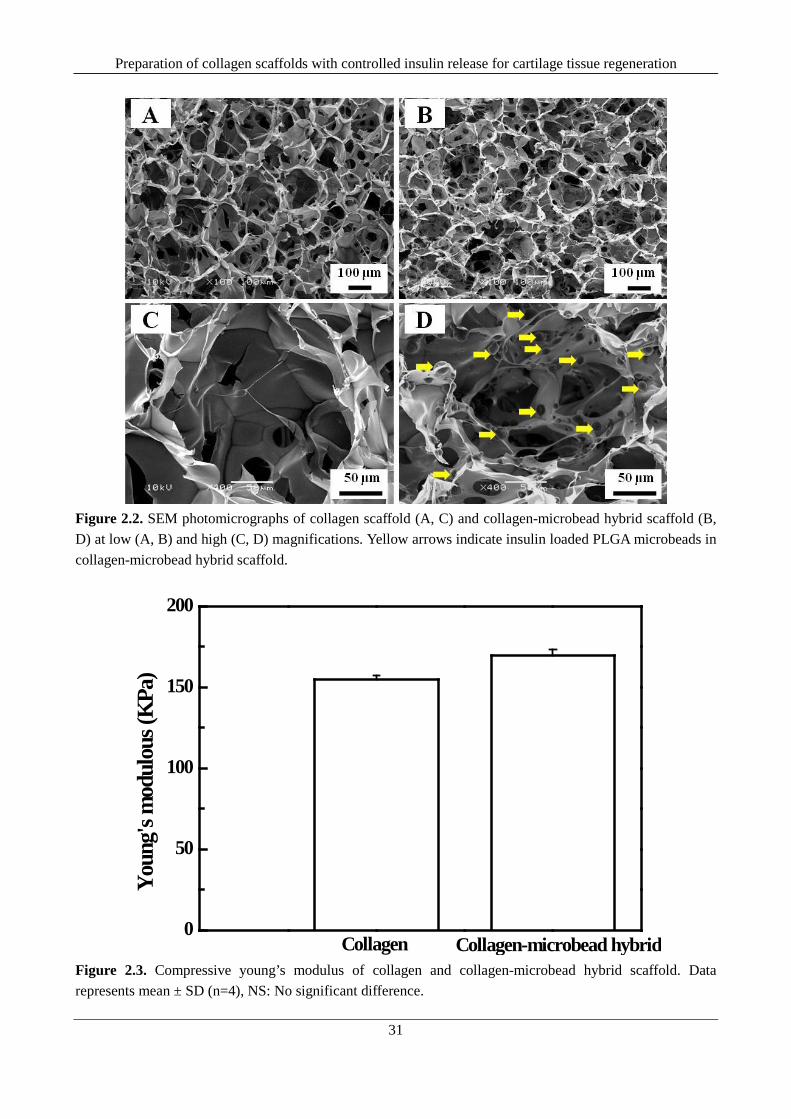
Preparation of collagen scaffolds with controlled insulin release for cartilage tissue regeneration
31
Figure 2.2. SEM photomicrographs of collagen scaffold (A, C) and collagen-microbead hybrid scaffold (B, D) at low (A, B) and high (C, D) magnifications. Yellow arrows indicate insulin loaded PLGA microbeads in collagen-microbead hybrid scaffold.
0
50
100
150
200
Collagen-microbead hybridCollagen
Youn
g's m
odul
ous (
KPa
)
Figure 2.3. Compressive young’s modulus of collagen and collagen-microbead hybrid scaffold. Data represents mean ± SD (n=4), NS: No significant difference.

Chapter 2
32
1 2 3 4 50
1020304050
Cum
ulat
ive i
nsul
in re
lease
(%)
Time (day)
Microbeads Hybrid scaffold
A
B
C
Figure 2.4. Cumulative insulin releases profile of microbeads and hybrid scaffold (A, B) and weight loss profile of microbeads in free and scaffold integrated state (C)
2.4.4. Cell adhesion, viability and proliferation Collagen (control) and collagen-microbead (experiment) hybrid scaffolds were cultured with bovine
articular chondrocytes. The scaffolds showed high cell seeding efficiencies and the seeding efficiencies of the scaffolds were 87.12±1.13% (control) and 86.99±1.38% (experiment). Introduction of microbead to collagen did not affect the pore structure of scaffold and therefore cell seeding efficiency. Figures 2.6 A-D present the SEM photomicrographs of adhered cells and their distribution in mid cross section of the scaffolds. The cells were well adhered to the pore wall surface. Homogeneous cell distribution was observed in entire cross section, which indicated the pore structure and interconnections facilitated the cells to reach in entire three dimensions of the scaffold. Cells could proliferate and migrate via interconnected pores and distributed in entire scaffold. Figure 2.6 E-H shows the SEM photomicrographs of mid cross section of the porous scaffolds after 1 week of chondrocyte culture. More cells were observed after 1 week period and indicating the cell were proliferated.
Figure 2.7 shows cell viability at mid cross sections of different scaffolds after 1 week of cell culture. More dead cells were detected in collagen scaffold cultured without insulin supplement compared to collagen scaffolds supplemented with 100 nM external insulin supplement and collagen-microbead hybrid scaffold. This indicated insulin had some effect on maintenance of chondrocyte viability. Furthermore, no dead cells were detected from the collagen-microbead hybrid scaffold. This might be due to the high local concentration of released insulin from the collagen-microbead hybrid scaffold.
Figure 2.8 shows the cell proliferation in different scaffolds. All the scaffolds facilitated cell proliferation observed from increased DNA quantities after 3rd and 7th day of culture period. A significant difference in the cell number among the scaffold groups was noticed after 3rd and 7th day of culture period. Cell proliferation in the scaffolds showed an increase order of collagen scaffold cultured without external insulin supplement < collagen scaffold cultured with 100 nM external insulin supplement < collagen-microbead hybrid scaffold. The result suggested that the insulin promoted chondrocyte proliferation. The insulin released from the spatially located microbeads in the collagen-microbead hybrid scaffold might have met the local insulin demand for the chondrocytes for their survival and usual proliferation. However, due to extensive cell proliferation with time, the diffusion of bioactive insulin from cell culture medium might not be sufficient to nourish the interior cells of the scaffold supplemented with
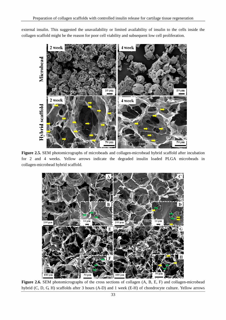
Preparation of collagen scaffolds with controlled insulin release for cartilage tissue regeneration
33
external insulin. This suggested the unavailability or limited availability of insulin to the cells inside the collagen scaffold might be the reason for poor cell viability and subsequent low cell proliferation.
Figure 2.5. SEM photomicrographs of microbeads and collagen-microbead hybrid scaffold after incubation for 2 and 4 weeks. Yellow arrows indicate the degraded insulin loaded PLGA microbeads in collagen-microbead hybrid scaffold.
Figure 2.6. SEM photomicrographs of the cross sections of collagen (A, B, E, F) and collagen-microbead hybrid (C, D, G, H) scaffolds after 3 hours (A-D) and 1 week (E-H) of chondrocyte culture. Yellow arrows

Chapter 2
34
indicate the insulin loaded PLGA microbeads in collagen-microbead hybrid scaffold and green arrows indicate adhered chondrocytes.
Figure 2.7. Live/dead staining of mid cross section of collagen scaffold cultured without external insulin (A), collagen scaffold cultured with 100 nM external insulin supplement (B) and collagen-microbead hybrid scaffold (C) after 1 week of chondrocyte culture. Green fluorescence indicates live cells and red fluorescent dots indicate dead cells.
NS
*
***
**
***
***
Figure 2.8. DNA amount in the cell-scaffold constructs of collagen scaffold, collagen scaffold supplemented

Preparation of collagen scaffolds with controlled insulin release for cartilage tissue regeneration
35
with 100 nM insulin and collagen-microbead hybrid scaffold after 1, 3 and 7 days of chondrocyte culture. Data represent mean ± SD (n=4),*significant (p<0.05), **significant (p<0.01), ***significant (p<0.001), NS: No significant difference.
The study presented a controlled release approach to solve the problem of cell death in porous collagen scaffold for prevention of necrosis and maintenance of usual cell proliferation. Owing to the importance of collagen porous scaffolds of controlled pore structure and improved mechanical strength in cartilage tissue regeneration, controlled release function via biodegradable PLGA microbeads was introduced in order to improve the regeneration potential of the prepared scaffold.
2.5. Conclusion Controlled insulin releasing porous collagen-microbead hybrid scaffold with controlled pore structure
was prepared by introduction of insulin loaded PLGA microbeads into porous collagen matrix. The collagen-microbead hybrid scaffold demonstrated a high mechanical strength and a stable release of insulin for 4 weeks. The released insulin demonstrated its effect on cultured chondrocytes for their survival and proliferation. The bioactive hybrid scaffold should be useful for maintenance of prolonged survival and proliferation of cultured chondrocytes towards the application in cartilage tissue engineering.
2.6. References 1. J. S. Temenoff, and A. G. Mikos, "Review: tissue engineering for regeneration of articular cartilage,"
Biomaterials, vol. 21, no. 5, pp. 431-440, 2000. 2. W. Kafienah, M. Jakob, O. Démarteau, A. Frazer, M. D. Barker, I. Martin, and A. P. Hollander,
"Three-dimensional tissue engineering of hyaline cartilage: comparison of adult nasal and articular chondrocytes," Tissue engineering, vol. 8, no. 5, pp. 817-826, 2002.
3. D. W. Hutmacher, "Scaffolds in tissue engineering bone and cartilage," Biomaterials, vol. 21, no. 24, pp. 2529-2543, 2000.
4. P. M. Van der Kraan, P. Buma, T. Van Kuppevelt, and W. B. Van den Berg, "Interaction of chondrocytes, extracellular matrix and growth factors: relevance for articular cartilage tissue engineering," Osteoarthritis and Cartilage, vol.10, no. 8, pp. 631-637, 2002.
5. D. Nesic, R. Whiteside, M. Brittberg, D. Wendt, I. Martin, and P. Mainil-Varlet, "Cartilage tissue engineering for degenerative joint disease," Advanced drug delivery reviews, vol. 58, no. 2, pp. 300-322, 2006.
6. T. Minas, and S. Nehrer, "Current concepts in the treatment of articular cartilage defects," Orthopedics, vol. 20, no. 6, pp. 525-538, 1997.
7. E. B. Hunziker, "Articular cartilage repair: basic science and clinical progress. A review of the current status and prospects," Osteoarthritis and cartilage, vol. 10, no. 6, pp. 432-463, 2002.
8. W-J Li, R. Tuli, C. Okafor, A. Derfoul, K. G. Danielson, D. J. Hall, and R. S. Tuan, "A three-dimensional nanofibrous scaffold for cartilage tissue engineering using human mesenchymal stem cells," Biomaterials, vol. 26, no. 6, pp. 599-609, 2005.
9. H. Lu, H. H. Oh, N. Kawazoe, K. Yamagishi, and G. Chen, "PLLA–collagen and PLLA–gelatin hybrid scaffolds with funnel-like porous structure for skin tissue engineering," Science and Technology of Advanced Materials, vol. 13, no. 6 (2012): 064210.
10. W. Dai, Z. Yao, J. Dong, N. Kawazoe, C. Zhang, and G. Chen, "Cartilage tissue engineering with controllable shape using a poly (lactic-co-glycolic acid)/collagen hybrid scaffold," Journal of Bioactive and Compatible Polymers, vol. 28, no. 3, pp. 247-257, 2013.

Chapter 2
36
11. S. J. Hollister, "Porous scaffold design for tissue engineering," Nature materials, vol. 4, no. 7, pp. 518-524, 2005.
12. A. Borzacchiello, A. Gloria, L. Mayol, S. Dickinson, S. Miot, I. Martin, and L. Ambrosio, "Natural/synthetic porous scaffold designs and properties for fibro-cartilaginous tissue engineering," Journal of Bioactive and Compatible Polymers, vol. 26, no. 5, pp. 437-451, 2011.
13. H. Lu, Y. G. Ko, N. Kawazoe, and G. Chen, "Cartilage tissue engineering using funnel-like collagen sponges prepared with embossing ice particulate templates," Biomaterials, vol. 31, no. 22, pp. 5825-5835, 2010.
14. Y. G. Ko, N. Kawazoe, T. Tateishi and G. Chen, "Preparation of chitosan scaffolds with a hierarchical porous structure," Journal of Biomedical Materials Research Part B: Applied Biomaterials, vol. 93, no. 2, pp. 341-350, 2010.
15. Q. Zhang, H. Lu, N. Kawazoe, and G. Chen, "Preparation of collagen scaffolds with controlled pore structures and improved mechanical property for cartilage tissue engineering," Journal of Bioactive and Compatible Polymers, vol. 28, no. 5, pp. 426-438, 2013.
16. K. Song, Y. Liu, H. M. Macedo, L. Jiang, C. Li, G. Mei, and T. Liu, "Fabrication and evaluation of a sustained-release chitosan-based scaffold embedded with PLGA microspheres," Materials Science and Engineering: C, 2012.
17. E. C. Ko, Y. Fujihara, T. Ogasawara, Y. Asawa, S. Nishizawa, S. Nagata, T. Takato, and K. Hoshi, "Administration of the insulin into the scaffold atelocollagen for tissue‐engineered cartilage," Journal of Biomedical Materials Research Part A, vol. 97, no. 2, pp. 186-192, 2011.
18. K. Lee, E. A. Silva, and D. J. Mooney, "Growth factor delivery-based tissue engineering: general approaches and a review of recent developments," Journal of The Royal Society Interface, vol. 8, no. 55, pp.153-170, 2011.
19. F. M. Chen, M. Zhang, and Z. F. Wu, "Toward delivery of multiple growth factors in tissue engineering," Biomaterials, vol.31, no. 24, pp. 6279-6308, 2010.
20. P. Tayalia, and D. J. Mooney, "Controlled growth factor delivery for tissue engineering," Advanced Materials, vol. 21, no. 32‐33, pp. 3269-3285, 2009.
21. K. Lee, E. A. Silva, and D. J. Mooney, "Growth factor delivery-based tissue engineering: general approaches and a review of recent developments," Journal of The Royal Society Interface, vol. 8, no. 55, pp 153-170, 2011.
22. K. Andreas, R. Zehbe, M. Kazubek, K. Grzeschik, N. Sternberg, H. Bäumler, H. Schubert, M. Sittinger, and J. Ringe, "Biodegradable insulin-loaded PLGA microspheres fabricated by three different emulsification techniques: Investigation for cartilage tissue engineering," Acta Biomaterialia, vol. 7, no. 4, pp. 1485-1495, 2011.
23. M. Ye, S. Kim, and K. Park, "Issues in long-term protein delivery using biodegradable microparticles," Journal of Controlled Release, vol. 146, no. 2, pp. 241-260, 2010.
24. C. Manoharan, and J. Singh, "Insulin loaded PLGA microspheres: effect of zinc salts on encapsulation, release and stability," Journal of pharmaceutical sciences, vol. 98, no. 2, pp. 529-542, 2009.
25. Lam, Xanthe M., Eileen T. Duenas, Ann L. Daugherty, Nancy Levin, and Jeffrey L. Cleland. "Sustained release of recombinant human insulin-like growth factor-I for treatment of diabetes." Journal of Controlled Release 67, no. 2 (2000): 281-292
26. M. Lee, T. T. Chen, M. L. Iruela-Arispe, B. M. Wu, and J. C. Dunn, "Modulation of protein delivery from modular polymer scaffolds," Biomaterials, vol. 28, no. 10, pp. 1862-1870, 2007.

Preparation of collagen scaffolds with controlled insulin release for cartilage tissue regeneration
37
27. F. Ungaro, M. Biondi, I. d'Angelo, L. Indolfi, F. Quaglia, P. A. Netti, and M I La Rotonda, "Microsphere-integrated collagen scaffolds for tissue engineering: effect of microsphere formulation and scaffold properties on protein release kinetics," Journal of controlled release, vol. 113, no. 2, pp. 128-136, 2006.

Chapter 3
38
Chapter 3
Preparation of collagen porous scaffold with sustained release of
insulin for skin tissue regeneration
3.1. Executive summary This chapter introduces the preparation of long-term insulin releasing collagen-microbead hybrid
scaffold for application in skin tissue engineering. Insulin incorporated PLGA microbeads of two distinct sizes such as 19.4±1.6 µm and 4.4±0.9 µm were prepared using double emulsion method and were used to prepare the collagen-microbead hybrid scaffolds. The hybrid scaffolds were prepared by freeze drying method using 150µm-250µm pre-prepared ice particulates. The hybrid scaffolds had controlled pore structure and interconnected pores. The PLGA microbeads were well distributed on pore walls of the hybrid scaffolds. The hybrid scaffolds showed a lower initial burst and more stable insulin than did the free microbeads. Culture of human dermal fibroblasts in the hybrid scaffolds demonstrated the bioactivity of released insulin. The hybrid scaffold prepared with 19.4±1.6 µm microbeads showed a more sustained release of insulin and had higher promotion effect on cell proliferation than did the other hybrid and control scaffolds. The hybrid scaffold prepared by incorporation of 19.4±1.6 µm microbeads should be useful for preparation of sustained insulin releasing collagen porous scaffold for skin regeneration in insulin dependant diabetes patients.
3.2. Introduction Chronic foot ulcers are commonly associated with diabetes [1]. It starts with simple breakdown of the
skin with formation of superficial open wound and frequently leads to osteomyelitis-a condition characterized by deep wound penetrating to tendon, bone and ligaments [2]. Failure of wound healing process at early stage of ulcers often leads to poor quality of life including high rate of mortality [3]. In order to combat with such life threatening problem, a wide variety of treatment strategies have been adopted by different clinicians in order to fasten the process of wound healing [3]. Despite of the progress, the problems of ulcer recurrence and high cost associated with hospitalization remains a clinical challenge [3].
Skin tissue engineering using cultured fibroblast has been evolved as a promising approach for wound repair through the regeneration of a functional skin tissue [4, 5]. Biodegradable three-dimensional porous scaffolds have been well studied for their ability to regenerate various types of tissues such as skin, cartilage, bone and so on [5-10]. Collagen porous scaffolds have been the most popular in context of skin tissue

Preparation of collagen porous scaffold with sustained release of insulin for skin tissue regeneration
39
engineering because of their excellent biocompatibity and minimal antigenicity [5, 11, 12]. Growth factors play an important part in tissue engineering. The area of controlled drug delivery has added significant advantage to engineered scaffolds to release the required bioactive factors in a controlled and sustained fashion for prolonged duration [13-16]. Therefore enhancing the functionality of the biomaterial scaffold by integrating appropriate controlled release devices may lead to development of novel functional platforms that can control and guide the tissue regeneration process in a larger fold [16]. In this approach, the material platform can be precisely tuned to meet the specific requirements of cells by presentation of bioactive molecules in a spatio-temporal fashion [16, 17]. This can further facilitate the control over the regenerating potential of engineered scaffolds [13, 16].
Growth factor microencapsulation through biodegradable PLGA microbeads has shown considerable interest in the area of controlled delivery of growth factors for tissue engineering [13, 16, 18-20]. Growth factor release from the PLGA microbeads can be programmed based on the control over their size and porosity. Controlled degradation and diffusion of encapsulated bioactive Growth factor from PLGA microbeads allows the controlled and sustain release of the bioactive molecule for a desired time frame [20-22]. Furthermore, the release can be additionally modulated after the introduction of such devices into porous biodegradable scaffold [23-25]. This rational design of tissue engineering scaffolds based on the combined approach of controlled growth factor release and three-dimensional pore structures can offer potential advantage to deliver the bioactive factors in pre-programmed rates as per cellular requirements in three-dimensional microenvironment [13, 16, 19].
Based on this concept, collagen porous scaffolds were hybridized with insulin releasing PLGA microbeads to control the release of bioactive insulin for sufficient duration during cell culture. Insulin incorporated PLGA microbeads were prepared by water-in-oil-in-water (w-o-w) double emulsion solvent evaporation method. The microbeads were hybridized with collagen by freeze-drying method using pre-prepared ice particulates as a porogen material. In vitro insulin release and degradation was studied under a shaking condition at 37 º C. The hybrid scaffolds were used for the culture of human dermal fibroblasts to investigate the effect of released insulin on cell functions.
3.3. Materials and methods
3.3.1. Materials PLGA (copolymer composition ratio of 50:50, weight average molecular weight 20 kDa and inherent
viscosity of 0.187 to 0.229 dL/g), methylene chloride (CH2Cl2), poly vinyl alcohol (86-90 mol % hydrolysis), recombinant human insulin, hydrochloric acid (HCl), sodium hydroxide (NaOH), absolute ethanol (99.5%), N-hydroxysuccinimide esters (NHS), 25% glutaraldehyde solution and sodium dihydrogen phosphate (NaH2PO4) were obtained from Wako Pure Chemicals Ltd., Japan. L-cysteine hydrochloride monohydrate (minimum 98%), ethylene diamine tetra acetic acid (EDTA), papain (from papaya latex) and DNA quantification kit were obtained from Sigma-Aldrich, USA. Neonatal human dermal fibroblast (NHDF), medium 106, growth supplement and antibiotics (gentamicin and amphotericin) were obtained from Cascade Biologics Inc., Japan. Phosphate buffer saline (10X, pH=7.4) was obtained from Nacali Tesque Inc., Japan. Porcine collagen type-1 was obtained from Nitta Gelatin, Japan. 1-Ethyl-3-[3-dimethylaminopropyl] carbodiimide hydrochloride (EDC/EDAC) was obtained from Peptide Institute Inc., Japan. Cellstain double Staining kit was obtained from Dojindo Laboratories, Japan. Micro BCA protein assay Kit was obtained from Pierce Biotechnology, USA. All the materials in this study were used as received without further purification. Molecular biology grade milli-Q water from millipore water system (Millipore Corporation.,
Chapter 3
40
USA) was used for preparation of all the solutions and reagents.
3.3.2. Methods
3.3.2.1. Preparation of insulin loaded PLGA microbeads Insulin loaded PLGA microbeads were prepared by w1-o-w2 double emulsion solvent evaporation
method [27]. PLGA solution at concentration of 0.5 g mL−1 was prepared by dissolving PLGA in methylene chloride. 50µL of insulin solution (insulin in 0.01 M HCl) at a concentration of 20 mg mL−1 (w1) was dispersed in 1 mL of PLGA (o) by homogenization at 8000 rpm for 1 minute. The resulted w1-o emulsion was further re-emulsified in 2 mL of 3% (w/v) saturated PVA (w2) prepared by 50 % (v/v) saturation with methylene chloride. The re-emulsification process was carried out under high speed homogenization for 10 minutes to prepare a double (w1-o-w2) emulsion. Two different homogenization speeds (1000 rpm and 8000 rpm) were used to control the microbead size. The resulted double emulsion was further added in drop wise to a large aqueous phase containing 200 mL of 0.5 % (w/v) PVA and stirred magnetically at 300 rpm in a hood for overnight to allow adequate solvent evaporation. The hardened microbeads were recovered after centrifugation (3500 rpm for 5 minutes) and wash (twice with 0.5 % (w/v) PVA and twice with milli-Q water). The collected microbeads were allowed to freeze at -80º C for 6 hours and freeze-dried for 48 hours in a freeze drier (Vir Tis AdVantage Benchtop Freeze Dryer, S P Industries Inc., Japan) below 5 kPa. The freeze dried microbeads were kept in tightly capped vials and stored in a desiccator for further use. Blank microbeads (control) were also prepared by similar procedure without the use of insulin.
3.3.2.2. Preparation of collagen microbead porous hybrid scaffold The hybrid porous scaffolds were prepared by a freeze-drying method using pre-prepared ice
particulates of a diameter range of 150-250 µm as a porogen material [28, 29]. Ice particulates of random sizes were prepared by spraying water droplets into liquid N2 and the particulates were further stabilized at -15º C in a low temperature chamber (WT-201, ESPEC Corp., Osaka, Japan) before sieving. Ice particulates in the diameter range of 150-250 µm were selectively sieved from the random size mixture of by a testing sieve (Tokyo screen co. ltd., Japan). Hybrid scaffolds were prepared from 2 % (w/v) collagen aqueous solution with a ratio of ice particulate to collagen as 50:50 (w/v). The collagen aqueous solution (2.2 % (w/v)) was prepared by dissolving dried collagen in a mixture solution of acetic acid (0.1 M, pH 3.0) and 10% ethanol. Dried insulin incorporated microbeads were dispersed in 10 % ethanol to prepare microbead suspension. The suspension was further sonicated for 1 minute in an ultrasonic water bath (Branson Ultrasonic Corporation., USA) to ensure free dispersion of microbeads. The required volume of microbead suspension was added to the 2.2 % (w/v) collagen aqueous solution and stirred magnetically for 1 hour at 4 º C for formation of microbead dispersed collagen aqueous solution of 2 % (w/v). The weight ratio of the microbeads was controlled as 50% of the total weight of collagen in mixture. 2% collagen solution with homogeneously dispersed microbeads was transferred to a low temperature chamber maintained at -5º C and stirred magnetically for another 2 hours in order to reduce the temperature of the solution to working temperature range (-2 º C). Subsequently, ice particulates were added to the microbead suspended collagen solution and mixed thoroughly to prepare a homogeneous ternary mixture of ice particulates, collagen and microbeads. The final mixture was molded in a silicone frame template with a dimension of 6 cm (length) × 4 cm (breadth) × 5 mm (thickness) placed over a perfluoroalkoxy (PFA) film wrapped copper plate. The temperature of the copper plate was balanced with the chamber temperature prior to molding step. The top surface of the mold was covered with a polyvinylidene chloride wrapped glass plate. The whole system was

Preparation of collagen porous scaffold with sustained release of insulin for skin tissue regeneration
41
transferred to -80º C for freezing. After 6 hours of freezing, the frozen mold was freeze-dried for at least 72 hours in a freeze-drier below 5 Pa. The freeze-dried scaffolds were cross-linked in 50 mM EDC and 20 mM NHS in 90% (v/v) ethanol for 24 hours at room temperature (RT). The cross-linked scaffolds were washed with milli-Q water for six times (approximate time≈2 hours) to remove the excess by-products from cross-linking reaction as well as additional non-reacted cross-linking reagents. The washed scaffolds were subjected to second freeze-drying for 72 hours to prepare the final scaffolds. In the present research, we prepared two different kinds of hybrid porous scaffold by hybridization of microbeads of two distinct sizes with collagen. Control collagen scaffold was also prepared by using the similar procedure without the use of microbeads. All the prepared scaffolds were stored in a desiccator for further use.
3.3.2.3. Scanning Electron Microscopy (SEM) The morphology of insulin incorporated microbead and scaffold microstructure was examined by a
scanning electron microscope (SEM, JSM-5610, JEOL Ltd., Tokyo, Japan). Freeze-dried microbeads were evenly dispersed over a carbon adhesive mounted over a copper stub and were sputtered with a thin layer of platinum by a sputter-coater (ESC-101, Elionix, Tokyo, Japan) for 500 seconds prior to SEM observation. For scaffold microstructure, the freeze-dried collagen scaffolds were cut into cross section with a sharp razor blade and mounted on a carbon adhesive over the SEM stub. The samples were sputter-coated with platinum for 300 seconds. The microbeads and scaffolds were observed at an acceleration potential of 5 kV and 10 kV respectively.
3.3.2.4. Microbead size analysis 10 mg of freeze-dried microbeads were suspended in 1 mL of milli-Q water. The free dispersion of
microbeads in water was ensured by sonication of the suspension for 30 seconds by using an ultrasonic water bath. The samples were analyzed for their average size as well as size distribution profile by using a laser diffraction particle size analyzer (SALD 7000, Shimadzu Corporation., Japan). The microbead size was measured for three batches of formulation and the mean average was calculated as means ± standard deviation (n = 3)
3.3.2.5. Insulin loading efficiency (LE) The confirmation of insulin loading as well as insulin quantification in prepared microbeads were
carried out by Micro-BCA protein assay using a Micro BCA assay Kit. 10 mg of dried insulin incorporated microbeads were dissolved in 1 mL of methylene chloride at RT. Insulin was extracted into 0.01 M HCl under vigorous shaking for 2 minutes in a high speed vortex device (Vortex genie, Fischer, Pittsburgh., PA) at setting 10. The suspension was allowed to settle for 5 minutes at RT and supernatant aqueous phase containing insulin was extracted by separating from the remaining organic phase. 150 µL of the supernatant solution was used for the insulin quantification. Briefly 150 µL of albumin (BSA) standard in duplicate and individual samples in triplicate were added to individual wells of a 96 well plate.150 µL of assay working agent was added to each of the wells containing standards as well as samples. The plate was covered with a sealing tape and the mixing of the solutions in the wells was ensured by shaking the plate in a micro plate shaker for 30 seconds. The micro plate was incubated at 37 º C in an incubator for 2 hour. Following the incubation period, the plate was cooled at RT and the absorbance was measured at 562 nm by a micro plate reader. The blank absorbance was subtracted and the insulin concentration (µg/mL) of the unknown samples was measured by comparing with standard curve (R2=0.998) obtained from BSA standard. Total quantity of the incorporated insulin was calculated. The loading efficiency of the formulations was calculated using

Chapter 3
42
following equation [22]. Loading efficiency (%) = [Weight of the incorporated insulin /weight of total insulin used for incorporation]
× 100
3.3.2.6. In vitro insulin release In vitro insulin release from microbeads as well as their corresponding hybrid scaffold was studied in
PBS (pH=7.4) at 37º C. Briefly, 30 mg of microbead from each formulations were weighed in 2 mL tubes. Hybrid scaffolds were cut into discs of dimension of Ø 10 mm (area) × H 5 mm (height) by a surgical biopsy punch and placed in 50 mL tubes. 1.2 mL of sterile PBS was added to each of the tubes containing microbeads and 1 mL PBS was added to each tube containing the hybrid scaffold. The tubes were tightly capped and incubated in a shaking water bath incubator (Taitec Corporation., Japan) at 37º C with a shaking speed of 50 rpm.The scaffolds were degassed before incubation to ensure the penetration of PBS to each of the scaffold pores. After pre-determined time points of 1, 2, 4, 8, 12, 16, 20, 24 and 28 days, the required volume of release medium (1mL from microbeads and 0.5 mL from scaffolds) was collected and replaced with equivalent volume of fresh PBS. Release experiment was performed for a period of 4 weeks. Control scaffolds (collagen scaffold with blank microbeads) were also used for the release experiment. The insulin amount in released medium was quantified by Micro BCA protein assay. The cumulative release (%) was calculated from insulin concentration obtained from Micro BCA protein assay and plotted against time (days) to get the release curve. The experiments were performed in triplicate for each sample and the data points in the curve were presented as means ± standard deviation (n=3). The scaffolds after the 4 week release period were collected and washed six times with milli-Q. The washed scaffolds were freeze dried and the microstructure was observed by SEM. The pH of the released medium was measured by using a digital pH meter (HORIBA Ltd, Japan). The mean pH value of triplicate samples was plotted against the time to get pH change profile during the release period. In order to monitor the degradation, the weight loss profile of the free microbeads as well as the microbeads in the hybrid collagen scaffold was determined. The microbeads as well as scaffolds were collected in predetermined time intervals of 7, 14, 21 and 28 days after incubation. The samples were washed and freeze dried. The freeze dried samples were weighed and the remaining microbead weight % was calculated at each time point. The weight % of triplicate samples was plotted against the time to get the degradation profile. Control collagen scaffolds were also used for degradation experiment.
3.3.2.7. In vitro cell culture and bioactivity evaluation Bioactivity of the hybrid scaffolds was evaluated by in-vitro culture of neonatal human dermal fibroblast
(NHDF) cells. Four groups of scaffolds including control (positive and negative) and two experimental groups (two categories of hybrid scaffolds) were used for cell culture experiment. NHDF cells were sub-cultured over 175 cm2 culture flasks (BD Biosciences., USA) in medium 106 supplemented with 2% fetal bovine serum (FBS), 10 ng/mL human recombinant epidermal growth factor (h-EGF), 3 ng/mL human recombinant fibroblast growth factor-basic (hFGF-b) and antibiotics (gentamicin and amphotericin). The cell culture was maintained in a CO2 incubator (Sanyo Corporation., Japan) equilibrated with 5% CO2 at 37º C. Cells were detached from the surface of the culture flask with the help of PBS and 0.25 % trypsin-EDTA once after the confluent monolayer has reached. The cell suspension was centrifuged (1100 rpm for 5 minutes) and the pellet was resuspended with fresh cell culture medium. The cell number was counted with a hemocytometer. The scaffolds were cut into discs with dimension of Ø 6 mm× H4 mm by a surgical biopsy punch and sterilized with 70% ethanol .The sterilized scaffolds were transferred to a clean bench and washed

Preparation of collagen porous scaffold with sustained release of insulin for skin tissue regeneration
43
with PBS for six times for 1 hour and exchanged with cell culture medium by incubating with medium for 3 hours. Once exchanged with cell culture medium, all the scaffolds were transferred to 12 well tissue culture plates. The excess medium was dried with sterile paper towel (Kimwipe) and 70 µL of cell suspension (cell density of 0.67×10⁶ cells/mL) was dropped to each scaffold and incubated for 3 hours for cell adhesion. Following 3 hours of pre-incubation, the cell-scaffold constructs were transferred to new 12 well tissue culture plates and cell culture medium was added. Culture medium with 50 nM insulin was added to the wells containing the normal collagen scaffold as a positive control. Cells were cultured for 2 week duration. Culture medium was changed in every three days.
The cell-scaffold constructs after culture for 3 hours and 14 days were washed thrice in PBS and fixed with 0.01 % glutaldehyde for 24 hours at RT. The fixed constructs were washed thrice in milli-Q and freeze dried. The cross-sections of the freeze-dried scaffolds were observed by SEM for studying cell adhesion, distribution and migration to inner cross sectional pores.
Cell viability was evaluated by performing live-dead staining assay using Cellstain double staining kit. The assay kit uses calcein-AM and propidium iodide to stain live and dead cells, respectively. After 1 and 14 days of cell culture, cell-scaffold constructs were washed with PBS (thrice) and incubated in 2 μM calcein-AM and 4 μM propidium iodide solution in cell culture medium for 10 minutes. The specimens were observed in a fluorescence microscope (Olympus Corp., Japan) for observation of live and dead cells.
The cell proliferation in the scaffolds was evaluated by quantifying the DNA amount in cell-scaffold constructs after being cultured for 1, 7 and 14 days. At each time point, the cell-scaffold constructs were collected and freeze-dried. The freeze-dried scaffolds were digested with papain solution. Papain was dissolved at 400 μg mL−1 in 0.1 M phosphate buffer (pH =6.0) prepared with sodium dihydrogen phosphate, L-cysteine hydrochloride monohydrate and ethylene diamine tetra acetic acid (EDTA). 500 µL of papain solution was added to each aliquot containing freeze-dried cell-scaffold construct. The aliquots were incubated in a shaking incubator at 60º C with shaking speed of 175 rpm (24 hours) for complete digestion. The digested samples were used to measure the DNA content by using a standard curve (R2=0.999984) prepared using calf thymus DNA standard and fluorescent dye (Hoechst 33258).The fluorescence emission was measured using FP-6500 spectrofluorometer (JASCO., Japan) at an excitation wavelength of 360 nm and emission wavelength of 460 nm. Four samples were used to calculate the average and SD (n = 4). Cell seeding efficiency for each scaffold was calculated by comparing the DNA content in initial volume of seeded cell suspension and the DNA amount as quantified from cell scaffold construct after 1 day culture. The DNA content in the initial volume of seeded cell suspension was also determined by similar method as above.
3.3.2.8. Statistical analysis All the data were expressed as the mean ± standard deviation (SD). One-way analysis of variance was
performed to reveal significant differences, followed by Tukey’s post hoc test for pair wise comparison. Statistical analysis was executed using Kyplot 2.0 beta 15. The difference was considered significant when the p-value was less than 0.05.
3.4. Results
3.4.1. Preparation and characterization of microbeads PLGA was used to microencapsulate human recombinant insulin. Double emulsion (w1-o-w2) method
was used for preparation of insulin incorporated microbeads. Two types of microbeads having different

Chapter 3
44
average size were prepared. SEM observation showed that the microbeads had spherical morphology with smooth surfaces (Figure 3.1A and 3.1B). Size distribution and size of the microbeads were measured by laser particle size analysis (Figure 3.1C). The microbeads showed a narrow size distribution. The average sizes of the two types of microbeads was 19.4±1.6 µm (large size) and 4.4±0.9 µm (small size), corresponding to emulsification speeds of 1000 rpm and 8000 rpm respectively. Insulin loading efficiencies of the microbeads were 87.0±2.0% and 85.1±2.8% corresponding to large and small size microbeads respectively. The result indicated that both microbeads showed high insulin loading.
0.1 1 10 100 10000
10
20
30
40
Freq
uenc
y (%
)
Diameter (µm)
19.4 µm 4.4 µm
19.4±1.6 µm 4.4±0.9 µmA
C
B
10 µm 10 µm
Figure 3.1. SEM photomicrographs of insulin incorporated PLGA microbeads prepared by two different w1-o-w2 emulsification speeds of (A) 1000 rpm and (B) 8000 rpm and size distribution of PLGA microbeads analyzed by a laser differentiation particles analyzer (C).
3.4.2. Preparation and characterization of collagen-microbead hybrid scaffolds Collagen-microbead hybrid scaffolds were prepared by freeze-drying the mixture of microbeads and
collagen aqueous solution using pre-prepared ice particulates as a porogen material. Figure 3.2 shows the gross appearance and microstructure of collagen scaffolds and collagen-microbead hybrid scaffolds. Evenly distributed large pores were formed in both the scaffolds of collagen and collagen-microbead. The size of large pores was in a range of 150 µm-250 µm as evident from the SEM photomicrograph. The large pores were interconnected to each other, which may facilitate cell penetration and distribution. The large pores should be the replica of the pre-prepared ice particulates used as a porogen material. Microbeads were observed in the collagen-microbead hybrid scaffolds (white arrows) and were homogeneously distributed throughout the scaffolds.
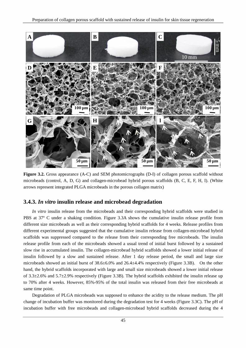
Preparation of collagen porous scaffold with sustained release of insulin for skin tissue regeneration
45
10 mm
5 mm
A CB
E FD
G H l
100 µm 100 µm 100 µm100 µm
50 µm 50 µm50 µm
Figure 3.2. Gross appearance (A-C) and SEM photomicrographs (D-I) of collagen porous scaffold without microbeads (control, A, D, G) and collagen-microbead hybrid porous scaffolds (B, C, E, F, H, I). (White arrows represent integrated PLGA microbeads in the porous collagen matrix)
3.4.3. In vitro insulin release and microbead degradation In vitro insulin release from the microbeads and their corresponding hybrid scaffolds were studied in
PBS at 37º C under a shaking condition. Figure 3.3A shows the cumulative insulin release profile from different size microbeads as well as their corresponding hybrid scaffolds for 4 weeks. Release profiles from different experimental groups suggested that the cumulative insulin release from collagen-microbead hybrid scaffolds was suppressed compared to the release from their corresponding free microbeads. The insulin release profile from each of the microbeads showed a usual trend of initial burst followed by a sustained slow rise in accumulated insulin. The collagen-microbead hybrid scaffolds showed a lower initial release of insulin followed by a slow and sustained release. After 1 day release period, the small and large size microbeads showed an initial burst of 38.6±6.0% and 26.4±4.4% respectively (Figure 3.3B). On the other hand, the hybrid scaffolds incorporated with large and small size microbeads showed a lower initial release of 3.3±2.6% and 5.7±2.9% respectively (Figure 3.3B). The hybrid scaffolds exhibited the insulin release up to 70% after 4 weeks. However, 85%-95% of the total insulin was released from their free microbeads at same time point.
Degradation of PLGA microbeads was supposed to enhance the acidity to the release medium. The pH change of incubation buffer was monitored during the degradation test for 4 weeks (Figure 3.3C). The pH of incubation buffer with free microbeads and collagen-microbead hybrid scaffolds decreased during the 4
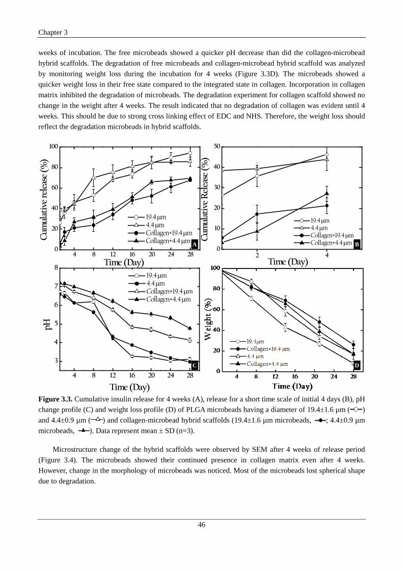
Chapter 3
46
weeks of incubation. The free microbeads showed a quicker pH decrease than did the collagen-microbead hybrid scaffolds. The degradation of free microbeads and collagen-microbead hybrid scaffold was analyzed by monitoring weight loss during the incubation for 4 weeks (Figure 3.3D). The microbeads showed a quicker weight loss in their free state compared to the integrated state in collagen. Incorporation in collagen matrix inhibited the degradation of microbeads. The degradation experiment for collagen scaffold showed no change in the weight after 4 weeks. The result indicated that no degradation of collagen was evident until 4 weeks. This should be due to strong cross linking effect of EDC and NHS. Therefore, the weight loss should reflect the degradation microbeads in hybrid scaffolds.
4 8 12 16 20 24 280
20
40
60
80
100
Cum
ulat
ive
rele
ase
(%)
Time (Day)
19.4 µm 4.4 µm Collagen+19.4 µm Collagen+4.4 µm
2 40
10
20
30
40
50
Cum
ulat
ive
Rel
ease
(%)
Time (Day)
19.4 µm 4.4 µm Collagen+19.4 µm Collagen+4.4 µm
4 8 12 16 20 24 28
3
4
5
6
7
8
pH
Time (Day)
19.4 µm 4.4 µm Collagen+19.4 µm Collagen+4.4 µm
A B
C D
Figure 3.3. Cumulative insulin release for 4 weeks (A), release for a short time scale of initial 4 days (B), pH change profile (C) and weight loss profile (D) of PLGA microbeads having a diameter of 19.4±1.6 µm (
) and 4.4±0.9 µm ( ) and collagen-microbead hybrid scaffolds (19.4±1.6 µm microbeads,
; 4.4±0.9 µm microbeads,
). Data represent mean ± SD (n=3).
Microstructure change of the hybrid scaffolds were observed by SEM after 4 weeks of release period (Figure 3.4). The microbeads showed their continued presence in collagen matrix even after 4 weeks. However, change in the morphology of microbeads was noticed. Most of the microbeads lost spherical shape due to degradation.

Preparation of collagen porous scaffold with sustained release of insulin for skin tissue regeneration
47
100 µm 100 µm
50 µm50 µm
A B
C DFE
20 µm 20 µm
Figure 3.4. SEM microstructure of the hybrid scaffolds of microbeads of size 19.4±1.6 µm (A, C, E) and 4.4±0.9 µm (B, D, F) after 4 weeks of incubation.
3.4.4. Bioactivity of hybrid scaffolds Bioactivity of the released insulin was evaluated by in vitro culture of human neonatal dermal
fibroblasts. The scaffolds were seeded with fibroblasts of equal cell numbers. Cell seeding efficiency in collagen scaffold and hybrid scaffolds incorporated with large and small microbeads was 86.33±0.85%, 88.32±2.08% and 87.43±2.8% respectively. The scaffolds showed same level of cell seeding efficiency because of their similar porous structures. Cell adhesion in different scaffolds was studied by SEM observation of the cross-sections of the hybrid scaffolds after 3 hours and 14 days of culture.A clear distinction among the cells (blue arrows) and microbeads (green arrows) was noticed in each of the scaffold after 3 hours culture (Figure 3.5).
Cells adhered to pore wall surfaces of the scaffolds. Seeded cells could migrate into the inner bulk pores via interconnected pore structures and distributed throughout the scaffolds. After culture for 2 weeks, cells proliferated and filled the spaces in the porous scaffolds (Figure 3.6).
Cell viability was evaluated after culture for 1 day and 14 days (Figure 3.7). Green fluorescence represents the live cells and red fluorescent dots indicate the dead cells. All the fluorescent images revealed the presence of very few dead cells in each of the scaffold and thus demonstrated the high viability of cells cultured in the porous scaffolds. After culture for 14 days, more cells were detected, which indicated cell proliferation during the culture period.
Cell proliferation was quantitatively evaluated by quantifying the DNA amount after culture for 1 day, 7 days and 14 days (Figure 3.8). The scaffolds showed an increased trend in DNA quantities till the 14 days period. Collagen scaffold with external insulin supplement showed significantly higher proliferation than the collagen scaffold without insulin. Similarly all the hybrids scaffolds showed increased DNA quantities than

Chapter 3
48
the control scaffolds after 7 days and 14 days period. After 14 days of cell culture, the hybrid scaffold with large size microbeads showed significantly increased DNA quantities compared to other scaffolds. The results indicated that insulin released from the hybrid scaffolds stimulated cell proliferation more effectively than did the insulin supplemented in the culture medium.
Figure 3.5. SEM photomicrographs of the cross-sections of control collagen scaffolds (A, D) and hybrid scaffolds of microbeads of size 19.4±1.6 µm (B, E) and 4.4±0.9 µm (C, F) after 3 hours of cell culture. Green arrows indicate scaffold integrated PLGA microbeads and blue arrows indicate adhered cells.
A B C D
E F G H
50 µm50 µm50 µm50 µm
100 µm100 µm100 µm100 µm
Figure 3.6. SEM photomicrographs of the cross section of control collagen scaffolds without insulin (A, E), control collagen scaffolds with insulin supplemented in medium (B, F) and hybrid scaffolds of microbeads of size 19.4±1.6 µm (C, G) and 4.4±0.9 µm (D, H) after 2 weeks of cell culture
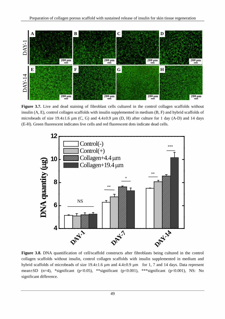
Preparation of collagen porous scaffold with sustained release of insulin for skin tissue regeneration
49
DAY
-1D
AY-1
4A B C D
E F G H
200 µm 200 µm 200 µm 200 µm
200 µm 200 µm 200 µm 200 µm
Figure 3.7. Live and dead staining of fibroblast cells cultured in the control collagen scaffolds without insulin (A, E), control collagen scaffolds with insulin supplemented in medium (B, F) and hybrid scaffolds of microbeads of size 19.4±1.6 µm (C, G) and 4.4±0.9 µm (D, H) after culture for 1 day (A-D) and 14 days (E-H). Green fluorescent indicates live cells and red fluorescent dots indicate dead cells.
4
6
8
10
12
DAY-14DAY-7
DAY-1
DNA
quan
tity
(µg)
Control(-) Control(+) Collagen+4.4 µm Collagen+19.4 µm
**
**
NS
***
*
Figure 3.8. DNA quantification of cell/scaffold constructs after fibroblasts being cultured in the control collagen scaffolds without insulin, control collagen scaffolds with insulin supplemented in medium and hybrid scaffolds of microbeads of size 19.4±1.6 µm and 4.4±0.9 µm for 1, 7 and 14 days. Data represent mean±SD (n=4), *significant (p<0.05), **significant (p<0.001), ***significant (p<0.001), NS: No significant difference.

Chapter 3
50
3.5. Discussion Growth factors play a critical role in transferring a wide range of biochemical signals in cellular
microenvironment for cell adhesion, proliferation, migration, cell recruitment, differentiation and gene expression [13, 16, 19]. The bioactive factors act on cell surface receptors and regulate complex biochemical pathways [13]. Therefore, local release of these factors inside 3D porous scaffolds may induce enhanced bio-activation of the cells which can further lead to higher cell proliferation. Furthermore, controlled and sustained release of these factors in bioactive form for prolonged duration may induce effective tissue regeneration [13, 16, 19]. Entrapment of the controlled release biodegradable PLGA microbeads incorporating specific growth factors within the porous scaffold is one of the promising and well established strategies to achieve the desired goal [13, 16, 24, 25, 30].
Based on such background, we have used human recombinant insulin as a powerful and potent applied factor for fibroblast proliferation in 3D porous collagen scaffold. The hypothesis was that controlled release of bioactive insulin from porous 3D material platform for a longer time frame may lead to enhanced fibroblast proliferation and subsequent tissue regeneration. To meet the purpose, we used PLGA microbeads as controlled drug delivery devices to microencapsulate the insulin for localization inside the collagens scaffolds of controlled pore structure. In order to evaluate the size effect of microbead for localization, PLGA microbeads of two distinct sizes 19.4±1.6 µm (large) and 4.4±0.9 µm (small) were prepared using w1-o-w2
emulsion with an aim to realize differential insulin release behavior. Double emulsion method was preferred for its ability to yield the microbeads with high encapsulation efficiency and low initial burst [18]. SEM observation of the prepared microbeads revealed smooth surface morphology with no visible pores (Figure 3.1A and 3.1B). Microbeads with least porous surface are usually considered as appropriate for growth factor delivery in tissue engineering because of their ability for high insulin loading which can release the incorporated growth factor for a prolonged duration [19]. The prepared microbeads showed more than 85% insulin loading efficiency and showed a sustained release of insulin for longer than 4 weeks (Figure 3.3A).
The microbeads of different size were introduced in collagen porous scaffolds to prepare the hybrid scaffolds. Collagen has been widely studied as excellent biodegradable and biocompatible materials for tissue scaffold design [5, 9, 11, 28, 31, 32]. It can be fabricated into the scaffolds with high porosity and good interconnectivity for facilitating cellular communication. Previously we used pre-prepared ice particulates of controlled diameter as a porogen material to fabricate collagen scaffolds with controlled and homogeneous pore structure [28, 29]. In the present research, we also used pre-prepared ice particulates to control the pore structure of the hybrid scaffolds. The collagen-microbead hybrid scaffolds showed well controlled pores with high interconnectivity for easy cell seeding and cell migration into the inner bulk pores of the scaffolds. SEM photomicrograph of the hybrid scaffolds revealed the presence of microbeads over porous collagen matrix without any surface erosion. The microbeads and collagen showed integrated structures.
In vitro Insulin release profile from each size microbeads and their corresponding hybrid scaffolds suggested the suppression of insulin release in presence of collagen (Figure 3.3A and 3.3B). An enhanced sustained release was noticed from each of the hybrid scaffolds compared to the release from the free microbeads. Presence of collagen was believed to delay the induction time for activation of initial protein release and also to retard the diffusion of insulin to surrounding medium [24]. Microbead size showed some effect on insulin release as reported elsewhere [33]. Furthermore, when being introduced into collagen matrix, the release profile was additionally modulated. Weight loss as well as pH change profiles from the hybrid scaffold demonstrated the delayed and slow degradation of microbeads inside the collagen matrix. Delayed degradation of microbeads might be due to the inhibitory effect of collagen matrix on water uptake.

Preparation of collagen porous scaffold with sustained release of insulin for skin tissue regeneration
51
The released insulin was tested for its ability to enhance fibroblast proliferation in hybrid scaffolds. The hypothesis was that continuous exposure of fibroblasts to released insulin via sustained release from localized microbeads could cause the enhanced bio-activation of the cells leading to enhanced cell proliferation. The proliferation of fibroblasts in presence of insulin is concentration dependant. In the present study, fibroblasts were cultured in the control collagen scaffold in presence of an external insulin concentration of 50 nM in order to investigate the effect of insulin on cultured fibroblasts. Significantly higher cell proliferation was observed in the control collagen scaffold when external insulin was supplemented in the culture medium. Furthermore, when cells were cultured in the hybrid scaffolds, interestingly all the hybrid scaffolds showed an enhanced cell proliferation compared to the control groups. This indicated that the sustained released insulin in the hybrid scaffolds more likely caused the enhanced activation of the fibroblasts by their local releasing action. The amount of insulin in the incorporated microbeads in the hybrid scaffolds was higher (≈5 µg) than that of the external insulin amount supplemented in the culture medium. Therefore it should obvious that a higher cell proliferation could be expected in the hybrid scaffolds only if the released insulin from the microbead was bioactive. From the cell proliferation result, the released insulin from the hybrid scaffolds was bioactive during the 14 days culture period. Overall, the hybrid scaffold of large size microbeads showed significantly higher cell proliferation than did the hybrid scaffold of small size microbead after 14 days of cell culture. The result can be explained by taking into account of the release profile from individual hybrid scaffold. As shown in the release curves, during the initial week concentration of released insulin from hybrid scaffold of small microbeads was higher than that of hybrid scaffold of large microbeads. This probably caused a concentration dependant cell proliferation which accounted for the high cell proliferation. However from day 7 to day 14, the insulin release curve from the hybrid scaffolds of large microbeads exhibited a higher linearity in insulin release than did the hybrid scaffold of small microbeads. The result indicated that the released insulin concentration between days 7 to day 14 should be higher from the hybrid scaffolds of large microbeads compared to the hybrid scaffold of small microbeads. Furthermore an early plateau was observed in the release profile of hybrid scaffold of small microbeads. On the contrary, hybrid scaffold with large size microbeads showed a continuous insulin release phase up to the period of 4 weeks. The results indicated that insulin release from hybrid scaffolds with large microbeads could be well maintained up to the 4 weeks.
3.6. Conclusion Porous collagen scaffolds incorporated with insulin releasing PLGA microbeads were prepared as a
controlled and sustained insulin delivery platform for skin tissue regeneration. The porous structure was controlled by using pre-prepared ice particulates as a porogen material. Microbeads of two distinct sizes were tested for their release feature from the hybrid scaffolds. The hybrid scaffolds showed slow release of insulin for more than 4 weeks and the released insulin showed high bioactivity. The incorporation of the microbeads of size 19.4±1.6 µm was proved as ideal for development of long term insulin releasing porous collagen scaffold for biomedical application.
3.7. References 1. Lipsky, Benjamin A., Anthony R. Berendt, H. Gunner Deery, John M. Embil, Warren S. Joseph, Adolf
W. Karchmer, Jack L. LeFrock et al. "Diagnosis and treatment of diabetic foot infections." Clinical Infectious Diseases 39, no. 7 (2004): 885-910.
2. Frykberg, Robert G., Brian Wittmayer, and Thomas Zgonis. "Surgical management of diabetic foot infections and osteomyelitis." Clinics in podiatric medicine and surgery 24, no. 3 (2007): 469-482.

Chapter 3
52
3. Jeffcoate, William J., and Keith G. Harding. "Diabetic foot ulcers." The Lancet 361, no. 9368 (2003): 1545-1551.
4. Priya, S. Geetha, Hans Jungvid, and Ashok Kumar. "Skin tissue engineering for tissue repair and regeneration." Tissue Engineering Part B: Reviews 14, no. 1 (2008): 105-118.
5. Lu, Hongxu, Hwan Hee Oh, Naoki Kawazoe, Kozo Yamagishi, and Guoping Chen. "PLLA–collagen and PLLA–gelatin hybrid scaffolds with funnel-like porous structure for skin tissue engineering." Science and Technology of Advanced Materials 13, no. 6 (2012): 064210.
6. Dai, Wenda, Zhenjun Yao, Jian Dong, Naoki Kawazoe, Chi Zhang, and Guoping Chen. "Cartilage tissue engineering with controllable shape using a poly (lactic-co-glycolic acid)/collagen hybrid scaffold." Journal of Bioactive and Compatible Polymers 28, no. 3 (2013): 247-257.
7. Hollister, Scott J. "Porous scaffold design for tissue engineering." Nature materials 4, no. 7 (2005): 518-524.
8. Borzacchiello, A., A. Gloria, L. Mayol, Sally Dickinson, S. Miot, I. Martin, and L. Ambrosio. "Natural/synthetic porous scaffold designs and properties for fibro-cartilaginous tissue engineering." Journal of Bioactive and Compatible Polymers 26, no. 5 (2011): 437-451.
9. Lu, Hongxu, Young-Gwang Ko, Naoki Kawazoe, and Guoping Chen. "Cartilage tissue engineering using funnel-like collagen sponges prepared with embossing ice particulate templates." Biomaterials 31, no. 22 (2010): 5825-5835.
10. Ko, Young‐Gwang, Naoki Kawazoe, Tetsuya Tateishi, and Guoping Chen. "Preparation of chitosan scaffolds with a hierarchical porous structure." Journal of Biomedical Materials Research Part B: Applied Biomaterials 93, no. 2 (2010): 341-350.
11. Chen, Guoping, Naoki Kawazoe, and Tetsuya Tateishi. "Hybrid porous scaffolds of biodegradable synthetic polymers and collagen for tissue engineering." Handbook of Intelligent Scaffolds for Tissue Engineering and Regenerative Medicine (2012): 417.
12. Ma, Lie, Changyou Gao, Zhengwei Mao, Jie Zhou, Jiacong Shen, Xueqing Hu, and Chunmao Han. "Collagen/chitosan porous scaffolds with improved biostability for skin tissue engineering." Biomaterials 24, no. 26 (2003): 4833-4841.
13. Chen, Fa-Ming, Min Zhang, and Zhi-Fen Wu. "Toward delivery of multiple growth factors in tissue engineering." Biomaterials 31, no. 24 (2010): 6279-6308.
14. Rajam, A. Merlin, P. Jithendra, C. Rose, and Asit B. Mandal. "In vitro evaluation of dual growth factor-incorporated chitosan nanoparticle impregnated collagen–chitosan scaffold for tissue engineering." Journal of Bioactive and Compatible Polymers 27, no. 3 (2012): 265-277.
15. Ekenseair, Adam K., F. Kurtis Kasper, and Antonios G. Mikos. "Perspectives on the interface of drug delivery and tissue engineering." Advanced Drug Delivery Reviews (2012).
16. Shi, Shanshan, Xiangrong Cheng, Jiawei Wang, Wei Zhang, Lin Peng, and Yufeng Zhang. "RhBMP-2 microspheres-loaded chitosan/collagen scaffold enhanced osseointegration: an experiment in dog." Journal of biomaterials applications 23, no. 4 (2009): 331-346.
17. Rice, Jeffrey J., Mikaël M. Martino, Laura De Laporte, Federico Tortelli, Priscilla S. Briquez, and Jeffrey A. Hubbell. "Engineering the regenerative microenvironment with biomaterials." Advanced healthcare materials 2, no. 1 (2013): 57-71.
18. Andreas, Kristin, Rolf Zehbe, Maja Kazubek, Karolina Grzeschik, Nadine Sternberg, Hans Bäumler, Helmut Schubert, Michael Sittinger, and Jochen Ringe. "Biodegradable insulin-loaded PLGA microspheres fabricated by three different emulsification techniques: Investigation for cartilage tissue engineering." Acta biomaterialia 7, no. 4 (2011): 1485-1495.

Preparation of collagen porous scaffold with sustained release of insulin for skin tissue regeneration
53
19. Tayalia, Prakriti, and David J. Mooney. "Controlled growth factor delivery for tissue engineering." Advanced Materials 21, no. 32‐33 (2009): 3269-3285.
20. Lee, Kangwon, Eduardo A. Silva, and David J. Mooney. "Growth factor delivery-based tissue engineering: general approaches and a review of recent developments." Journal of The Royal Society Interface 8, no. 55 (2011): 153-170.
21. Sinha, V. R., and Aman Trehan. "Biodegradable microspheres for protein delivery." Journal of Controlled Release 90, no. 3 (2003): 261-280.
22. Ye, Mingli, Sungwon Kim, and Kinam Park. "Issues in long-term protein delivery using biodegradable microparticles." Journal of Controlled Release 146, no. 2 (2010): 241-260.
23. Lee, Min, Tom T. Chen, M. Luisa Iruela-Arispe, Benjamin M. Wu, and James CY Dunn. "Modulation of protein delivery from modular polymer scaffolds." Biomaterials 28, no. 10 (2007): 1862-1870.
24. Ungaro, Francesca, Marco Biondi, Ivana d'Angelo, Laura Indolfi, Fabiana Quaglia, Paolo Antonio Netti, and Maria Immacolata La Rotonda. "Microsphere-integrated collagen scaffolds for tissue engineering: effect of microsphere formulation and scaffold properties on protein release kinetics." Journal of controlled release 113, no. 2 (2006): 128-136.
25. Biondi, Marco, Laura Indolfi, Francesca Ungaro, Fabiana Quaglia, Maria Immacolata La Rotonda, and Paolo A. Netti. "Bioactivated collagen-based scaffolds embedding protein-releasing biodegradable microspheres: tuning of protein release kinetics." Journal of Materials Science: Materials in Medicine 20, no. 10 (2009): 2117-2128.
26. Monaco, Sara, Maddalena Illario, Maria Rosaria Rusciano, Giovanni Gragnaniello, Gaetano Di Spigna, Eleonora Leggiero, Lucio Pastore, Gianfranco Fenzi, Guido Rossi, and Mario Vitale. "Insulin stimulates fibroblast proliferation through calcium-calmodulin-dependent kinase II." Cell Cycle 8, no. 13 (2009): 2024-2030.
27. Cui, Fude, Dongmei Cun, Anjin Tao, Mingshi Yang, Kai Shi, Min Zhao, and Ying Guan. "Preparation and characterization of melittin-loaded poly (DL-lactic acid) or poly (DL-lactic-co-glycolic acid) microspheres made by the double emulsion method." Journal of controlled release 107, no. 2 (2005): 310-319.
28. Zhang, Qin, Hongxu Lu, Naoki Kawazoe, and Guoping Chen. "Preparation of collagen scaffolds with controlled pore structures and improved mechanical property for cartilage tissue engineering." Journal of Bioactive and Compatible Polymers (2013).
29. Zhang, Qin, Hongxu Lu, Naoki Kawazoe, and Guoping Chen. "Preparation of collagen porous scaffolds with a gradient pore size structure using ice particulates." Materials Letters (2013).
30. Perets, Anat, Yaacov Baruch, Felix Weisbuch, Gideon Shoshany, Gera Neufeld, and Smadar Cohen. "Enhancing the vascularization of three‐dimensional porous alginate scaffolds by incorporating controlled release basic fibroblast growth factor microspheres." Journal of Biomedical Materials Research Part A 65, no. 4 (2003): 489-497.
31. Ko, Young-Gwang, Naoki Kawazoe, Tetsuya Tateishi, and Guoping Chen. "Preparation of novel collagen sponges using an ice particulate template." Journal of Bioactive and Compatible Polymers 25, no. 4 (2010): 360-373.
32. Oh, Hwan Hee, Young‐Gwang Ko, Hongxu Lu, Naoki Kawazoe, and Guoping Chen. "Preparation of Porous Collagen Scaffolds with Micropatterned Structures." Advanced Materials 24, no. 31 (2012): 4311-4316.
Chapter 3
54
33. Manoharan, Chandrasekar, and Jagdish Singh. "Insulin loaded PLGA microspheres: effect of zinc salts on encapsulation, release, and stability." Journal of pharmaceutical sciences 98, no. 2 (2009): 529-542.

Preparation of dexamethasone loaded collagen microbead functionalized PLLA-collagen hybrid scaffold for osteogenic differentiation of mesenchymal stem cells
55
Chapter 4
Preparation of dexamethasone loaded collagen microbead
functionalized PLLA-collagen hybrid scaffold for osteogenic
differerntiation of mesenchymal stem cells
4.1 Executive summary This chapter introduces the preparation of a dexamethasone (Dex) releasing collagen microbead
functionalized poly(L-Lactide) (PLLA)-collagen hybrid scaffold with controlled pore structure for osteogenic differentiation of human bone marrow derived mesenchymal stem cells (MSCs). Dex/collagen microbead functionalized hybrid scaffolds of PLLA-collagen were prepared by a combined method of emulsion freeze drying and porogen leaching using pre-prepared ice collagen particulates as porogen material. Dex release from hybrid scaffold was studied at 37 ºC under shaking condition and the impact of released Dex towards osteogenic lineage differentiation was investigated by 3 week in vitro culture of MSCs. The results showed that hybrid scaffold had controlled pore structure and interconnected pores deposited with collagen fibers. The hybrid scaffold facilitated cell seeding and the spatial localization of Dex/collagen microbeads facilitated a microgel-assisted spatio-temporal control of Dex release. The released Dex was useful for osteogenic differentiation of MSCs confirmed from the elevated expression of osteogenic specific gene encoded proteins. The hybrid scaffold should be useful for regeneration of a functional bone tissue.
4.2. Introduction The clinical need of an ideal bone graft substitute for bone defects has attracted bone tissue engineering
as an alternative and promising therapeutic approach [1-3]. Functional bone regeneration using porous scaffolds, stem cells and bioactive instructive cues has been evolved as a promising technique for restoring the function of a damaged or lost bone [4-6]. In tissue engineering, porous scaffolds provide structural support as well as functional platform for release of bioactive molecules such as growth factors and drugs [7-10]. Preparation of porous scaffolds with controlled pore structure and controlled drug release has been addressed to improve the regeneration potential of porous scaffolds [11-13]. Spatial localization of drug releasing microbeads carrying the required drug molecules in porous scaffold was used as an important tool to achieve the sustained drug release [11, 13].
Osteogenic differentiation of stems cells over porous scaffold is desirable for functional bone

Chapter 4
56
regeneration. It requires the preparation of porous scaffolds that can be able to release osteoinductive molecule in a therapeutic concentration for prolonged duration [12]. Bioactive molecules such dexamethasone (Dex), osteogenin, bone morphogenic protein (BMP), β-glycerophosphate dehydrates (β-GP) and ascorbic acid have been reported to induce the osteogenic differentiation of mesenchymal stem cells (MSCs) and used as osteoinductive molecules for bone tissue engineering [14, 15]. Dex is considered as a well established osteogenic differentiation agent for MSCs [14]. Dex releasing porous scaffold can be promising for directed osteogenic differentiation of MSCs [15-18]. A few investigations have already reported the preparation of a micro-and nano-structured material platform with controlled Dex-releasing ability. Dex-incorporated polycaprolactone (PCL) nanofibrous mesh and core shell poly(L-lactide-co-caprolactone) (PLLACL)-collagen nanofibrous scaffolds have been addressed as an osteoinductive material for bone marrow derived MSCs [16, 17]. However, the poor mechanical strength and lack of adequate porous architecture in nanofibrous scaffold do not mimic the true bone architecture. Furthermore, the Dex-release from these scaffolds is sustained for a limited duration. Dex-loaded microporous calcium phosphate cements have been investigated as potential osteoinductive platform for bone tissue engineering [18-20]. Despite of impressive mechanical strength and controlled Dex-releasing ability, ceramic scaffolds lack controlled pore structure and interconnected pores. Porous poly(lactic-co-glycolic acid) (PLGA) scaffolds prepared using porogen leaching and gas foaming technique have also been investigated as a controlled delivery platform for Dex [21]. However, the hydrophobic nature of PLGA hinders homogeneous cell seeding and results in low seeding efficiency. To address the issues, we have designed and prepared a Dex/collagen microbead-incorporated PLLA-collagen hybrid scaffold with controlled pore structure as an osteoinductive platform for MSCs. The hypothesis behind the design was to use Dex/collagen microbeads as microgels for controlled release of Dex for osteogenic differentiation of MSCs [14].
Aqueous collagen solution containing Dex was used to introduce Dex/collagen microbeads into a PLLA-collagen hybrid scaffold with controlled pore structure. The hybrid scaffold was prepared by simultaneous freeze-drying and porogen leaching technique using ice collagen particulates as a porogen material. In vitro Dex release from the hybrid scaffold was studied under shaking condition at 37 ºC. Proliferation and osteogenic differentiation of MSCs in the hybrid scaffolds were investigated.
4.3. Materials and methods
4.3.1. Preparation of ice collagen particulates Ice collagen particulates were prepared by spraying 0.05 wt% aqueous collagen (Nitta Gelatin, Japan)
solution into liquid N2. The prepared ice collagen particulates were stabilized at -15 ºC for 6 hours in a low temperature chamber (WT-201, ESPEC Corp., Osaka, Japan). Particulates in a range of 425 µm-500 µm in diameters were sieved by using two testing sieves (Tokyo screen co. ltd., Japan) having mesh pores of 425 µm and 500 µm.
4.3.2. Preparation of porous scaffold The preparation scheme of Dex-loaded PLLA-collagen hybrid scaffold is illustrated in Figure 4.1.
Dex/collagen mixture solution was prepared by dissolving Dex (Sigma Aldrich, USA) in 0.5% (w/v) collagen solution at two concentrations of 16.5 mg/mL and 33.0 mg/mL. PLLA solution was prepared by dissolving PLLA (Mw=75,000-120,000; Sigma Aldrich, USA) in methylene chloride at a concentration of 133.3 mg/mL. The Dex/collagen mixture solution was emulsified in PLLA solution at a ratio of 1:5 (v/v).

Preparation of dexamethasone loaded collagen microbead functionalized PLLA-collagen hybrid scaffold for osteogenic differentiation of mesenchymal stem cells
57
The emulsification process was carried out at a high speed homogenization of 8000 rpm for 5 minutes. The amount of Dex in the prepared emulsion was controlled as 0.0%, 2.5% and 5.0% of the total dry weight of polymers (PLLA and collagen). The resulted emulsion was cooled at -5 ºC in a low temperature chamber and mixed with 85% weight fraction of pre-prepared ice collagen particulates. The ice collagen particulates were homogeneously mixed in pre-cooled emulsion and the mixture was casted in a thick silicone frame with a dimension of 6 cm (length) × 4 cm (breadth) × 5 mm (thickness) placed over a perfluoroalkoxy (PFA) film-wrapped copper plate. The construct was kept in the low temperature chamber at -10 ºC for 4 hours and subsequently frozen in liquid nitrogen. The frozen construct was freeze-dried in a freeze-drier (Tokyo Rikakikai, Tokyo) below 5 Pa to prepare collagen microbead incorporated PLLA-collagen hybrid scaffolds with different loads of Dex. PLLA-C-Dex 0%, PLLA-C-Dex 2.5% and PLLA-C-Dex 5% represent the collagen microbead incorporated PLLA-collagen hybrid scaffolds with Dex loads of 0.0%, 2.5% and 5.0% respectively. PLLA-collagen hybrid scaffold with direct addition of 2.5% of powdered Dex were also prepared by similar method without using collagen as micro-carriers and is represented by PLLA-Dex 2.5%.
Freeze dry
PLLA solution PLLA –Dex/collagen emulsion Dex loaded PLLA-collagen hybrid scaffold
Add
Add Dex/collagen solution+
homogenization
PLLA solution Ice collagen particulates
Figure 4.1. Preparation scheme of Dex-loaded PLLA-collagen hybrid scaffold.
4.3.3. SEM observation The scaffold microstructure was examined by a scanning electron microscope (SEM, JSM-5610, JEOL
Ltd., Tokyo, Japan). Freeze-dried scaffolds were cut into cross sections and mounted on a carbon adhesive over the SEM stub. The samples were sputter-coated with platinum for 300 seconds and were observed at an acceleration voltage of 10 kV.
4.3.4. In vitro Dex release In vitro Dex-release from hybrid scaffolds was studied in PBS (pH=7.4) at 37 ºC. Scaffolds were cut in
cuboids with a dimension of 8 mm (length) × 8 mm (breadth) × 4 mm (thickness) and weighed. The total incorporated Dex was estimated using the weight of cuboids and Dex% in the scaffolds. Each scaffold was kept in a 15 mL tube. 5 mL sterile PBS was added to each of the tubes and degassed until complete immersion. The immersed scaffolds were incubated in a shaking water bath incubator (Taitec Corporation., Japan) at 37 ºC with a shaking speed of 50 rpm. After pre-determined time points of 1, 2, 4, 8, 12, 16, 20, 24 and 28 days, 3 ml of released PBS was collected from each tube and replaced with equivalent volume of fresh PBS. The Dex amount in released PBS was quantified by UV-Visible spectroscopy at the highest absorption peak of Dex (242 nm) using a standard curve (R2=0.99). The release profiles were obtained by plotting the cumulative release (%) against time (day). The experiments were performed in triplicate for each type of scaffold and the data points in the curves were presented as mean ± standard deviation (n=3). The
Chapter 4
58
scaffolds after 28 days of incubation were washed and freeze-dried. The cross sections of the freeze-dried scaffolds were observed by SEM to analyze the changes in their microstructures.
4.3.5. In vitro cell culture All the hybrid scaffolds were cut in cuboids with a dimension of 5 mm (length) × 5 mm (breadth) × 3 mm
(thickness) and subjected to ethylene oxide gas (EOG) sterilization. The sterilized scaffolds were pre-wetted with ethanol and exchanged with warm PBS followed by warm DMEM medium. MSCs (Lonza, Walkersville, MD) were subcultured in culture medium (DMEM containing 10% fetal bovine serum, 1000 mg/L glucose, 584 mg/L glutamine, 100 U/mL penicillin, 100 µg/mL streptomycin, 0.1 mM nonessential amino acids, 0.4 mM proline, 1 mM sodium pyruvate and 50 µg/mL ascorbic acid) under an atmosphere of 5% CO2 at 37 °C. The passage four (P4) MSCs were harvested by treatment with a trypsin/EDTA solution after 90% confluence. The harvested cells were seeded into the scaffolds by adding 60 µL of the cell suspension to each scaffold (1.0×10⁵ cells/scaffold). The cell seeding was performed to both sides of the scaffold (30 µl of cell suspension to each side). The second seeding was carried out after 3 hours of culture since the first cell seeding. The cell/scaffold constructs were cultured in the following media under an atmosphere of 5% CO2 at 37 °C for 3 weeks. The media were changed on every third day. DMEM medium supplemented with 10 mM β-glycerophosphate (β-GP) was used for culture of PLLA-Dex 2.5%, PLLA-C-Dex 2.5% and PLLA-C-Dex 5%. DMEM medium supplemented with 10 mM β-GP and 100 nM Dex was used to culture of PLLA-C-Dex 0%. The cell seeding efficiency in the hybrid scaffolds was evaluated by counting the non-adhered cells using a hemocytometer as following equation. Cell seeding efficiency (%) = [(number of seeded cells - number of non adhered cells to the scaffold) / number
of seeded cells] × 100
4.3.5.1. Cell adhesion, viability and proliferation The cell/scaffold constructs collected after 1 day, 7 days and 21 days of cell culture were fixed with
0.01 % glutaldehyde for 24 hours at room temperature. The fixed constructs were washed and freeze dried. The cross sections of the freeze dried scaffolds were observed using SEM for cell adhesion and distribution.
Cell viability was evaluated after 1 day, 7 days and 21 days of culture by live/dead staining assay using a Cellstain Double Staining kit (Dojindo Laboratories., Japan). Cell/scaffold constructs were washed in PBS and cut into cross sections. The slices were incubated in 2 μM calcein-AM and 4 μM propidium iodide solution in PBS for 10 minutes. The specimens were observed in a fluorescence microscope (Olympus Corp., Japan) for observation of live and dead cells.
The cell proliferation was evaluated by quantifying the DNA amount in cell/scaffold constructs after 1 day, 7 days and 21 days of culture period using a DNA quantification Kit (Sigma Aldrich, USA). The cell/scaffold constructs after each time point were collected and freeze dried. The freeze dried scaffolds were digested with papain solution. The digested samples were used to measure the DNA content by using a standard curve (R2=0.99) prepared using calf thymus DNA standard and fluorescent dye (Hoechst 33258). The fluorescence emission was measured using a FP-6500 spectrofluorometer (JASCO, Japan) at an excitation wavelength of 360 nm and emission wavelength of 460 nm. Three samples were used to calculate the average and SD (n = 3).
4.3.5.2 Osteogenic differentiation The osteogenic differentiation of cells was evaluated using genotypic as well as phenotypic analysis.
The genotypic analysis was performed by gene expression of osteogenic specific marker proteins using a

Preparation of dexamethasone loaded collagen microbead functionalized PLLA-collagen hybrid scaffold for osteogenic differentiation of mesenchymal stem cells
59
real-time polymerase chain reaction (RT-PCR) and the phenotypic information was obtained using ALP and Alizarin red S staining for detection of early and late osteogenic markers respectively.
The cell/scaffold constructs after 7 days and 21 days of cell culture were collected and washed in PBS. Total RNA was extracted by using RNeasy Plus mini kit (Qiagen, U.S.A). Total RNA (1.0 µg) was used as a first strand reaction that included random hexamer primers and murine leukaemia virus reverse transcriptase (Applied Biosystems, Foster city, CA). RT-PCR was amplified for 18s r-RNA, glyceraldehydes-3-phosphate dehydrogenase (GAPDH), alkaline phosphatase (ALP), integrin binding sialoprotein (IBSP), osteopontin (SPP1), runt-related transcription factor 2 (RUNX2). The reaction was performed with 10 ng of c-DNA, 90 nM PCR primers, 25 nM PCR probe and FAST q-pcr Master Mix plus Low ROX (Eurogentec). The expression levels of 18s r-RNA were used as an endogenous control and gene expression levels relative to GAPDH were calculated using the comparative Ct method. The sequences of primers and probes are listed in Table 1. The primers and probes were obtained from Applied Biosystems and Hokkaido System Science (Sapporo, Japan).
Table 4.1. Primers and probes for real-time PCR analysis. m-RNA Oligonucleotide 18s r-RNA GAPDH ALP IBSP SPP1 RUNX2
Forward Reverse Probe Forward Reverse Probe Forward Reverse Probe
Hs99999901_s1 Hs99999905_m1 5’-GACCCTTGACCCCCACAAT-3’ 5’-GCTCGTACTGCATGTCCCCT-3’ 5’-TGGACTACCTATTGGGTCTCTTCGAGCCA-3’ 5’-TGCCTTGAGCCTGCTTCC-3’ 5’-GCAAAATTAAAGCAGTCTTCATTTTG-3’ 5’-CTCCAGGACTGCCAGAGGAAGCAATCA-3’ 5’-CTCAGGCCAGTTGCAGCC-3’ 5’-CAAAAGCAAATCACTGCAATTCTC-3’ 5’-AAACGCCGACCAAGGAAAACTCACTACC-3’ Hs00231692_m1
Cell/scaffold constructs after 21 days of cell culture were fixed in 4% paraformal dehyde for 10 minutes
at room temperature. The fixed constructs were washed in PBS and soaked in 0.1% naphthol AS-MX phosphate and 0.1% fast red violet LB salt (Fast blue RR salt) in 56 mM 2-amino-2-methyl-1,3-propanediol (pH 9.9) for 10 minutes at room temperature. The stained samples were washed for six times before observation. The scanned image of mid cross section was captured for observation of ALP expression pattern.
Cell/scaffold constructs after 21 days of cell culture were fixed in 4% paraformal dehyde for 10 minutes at room temperature. The constructs were stained with 0.5% freshly prepared Alizarin red S solution (PH =4.2) and incubated at room temperature for 10 minutes. Stained constructs were washed for six times before observation. The scanned image of mid cross section was captured for observation of calcium deposition.
4.3.6. Statistical analysis All the data were expressed as the mean±standard deviation (SD). One-way analysis of variance was
performed to reveal significant differences, followed by Tukey’s post hoc test for pair wise comparison. Statistical analysis was executed using Kyplot 2.0 beta 15. The difference was considered significant when

Chapter 4
60
the p-value was less than 0.05.
4.4 Results
4.4.1 Porous scaffold characterization A combined method of porogen leaching and emulsion freeze drying was used to prepare hybrid
scaffolds of PLLA-collagen using 425 µm-500 µm ice collagen particulates as a porogen material. Dex was introduced to the hybrid scaffolds via two routes. One was via dispersed collagen microbeads in PLLA-collagen/Dex emulsion (Figure 4.1) and the other was direct mixing of powdered Dex to PLLA solution (as discussed in 4.3.2). Figure 4.2 shows the gross appearance (Figure 4.2a-d) and SEM photomicrographs (Figure 4.2e-l) of the prepared hybrid scaffolds. The hybrid scaffolds had a controlled pore structure with large pore size equivalent to that of ice collagen particulates. Moreover, the pores were interconnected and demonstrated more or less deposition of collagen fibers. The collagen fibers on the pore surface in the hybrid scaffolds should be the result of freeze dried ice collagen particulates. The collagen fibers could enhance the wettability of the inner pores in the hybrid scaffolds to facilitate a homogeneous cell seeding. Introduction of different amount of Dex did not affect the microstructure of PLLA-C-Dex hybrid scaffolds.
4.4.2 In vitro Dex release and degradation In vitro Dex release profile from different hybrid scaffolds indicated that Dex release was sustained for
28 days (Figure 4.3). The PLLA-C-Dex hybrid scaffolds demonstrated a quicker release of Dex than did the PLLA-Dex 2.5%. This could be due to the presence of interstitial collagen microbeads in PLLA-C-Dex hybrid scaffolds. The release profiles demonstrated an initial burst followed by a sustained release phase. The initial burst release of Dex was increased with increase in incorporated Dex amount in PLLA-C-Dex hybrid scaffolds. The quicker and sustained release of Dex from PLLA-C-Dex hybrid scaffolds could be useful for constant supply of Dex in required concentration. The Dex/collagen microbeads in hybrid scaffolds might became microgel to assist the sustained release of Dex from PLLA-C-Dex scaffolds. The Dex release from PLLA-Dex 2.5% hybrid scaffold indicated a low initial burst and slowest Dex release during the sustained phase. This could be due to the delayed initial activation of Dex release due to string hydrophobic nature of PLLA. Figure 4.4 shows the microstructures of the hybrid scaffolds after 28 days of degradation. The walls of degraded PLLA-C-Dex hybrid scaffolds showed higher porosity than did the PLLA-Dex 2.5% hybrid scaffold. This could be due to the quick degradation of collagen microbeads in the PLLA-C-Dex hybrid scaffolds. The larger number of micro-pores in pore walls of PLLA-C-Dex might facilitate the faster degradation of PLLA-C-Dex hybrid scaffolds than PLLA-Dex 2.5% hybrid scaffold and could shorten the overall degradation time of the hybrid scaffolds.

Preparation of dexamethasone loaded collagen microbead functionalized PLLA-collagen hybrid scaffold for osteogenic differentiation of mesenchymal stem cells
61
500 µm
100 µm
500 µm
100 µm
500 µm
100 µm
e f g h
i j k l
500 µm
100 µm
b c da
1 cm
Figure 4.2. Gross appearance (a-d), SEM photomicrographs of cross sections of hybrid scaffolds at low (e-h) and high (i-l) magnifications: PLLA-Dex 2.5% (a, e, i), PLLA-C-Dex 0% (b, f, j), PLLA-C-Dex 2.5% (c, g, k) and PLLA-C-Dex 5% (d, h, l).
0 4 8 12 16 20 24 280
20
40
60
80
100
Cum
ulat
ive r
eleas
e (%
)
Time(Day)
PLLA-Dex 2.5% PLLA-C-Dex 2.5% PLLA-C-Dex 5%
Figure 4.3. Cumulative Dex release profiles from PLLA-Dex 2.5%, PLLA-C-Dex 2.5% and PLLA-C-Dex 5%. Data represents mean ± SD (n=3).

Chapter 4
62
a b c d
e f g h
100 µm 100 µm 100 µm 100 µm
500 µm 500 µm 500 µm 500 µm
Figure 4.4. SEM photomicrographs of cross sections of hybrid scaffolds after 4 weeks of degradation at low (a-d) and high (e-h) magnifications: PLLA-Dex 2.5% (a, e), PLLA-C-Dex 0% (b, f), PLLA-C-Dex 2.5% (c, g) and PLLA-C-Dex 5% (d, h).
4.4.3 In vitro cell culture
4.4.3.1 Cell adhesion, viability and proliferation An ideal scaffold for bone tissue regeneration should facilitate cell adhesion and proliferation [18].
Moreover, the cells should be viable even after long culture period. The entire hybrid scaffolds demonstrated a good cell seeding efficiency. The cell seeding efficiency of the PLLA-C-Dex hybrid scaffolds was ≈80% and PLLA-Dex 2.5% hybrid scaffold was ≈73%. No significant differences in seeding efficiencies were observed among PLLA-C-Dex hybrid scaffolds with different loads of Dex. The presence of collagen fibers in the micropores could be one of the reasons for high cell seeding efficiency in entire hybrid scaffolds. The presence of interstitial collagen microbeads could be the reason for high cell seeding efficacy in PLLA-C-Dex hybrid scaffolds than PLLA-Dex 2.5% hybrid scaffold. The seeded cells adhered to the pore walls and uniformly distributed throughout the hybrid scaffolds (Figure 4.5). After 21 days of culture, the cells proliferated and produced extracellular matrices to occupy the spaces in the scaffolds (Figure 4.6). Figure 4.7 shows the live/dead staining images of the cells over hybrid scaffold after 1 day, 7 days and 21 days of culture. The results showed that most of cells were viable after all the time points of cell culture. Cell proliferation in the hybrid scaffolds was measured by quantifying the DNA amounts in cell/scaffold constructs. The increase in DNA quantity with time indicated the high proliferation potential of BMSCs in the hybrid scaffolds (Figure 4.8). The cell proliferation potential in PLLA-C-Dex hybrid scaffolds was significantly higher than PLLA-Dex 2.5%. This could be due to high initial cell seeding efficiency of PLLA-C-Dex hybrid scaffolds. Furthermore Dex incorporation could increase the bioactivity of the PLLA-C-Dex hybrid scaffolds. PLLA-C-Dex 2.5% hybrid scaffold demonstrated highest cell proliferation among the entire hybrid scaffolds after 21 days of culture. The comparatively low proliferation potential of PLLA-C-Dex 5% could be due to the potential toxicity of culture medium by high initial burst release of Dex [14, 16, 19].

Preparation of dexamethasone loaded collagen microbead functionalized PLLA-collagen hybrid scaffold for osteogenic differentiation of mesenchymal stem cells
63
Figure 4.5. SEM photomicrographs of cross sections of hybrid scaffolds at low (a-d) and high (e-h) magnifications after one day cell culture: PLLA-Dex 2.5% (a, e), PLLA-C-Dex 0% (b, f), PLLA-C-Dex 2.5% (c, g) and PLLA-C-Dex 5% (d, h).
10 µm 10 µm 10 µm 10 µm
e f g h
500 µm 500 µm 500 µm 500 µm
a b c d
Figure 4.6. SEM photomicrographs of cross sections of hybrid scaffolds at low (a-d) and high (e-h) magnifications after 21 days cell culture: PLLA-Dex 2.5% (a, e), PLLA-C-Dex 0% (b, f), PLLA-C-Dex 2.5% (c, g) and PLLA-C-Dex 5% (d, h).

Chapter 4
64
Figure 4.7. Live/dead staining of cells after culture in the hybrid scaffolds for 1 day (a-d), 7 days (e-h) and 21 days (i-l) of cell culture: PLLA-Dex 2.5% (a, e & i), PLLA-C-Dex 0% (b, f & j), PLLA-C-Dex 2.5% (c, g & k) and PLLA-C-Dex 5% (d, h & i). Green fluorescence represents live cells and red fluorescent represents dead cells.
.

Preparation of dexamethasone loaded collagen microbead functionalized PLLA-collagen hybrid scaffold for osteogenic differentiation of mesenchymal stem cells
65
Figure 4.8. DNA quantification of cell/scaffold constructs after 1 day, 7days and 21 days of cell culture. Data represent mean ± SD (n=4),*significant (p<0.05), **significant (p<0.01), ***significant (p<0.001), Ø: significantly different from all other scaffold groups (p<0.05), NS: No significant difference.
4.4.3.2. Osteogenic differentiation: RT-PCR, ALP and Alizarin red S staining Induction of osteogenic differentiation in stem cells is necessary for the use of hybrid scaffolds in bone
tissue engineering. Osteogenic differentiation are usually studied by examining the expression level of osteogenic marker proteins such as ALP, SPP1, IBSP and an osteoblast-relating transcription factor, RUNX2 [19]. In this study, the osteogenic differentiation of BMSCs in hybrid scaffolds were compared by investigating the expression of osteogenic genes, ALP, SPP1, IBSP and RUNX2 by RT-PCR analysis after 7 days and 21 days of cell culture. The cells cultured in PLLA-C-Dex 0% hybrid scaffold supplied with normal osteogenic differentiation medium showed highest expression level of ALP, SPP1, IBSP and RUNX2 than did cells cultured in other hybrid groups after 7 days of culture period (Figure 4.9). Cells cultured in PLLA-Dex 2.5% and PLLA-C-Dex 2.5% hybrid scaffolds supplied by Dex free culture medium exhibited the similar expression level of the osteogenic genes. This indicated the controlled release of Dex from both the scaffolds might elevated the osteogenic differentiation of the BMSCs. Cells cultured in PLLA-C-Dex 5% hybrid scaffold showed the lowest level of gene expression which could be due to over released Dex in cell culture medium. After 21 days of cell culture, the cells in all the hybrid scaffolds exhibited similar expression level of the osteogenic genes, while cells cultured in PLLA-C-Dex 2.5% showed the highest expression of COL1. From day 7 to day 21, increased COL1 expression was observed in all the hybrid scaffolds, while the expression of all osteogenesis genes were found to be decreased, which a quite common phenomenon observed in previous studies [19]. After 21 days of culture period, the ALP staining showed positive for all the hybrid scaffolds (Figure 4.10a-d). Alizarin red S staining showed positive for

Chapter 4
66
PLLA-C-Dex 0% and PLLA-C-Dex 2.5% hybrid scaffolds (4.10e-h). PLLA-C-Dex 2.5% hybrid scaffold showed a more pronounced staining than did the PLLA-C-Dex 2.5%, indicating high mineral deposition. Homogeneous ALP expression and pronounced bone mineralization in PLLA-C-Dex 2.5% hybrid scaffold could facilitate the formation of a homogeneous bone tissue. The overall gene expression and staining results indicated that the Dex/collagen microbead incorporated PLLA-collagen hybrid scaffold promoted the expression of osteogenic genes even in a Dex free osteogenic medium and the effect was more significant when hybrid scaffolds were prepared from a load of 2.5% Dex.
Figure 4.9. Gene expression of cells after 7 days and 21 days culture in the hybrid scaffolds. Data represent mean ± SD (n=3),*significant (p<0.05), **significant (p<0.01), ***significant (p<0.001).

Preparation of dexamethasone loaded collagen microbead functionalized PLLA-collagen hybrid scaffold for osteogenic differentiation of mesenchymal stem cells
67
Figure 4.10. ALP (a-d) and Alizarin red S (e-h) staining images of PLLA-Collagen hybrid scaffold after 21 days of cell culture: PLLA-Dex 2.5% (a. e), PLLA-C-Dex 0% (b, f), PLLA-C-Dex 2.5% (c, g) and PLLA-C-Dex 5% (d, h).
4.5 Discussion In order to design the appropriate osteoinductive hybrid scaffold, we checked the performance of
PLLA-C-Dex hybrid scaffolds by introducing different loads of Dex (2.5% or 5.0%). The appropriate formulation condition for preparation of an ideal Dex/collagen microbead-incorporated PLLA-collagen hybrid osteoinductive scaffold was evaluated using the proliferation and differentiation potential of MSCs. The cell proliferation results indicated that the PLLA-C-Dex hybrid scaffolds had a higher cell proliferation potential than did the PLLA-Dex 2.5% hybrid scaffolds. When Dex amount was increased from 0.0% to 2.5%, the proliferation potential of PLLA-C hybrid scaffold was significantly increased with culture time. However the similar trend was lost when the Dex concentration was further increased from 2.5% to 5.0%. According to the previous reports, the effective concentration of Dex for osteogenic differentiation should be with in the range of 10 nM-100 nM and the concentration at the range higher than 1000 nM is toxic to cells [14, 16, 19]. It has also been reported that the transient exposure of Dex at initial week of cell culture is very important for introducing and maintaining the differentiation process [14, 16]. From our results, it was observed that the released Dex concentration from PLLA-C-Dex 5% hybrid scaffold was very high during initial week of release. After initial burst, the release concentration of Dex in cell culture medium was 870±29 nM which is an approachable value to the toxic limit. However, the release concentration from the PLLA-C-Dex 2.5% hybrid scaffold was significantly lower than the PLLA-C-Dex 5% hybrid scaffold and was approachable to a therapeutic limit. After the initial burst release, the Dex concentration in cell culture medium was 325±27 nM and the cells exposed to near therapeutic Dex concentration might have caused high cell proliferation as well as enhanced expression of osteogenic marker proteins. The cells cultured in PLLA-C-Dex 2.5% showed the highest expression of COL 1 and higher expression of ALP than the cells cultured in PLLA-C-Dex 5%, although not significantly different. Overall, PLLA-C-Dex 2.5% hybrid
Chapter 4
68
scaffold had demonstrated higher proliferation as well as higher expression of osteogenic specific genes. Furthermore, the pronounced bone mineral deposition in PLLA-C-Dex 2.5% hybrid scaffold could clearly demonstrate the effect of a spatio-temporal controlled release of Dex from Dex/collagen microbeads. The results indicated that the Dex/collagen microbead-incorporated PLLA-collagen hybrid scaffold should be a good scaffold for bone tissue engineering application. And the use of ice collagen particulates in creation of hydrophilic interconnected pore structures in hydrophobic scaffolds like PLLA or PLGA could be useful to facilitate cell seeding and cell distribution.
4.6. Conclusion Osteoinductive PLLA-collagen hybrid scaffolds with controlled pore structure were prepared using ice
collagen particulates. Incorporation of dexamethasone via collagen microbeads facilitated a microgel-assisted controlled delivery of dexamethasone in appropriate concentration for an osteogenic differentiation. Hybrid scaffolds prepared with 2.5 wt% load of dexamethasone exhibited high cell proliferation and osteogenic differentiation potential. The hybrid scaffolds should be of great importance in bone tissue engineering.
4.7. References 1. Burg, Karen JL, Scott Porter, and James F. Kellam. "Biomaterial developments for bone tissue
engineering." Biomaterials 21, no. 23 (2000): 2347-2359. 2. Hutmacher, Dietmar W. "Scaffolds in tissue engineering bone and cartilage." Biomaterials 21, no. 24
(2000): 2529-2543. 3. Oryan, Ahmad, Soodeh Alidadi, Ali Moshiri, and Nicola Maffulli. "Bone regenerative medicine:
classic options, novel strategies, and future directions." Journal of orthopaedic surgery and research 9, no. 1 (2014): 18.
4. Lee, Christopher SD, Christopher D. Hermann, Rolando Gittens, Rene Olivares‐Navarrete, Zvi Schwartz, and Barbara D. Boyan. "Bone Bioengineering: Scaffolds, Growth Factors, and Stem Cells." Stem Cells in Craniofacial Development and Regeneration (2013): 339-366.
5. Hwang, Nathaniel S., Shyni Varghese, H. Janice Lee, Zijun Zhang, and Jennifer Elisseeff. "Biomaterials directed in vivo osteogenic differentiation of mesenchymal cells derived from human embryonic stem cells." Tissue Engineering Part A 19, no. 15-16 (2013): 1723-1732.
6. Jin, Guang‐Zhen, Tae‐Hyun Kim, Joong‐Hyun Kim, Jong‐Eun Won, So‐Young Yoo, Seong‐Jun Choi, Jung Keun Hyun, and Hae‐Won Kim. "Bone tissue engineering of induced pluripotent stem cells cultured with macrochanneled polymer scaffold." Journal of Biomedical Materials Research Part A 101, no. 5 (2013): 1283-1291.
7. Chen, Guoping, Takashi Ushida, and Tetsuya Tateishi. "Scaffold design for tissue engineering." Macromolecular Bioscience 2, no. 2 (2002): 67-77..
8. Hollister, Scott J. "Porous scaffold design for tissue engineering." Nature materials 4, no. 7 (2005): 518-524.
9. Freed, Lisa E., Gordana Vunjak-Novakovic, Robert J. Biron, Dana B. Eagles, Daniel C. Lesnoy, Sandra K. Barlow, and Robert Langer. "Biodegradable polymer scaffolds for tissue engineering." Nature Biotechnology 12, no. 7 (1994): 689-693.
10. Custódio, Catarina A., Rui L. Reis, and João F. Mano. "Engineering Biomolecular Microenvironments for Cell Instructive Biomaterials." Advanced healthcare materials (2014).

Preparation of dexamethasone loaded collagen microbead functionalized PLLA-collagen hybrid scaffold for osteogenic differentiation of mesenchymal stem cells
69
11. Nanda, Himansu Sekhar, Naoki Kawazoe, Qin Zhang, Shangwu Chen, and Guoping Chen. "Preparation of collagen porous scaffolds with controlled and sustained release of bioactive insulin." Journal of Bioactive and Compatible Polymers: Biomedical Applications 29, no. 2 (2014): 95-109.
12. Mouriño, Viviana, and Aldo R. Boccaccini. "Bone tissue engineering therapeutics: controlled drug delivery in three-dimensional scaffolds." Journal of the Royal Society Interface 7, no. 43 (2010): 209-227..
13. Lee, Jue-Yeon, Sung-Heon Nam, Su-Yeon Im, Yoon-Jeong Park, Yong-Moo Lee, Yang-Jo Seol, Chong-Pyoung Chung, and Seung-Jin Lee. "Enhanced bone formation by controlled growth factor delivery from chitosan-based biomaterials." Journal of Controlled Release 78, no. 1 (2002): 187-197..
14. Jaiswal, Neelam, Stephen E. Haynesworth, Arnold I. Caplan, and Scott P. Bruder. "Osteogenic differentiation of purified, culture‐expanded human mesenchymal stem cells in vitro." Journal of cellular biochemistry 64, no. 2 (1997): 295-312.
15. Lee, Soo-Hong, and Heungsoo Shin. "Matrices and scaffolds for delivery of bioactive molecules in bone and cartilage tissue engineering." Advanced drug delivery reviews 59, no. 4 (2007): 339-359.
16. Martins, Albino, Ana Rita C. Duarte, Susana Faria, Alexandra P. Marques, Rui L. Reis, and Nuno M. Neves. "Osteogenic induction of hBMSCs by electrospun scaffolds with dexamethasone release functionality." Biomaterials 31, no. 22 (2010): 5875-5885.
17. Su, Yan, Qianqian Su, Wei Liu, Marcus Lim, Jayarama Reddy Venugopal, Xiumei Mo, Seeram Ramakrishna, Salem S. Al-Deyab, and Mohamed El-Newehy. "Controlled release of bone morphogenetic protein 2 and dexamethasone loaded in core–shell PLLACL–collagen fibers for use in bone tissue engineering." Acta biomaterialia 8, no. 2 (2012): 763-771..
18. Bose, Susmita, and Solaiman Tarafder. "Calcium phosphate ceramic systems in growth factor and drug delivery for bone tissue engineering: a review." Acta biomaterialia 8, no. 4 (2012): 1401-1421.
19. Forouzandeh, Azin, Saeed Hesaraki, and Ali Zamanian. "The releasing behavior and in vitro osteoinductive evaluations of dexamethasone-loaded porous calcium phosphate cements." Ceramics International 40, no. 1 (2014): 1081-1091.
20. Son, Jun Sik, Mark Appleford, Joo L. Ong, Joseph C. Wenke, Jong Min Kim, Seok Hwa Choi, and Daniel S. Oh. "Porous hydroxyapatite scaffold with three-dimensional localized drug delivery system using biodegradable microspheres." Journal of Controlled Release 153, no. 2 (2011): 133-140.
21. Yoon, Jun Jin, Jung Hoe Kim, and Tae Gwan Park. "Dexamethasone-releasing biodegradable polymer scaffolds fabricated by a gas-foaming/salt-leaching method." Biomaterials 24, no. 13 (2003): 2323-2329

Chapter 5
70
Chapter 5
Concluding remarks and future prospects
The prime focus of this thesis work was to design few kinds of porous scaffold with controlled pore structure and controlled drug release function for application in various types of tissue regeneration. Biodegradable porous scaffolds were prepared from FDA approved natural and synthetic degradable polymers. Therefore, the prepared scaffolds could be useful for clinical applications. The scaffolds were prepared by freeze drying which is a simple, cost effective and versatile method for preparation of porous scaffolds. The pore structure in scaffolds were created using pre-prepared ice particulates made from either pure water or aqueous collagen solution and controlled using the particulates of selective diameters. Controlled release function was introduced using precise integration of drug delivery devices (biodegradable microbeads from natural and synthetic polymers) into the porous scaffolds. The drug delivery devices were spatially integrated in entire three dimensions of the porous scaffolds with controlled pore structure. Three different kind of porous scaffolds of controlled drug releasing function were prepared and presented in this thesis and were discussed in chapter 2, 3 and 4.
Chapter 2 described the preparation of a collagen porous scaffold with controlled insulin release function for survival and proliferation of bovine articular chondrocytes in thick porous scaffolds. The ultimate research goal was to prevent nutrient depletion aided necrosis in thick scaffolds meant for the regeneration of large cartilage defects. In chapter 3, it was described the preparation of a sustained insulin releasing collagen porous scaffold for application in ulcer skin regeneration. In chapter 2 and 3, insulin loaded PLGA microbeads were prepared using double emulsion method and were used as drug delivery devices for incorporation in collagen porous scaffolds. PLGA microbeads of different size should release the insulin in different release rates. Therefore, the release kinetics should be controlled by variation of size of microbeads. In chapter 3, the size of the microbeads was controlled using the differential homogenization speeds of double emulsion formation and were used as a tool to achieve differential insulin release from collagen porous scaffolds. Collagen porous scaffolds functionalized with insulin loaded PLGA microbeads were prepared by precise introduction of the microbeads into collagen matrix during the process of scaffold preparation. Pore structure was created and controlled using pre-prepared ice micro particulates of 150 µm-250 µm diameters. Ice particulates were developed as an innovative porogen material since decades and have several advantages such as complete removal of porogen from scaffolding material as well as a nontoxic porogen. The prepared scaffolds were characterized (microstructure, mechanical test, degradation, in vitro insulin release) and biologically evaluated using culture of bovine articular chondrocytes (for cartilage tissue engineering; chapter 2) and neonatal human dermal fibroblasts (for skin tissue engineering;

Concluding remarks and future prospects
71
chapter-3). The prepared scaffolds had porous architecture with controlled pore structure and interconnected pores. The intricate porous network along with interconnected pores in 3D scaffolds facilitated effective cell penetration, migration and high cell proliferation. The drug delivery devices (insulin loaded PLGA microbeads) showed homogeneous spatial distribution in entire three dimensions of porous scaffolds. The prepared microbead functionalized collagen scaffolds with controlled pore structure demonstrated very high mechanical strength (approx. 15 fold higher mechanical strength than previously reported collagen scaffold prepared using freeze drying as first demonstrated by Zhang et al.).The high mechanical integrity of the collagen scaffolds facilitated easy cell manipulation and protected the deformation of scaffold during in vitro cell culture. More importantly, the porous scaffolds were very efficient to deliver bioactive insulin in a controllable manner (Chapter 2) and an enhanced sustained release was achieved by integration of the microbeads with suitable average size (Chapter 3). The controlled and prolonged insulin releasing ability of the collagen porous scaffolds should be an useful tool for design of natural polymer based high mechanical strength porous scaffolds of controlled drug release function using several other drugs. From the bioactivity studies, the results demonstrated the released insulin was bioactive for quite a longer duration and was useful for cell survival (chapter 2) and proliferation (chapter 3). This suggested that the released bioactive molecule should be used as either growth factor (to stimulate cell proliferation) or as a bioactive nutrient (for survival of cells under starvation). Both the results demonstrated, the integration of controlled release function in porous scaffolds with controlled pore structure prepared from natural polymers could be promising for improving cell function in tissue engineering.
In chapter 4, it was demonstrated, the preparation of a PLLA-collagen hybrid scaffold with controlled pore structure as a dexamethasone delivery platform for differentiation of bone marrow derived mesenchymal stem cells. In this research, dexamethasone was used as an osteoinductive molecule to deliver from porous PLLA-Collagen hybrid platform via collagen microbeads embedded in the matrix of PLLA. Pre-prepared ice collagen micro particulates of selective diameter (425 µm-500 µm) were used as porogen material. Ice collagen micro particulates prepared from aqueous collagen solution (0.05% (w/v)) was used to increase the wettability in pores of the hybrid scaffold. Collagen fibers deposited in the pores of hybrid scaffold had facilitated improved cell seeding and homogeneous cell distribution for tissue regeneration. The spatially distributed collagen microbeads containing dexamethasone became microgel at physiological temperature during cell culture. The diffusion and degradation aided release of dexamethasone from collagen microgel together with external beta glycerophosphate in cell culture medium demonstrated osteogenic differentiation of mesenchymal stem cells. The prepared microgel functionalized PLLA-Collagen hybrid scaffolds with controlled pore structure should be used as an effective osteoinductive platform for bone regeneration The incorporation of bioactive molecules via collagen microgels should be promising for controlled delivery of growth, differentiation factor and therapeutics from synthetic degradable scaffolds with controlled pore structures for stem cell proliferation, directed differentiation and subsequent tissue regeneration.
In conclusion, it was demonstrated the preparation of functional scaffolds approachable to ideal ECM for use in variety of tissue regeneration and stem cell differentiation studies. This intersectional studies on two different technologies such as scaffold based tissue engineering (porous scaffold design) and controlled drug delivery (design of controlled release carriers with controllable release profile) can be greatly beneficial to incorporate multiple functions in a single material platform to achieve a greater clinical outcome. Such multifunctional porous platform can be extremely useful for tissue regeneration and drug therapy. The whole study in this thesis should provide an ideal model for development of next generation porous scaffolds for biology and medicine.

72
List of publications
Journal contributions
Himansu Sekhar Nanda, Nakamoto Tomoko, Shangwu Chen, Rong Cai, Naoki Kawazoe, Guoping Chen. “Collagen microgel assisted dexamethasone release from PLLA-Collagen hybrid scaffold of controlled pore strucrure for osteogenic differentiation of mesenchymal stem cells” (Submitted to Journal of Biomaterials Science, Polymer Edition)
Himansu Sekhar Nanda, Naoki Kawazoe, Qin Zhang, Shangwu Chen, Guoping Chen. “Preparation of collagen porous scaffold for controlled and sustained release of bioactive insulin” Journal of Bioactive and Compatible Polymers: Biomedical Applications March 2014 29: 95-109 DOI: http://dx.doi.org/10.1177/0883911514522724
Himansu Sekhar Nanda, Shangwu Chen, Qin Zhang, Naoki Kawazoe, Guoping Chen. “Collagen scaffolds with controlled insulin release and controlled pore structure for cartilage tissue engineering” BioMed Research International, vol. 2014, Article ID 623805, 10 pages, 2014 DOI: http://dx.doi.org/10.1155/2014/623805
Conference contributions Himansu Sekhar Nanda, Naoki Kawazoe, Qin Zhang, Shangwu Chen, Guoping Chen. “Preparation
of a long term insulin releasing porous collagen scaffold for skin tissue regeneration”, 2nd Hoffman family symposium: International Symposium on smart biomaterials at National Institute for Materials Science, Japan, 24th-25th March 2014 (poster)
Himansu Sekhar Nanda, Naoki Kawazoe, Guoping Chen. Preparation of protein incorporated biodegradable microbeads with controllable release profile, International symposium on Biocompatibility and Applications of Nanocarbons jointly with 6th annual meeting of Nano-Biomedical society at National Institute of Advanced Industrial Science and Technology (NAIST), Japan, 9th-10th July 2012 (Poster)
Himansu Sekhar Nanda, Naoki Kawazoe, Guoping Chen. Preparation of PLGA microbeads for controlled delivery of insulin., 9th World Biomaterials Congress at Chengdu, China, 1st – 5th June 2012 (Poster)

73
Acknowledgements
It’s my great privilege to acknowledge each and every individual, whose kind contribution in the form
of sincere effort and encouragement are included for the successful completion of this Ph.D thesis. I would like to extend my sincere gratitude to Professor Guoping Chen (Unit Director and Principal Investigator, Tissue Regeneration Materials Unit, National Institute for Materials Science (NIMS)) for his kind invitation to his laboratory to impart high quality academic training. His consistent inspiration and effort in building a strong moral character and good scientific attitude is noteworthy. I would like express my sincere thank to Dr. Naoki Kawazoe (MANA scientist) for providing valuable support and assistance during my tenure at NIMS. I am also thankful for his sincere effort to inoculate the character of honesty, deep compassion and kindness. I would like to express my deepest love to each of the former as well as present group members (Dr. Hongxu Lu, Dr. Takashi Hoshiba, Dr. Hwan Hee Oh, Dr. Wei Song, Dr. Qin Zhang, Dr. Lingfeng Guo, Dr. Jasmine Li, Dr. Ida Dulinska Molak, Dr. Tomoko Nakamoto, Mr. Yuichi Hirayama, Ms. Megumi Nozato, Mr. Koki Hagiwara, Mr. Radyum Ikono, Mr. Hongli Mao, Mr. Shangwu Chen, Mr. Xinlong Wang, Ms. Rong Cai, Ms. Jia Hui NG, Ms. Hu Xiaohong, Mr. Jing Chao Li, Ms. Jing Zhang, Ms. Harue Nagata, Ms. Nobue Kobayashi and Ms. Akemi Tateno) for their consistent help, support and inspiration. Special thanks to Dr. Sandeep Sharma for being a wonderful room-mate at Japan for 1.5 years. Your valuable contribution to my academic life will be always memorable. Thanks to Dr. Subhadip Bodhak, Dr. Debabrata Payara, Dr. Anuj Dhiman, Dr. Neeraj Goyal, Dr. Chinnamuthu P, Dr. Satyajit Sahu, Dr. Venkata Krishnan, Dr. Saumya R Mohapatra, Dr. Batu Ghosh, Dr. Mrinal Dutta, Dr. S P Singh, Dr. Mrinalini Mishra, Dr. Jaivardhan Sinha, Mr. M. Obulesu, Mr. Ranjit, Mr. C I Satish, Mr. Abhisekh Pathak, Mr. Tilak Bhattacharjee, Mr. E. Ganeshan, Ms. Shipra Chauhan, Mr. U Rajesh Kumar, Mr. Dhananjaya Pal, Mr. Rajani kanta Nahak, Mr. Lelin Patel, Mr. TSR Mouli, Mr. Sunil Kumar Pradhan, Mr. Jitendra Pal, Mr Rajkaran Nayar, Mr. Ankur Anand, Mr. Rajeshkumar Mohanraman and Mr. Apurba Samal, who are like gifted people around the globe to co-operate me in my difficulties. I would like to thank my student Ms. Nidhi Gaur for her valuable co-operation during my doctoral period at Japan. I am immensely grateful to my sisters (Mauni and Tuni), brother-in-law (Govind) for their kind support in my need. I am also thankful to my past academic advisors Prof. Narayan Chandra Mishra, Professor R. Jayaganthan and my industrial collaborator NEW Vision CEO Dr. J T Lin for their necessary moral support during the career at NIMS.
More importantly, I would like to express my deepest love and gratitude to my beloved parents for their consistent support, motivation and sincere prayer for me to overcome all the difficulties. Last but not the least, I would like to call Maa for helping me to defend all the obstacles during the past and present.
The financial support from international joint doctoral program in Materials Science and Engineering hosted by National Institute for Materials Science and Graduate School of Pure and Applied Science, University of Tsukuba is sincerely acknowledged. Sincerely Himansu Sekhar Nanda National Institute for Materials Science Date: 27.05.2014