problem session - 東京大学
TRANSCRIPT

1
Problem Session
2020. 10.10.
Aoi Takeuchi
Cyclopeptide 1 was isolated from roots of Galianthe thalictroides. Major fragment ions of 1 observed in ESI-MS/MS
analysis were listed in Table 1. Please provide structures of fragment ions listed in Table 1 and describe possible
mechanisms of fragmentation.
1
exact mass: 802.3538
Table 1. major fragment ions in ESI-MS/MS analysis
Note: The fragmentation was carried out in low-energy CID (collision-induced dissociation), so satellite ions like
d-, v-, or w- ions were not generated.
m/z fragment ions
825.3448 [1+Na]+
803.3634 [1+H]+
785.3556 [1+H-H2O]+
714.3138
643.2819
541.2301
523.2172
470.1916
454.1978
452.1810
383.1591
355.1640

2
Supplementary Note:
It is often the case that CID of bn ion (n≥5) leads to anomalous sequence fragment ions that cannot directly be
derived from the original peptide structure (S1). The linear bn ion with a C-terminal oxazolone ring (S2) is attacked
by the N-terminal amino group to form a cyclic peptide bn isomer (S3). The cyclic intermediate undergoes various
proton transfer reactions, then get cleaved to form other fragment ions (S2’) leading to scrambling of sequence
information.
For details, see also;
Harrison, A. G.; Young, A. B.; Bleiholder, C.; Suhai, S.; Paizs, B. J. Am. Chem. Soc. 2006, 128, 10364.

3
Problem Session Answer
2020. 10.10.
Topic: de novo MS/MS sequencing of cyclic peptides Aoi Takeuchi
Contents:
1. Rubiaceae-type cyclopeptide (RA)
2. Nomenclature for peptidic fragment ions
3. Reaction mechanisms of fragmentation
4. Solution for the problem
1. Rubiaceae-type cyclopeptide (RA)
The structure of 1 was determined by 1H and 13C NMR, and HRESI-MS/MS data. The absolute configurations of
residues were characterized by applying Marfey’s method1,4.
Figure 1. HRESI-MS/MS spectrum of 11
In general, tandem mass spectrometry (MS/MS) is an attractive method for structure elucidation of peptidic
natural products as it can access to peptide sequence information from picograms of non-purified material5,6.
In the case of cyclopeptides, it is sometimes complicated to assign of MS/MS spectrum as their propensity to
break at all pairs of points in their cyclic backbone gives a far more complex series of ions than in linear peptides7,8.
Also, it is possible that yielding fragment ions undergo sequence scrambling9 (Supplementary Notes, colored in
red in Figure 1), sometimes making the resulting MS/MS spectrum more complex. (Figure 2)
643.2819
470.1916
452.1810 red: unaccountable in typical fragmentation
4
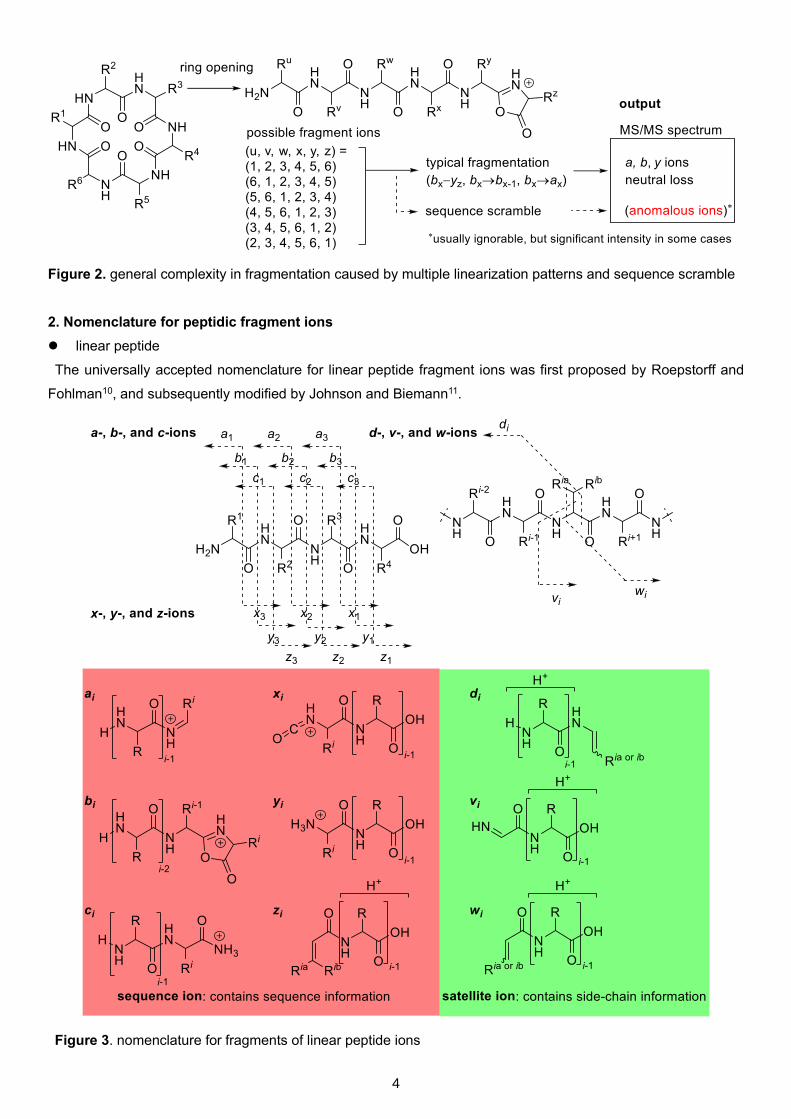
Figure 2. general complexity in fragmentation caused by multiple linearization patterns and sequence scramble
2. Nomenclature for peptidic fragment ions
⚫ linear peptide
The universally accepted nomenclature for linear peptide fragment ions was first proposed by Roepstorff and
Fohlman10, and subsequently modified by Johnson and Biemann11.
Figure 3. nomenclature for fragments of linear peptide ions
5
In low energy CID, cleavage of carbonyl C-N dominantly occurs to give b- and y- ions while charge-remote
fragmentation at C-carbonyl C gives a- and x-ions in high energy CID12. a- Ions can also be generated from the
degradation of b- ions. c- And z- ions dominantly appears at electron transfer dissociation (ETD)13 and electron
capture dissociation (ECD)14 spectrum. Formation of satellite ions require high collision energy and/or radical
processes. Thus, they can be observed in high energy CID and in some ECD/ETD experiments12.
To sum up, a-, b-, and y- ions are dominantly observed in low energy CID.
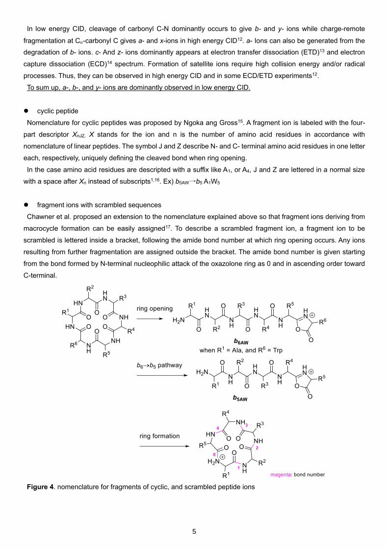
⚫ cyclic peptide
Nomenclature for cyclic peptides was proposed by Ngoka ang Gross15. A fragment ion is labeled with the four-
part descriptor XnJZ. X stands for the ion and n is the number of amino acid residues in accordance with
nomenclature of linear peptides. The symbol J and Z describe N- and C- terminal amino acid residues in one letter
each, respectively, uniquely defining the cleaved bond when ring opening.
In the case amino acid residues are descripted with a suffix like A1, or A4, J and Z are lettered in a normal size
with a space after Xn instead of subscripts1,16. Ex) b5AW→b5 A1W5
⚫ fragment ions with scrambled sequences
Chawner et al. proposed an extension to the nomenclature explained above so that fragment ions deriving from
macrocycle formation can be easily assigned17. To describe a scrambled fragment ion, a fragment ion to be
scrambled is lettered inside a bracket, following the amide bond number at which ring opening occurs. Any ions
resulting from further fragmentation are assigned outside the bracket. The amide bond number is given starting
from the bond formed by N-terminal nucleophilic attack of the oxazolone ring as 0 and in ascending order toward
C-terminal.
Figure 4. nomenclature for fragments of cyclic, and scrambled peptide ions

6
Figure 4 (continue). nomenclature for fragments of cyclic, and scrambled peptide ions
3. Reaction mechanisms of fragmentation
In low energy CID, peptides undergo three types of dominant fragmentation pathway18. First, ring opening takes
place via the bx-yz pathway with oxazolone formation, resulting in a linear peptide ion having a free N-terminus and
an oxazolone ring at the C-terminus19. The resulting linear ion can originate a smaller b fragment via bx→bx-1
pathway20,21, which in turn loses CO via the bx→ax pathway19,22.
The bx-yz pathway is explained in “mobile proton model”. In this model, fragmentation requires the transfer of a
proton from a basic site to the amide nitrogen23. (for rationale of this model, see 190914_PS_Hiroaki_Itoh24)
Figure 5. mechanisms of fragmentations of a cyclic peptide in low-energy CID.

7
A particularly common loss of H2O occurs for protonated peptides containing a serine or threonine residue where
there is a side-chain hydroxyl group25. Ser residue in N-terminal is likely to undergo dehydration in neighboring
group participation, while dehydration of Ser residue in the peptide sequence is likely to proceed in cis 1,2
elimination26,27. Dehydroalanine (ΔAla, 3), oxazoline (5), or aziridine (6) are possible products of dehydration
(Figure 6). (Dehydroalanine products are suggested for structures of fragment ions in solutions of this session.)
Rationale of mechanisms of dehydration of Ser residue26
(i) MS3 intensity patterns of dehydrated ions suggested generation of 6 from Ser, and 3 from Ac-Ser. (Table 2.)
(ii) H/D exchange experiment indicated dehydration of Ser proceeded in neighboring group participation, while
that of Ac-Ser proceeded both in neighboring group participation, and cis 1,2 elimination. (Table 3.)
Figure 6. mechanisms of dehydration of Ser residue
Table 2. CID MS3 spectra of side chain loss MS/MS product ions
Table 3. CID MS/MS spectra of fully deuterated serine and N-acetyl serine

8
4. Solution for the problem
For sake of simplicity, the structure of [1+H]+ is described as a 6-membered sequence information. (Figure 7)
Five possible reactions in low-energy CID are listed in Table 4. They provide two types of information, order of
sequence of fragment ions, and mass numbers of leaving components from fragment ions. The latter can be
accountable from mass numbers of fragment ions in Table 1, the only information we get from MS/MS analysis.
Figure 7. simplified sequence information of [1+H]+
Table 4. possible reactions in low-energy CID
CID reaction providing information accountability
bx-yz order of sequence not directly accountable from Table 1
sequence scramble
bx→bx-1 leaving component accountable from Table 1
bx→ax
dehydration
Losses of masses of all fragment ions listed in Table 1 are derived from either the bx→bx-1 pathway, the bx→ax
pathway, or dehydration. Only 5 mass number patterns are possible to be lost in the processes (Figure 8). This
means all fragment ions in Table 1 can be bridged with the 5 patterns. If a difference between mass numbers of
two fragment ions is 71, it is probable that these two ions are bx and bx-1, and the leaving residue is Ala. All mass
numbers in Table 1 are thus arranged into two fragmentation pathways shown in Figure 9, starting from [1+H]+,
or [1+H-H2O]+.
Figure 8. possible leaving fragments with exact masses during low energy CID fragmentation processes of [1+H]+

9
Figure 9. possible fragmentation pathways of [1+H]+ (mass numbers are displayed in an integer)
It is necessary to assume sequence scramble to explain successive leaving of two Ala in pathway 2. As sequence
scramble is reported to potentially occur in bn ion (n≥5) so far28, only possible combination of (x,y) is (1,4) in the
case of pathway 2. In the same way, (x,y) is uniquely determined as (4,1) in the case of pathway 1 to avoid
sequence scrambling of too short fragment ions. Thus, initial b-y pathway to lead ring opening occurs at Ala4-Tyr5
in the case of pathway 1, and Ala1-Ser2 in the case of pathway 2.
Fragmentation pathway and structures of fragment ions of pathway 1, and 2 are displayed in Figure 10, 11,
respectively.

10
Figure 10. fragmentation pathway 1, and structures of fragment ions

11
Figure 11. fragmentation pathway 2, and structures of fragment ions

12
Note 1. trends of site-selectivity of ring opening
Bond cleavage can potentially occur at any amide
bonds and the fragility or stability of an individual
peptide bond is dependent upon the amino acid
residues flanking it. Statistical study of CID
fragmentation of 1,465 tryptic peptides exhibited
residual bond cleavage trends in bx-yz pathway28,
which is supporting that ring opening of 1 occurred
at C-terminal amide bond of Ala.
Figure 12 (right). the extent to which each residue
directionally enhances cleavage at its N-terminal amide bond (N-bias),
which is calculated by subtracting intensity of the C-terminal fragment peak from that of N terminal
Note 2. reaction mechanism and rationale of sequence scramble
Figure 13. reaction mechanism of sequence scramble in pathway 2.
(i) MS3 analysis: several anomalous di-, tri-, tetrapeptide were expulsed from b5 ion of oligopeptide29
(ii) response to variable collisional energy: breakdown graph for the b5 ion of linear hexapeptide
YAGFL was similar to that of corresponding cyclopentapeptide9.
(iii) N-acetylation: acetyl-capping of N-terminal prevented sequence scrambling30.
rela
tive in
tensity d
iffe
rence (
N-b
ias)
residue

13
Alternative answers at m/z = 714.3138, 643.2819, 541.2301, and 454.1978
It also gives corresponding mass numbers to assume that the combination of (x,y) in Figure 9 gets
reversed. Note that fragmentation pathways of these fragments do not account for the entire ion series
shown in Table 1.
Table 1 (completed). major fragment ions in ESI-MS/MS analysis
m/z fragment ions
825.3448 [1+Na]+
803.3634 [1+H]+
785.3556 [1+H-H2O]+
714.3138 b5 S2A1 -H2O; b5 T5A4 -H2O
643.2819 [b5 S2A13]b4 -H2O; [b5 T5A43]b4 -H2O
541.2301 b4 T5A4; [b5 S2A13]b4
523.2172 b4 T5A4 -H2O
470.1916 [b5 S2A13]b3
454.1978 b3 T5A4; b3 A4T3
452.1810 [b5 S2A13]b3 -H2O
383.1591 b2 T5A4
355.1640 a2 T5A4

14
References
1. Patrícia, O. F.; Maria, F. C. M.; Renata, T. P.; Wilson, H. K.; Marcos, V. G. O. B.; Fernanda, R. G.;
Walmir, S. G. J. Nat. Prod. 2016, 79, 1165.
2. Tan, N.-H.; Zhou, J. Chem. Rev. 2006, 106, 840.
3. Xuejia, Z.; Qirui B.; Xingdong, W.; Zhe W.; Yuanyuan M.; Ninghua, T. J. Chromatogr. A 2018, 1581-
1582, 43.
4. Bhushan, R.; Brückner, H. Amino Acids 2004, 27, 231.
5. Li, J.; Vederas, J. Science 2009, 325, 161.
6. Mohimani, H.; Liu, W.-T.; Mylne, J. S.; Poth, A. G.; Colgrave, M. L.; Tran, D.; Selsted, M. E.; Dorrestein, P.
C.; Pevzner, P. A. J. Proteome Res. 2011, 10, 4505.
7. Tomer, K. B.; Crow, F. W.; Gross, M. L.; Kopple, K. D. Anal. Chem. 1984, 56, 880.
8. Eckart, K.; Schwarz, H.; Tomer, K. B.; Gross, M. L. J. Am. Chem. Soc. 1985, 107, 6765.
9. Harrison, A. G.; Young, A. B.; Bleiholder, C.; Suhai, S.; Paizs, B. J. Am. Chem. Soc. 2006, 128, 10364.
10. Roepstorff, P.; Fohlman, J. Biomed. Mass Spectrom. 1984, 11, 601.
11. Johnson, R. S.; Martin, S. A.; Biemann, K.; Stults, J. T.; Watson, J. T. Anal. Chem. 1987, 59, 2621.
12. Medzihradszky, K. F.; Chalkley, R. J. Mass Spectrom. Rev. 2015, 34, 43.
13. Han, H.; Xia, Y.; McLuckey, S. A. J. Proteome Res. 2007, 6, 3062.
14. Zubarev, R. A.; Kelleher, N. L.; McLafferty, F. W. J. Am. Chem. Soc. 1998, 120, 3265.
15. Ngoka, L. C. M.; Gross, M. L. J. Am. Soc. Mass Spectrom. 1999, 10, 360.
16. Priscila, M. C. -B.; Patrícia, O. F.; Maria, F. C. M.; Fernanda, R. G.; Walmir, S. G. J. Nat. Prod. 2015, 78,
2754.
17. Chawner, R.; Gaskell, S. J.; Eyers, C. E. Rapid Commun. Mass Spectrom. 2012, 26, 205.
18. Jegorov, A.; Paizs, B.; Žabka, M.; Kuzma, M.; Giannakopulos, A, E.; Derrick, P, J.; Havlíček, V. Eur. J.
Mass. Spectrom. 2003, 9, 105.
19. Paizs, B.; Suhai, S. Mass Spectrom. Rev. 2005, 24, 508.
20. Harrison, A. G.; Young, A. B.; Bleiholder, C.; Suhai, S.; Paizs, B. J. Am. Chem. Soc. 2006, 128, 10364.
21. Yalcin, T.; Harrison, A. G. J. Mass. Spectrom. 1996, 31, 1237.
22. Yalcin, T.; Khouw, C.; Csizmadia, I. G.; Peterson, M. R.; Harrison, A. G. J. Am. Soc. Mass. Spectrom.
1995, 6, 1165.
23. Tsaprailis, G.; Nair, H.; Somogyi, A.; Wysocki, V. H.; Zhong, W. Q.; Futrell, J. H.; Summerfield, S. G.;
Gaskell, S. J. J. Am. Chem. Soc. 1999, 121, 5142.
24. 190914_PS_Hiroaki_Itoh
25. Neta, P.; Pu, Q.-L.; Yang, X.; Stein, S. E. Int. J. Mass Spectrom. 2007, 267, 295.
26. Reid, G. E.; Simpson, R. J.; O’Hair, R. A. J. J. Am. Soc. Spectrom. 2000, 11, 1047.
27. DeGnore, J. P.; Qin J. J. Am. Soc. Mass Spectrom. 1998, 9, 1175.
28. Tabb, D. L.; Smith, L. L.; Breci, L. A.; Wysocki, V. H.; Lin, D.; Yates, J. R. Anal. Chem. 2003, 75, 1155.
29. L. Mouls.; Aubagnac, J. L.; Martinez. J.; Enjalbal, C. J. Proteome Res. 2007, 6, 1378.
30. Harrison, A. G. J. Am. Soc. Mass Spectrom. 2008, 19, 1776.