real-time observation of formation of indium phosphide nanowires
TRANSCRIPT

Journal of Physics Conference Series
OPEN ACCESS
Real-time observation of formation of indiumphosphide nanowires by means of GISAXSTo cite this article T Kawamura et al 2007 J Phys Conf Ser 83 012035
View the article online for updates and enhancements
Related contentFlexible Electronics Volume 2 NanowireFETV K Khanna
-
Real-Time Observation of Growth ofTungsten Oxide Nanowires with aScanning Electron MicroscopeKeigo Kasuya Takeshi Ooi YusukeKojima et al
-
Preparation and characterization of CuOnanowire arraysYu Dongliang Ge Chuannan and DuYouwei
-
This content was downloaded from IP address 11923669115 on 20092021 at 1955
Real-time observation of formation of indium phosphide nanowires by means of GISAXS
T Kawamura S Bhunia S Fujikawa Y Watanabe J Matsui Y Kagoshima and Y Tsusaka NTT Basic Research Laboratories NTT Corporation Wakamiya Morinosato Atsugi Kanagawa 243-0198 Japan
University of Hyogo 1-1-1 Koto Kamigori Hyogo 678-1297 Japan
e-mail tomoakikawamuranichiacojp
Abstract Real-time observation of InP nanowire formation was performed using grazing incidence small angle x-ray scattering (GISAXS) Prior to the nanowire growth gold colloidal particles were spread on the substrate as the catalyst and annealed at 500 Changes of GISAXS images were clearly observed after annealing suggesting the formation of molten metal droplets which were used for nanowire growth After staring the growth little change except for the increase of GISAXS intensity was observed suggesting that x-ray scattering from nanowires overlapped with that from the catalysts From the GISAXS images calculated using a sphere and cylinder model scattering from the nanowires shows the streak along the qydirection and that from droplets shows an increase of intensity around the specular reflection which qualitatively explains the measured GISAXS image after the growth
1 Introduction Recent developments in semiconductor nanowires have been shown their diversity for optical [1] and Electrical [2] devices using quantum effects Ever since the fabrication of Si sub-micrometer whiskers by means of the vapor-liquid-solid (VLS) mechanism [3] various kinds of nanowires including Si [45] GaAs [6] and InP [1] have been grown using chemical vapor deposition chemical beam epitaxy and laser-assisted catalytic growth Figures 1(a)-(d) schematically illustrate the principle of VLS growth First catalyst metals are evaporated on the substrate as a thin film (Fig 1(a)) After the formation of metal layer the substrate is annealed to make small droplets of catalyst metal (Fig1 (b)) It is noted that catalyst metal usually melts at a temperature lower than the usual melting point of metal because of the size effect After molten metal balls are formed material gases are supplied for nanowire growth (Fig 1(c)) Since
(a) (b)
(c)(d)
Fig1 Schematic drawing of the vapour-liquid-solid (VLS) growth mechanism
lsquoBuriedrsquo Interface Science with X-rays and Neutrons 2007 IOP PublishingJournal of Physics Conference Series 83 (2007) 012035 doi1010881742-6596831012035
ccopy 2007 IOP Publishing Ltd 1
atoms tend to be incorporated into the small balls because of the dependence of local chemical potential on ball size [4 7] growth of small nanowires is more dominant than that of larger ones When the incorporation process starts the catalytic balls become saturated and the super-cooling sometimes occurs in them inducing crystallization of the nanowires from the bottom of the balls Once the crystallization process starts it continues until the materials gas supply is stopped or the catalytic metals are exhausted (Fig 1(d))
Although the previous researches the detailed growth mechanism is not yet well understood because nanowire growth depends on several factors such as the growth temperature vapor pressure local chemical potential of the catalytic balls substrate and vapors and the size of the balls which dynamically changes during the growth process Additionally ordinary evaluation techniques such as SEM and TEM can not be applied in the gas-phase environment which is very important for VLS growth In contrast to these electron-based techniques x-ray based techniques can be easily adapted to growth in the gas-phase environment and provide much structural information such as the size and shape of particles [8] and nanowires In particular grazing incidence small angle x-ray scattering (GISAXS) is effective for monitoring nanowire growth since the growth time using the VLS mechanism is usually very short It allows us to observe structural changes in catalysts and nanowires very quickly with a two-dimensional detector In this article we report the preliminary results of real-time GISAXS measurement of InP nanowire formation in the gas-phase
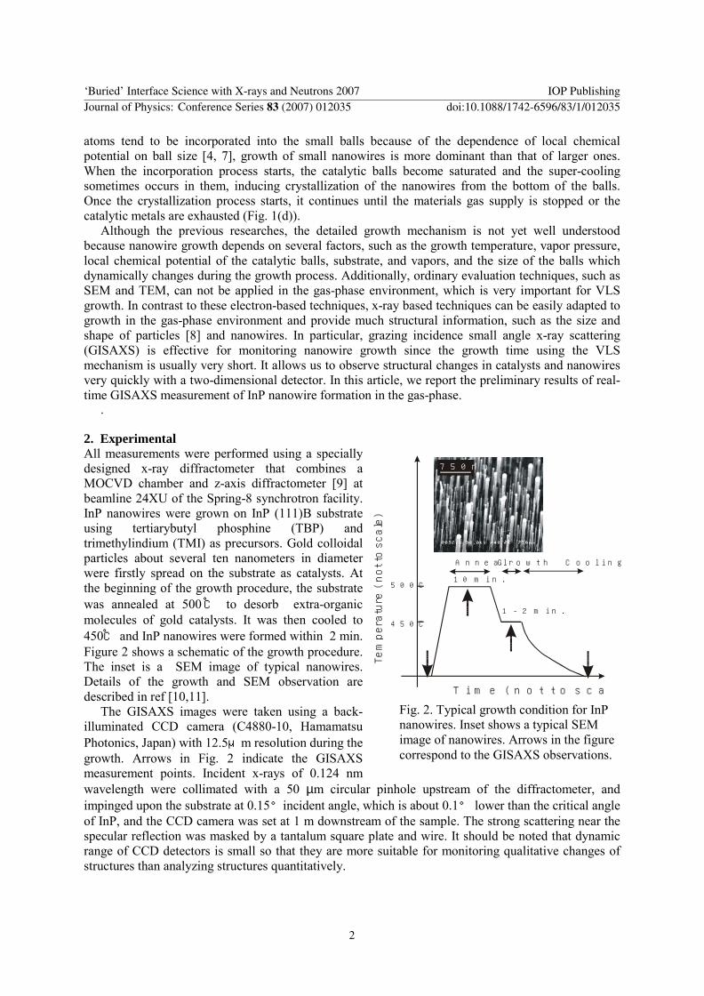
2 Experimental All measurements were performed using a specially designed x-ray diffractometer that combines a MOCVD chamber and z-axis diffractometer [9] at beamline 24XU of the Spring-8 synchrotron facility InP nanowires were grown on InP (111)B substrate using tertiarybutyl phosphine (TBP) and trimethylindium (TMI) as precursors Gold colloidal particles about several ten nanometers in diameter were firstly spread on the substrate as catalysts At the beginning of the growth procedure the substrate was annealed at 500 to desorb extra-organic molecules of gold catalysts It was then cooled to 450 and InP nanowires were formed within 2 min Figure 2 shows a schematic of the growth procedure The inset is a SEM image of typical nanowires Details of the growth and SEM observation are described in ref [1011]
The GISAXS images were taken using a back-illuminated CCD camera (C4880-10 Hamamatsu Photonics Japan) with 125μm resolution during the growth Arrows in Fig 2 indicate the GISAXS measurement points Incident x-rays of 0124 nm wavelength were collimated with a 50 microm circular pinhole upstream of the diffractometer and impinged upon the substrate at 015degincident angle which is about 01deg lower than the critical angle of InP and the CCD camera was set at 1 m downstream of the sample The strong scattering near the specular reflection was masked by a tantalum square plate and wire It should be noted that dynamic range of CCD detectors is small so that they are more suitable for monitoring qualitative changes of structures than analyzing structures quantitatively
(a)
(b )
7 50nm
A nneal
500C
450C
10 m in
1 -2 m in
G row th C ooling
Tim e (notto scale)
Tempera
ture
(notto
scale)
Fig 2 Typical growth condition for InP nanowires Inset shows a typical SEM image of nanowires Arrows in the figure correspond to the GISAXS observations
lsquoBuriedrsquo Interface Science with X-rays and Neutrons 2007 IOP PublishingJournal of Physics Conference Series 83 (2007) 012035 doi1010881742-6596831012035
2
3 Results and Discussion Figure 3 shows a GISAXS image taken at room temperature 100 sec before starting the growth procedure In the figure coordinates of qy and ∆qz (= qz - qz
specular) correspond to the directions of in-plane
and out-of-plane scatterings A gray region in Fig 3 shows the masked area on the CCD detector Obviously x-ray scatterings from the gold particles are clearly observed and the scattering is broader along the qy direction than along the qz direction Considering that the average particle size was about 3 nm and the number of particles was larger than the number that would form a single layer on the substrate it seems that gold particles segregated and formed a stacked layer of particles
Figure 4 shows the GISAXS image taken at 500during the annealing process During the annealing process GISAXS images were taken every 6 seconds with 1 sec irradiation time to observe the time evolution of GISAXS images After the measurements all data were summed to increase signal counts since the acquired signals in each measurement were too weak Owing to the dark current and reading error of CCD signals artifacts appeared in the x-ray image along the qy =plusmn007 as shown by the light gray area in Fig 4 The dark gray region in the figure again corresponds to the masked area on the CCD detector and all GISAXS images are normalized so that we can compare each one directly During the annealing process the broadening of scattering along the qz direction was observed although the intensity along the qy direction was almost the same This suggests a decrease of the heightradius ratio of droplets and the forming of wetting layer on the substrate
After annealing substrate temperature was lowered to 450 and GISAXS images were taken during a 2-min nanowire growth The measurement condition for the CCD detector was the same as that during annealing Figures 5(a) and (b) show the GISAXS images taken during the nanowire growth and (c) which taken after the growth Light and dark gray regions correspond to the artifacts of the detector and the tantalum-masked area respectively The GISAXS patterns during the growth as shown in Fig 5(a) and (b) were basically the same as that after annealing except for an increase of scattering intensity This suggests that scattering mainly came from the catalyst droplets on the surface which did not contribute to the growth of nanowires After the growth was stopped the shape of the GISAXS image taken in the first 1 min changed slightly as shown as Fig 5(c) The major change was the appearance of a spear-head like corner at ∆qz = 00nm-1 suggesting that scattering from the nanowires was added to the GISAXS image
Finally a GISAXS image was taken after cooling the substrate to room temperature (Fig 6(a)) The integration time was 100~sec and the masked area is again the dark gray region Changes in the GISAXS image from that before the growth (Fig 3) were clearly observed The main features are the broadening of scattering along the qz direction and the narrowing of it along qy direction Considering
-04 -02 00 02-02
00
02
04
06
qy (1nm)
∆qz
(1n
m)
04
-04 -02 00 02 04-02
00
02
04
06
qy (1nm)
∆qz
(1n
m)
Fig 3 GISAX image taken before the growth at room temperature with catalyst particles The streaky pattern along ∆qz=00 suggests the segregation of gold particles
Fig 4 GISAX image taken during annealing The broadening of the image suggests the segregation and formationof large molten balls for nanowire growth at room temperature with catalyst particles
lsquoBuriedrsquo Interface Science with X-rays and Neutrons 2007 IOP PublishingJournal of Physics Conference Series 83 (2007) 012035 doi1010881742-6596831012035
3
the change in GISAXS images during the growth these changes are mainly caused by the formation of large alloyed droplets and InP nanowires
To estimate the scatterings from nanowires and droplets GISAXS images were calculated using a sphere and cylinder models Figures 6(a) (b) and (c) show the measured and calculated GISAXS images The number of contour lines in Fig 6 (a) is increased to show the scatterings far from the specular reflection The calculation was performed using the IsGISAXS program [12] with parameters of a 60 nm radius for the sphere model and a 15 nm radius and 150 nm heights for the cylinder model Scatterings from the spheres have symmetry corresponding to the specular point and considering the form factor of the spherical shape the scattering pattern does not depend on the size of particles In contrast to the calculation using the sphere mode sharp streaks along qy direction were observed in the
-04 -02-02
00
02
04
qy (1nm)
∆qz
(1n
m)
00 02 04
(a)
-04 -02-02
00
02
04
qy (1nm)
∆qz
(1n
m)
00 02 04
(b)
-04 -02-02
00
02
04
qy (1nm)
∆qz
(1n
m)
00 02 04
(c)
Fig 6 Measured and calculated GISAXS images (a) Measured image after the growth and (b) sphere model and (c) cylinder model were used for the calculation
-04 -02 00 02
qy (1nm )
∆qz(1
nm)
-02
00
02
04
06-02
00
02
04
06
-02
00
02
04
06 (a)
(b)
(c)
04
Fig 5 Changes in GISAX images during nanowire growth After the growth process a spear-head like corner was observedalong ∆qz = 00 suggesting scattering of nanowires
lsquoBuriedrsquo Interface Science with X-rays and Neutrons 2007 IOP PublishingJournal of Physics Conference Series 83 (2007) 012035 doi1010881742-6596831012035
4
GISAXS image calculated using the cylinder model Although the dispersions of size and height for each model were not included calculated GISAXS images of the cylinder model is similar to the measured one suggesting that the scattering from nanowires overlapped in the GISAXS images after the growth which would explain the spear-like corners along the qy direction
4 Summary In summary we observed the formation of InP nanowires by means of GISAXS The GISAXS image taken before the growth shows that colloidal particles were stacked and segregated After the annealing the gold particles melted forming droplets which act as catalysts for nanowire growth When the nanowire growth started additional scatterings occurred along qy direction From the result of GISAXS calculations using a sphere and cylinder models the origin of this streak is estimated to the formation of nanowires Although much information could be obtained from the real-time measurement a quantitative analysis has not yet been performed because large dynamic-range measurement using CCD detectors is difficult This difficulty would be overcome by combining different types of detectors such as CCD detectors and imaging plates
References [1] Duan X Huang Y Cui Y Wang J and Lieber C M 2001 Nature 409 66[2] Bjork M T Ohlsson B J Thelander C Persson A I Deppert K Wallenberg L R and Samuelson
L 2002 Appl Phys Lett 81 4458 [3] Wagner R S and Ellis W C 1964 Appl Phys Lett 4 89A [4] Westwater J Gosain D P Tomiya S Usui S and Ruda H 1997 J VAc Sci Technol B15 554[5] Morales A M and Lieber C M 1998 Science 279 208[6] Hiruma K Yazawa M Katsuyama T Ogawa K Haraguchi K Koguchi M and Kakibayashi H
1995 J Appl Phys 77 447[7] Givargizov E I 1975 J Cryst Growth 31 20[8] Glatter O and Kratky O 1982 Small Angle X-ray Scattering [9] Kawamura T Watanabe Y Utsumi Y Uwai K Matsui J Kagoshima Y Tsusaka Y and
Fujikawa S 2000 J Cryst Growth 221 106[10] Bhunia S Kawamura T Watanabe Y Fujikawa S and Tokushima K 2003 Appl Phys Lett 83
3371 [11] Bhunia S Kawamura T Fujikawa S and Watanabe Y 2004 Physica E 21 583[12] IsGISAXSa program for Grazing-Incidence Small-Angle X-Ray Scattering analysis of
supported islands Lazzari R 2002 Appl Cryst 35 406
lsquoBuriedrsquo Interface Science with X-rays and Neutrons 2007 IOP PublishingJournal of Physics Conference Series 83 (2007) 012035 doi1010881742-6596831012035
5

Real-time observation of formation of indium phosphide nanowires by means of GISAXS
T Kawamura S Bhunia S Fujikawa Y Watanabe J Matsui Y Kagoshima and Y Tsusaka NTT Basic Research Laboratories NTT Corporation Wakamiya Morinosato Atsugi Kanagawa 243-0198 Japan
University of Hyogo 1-1-1 Koto Kamigori Hyogo 678-1297 Japan
e-mail tomoakikawamuranichiacojp
Abstract Real-time observation of InP nanowire formation was performed using grazing incidence small angle x-ray scattering (GISAXS) Prior to the nanowire growth gold colloidal particles were spread on the substrate as the catalyst and annealed at 500 Changes of GISAXS images were clearly observed after annealing suggesting the formation of molten metal droplets which were used for nanowire growth After staring the growth little change except for the increase of GISAXS intensity was observed suggesting that x-ray scattering from nanowires overlapped with that from the catalysts From the GISAXS images calculated using a sphere and cylinder model scattering from the nanowires shows the streak along the qydirection and that from droplets shows an increase of intensity around the specular reflection which qualitatively explains the measured GISAXS image after the growth
1 Introduction Recent developments in semiconductor nanowires have been shown their diversity for optical [1] and Electrical [2] devices using quantum effects Ever since the fabrication of Si sub-micrometer whiskers by means of the vapor-liquid-solid (VLS) mechanism [3] various kinds of nanowires including Si [45] GaAs [6] and InP [1] have been grown using chemical vapor deposition chemical beam epitaxy and laser-assisted catalytic growth Figures 1(a)-(d) schematically illustrate the principle of VLS growth First catalyst metals are evaporated on the substrate as a thin film (Fig 1(a)) After the formation of metal layer the substrate is annealed to make small droplets of catalyst metal (Fig1 (b)) It is noted that catalyst metal usually melts at a temperature lower than the usual melting point of metal because of the size effect After molten metal balls are formed material gases are supplied for nanowire growth (Fig 1(c)) Since
(a) (b)
(c)(d)
Fig1 Schematic drawing of the vapour-liquid-solid (VLS) growth mechanism
lsquoBuriedrsquo Interface Science with X-rays and Neutrons 2007 IOP PublishingJournal of Physics Conference Series 83 (2007) 012035 doi1010881742-6596831012035
ccopy 2007 IOP Publishing Ltd 1
atoms tend to be incorporated into the small balls because of the dependence of local chemical potential on ball size [4 7] growth of small nanowires is more dominant than that of larger ones When the incorporation process starts the catalytic balls become saturated and the super-cooling sometimes occurs in them inducing crystallization of the nanowires from the bottom of the balls Once the crystallization process starts it continues until the materials gas supply is stopped or the catalytic metals are exhausted (Fig 1(d))
Although the previous researches the detailed growth mechanism is not yet well understood because nanowire growth depends on several factors such as the growth temperature vapor pressure local chemical potential of the catalytic balls substrate and vapors and the size of the balls which dynamically changes during the growth process Additionally ordinary evaluation techniques such as SEM and TEM can not be applied in the gas-phase environment which is very important for VLS growth In contrast to these electron-based techniques x-ray based techniques can be easily adapted to growth in the gas-phase environment and provide much structural information such as the size and shape of particles [8] and nanowires In particular grazing incidence small angle x-ray scattering (GISAXS) is effective for monitoring nanowire growth since the growth time using the VLS mechanism is usually very short It allows us to observe structural changes in catalysts and nanowires very quickly with a two-dimensional detector In this article we report the preliminary results of real-time GISAXS measurement of InP nanowire formation in the gas-phase
2 Experimental All measurements were performed using a specially designed x-ray diffractometer that combines a MOCVD chamber and z-axis diffractometer [9] at beamline 24XU of the Spring-8 synchrotron facility InP nanowires were grown on InP (111)B substrate using tertiarybutyl phosphine (TBP) and trimethylindium (TMI) as precursors Gold colloidal particles about several ten nanometers in diameter were firstly spread on the substrate as catalysts At the beginning of the growth procedure the substrate was annealed at 500 to desorb extra-organic molecules of gold catalysts It was then cooled to 450 and InP nanowires were formed within 2 min Figure 2 shows a schematic of the growth procedure The inset is a SEM image of typical nanowires Details of the growth and SEM observation are described in ref [1011]
The GISAXS images were taken using a back-illuminated CCD camera (C4880-10 Hamamatsu Photonics Japan) with 125μm resolution during the growth Arrows in Fig 2 indicate the GISAXS measurement points Incident x-rays of 0124 nm wavelength were collimated with a 50 microm circular pinhole upstream of the diffractometer and impinged upon the substrate at 015degincident angle which is about 01deg lower than the critical angle of InP and the CCD camera was set at 1 m downstream of the sample The strong scattering near the specular reflection was masked by a tantalum square plate and wire It should be noted that dynamic range of CCD detectors is small so that they are more suitable for monitoring qualitative changes of structures than analyzing structures quantitatively
(a)
(b )
7 50nm
A nneal
500C
450C
10 m in
1 -2 m in
G row th C ooling
Tim e (notto scale)
Tempera
ture
(notto
scale)
Fig 2 Typical growth condition for InP nanowires Inset shows a typical SEM image of nanowires Arrows in the figure correspond to the GISAXS observations
lsquoBuriedrsquo Interface Science with X-rays and Neutrons 2007 IOP PublishingJournal of Physics Conference Series 83 (2007) 012035 doi1010881742-6596831012035
2
3 Results and Discussion Figure 3 shows a GISAXS image taken at room temperature 100 sec before starting the growth procedure In the figure coordinates of qy and ∆qz (= qz - qz
specular) correspond to the directions of in-plane
and out-of-plane scatterings A gray region in Fig 3 shows the masked area on the CCD detector Obviously x-ray scatterings from the gold particles are clearly observed and the scattering is broader along the qy direction than along the qz direction Considering that the average particle size was about 3 nm and the number of particles was larger than the number that would form a single layer on the substrate it seems that gold particles segregated and formed a stacked layer of particles
Figure 4 shows the GISAXS image taken at 500during the annealing process During the annealing process GISAXS images were taken every 6 seconds with 1 sec irradiation time to observe the time evolution of GISAXS images After the measurements all data were summed to increase signal counts since the acquired signals in each measurement were too weak Owing to the dark current and reading error of CCD signals artifacts appeared in the x-ray image along the qy =plusmn007 as shown by the light gray area in Fig 4 The dark gray region in the figure again corresponds to the masked area on the CCD detector and all GISAXS images are normalized so that we can compare each one directly During the annealing process the broadening of scattering along the qz direction was observed although the intensity along the qy direction was almost the same This suggests a decrease of the heightradius ratio of droplets and the forming of wetting layer on the substrate
After annealing substrate temperature was lowered to 450 and GISAXS images were taken during a 2-min nanowire growth The measurement condition for the CCD detector was the same as that during annealing Figures 5(a) and (b) show the GISAXS images taken during the nanowire growth and (c) which taken after the growth Light and dark gray regions correspond to the artifacts of the detector and the tantalum-masked area respectively The GISAXS patterns during the growth as shown in Fig 5(a) and (b) were basically the same as that after annealing except for an increase of scattering intensity This suggests that scattering mainly came from the catalyst droplets on the surface which did not contribute to the growth of nanowires After the growth was stopped the shape of the GISAXS image taken in the first 1 min changed slightly as shown as Fig 5(c) The major change was the appearance of a spear-head like corner at ∆qz = 00nm-1 suggesting that scattering from the nanowires was added to the GISAXS image
Finally a GISAXS image was taken after cooling the substrate to room temperature (Fig 6(a)) The integration time was 100~sec and the masked area is again the dark gray region Changes in the GISAXS image from that before the growth (Fig 3) were clearly observed The main features are the broadening of scattering along the qz direction and the narrowing of it along qy direction Considering
-04 -02 00 02-02
00
02
04
06
qy (1nm)
∆qz
(1n
m)
04
-04 -02 00 02 04-02
00
02
04
06
qy (1nm)
∆qz
(1n
m)
Fig 3 GISAX image taken before the growth at room temperature with catalyst particles The streaky pattern along ∆qz=00 suggests the segregation of gold particles
Fig 4 GISAX image taken during annealing The broadening of the image suggests the segregation and formationof large molten balls for nanowire growth at room temperature with catalyst particles
lsquoBuriedrsquo Interface Science with X-rays and Neutrons 2007 IOP PublishingJournal of Physics Conference Series 83 (2007) 012035 doi1010881742-6596831012035
3
the change in GISAXS images during the growth these changes are mainly caused by the formation of large alloyed droplets and InP nanowires
To estimate the scatterings from nanowires and droplets GISAXS images were calculated using a sphere and cylinder models Figures 6(a) (b) and (c) show the measured and calculated GISAXS images The number of contour lines in Fig 6 (a) is increased to show the scatterings far from the specular reflection The calculation was performed using the IsGISAXS program [12] with parameters of a 60 nm radius for the sphere model and a 15 nm radius and 150 nm heights for the cylinder model Scatterings from the spheres have symmetry corresponding to the specular point and considering the form factor of the spherical shape the scattering pattern does not depend on the size of particles In contrast to the calculation using the sphere mode sharp streaks along qy direction were observed in the
-04 -02-02
00
02
04
qy (1nm)
∆qz
(1n
m)
00 02 04
(a)
-04 -02-02
00
02
04
qy (1nm)
∆qz
(1n
m)
00 02 04
(b)
-04 -02-02
00
02
04
qy (1nm)
∆qz
(1n
m)
00 02 04
(c)
Fig 6 Measured and calculated GISAXS images (a) Measured image after the growth and (b) sphere model and (c) cylinder model were used for the calculation
-04 -02 00 02
qy (1nm )
∆qz(1
nm)
-02
00
02
04
06-02
00
02
04
06
-02
00
02
04
06 (a)
(b)
(c)
04
Fig 5 Changes in GISAX images during nanowire growth After the growth process a spear-head like corner was observedalong ∆qz = 00 suggesting scattering of nanowires
lsquoBuriedrsquo Interface Science with X-rays and Neutrons 2007 IOP PublishingJournal of Physics Conference Series 83 (2007) 012035 doi1010881742-6596831012035
4
GISAXS image calculated using the cylinder model Although the dispersions of size and height for each model were not included calculated GISAXS images of the cylinder model is similar to the measured one suggesting that the scattering from nanowires overlapped in the GISAXS images after the growth which would explain the spear-like corners along the qy direction
4 Summary In summary we observed the formation of InP nanowires by means of GISAXS The GISAXS image taken before the growth shows that colloidal particles were stacked and segregated After the annealing the gold particles melted forming droplets which act as catalysts for nanowire growth When the nanowire growth started additional scatterings occurred along qy direction From the result of GISAXS calculations using a sphere and cylinder models the origin of this streak is estimated to the formation of nanowires Although much information could be obtained from the real-time measurement a quantitative analysis has not yet been performed because large dynamic-range measurement using CCD detectors is difficult This difficulty would be overcome by combining different types of detectors such as CCD detectors and imaging plates
References [1] Duan X Huang Y Cui Y Wang J and Lieber C M 2001 Nature 409 66[2] Bjork M T Ohlsson B J Thelander C Persson A I Deppert K Wallenberg L R and Samuelson
L 2002 Appl Phys Lett 81 4458 [3] Wagner R S and Ellis W C 1964 Appl Phys Lett 4 89A [4] Westwater J Gosain D P Tomiya S Usui S and Ruda H 1997 J VAc Sci Technol B15 554[5] Morales A M and Lieber C M 1998 Science 279 208[6] Hiruma K Yazawa M Katsuyama T Ogawa K Haraguchi K Koguchi M and Kakibayashi H
1995 J Appl Phys 77 447[7] Givargizov E I 1975 J Cryst Growth 31 20[8] Glatter O and Kratky O 1982 Small Angle X-ray Scattering [9] Kawamura T Watanabe Y Utsumi Y Uwai K Matsui J Kagoshima Y Tsusaka Y and
Fujikawa S 2000 J Cryst Growth 221 106[10] Bhunia S Kawamura T Watanabe Y Fujikawa S and Tokushima K 2003 Appl Phys Lett 83
3371 [11] Bhunia S Kawamura T Fujikawa S and Watanabe Y 2004 Physica E 21 583[12] IsGISAXSa program for Grazing-Incidence Small-Angle X-Ray Scattering analysis of
supported islands Lazzari R 2002 Appl Cryst 35 406
lsquoBuriedrsquo Interface Science with X-rays and Neutrons 2007 IOP PublishingJournal of Physics Conference Series 83 (2007) 012035 doi1010881742-6596831012035
5