resocontoattivitàgruppodistudio(gds)epilessie(disimmuni ... · 7unità di neurologia, ospedale s....
TRANSCRIPT

Resoconto attività Gruppo di Studio (GdS) Epilessie Disimmuni per il triennio 2015-‐2017
Membri del GdS: Flavio Villani (chairman), Stefano Sartori (chairman), Carlo Antozzi, Elena Freri, Antonio Gambardella, Sara Matricardi, Roberto Michelucci, Luigi Zuliani
Obiettivi del GdS
Il GdS Epilessie disimmuni si è costituito con i seguenti obiettivi:
-‐ Identificare i fenotipi epilettici a possibile genesi autoimmune -‐ Analizzare la presentazione clinica, gli aspetti diagnostici e il trattamento di tali forme -‐ Delineare l’attuale condotta diagnostica e terapeutica dei Centri italiani per l’epilessia -‐ Formulare raccomandazioni per l’approccio diagnostico e terapeutico a tali forme -‐ Formulare PDTA condivisi a livello nazionale -‐ Diffondere la conoscenza su questo argomento
Attività del GdS
-‐ Incontri scientifici e organizzativi -‐ Attività di raccolta dati -‐ Attività di diffusione dei risultati fino a qui ottenuti
A) Incontri
1. Il primo incontro è avvenuto il 21 Aprile 2015 presso la Fondazione I.R.C.C.S. Istituto Neurologico Carlo Besta, con il seguente ordine del giorno:
-‐ Razionale, obiettivi, organizzazione e progetti del gruppo di studio
-‐ Presentazioni scientifiche
Presenti: Flavio Villani, Stefano Sartori, Carlo Antozzi, Elena Freri, Antonio Gambardella, Sara Matricardi, Roberto Michelucci, Luigi Zuliani, Francesco Deleo, Giuseppe Didato, Raffaele Iorio. Ospite Sarosh Irani.
Programma della giornata:
Sessione mattino
h. 10:30-‐11:30 Introduzione: razionale, obiettivi e organizzazione del GdS (Sartori e Villani)
h. 11:30-‐13:00 Seminario Sarosh Irani e discussione
h. 13:00-‐14:00 Intervallo pranzo
Sessione pomeriggio
h. 14:00-‐14:30 Linee guida, percorsi diagnostici e terapeutici nell’ambito delle encefaliti autoimmuni: la necessità della condivisione sul territorio nazionale (Zuliani)
h. 14:30-‐14:50 Il problema dei casi non definiti (Matricardi)
h. 14:50-‐15:05 Caso clinico esemplificativo (Freri)
h. 15:05-‐15:20 Caso clinico esemplificativo (Deleo)
h. 15:20-‐16:20 Progetti (PDTA, banche dati e materiale biologico) (moderazione: Antozzi, Gambardella, Michelucci)

h. 16:20-‐16:30 Discussione generale, varie e saluti
Sommario dei contenuti dell’incontro:
Durante l’incontro si è discussa la definizione del tema oggetto di studio, in particolare dell’equivalenza della terminologia “epilessia autoimmune” ed “encefalite autoimmune", sottolineando l’importanza di una “definizione” per l’epilettologo e l’immunologo come supplemento d’indagine e base per lavori prospettici.
L’epilessia autoimmune viene, quindi, intesa come entità clinica distinta, ma strettamente connessa con l’encefalite autoimmune, nella quale l’epilessia rappresenta il sintomo predominante, ma non esclusivo, del quadro neurologico. Si è pertanto sottolineata l’importanza di delineare criteri diagnostici condivisi per individuare quadri clinici con epilessia a possibile genesi disimmune, definendo altresì un algoritmo diagnostico e terapeutico.
È stata sottolineata l’opportunità di coinvolgere altri centri ed estendere collaborazioni nazionali e internazionali, al fine di costituire banche dati che includano retrospettivamente pazienti con diagnosi di epilessia disimmune con o senza encefalopatia associata, e diagnosi anticorpale sia positiva che negativa. Oltre alla raccolta retrospettiva dei casi già diagnosticati, fine del gruppo di studio è anche quello di strutturare un lavoro prospettico, includendo pazienti con epilessia di nuova diagnosi a possibile genesi disimmune secondo i criteri diagnostici definiti e condivisi, e la creazione di un registro (sul modello delle PME). Si evidenzia, inoltre, la necessità di creare banche locali di campioni biologici secondo protocolli condivisi di raccolta e conservazione dei materiali biologici.
Obiettivo del gruppo di studio è, infine, quello di stilare Raccomandazioni per l’epilettologo per la diagnosi e il trattamento di forme di epilessia ad eziologia disimmune.
2. Il secondo incontro è stato effettuato il 14 dicembre 2015 presso la Clinica Pediatrica dell’Università di Padova.
Presenti: Flavio Villani, Stefano Sartori, Elena Freri, Luigi Zuliani e Sara Matricardi.
Odg: sviluppo di progetti collaborativi multicentrici in base alla “call” ministeriale. Aggiornamento scientifico.
Sommario contenuti dell’incontro:
Il primo punto discusso ha riguardato il tipo di progetto che vorremmo mettere in cantiere: a tale proposito abbiamo tutti concordato che un progetto di Rete ha le caratteristiche più adatte a mettere insieme i diversi gruppi di ricerca che compongono il nostro GdS. La complessità e la necessità di coordinare diversi gruppi di ricerca rendono la concorrenza meno pesante rispetto ai progetti convenzionali. Ovviamente, in merito a tale scelta, attendiamo il parere di tutti i membri del GdS.
I progetti di Rete presuppongono l’organizzazione di Workpackages (WP) autonomi e coordinati che abbiano ciascuno un progetto specifico. Ogni WP avrà quindi “aims” specifici che dovranno essere raggiunti in autonomia rispetto agli altri WP, ma che concorreranno al raggiungimento degli obiettivi generali del progetto. La stesura della Lettera d’Intenti (LOI) comprenderà quindi una parte generale con obiettivi e quadro economico generale, e parti specifiche in numero pari ai WP. Per esperienza diretta (ma questo punto può essere discusso) riteniamo che tre WP possano essere sufficienti e non tali da complicare troppo il coordinamento. Ogni WP avrà il proprio PI e potrà aggregare altri gruppi di ricerca.
Al momento avremmo identificato 3 possibili WP: WP 1 Besta (Villani); WP 2 Padova (Sartori); WP 3 Bologna (Michelucci).
Nel corso della riunione abbiamo tentato di abbozzare un titolo e gli obiettivi generali del progetto:

Towards the recognition of autoimmune epilepsy in children and adult patients. A prospective cohort study for the characterization of biological markers of immune activation and advanced non-‐conventional 3T MRI analysis in different forms of new onset epilepsies.
Aim 1: To determine the prevalence of known anti-‐neuronal antibodies in an adult and pediatric cohort of patients with new onset epilepsies.
Aim 2: To identify a subgroup of patients with possible autoimmune epilepsies by means of clinical and paraclinical criteria defined on the basis of an extensive literature search.
Aim 3: To define the diagnostic gain of an advanced neuroimaging analysis (3T MRI) compared to conventional neuroimaging in the subsample of patients with possible autoimmune epilepsy.
Infine è stato discussa l’organizzazione della raccolta e della gestione dei dati (registro, piattaforma, analisi biostatistica), senza giungere ad una definizione conclusiva.
Aggiornamento clinico-‐scientifico:
Scopi: definire il ruolo dell’immunità (infiammazione) nelle epilessie di nuova diagnosi.
-‐ Criteri di reclutamento: pazienti (di ogni età) con epilessia di nuova diagnosi (Classificazione ILAE)
-‐ Consenso
1) Diagnosi di epilessia
2) Inquadramento clinico (anamnesi, Es. Obiettivo, EEG, RM, ecc… altre indagini laboratoristiche e strumentali a seconda del contesto clinico)
3) Raccolta di siero di tutti i pz reclutati (+/-‐ liquor) e ricerca Ab noti
4) Prima stratificazione eziologica in base ai dati raccolti:
• Epi idiopatiche • Epi sintomatiche ad eziologia nota • Epi non idiopatiche ad eziologia non nota (à indagini utili a stabilire una possibile natura
autoimmune – Liquor, ecc…-‐) 5) Seconda stratificazione: eziologia autoimmune sospettata in base a:
Criteri di letteratura…. Es:
• Epilessia ad esordio acuto o subacuto; • Epilessia come sintomo esclusivo o predominante il quadro clinico • Epi responsiva alla Tp. Immunomodulante • Evidenza di infiammazione SNC • Altri segni liquorali, clinici (es. comportamentali) • Ecc… (Definizione criteri di probabilità)
3. Il terzo incontro si è tenuto in occasione del Policentrico di quest’anno, il 26 gennaio 2017.
Presenti: Flavio Villani, Stefano Sartori, Elena Freri, Sara Matricardi, Roberto Michelucci, Antonio Gambardella, Patrizia Riguzzi.
Sommario contenuti dell’incontro:
l’obiettivo è sempre l’identificazione di fenotipi epilettici accomunati da una possibile eziologia autoimmune. La letteratura, pur ipotizzando l’esistenza di condizioni epilettiche autoimmuni in cui la sintomatologia critica è predominante rispetto al quadro clinico complessivo, non fornisce dati
conclusivi per una loro sicura identificazione e, di conseguenza, non dà indicazioni condivise rispetto al loro possibile trattamento.
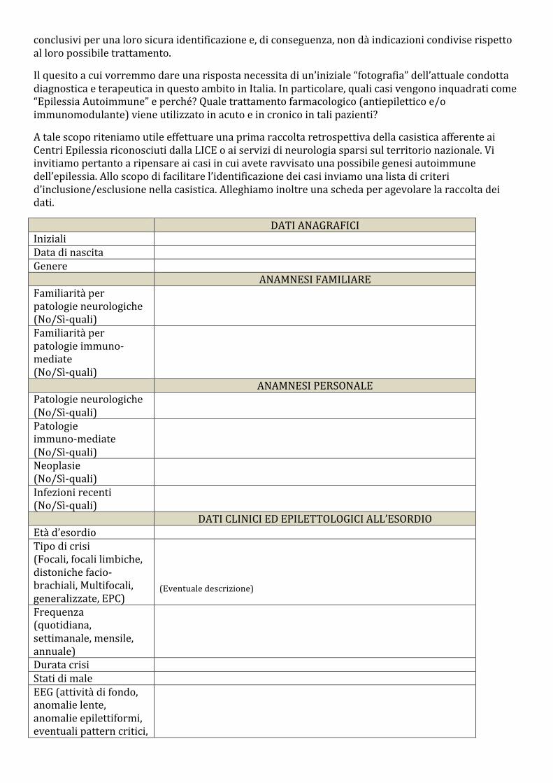
Il quesito a cui vorremmo dare una risposta necessita di un’iniziale “fotografia” dell’attuale condotta diagnostica e terapeutica in questo ambito in Italia. In particolare, quali casi vengono inquadrati come “Epilessia Autoimmune” e perché? Quale trattamento farmacologico (antiepilettico e/o immunomodulante) viene utilizzato in acuto e in cronico in tali pazienti?
A tale scopo riteniamo utile effettuare una prima raccolta retrospettiva della casistica afferente ai Centri Epilessia riconosciuti dalla LICE o ai servizi di neurologia sparsi sul territorio nazionale. Vi invitiamo pertanto a ripensare ai casi in cui avete ravvisato una possibile genesi autoimmune dell’epilessia. Allo scopo di facilitare l’identificazione dei casi inviamo una lista di criteri d’inclusione/esclusione nella casistica. Alleghiamo inoltre una scheda per agevolare la raccolta dei dati.
DATI ANAGRAFICI Iniziali Data di nascita Genere ANAMNESI FAMILIARE Familiarità per patologie neurologiche (No/Sì-‐quali)
Familiarità per patologie immuno-‐mediate (No/Sì-‐quali)
ANAMNESI PERSONALE Patologie neurologiche (No/Sì-‐quali)
Patologie immuno-‐mediate (No/Sì-‐quali)
Neoplasie (No/Sì-‐quali)
Infezioni recenti (No/Sì-‐quali)
DATI CLINICI ED EPILETTOLOGICI ALL’ESORDIO Età d’esordio Tipo di crisi (Focali, focali limbiche, distoniche facio-‐brachiali, Multifocali, generalizzate, EPC)
(Eventuale descrizione)
Frequenza (quotidiana, settimanale, mensile, annuale)
Durata crisi Stati di male EEG (attività di fondo, anomalie lente, anomalie epilettiformi, eventuali pattern critici,

organizzazione del sonno se disponibile) MRI Liquor (Leucociti, proteine, bande OC)
Auto-‐anticorpi (siero e liquor): immunità “classica”, anticorpi onconeuronali e antigeni intracellulari, anticorpi anti-‐antigeni di superficie neuronale
Altri segni o sintomi associati (cognitivi, psichiatrici, neurologici)
Terapia con AEDs (quali AEDs – risposta)
Terapia immunomodulante (tipo – risposta)
DATI CLINICI ED EPILETTOLOGICI AL FOLLOW-‐UP Durata Follow-‐up Tipo di crisi (Focali, focali limbiche, distoniche facio-‐brachiali, Multifocali, generalizzate, EPC)
(Eventuale descrizione)
Frequenza (quotidiana, settimanale, mensile, annuale)
Durata Stati di male EEG (attività di fondo, anomalie lente, anomalie epilettiformi, eventuali pattern critici, organizzazione del sonno se disponibile)
MRI Liquor (Leucociti, proteine, bande OC)
Auto-‐anticorpi (siero e liquor): immunità “classica”, anticorpi onconeuronali e antigeni intracellulari, anticorpi anti-‐antigeni di superficie neuronale
Altri segni o sintomi associati (cognitivi, psichiatrici, neurologici)
Terapia con AEDs (quali AEDs – risposta)
Terapia immunomodulante (tipo – risposta)
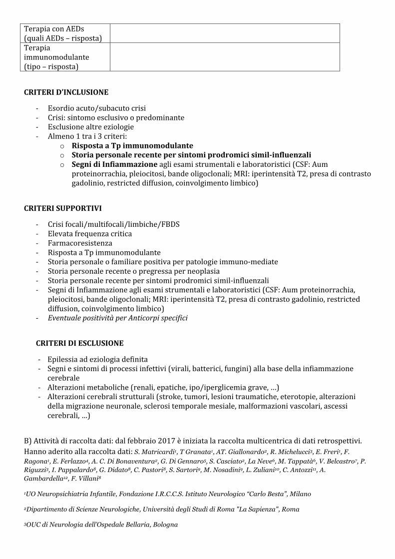
CRITERI D’INCLUSIONE
-‐ Esordio acuto/subacuto crisi -‐ Crisi: sintomo esclusivo o predominante -‐ Esclusione altre eziologie -‐ Almeno 1 tra i 3 criteri:
o Risposta a Tp immunomodulante o Storia personale recente per sintomi prodromici simil-‐influenzali o Segni di Infiammazione agli esami strumentali e laboratoristici (CSF: Aum
proteinorrachia, pleiocitosi, bande oligoclonali; MRI: iperintensità T2, presa di contrasto gadolinio, restricted diffusion, coinvolgimento limbico)
CRITERI SUPPORTIVI
-‐ Crisi focali/multifocali/limbiche/FBDS -‐ Elevata frequenza critica -‐ Farmacoresistenza -‐ Risposta a Tp immunomodulante -‐ Storia personale o familiare positiva per patologie immuno-‐mediate -‐ Storia personale recente o pregressa per neoplasia -‐ Storia personale recente per sintomi prodromici simil-‐influenzali -‐ Segni di Infiammazione agli esami strumentali e laboratoristici (CSF: Aum proteinorrachia,
pleiocitosi, bande oligoclonali; MRI: iperintensità T2, presa di contrasto gadolinio, restricted diffusion, coinvolgimento limbico)
-‐ Eventuale positività per Anticorpi specifici
CRITERI DI ESCLUSIONE
-‐ Epilessia ad eziologia definita -‐ Segni e sintomi di processi infettivi (virali, batterici, fungini) alla base della infiammazione cerebrale
-‐ Alterazioni metaboliche (renali, epatiche, ipo/iperglicemia grave, …) -‐ Alterazioni cerebrali strutturali (stroke, tumori, lesioni traumatiche, eterotopie, alterazioni della migrazione neuronale, sclerosi temporale mesiale, malformazioni vascolari, ascessi cerebrali, …)
B) Attività di raccolta dati: dal febbraio 2017 è iniziata la raccolta multicentrica di dati retrospettivi. Hanno aderito alla raccolta dati: S. Matricardi1, T Granata1, AT. Giallonardo2, R. Michelucci3, E. Freri1, F.
Ragona1, E. Ferlazzo4, A. C. Di Bonaventura2, G. Di Gennaro5, S. Casciato2, La Neve6, M. Tappatà6, V. Belcastro7, P. Riguzzi3, I. Pappalardo8, G. Didato8, C. Pastori8, S. Sartori9, M. Nosadini9, L. Zuliani10, C. Antozzi11, A. Gambardella12, F. Villani8
1UO Neuropsichiatria Infantile, Fondazione I.R.C.C.S. Istituto Neurologico “Carlo Besta”, Milano
2Dipartimento di Scienze Neurologiche, Università degli Studi di Roma "La Sapienza", Roma
3OUC di Neurologia dell’Ospedale Bellaria, Bologna

4Dipartimento di scienze mediche e chirurgiche, Università “Magna Graecia”, Catanzaro, Centro Regionale Epilessie, Azienda Ospedaliera BMM di Reggio Calabria
5I.R.C.C.S. Neuromed, Pozzilli
6Clinica Neurologica, Università di Bari, Bari
7Unità di Neurologia, Ospedale S. Anna, Como
8UO Epilettologia Clinica e Neurofisiologia Sperimentale, Fondazione I.R.C.C.S. Istituto Neurologico “Carlo Besta”, Milano
9Unità di Neurologia Pediatrica e Neurofisiologia, Università degli Studi di Padova, Padova
10Dipartimento di Neurologia, Ospedale Ca’ Foncello, Treviso
11UO Neuroimmunologia e Malattie Neuromuscolari, Fondazione I.R.C.C.S. Istituto Neurologico “Carlo Besta”, Milano
12Istituto di Neurologia, Università “Magna Grecia”, Catanzaro
C) Attività di diffusione conoscenze e risultati fino a qui ottenuti: si allegano documento sulle Epilessie autoimmuni (Margherita Nosadini) e abstract presentato alla LICE 2017 che riassume i dati fino ad ora raccolti dal GdS.

FENOTIPI EPILETTICI A POSSIBILE GENESI AUTOIMMUNE: UNO STUDIO RETROSPETTIVO DEL GRUPPO DI STUDIO LICE SULLE EPILESSIE DISIMMUNI
S. Matricardi1, T Granata1, AT. Giallonardo2, R. Michelucci3, E. Freri1, F. Ragona1, E. Ferlazzo4, A. C. Di Bonaventura2, G. Di Gennaro5, S. Casciato2, La Neve6, M. Tappatà6, V. Belcastro7, P. Riguzzi3, I. Pappalardo8, G. Didato8, C. Pastori8, S. Sartori9, M. Nosadini9, L. Zuliani10, C. Antozzi11, A. Gambardella12, F. Villani8
1UO Neuropsichiatria Infantile, Fondazione I.R.C.C.S. Istituto Neurologico “Carlo Besta”, Milano 2Dipartimento di Scienze Neurologiche, Università degli Studi di Roma "La Sapienza", Roma 3OUC di Neurologia dell’Ospedale Bellaria, Bologna 4Dipartimento di scienze mediche e chirurgiche, Università “Magna Graecia”, Catanzaro, Centro Regionale Epilessie, Azienda Ospedaliera BMM di Reggio Calabria 5I.R.C.C.S. Neuromed, Pozzilli 6Clinica Neurologica, Università di Bari, Bari 7Unità di Neurologia, Ospedale S. Anna, Como 8UO Epilettologia Clinica e Neurofisiologia Sperimentale, Fondazione I.R.C.C.S. Istituto Neurologico “Carlo Besta”, Milano 9Unità di Neurologia Pediatrica e Neurofisiologia, Università degli Studi di Padova, Padova 10Dipartimento di Neurologia, Ospedale Ca’ Foncello, Treviso 11UO Neuroimmunologia e Malattie Neuromuscolari, Fondazione I.R.C.C.S. Istituto Neurologico “Carlo Besta”, Milano 12Istituto di Neurologia, Università “Magna Grecia”, Catanzaro
Razionale e obiettivi: Identificazione di fenotipi epilettici a possibile eziopatogenesi autoimmune, per tentare una
classificazione per gruppi omogenei e per delineare in tale ambito l’attuale condotta diagnostica e terapeutica dei
Centri italiani per l’epilessia.
Metodi: Sono stati retrospettivamente arruolati 113 pazienti (18 bambini, 95 adulti) in cui le crisi epilettiche erano il
sintomo d’esordio o predominante il quadro clinico, e per i quali è stata identificata (positività di anticorpi
antineuronali) o ipotizzata (sulla base dei dati clinici e paraclinici suggestivi di infiammazione del SNC, o della risposta
alla terapia immunomodulante) una eziopatogenesi autoimmune.
Risultati: I pazienti sono stati seguiti per un periodo di almeno 24 mesi. Le crisi focali, farmacoresistenti nell’ 82%
dei casi, erano il tipo di crisi più frequente. La frequenza delle crisi era elevata, soprattutto all’esordio nel 73% dei casi,
con episodi di stato epilettico nel 45%. Nel 36% dei casi, la ricerca per anticorpi specifici è risultata positiva su siero
e/o su liquor. Nella maggior parte dei pazienti erano presenti anche altri segni e sintomi di coinvolgimento del SNC.
La terapia immunomodulante (con farmaci di I e II linea) ha determinato una riduzione significativa della frequenza
critica nel 60 % dei casi. Sono state confrontate le caratteristiche cliniche e paracliniche, nonché la risposta alla terapia
dei pazienti con e senza anticorpi.
Conclusioni: Il ruolo patogenetico dell’infiammazione nel determinare e sostenere l’attività critica è riconosciuto.
Una più certa classificazione dei fenotipi clinici e la definizione di modelli di trattamento possono migliorare la
prognosi di condizioni potenzialmente trattabili.
Bibliografia:
1 Toledano M, Pittock SJ. Autoimmune Epilepsy. Semin Neurol. 2015;35(3):245-58 2 Spatola M, Dalmau J. Seizures and risk of epilepsy in autoimmune and other inflammatory encephalitis. Curr Opin Neurol 2017;22. [Epub ahead of print]

AUTOIMMUNE EPILEPSY
INTRODUZIONE
Activation of the immune system is observed in many disease processes of the CNS, although discriminating a
primary (causal) immune response from a secondary (reactive) immune response to tissue damage is not
straightforward. There is a large and complex literature describing the presence of inflammation and immune
activation in seizures and epilepsy [Suleiman, 2015].
Although epilepsy is one of the most common neurologic disorders affecting millions of people worldwide, in a
substantial number of individuals the etiology remains unknown [Bien and Scheffer, 2011].
Over the last few years there has been increasing support for the hypothesis that some forms of epilepsy may have an
autoimmune component. Circumstantial evidence in support of this idea includes the apparent association of seizures
with certain autoimmune diseases (e.g., systemic lupus erythematosus and Hashimoto’s encephalopathy) and, in some
patients, an acute or subacute onset of the seizures, a rapidly progressive course, and a favourable response to
immunotherapy [Bien and Scheffer, 2011].
A large population-based study (n>2,000,000) showed that patients with autoimmune disease constituted 17.5% of
patients with epilepsy, and that the presence of an autoimmune disorder may contribute to a fourfold increased risk of
epilepsy [Ong, 2014].
Recently, clinically relevant autoantibodies have been detected in a number of CNS disorders that often present with
recurrent seizures [Bien and Scheffer, 2011]. The confident diagnosis of autoimmune encephalitis and epilepsy has
improved substantially owing to the discovery of pathogenic autoantibodies that seem to be discriminating biomarkers
of disease [Suleiman, 2015].
Specific neuronal auto-antibodies with pathogenic potential may be present in a subset of patients with epilepsy.
Importantly, it has recently been shown that some patients with these serum auto-antibodies are often refractory to
treatment with standard anti-epileptic drugs (AEDs) and, in contrast, may respond well to immunomodulatory
therapies [Irani, 2011].
AUTOIMMUNE ENCEPHALITIS WITH SEIZURES
Seizures are a common feature of autoimmune encephalitis, where patients characteristically have other clinical
features such as encephalopathy, behavioural alteration, and movement disorders, in addition to seizures [Suleiman,
2015].
Autoimmune encephalitis can be broadly separated into focal (i.e. limbic encephalitis), multifocal (i.e. (anti-GABAAR
encephalitis), or diffuse processes (i.e. anti-NMDAR encephalitis).
Autoantibodies to neuronal surface antigens described in autoimmune encephalitis associated with seizures are
reported in Table 1.
Epilessia e anticorpi anti-antigeni di superficie neuronale
Tipo di anticorpo
Descrizione del fenotipo Descrizione di crisi epilettiche (in presenza o
Descrizione di epilessia isolata (o come

meno di altre manifestazioni, es encefalite) in pazienti con anticorpi positivi
prevalente manifestazione) in pazienti con anticorpi positivi
NMDAR Anti-NMDAR encephalitis (multiphasic disease with behavioural and psychiatric changes, movement disorders, seizures, hyporesponsive state and dysautonomia)
Yes Yes
LGI1 Limbic encephalitis (confusion, agitation, memory loss and seizures) Isolated epilepsy (faciobrachial dystonic seizures) Progressive encephalomyelitis with rigidity and myoclonus Isolated chorea Hemianaesthesia Neurocardiac prodromes
Yes Yes [Quek, 2012; Irani, 2013] (adults)
Caspr2 Limbic encephalitis Morvan's syndrome Neuromyotonia Isolated epilepsy Encephalopathy
Yes Yes [Irani, 2010; Lancaster, 2011; Sunwoo, 2015] (adults)
AMPAR Limbic encephalitis Other encephalitis (multifocal/diffuse encephalopathy, hyponatremia, limbic encephalitis preceded by motor deficits, or a predominantly psychiatric syndrome)
Yes No
GABAAR Limbic encephalitis Other encephalitis Isolated epilepsy Isolated psychiatric disturbances Isolated cognitive impairment Stiff-person-syndrome Opsoclonus myoclonus ataxia syndrome
Yes Yes [Pettingill, 2015] (children and adults)
GABABR Limbic encephalitis Cerebellar ataxia Rapidly progressive encephalomyelopathy Opsoclonus myoclonus ataxia syndrome Isolated epilepsy
Yes Yes [Höftberger, 2013] (children and adults)
GlyR Limbic encephalitis Epileptic encephalopathy Isolated epilepsy Progressive encephalomyelitis with rigidity and myoclonus Stiff-person-syndrome Cerebellar ataxia Optic neuritis
Yes Yes [Brenner, 2013; Ekizoglu, 2014; Gresa-Arribas 2015] (children and adults)
DPPX Encephalitis Progressive encephalomyelitis with rigidity and myoclonus
No No
IgLON5 Syndrome with atypical sleep disorder with abnormal sleep movements and behaviour, obstructive sleep apnoea, dysautonomia, movement disorder
No No
D2R Basal ganglia encephalitis (encephalopathy, movement disorder, psychiatric symptoms, sleep disorder) No No mGluR5 Ophelia syndrome (Hodgkin lymphoma and limbic encephalitis)
Limbic encephalitis and prosopagnosia, without tumour Yes No
Tabella 1. Presenza di crisi epilettiche (in presenza o meno di altre manifestazioni, ad esempio encefalite), e di
epilessia isolata in pazienti con positivita’ per anticorpi anti antigeni di superficie neuronale.
AUTOIMMUNE EPILEPSY WITHOUT ENCEPHALITIS (paragrafo tratto da [Suleiman, 2015])
There are now many reports and accumulating data to define a group of patients with an autoimmune basis for their
seizures including those without typical ‘autoimmune encephalitis’ phenotype both in adults and in children. These
patients present primarily with seizures in the absence of other features of encephalitis such as encephalopathy,
although the seizures and electrographic abnormalities might be severe enough to produce an ‘epileptic’
encephalopathy.
Neuronal autoantibodies are found in many reports of adults and children with epilepsy, supporting the hypothesis that
the epilepsy is ‘autoimmune’ in these cases. The emerging theme in these reports suggests that autoantibodies are
more likely to be found in patients with focal seizures, particularly those who are refractory to antiepileptic drugs, and
those previously classified as having ‘unknown
cause’ [Quek, 2012; Brenner, 2013; Ekizoglu , 2014].
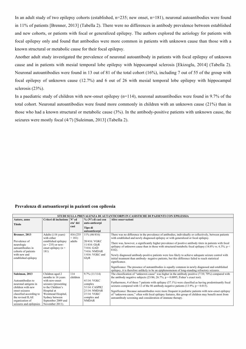
In an adult study of two epilepsy cohorts (established, n=235; new onset, n=181), neuronal autoantibodies were found
in 11% of patients [Brenner, 2013] (Tabella 2). There were no differences in antibody prevalence between established
and new cohorts, or patients with focal or generalized epilepsy. The authors explored the aetiology for patients with
focal epilepsy only and found that antibodies were more common in patients with unknown cause than those with a
known structural or metabolic cause for their focal epilepsy.
Another adult study investigated the prevalence of neuronal autoantibody in patients with focal epilepsy of unknown
cause and in patients with mesial temporal lobe epilepsy with hippocampal sclerosis [Ekizoglu, 2014] (Tabella 2).
Neuronal autoantibodies were found in 13 out of 81 of the total cohort (16%), including 7 out of 55 of the group with
focal epilepsy of unknown cause (12.7%) and 6 out of 26 with mesial temporal lobe epilepsy with hippocampal
sclerosis (23%).
In a paediatric study of children with new-onset epilepsy (n=114), neuronal autoantibodies were found in 9.7% of the
total cohort. Neuronal autoantibodies were found more commonly in children with an unknown cause (21%) than in
those who had a known structural or metabolic cause (3%). In the antibody-positive patients with unknown cause, the
seizures were mostly focal (4/7) [Suleiman, 2013] (Tabella 2).
Prevalenza di autoanticorpi in pazienti con epilessia
STUDI SULLA PREVALENZA DI AUTANTICORPI IN CASISTICHE DI PAZIENTI CON EPILESSIA Autore, anno
Titolo
Criteri di inclusione Nº ed eta’ dei casi
% (Nº) di casi con auto-anticorpi
Tipo di autoanticorpi
Altre osservazioni
Brenner, 2013 Prevalence of neurologic autoantibodies in cohorts of patients with new and established epilepsy
Adults (≥16 years) with either established epilepsy (n = 235) or new-onset epilepsy (n = 181)
416 (235 + 181) adults
11% (46/416) 20/416: VGKC 11/416: GlyR 7/416: GAD 7/416: NMDAR 1/416: VGKC and GlyR
There was no difference in the prevalence of antibodies, individually or collectively, between patients with established and newly diagnosed epilepsy or with generalized or focal epilepsy.
There was, however, a significantly higher prevalence of positive antibody titers in patients with focal epilepsy of unknown cause than in those with structural/metabolic focal epilepsy (14.8% vs. 6.3%; p < 0.02).
Newly diagnosed antibody-positive patients were less likely to achieve adequate seizure control with initial treatment than antibody- negative patients, but this difference failed to reach statistical significance.
Significance: The presence of autoantibodies is equally common in newly diagnosed and established epilepsy, it is therefore unlikely to be an epiphenomenon of long-standing refractory seizures.
Suleiman, 2013 Autoantibodies to neuronal antigens in children with new onset seizures classified according to the revised ILAE organization of seizures and epilepsies
Children aged 2 months to 16 years with new-onset seizures (presenting to the Children’s Hospital at Westmead Hospital, Sydney between September 2009 and November 2011).
114 children
9.7% (11/114) 4/114: VGKC complex 3/114: CASPR2 2/114: NMDAR 2/114: VGKC complex and NMDAR
The classification of “unknown cause” was higher in the antibody positive (7/10; 70%) compared with the antibody negative subjects (23/86; 26.7%; p = 0.0095, Fisher’s exact test).
Furthermore, 4 of these 7 patients with epilepsy (57.1%) were classified as having predominantly focal seizures compared with 12 of the 86 antibody-negative patients (13.9%; p = 0.015).
Significance: Because autoantibodies were more frequent in pediatric patients with new-onset epilepsy of “unknown cause,” often with focal epilepsy features, this group of children may benefit most from autoantibody screening and consideration of immune therapy.

Ekizoglu, 2014 Investigation of neuronal autoantibodies in two different focal epilepsy syndromes
Consecutive adult patients diagnosed with focal epilepsy of unknown cause (FEoUC) or mesial temporal lobe epilepsy with hippocampal sclerosis (MTLE-HS), followed for more than 1 year
81 adults 16% (13/81) 5/81: GlyR 4/81: CASPR2 2/81: NMDAR 2/81: : VGKC complex
Psychotic attacks and nonspecific MRI white matter changes showed significant associations in seropositive patients (p = 0.003 and p = 0.03, respectively).
Poor drug-response rates and total seizure counts were also higher in the seropositive patients but without reaching statistical significance. Three seropositive patients with previous epilepsy surgery showed typical histopathologic results for MTLE-HS, but not inflammatory changes. Moreover, some patients harbouring these antibodies partly benefited from immunotherapy.
Significance: We detected neuronal antibodies in one sixth of patients with focal epilepsy, GLY-R antibodies being the leading one. Psychosis or nonspecific MRI WMCs were frequent in the seropositive group. Our results suggested that relevant antibodies should be screened for a treatment possibility in these groups.
Wright, 2016
Children (aged 1 month to 16 years) were enrolled into the Dutch Study of Epilepsy in Childhood (DSEC) from four participating centers in The Netherlands between 1988 and 1992. Children with a presumed “acute symptomatic” etiology for their epilepsy were excluded.
178 children
9.5% (17/178) 7/178: NMDAR 4/178: CASPR2 3/178: VGKC complex 2/178: Contactin-2
Seventeen patients (9.5%) were positive for VGKC complex (n = 3), NMDAR (n = 7), CASPR2 (n = 4), and contactin-2 (n = 3), compared to three (3/112; 2.6%) healthy controls (VGKC complex [n = 1], NMDAR [n = 2]; p = 0.03; Fisher’s exact test). Low levels of neuronal antibodies are present in ~10% of patients with pediatric epilepsy at onset but are not associated with poor long-term outcomes or treatment intractability Antibodies can develop during the course of epilepsy and are not likely to be the sole cause of epilepsy in pediatric patients However, if associated with clinical features suggestive of autoimmune encephalitis, this “secondary inflammation” may be immunotherapy responsive as seen in other antibody-mediated diseases
Tabella 2. Studi sulla prevalenza di autoanticorpi in pazienti con epilessia.
QUANDO SOSPETTARE UN’EPILESSIA AUTOIMMUNE?
Despite increased recent research interest, no clear guidelines exist for the diagnosis or management of autoimmune
epilepsy [Dubey, 2015]. Different studies have adopted slightly different operative definitions for autoimmune
encephalitis (see next paragraph, “Definizioni operative”, and relative Table). Guidelines for the recognition of
autoimmune epilepsy have been proposed in children [Suleiman, 2013], based on modified guidelines for the
recognition, of suspected autoimmune CNS disorders by Zuliani et al [Zuliani, 2012] (see paragraph “Proposed
modified guidelines for the recognition of autoimmune epilepsy in children”).
- The features of autoimmune epilepsy include acute or subacute onset of seizures, usually in the context of
encephalopathy, and inflammation of the central nervous system on testing cerebrospinal fluid or magnetic
resonance imaging. Neuronal antibodies associated with autoimmune encephalitis and seizures in children
include NMDAR, voltage-gated potassium channel complex, glycine receptor, c-Aminobutyric acid type A
receptor (GABAAR), c-Aminobutyric acid type B receptor (GABABR), and glutamic acid decarboxylase
antibodies. These antibodies support the diagnosis of autoimmune epilepsy, but are not essential for diagnosis
[Suleiman, 2015].
- Autoimmune epilepsy is increasingly recognized in the spectrum of neurological disorders characterized by
detection of neural autoantibodies in serum or spinal fluid and responsiveness to immunotherapy. An
autoimmune cause is suspected based on frequent or medically intractable seizures and the presence of at least
one neural antibody, inflammatory changes indicated in serum or spinal fluid or on MRI, or a personal or
family history of autoimmunity. It is essential that an autoimmune etiology be considered in the initial
differential diagnosis of new onset epilepsy, because early immunotherapy assures an optimal outcome for the
patient [Greco, 2015].

Definizioni operative di sospetta epilessia autoimmune adottate ed anticorpi identificati in alcune delle
maggiori casistiche di pazienti con sospetta epilessia autoimmune
Le casistiche di pazienti con epilessia autoimmune utilizzano generalmente una definizione di epilessia autoimmune
basata su una combinazione dei seguenti criteri di inclusione ed esclusione (dettagliati nella tabella sottostante):
Criteri di inclusione:
- Epilepsy as the exclusive or predominant presenting concern [anche se in alcune casistiche questo non appare
invece tra i criteri di inclusione]
- Epilepsy with an acute or subacute onset
- Epilepsy that responded well to steroids or immunomodulatory agents
- Autoimmune pathogenesis suspected by the treating physicians based on:
o Detection of a neural autoantibody in serum or CSF which have been associated with autoimmune
encephalitis (any neuronal nuclear/cytoplasmic antibody such as anti-Hu or anti-CRMP-5, any
neuronal membrane antibody including anti-VGKC, anti- NMDA-R, anti-GABAB-R, anti-ganglionic
AChR, or anti-glutamic acid decarboxylase (GAD) antibody),
o Inflammatory CSF (leucocytosis, elevated proteins, or CSF-exclusive oligoclonal immunoglobulin
bands)
o MRI characteristics suggesting inflammation (T2 hyperintensities, contrast enhancement on
gadolinium studies, and/or restricted diffusion; limbic involvement).
Criteri di esclusione: presenza di altra eziologia che possa spiegare l’epilessia:
- Presence of CSF viral/bacterial/fungal antigens or antibodies or DNA PCR which could explain underlying
acute inflammatory brain parenchymal changes,
- Presence of metabolic abnormalities which could have precipitated seizures (severe renal or hepatic failure,
malignant hypertension, severe hypo/ hyperglycemia),
- Presence of brain structural lesions such as stroke, tumor, traumatic lesions, heterotopias, neuronal migration
anomalies, mesial temporal sclerosis, vascular malformation, abscess or infectious lesion which could have
precipitated the presenting seizure
- Presence of genetic backgrounds epilepsies (e.g., SCN1A mutations),
- Possibility of side effects to drugs
CASISTICHE DI PAZIENTI CON SOSPETTA EPILESSIA AUTOIMMUNE
Autore, anno Titolo
Definizione operativa di sospetta epilessia autoimmune Nº ed eta’ dei casi
% (Nº) di casi con auto-anticorpi
Tipo di autoanticorpi Quek, 2012
Autoimmune Epilepsy: Clinical Characteristics and Response to Immunotherapy
Autoimmune epilepsy was defined as (1) epilepsy as the exclusive (n=11) or predominant (n=21) presenting concern and (2) autoimmune pathogenesis suspected by the treating physicians based on detection of a neural autoantibody, inflammatory cerebrospinal fluid (CSF) (leukocytosis or CSF-exclusive oligoclonal immunoglobulin bands), or magnetic resonance imaging (MRI) characteristics suggesting inflammation (T2 hyperintensities, contrast enhancement on gadolinium studies, and/or restricted diffusion).
32 children and adults
91% (29/32) 18/32: VGKC complex (14 Lgi1, 1 Caspr2, and 3 were of unknown specificity) 7/32: GAD 2/32: CRMP-5 1/32: Ma 1/32: NMDAR 1/32: neuronal nicotinic acetylcholine receptor, ganglionic type
Bektaş, 2015 Epilepsy and Autoimmunity in Pediatric Patients
The patients were chosen from among epilepsy patients with undetermined etiology and susceptible autoimmunity who were referred or followed. Patients who met one of the following inclusion criteria were included in this study: (1) epilepsy that responded well to steroids or immunomodulatory agents and (2) epilepsy with an acute or subacute (<12 weeks) onset of symptoms whose underlying cause could not be determined. Exclusion criteria included neuronal migration anomalies, genetic backgrounds epilepsies (e.g., SCN1A mutations), diabetes (GAD Abs can be elevated), oncologic diseases, metabolic diseases, stroke, rheumatologic disease (e.g., systemic lupus erythematosus [SLE]), and side effects to drugs.
80 children
45% (36/80) 15/80: ANA 3/80: TPO 3/80: Antiphospholipid 1/80: Anticardiolipine 7/80: GAD 2/80: Ma2 2/80: Yo 13/80: VGKC complex

Dubey, 2015 Retrospective case series of the clinical features, management and outcomes of patients with autoimmune epilepsy
Cases included were patients presenting with new onset electrographic seizure activity, plus ≥2 of the following: (1) CSF findings consistent with inflammation (elevated CSF protein >50 and/or lymphocytic pleocytosis), (2) brain MRI showing signal changes consistent with limbic encephalitis, (3) autoimmune/paraneoplastic antibodies in serum or CSF which have been associated with autoimmune encephalitis in previous studies (any neuronal nuclear/cytoplasmic antibody such as anti-Hu or anti-CRMP-5, any neuronal membrane antibody including anti-VGKC, anti- NMDA-R, anti-GABAB-R, anti-ganglionic AChR, or anti-glutamic acid decarboxylase (GAD) antibody), (4) new onset seizure responding to immunomodulatory therapies. Cases were excluded if there was evidence of another identified cause of the patient’s seizures: (1) Presence of CSF viral/bacterial/fungal antigens or antibodies or DNA PCR which could explain underlying acute inflammatory brain parenchymal changes, (2) Presence of metabolic abnormalities which could have precipitated seizures (severe renal or hepatic failure, malignant hypertension, severe hypo/ hyperglycemia), (3) Presence of brain structural lesions such as stroke, tumor, traumatic lesions, heterotopias, mesial temporal sclerosis, vascular malformation, abscess or infectious lesion which could have precipitated the presenting seizure
34 adults
76.5% (26/34) 8/34: VGKC 7/34: NMDAR 4/34: GAD 2/34: GABAB 5/34: Anti-thyroid
Tabella 3. Definizione operativa di epilessia autoimmune utilizzata in alcune recenti casistiche

Proposed modified guidelines for the recognition of autoimmune epilepsy in children (paragrafo e Tabelle tratti
da [Suleiman, 2013])
Zuliani et al. proposed guidelines for the recognition, testing, and treatment of suspected autoimmune CNS disorders.
They used clinical criteria, supportive features, neuronal antibody testing, and the response to immune therapy to
classify patients into categories of definite, probable, and possible NSAS [Zuliani, 2012].
To improve recognition and diagnosis of children with suspected autoimmune epilepsy, we modified the guidelines
proposed by Zuliani et al. for identification of children with neuronal surface antibodies syndromes (Table 1). Then,
based on antibody testing and the response to immunotherapy (when given), we proposed five categories for
classification (in descending order of likelihood of autoimmune epilepsy) including definite, probable, possible,
unlikely, or unknown autoimmune epilepsy.
Tabelle tratte da [Suleiman, 2013]

ALTRE OSSERVAZIONI
Le caratteristiche EEG permettono di differenziare epilessia autoimmune da non autoimmune?
Le caratteristiche EEG non consentono di effettuare la distinzione tra epilessie autoimmuni e non autoimmune
[Baysal-Kirac, 2015].
In uno studio condotto su 20 pazienti adulti con epilessia e autoanticorpi (in alcuni casi epilessia isolata, in altri in
corso di encefalite autoimmune) (anticorpi anti NMDAR, GlyR, Caspr2, VGKC, GAD, Hu, amfifisina), e 21 controlli
seronegativi: non differenze significative nell’EEG dei pazienti seropositive e seronegativi. Altri dati emersi da questo
studio:
- Onset con stato di male non convulsivo (NCSE) o con stato di male focale motorio: 20% dei pazienti seorpositivi,
0% di quelli seronegativi
- Ritmo theta e delta continuo: in 71% pazienti seropositivi, e in 25% di quelli seronegativi
- Frontal intermittent rhythmic delta activity (FIRDA): in 40% pazienti seropositivi, e in 24% di quelli seronegativi
[Baysal-Kirac, 2015]
Efficacia dell’immunoterapia nelle epilessie autoimmuni
Art
icol
o Nº ed eta’ dei casi
Tipo di pazienti Risultati e osservazioni
Que
k, 2
012#
32 adulti Exclusive (n=11) or predominant (n=21) seizure presentation in whom an autoimmune etiology was suspected (on the basis of neural autoantibody, inflammatory CSF, or MRI suggesting inflammation) Autoantibodies in 91% (29/32)
After a median interval of 17 months (range, 3–72 months), 22 of 27 (81%) reported improvement post-immunotherapy; 18 were seizure free. The median time from seizure onset to initiating immunotherapy was 4 months for responders and 22 months for nonresponders (P<.05). All voltage-gated potassium channel complex antibody–positive patients reported initial or lasting benefit (P<.05). One voltage-gated potassium channel complex antibody–positive patient was seizure free after thyroid cancer resection; another responded to antiepileptic drug change alone. When clinical and serological clues suggest an autoimmune basis for medically intractable epilepsy, early-initiated immunotherapy may improve seizure outcome.
Iran
i, 20
13*#
10 adulti Facio-brachial dystonic seizures (cognitive impairment in 8/10) Autoantibodies in 100% (10%)
Facio-brachial dystonic seizures were controlled more effectively with immunotherapy than anti-epileptic drugs (P = 0.006). Strikingly, in the nine cases who remained anti-epileptic drug refractory for a median of 30 days (range 11–200), the addition of corticosteroids was associated with cessation of faciobrachial dystonic seizures within 1 week in three and within 2 months in six cases. VGKC antibodies persisted in the four cases with relapses of faciobrachial dystonic seizures during corticosteroid withdrawal. Time to recovery of baseline function was positively correlated with time to immunotherapy (r = 0.74; P = 0.03) but not time to anti-epileptic drug administration (r = 0.55; P = 0.10). Of 10 cases, the eight cases who received anti-epileptic drugs (n = 3) or no treatment (n = 5) all developed cognitive impairment. By contrast, the two who did not develop cognitive impairment received immunotherapy to treat their faciobrachial dystonic seizures (P = 0.02). In eight cases without clinical magnetic resonance imaging evidence of hippocampal signal change, cross-sectional volumetric magnetic resonance imaging post-recovery, after accounting for age and head size, revealed cases (n = 8) had smaller brain volumes than healthy controls (n = 13) (P50.001).
Dub
ey, 2
015#
34 adulti Hospitalized patients who presented predominantly due to seizures with concern for autoimmune etiology Autoantibodies in 76.5% (26/34)
Time from symptom onset to diagnosis (p < 0.005) and symptom onset to immunomodulation (p < 0.005) was significantly lower among patients who achieved responder rate (RR). Conclusion: This study highlights certain important clinical and electrographic aspects of autoimmune epilepsy, and the significance of early diagnosis and initiation of immunomodulatory therapy.
Tabella 4. Efficacia dell’immunoterapia rispetto ai farmaci antiepilettici nelle epilessie autoimmune, e beneficio di
immunoterapia precoce rispetto a tardiva.
Legenda: *L’immunoterapia e’ piu’ efficace degli AED nelle epilessie autoimmuni (con significativita’ statistica);
#Beneficio di immunoterapia early vs late (con significativita’ statistica)
Tra gli anticorpi anti antigeni intraneuronali, crisi epilettiche (in presenza o meno di altre manifestazioni) sono state
frequentemente descritte anche nei pazienti con anticorpi anti-GAD (associati alle seguenti sindromi cliniche: limbic
encephalitis, stiff-person-syndrome; cerebellar ataxia; downbeat nystagmus; palatal tremor; brainstem dysfunction;
isolated epilepsy (mostly temporal lobe epilepsy). Epilessia isolata o come prevalente manifestazione e’ stata descritta

in pazienti con positivita’ per anticorpi anti-GAD [Liimatainen, 2010; Lilleker, 2014; Bektaş, 2015; Akaishi, 2015-
(not exhaustive) (children and adults)]
In un recente articolo su anticorpi anti-GAD ed epilessia, viene descritto nessun miglioramento delle crisi nei pazienti
trattati con immunoterapia [Lilleker, 2014]. Five patients received immunotherapy. No improvement in seizures was
observed in any. One patient with equivocal MRI evidence of hippocampal sclerosis and concordant video EEG and
PET scan, achieved 12 months seizure freedom following temporal lobectomy. Conclusions: The relevance of GAD
Abs to epilepsy remains uncertain. Our experience does not support the routine use of immunotherapy in patients with
epilepsy and GAD Abs [Lilleker, 2014].

References Baysal-Kirac L, Tuzun E, Altindag E, Ekizoglu E, Kinay D, Bilgic B, Tekturk P, Baykan B. Are There Any Specific EEG Findings in Autoimmune Epilepsies? Clin EEG Neurosci. 2016 Jul;47(3):224-34.
Bektaş Ö, Jacobson L, Tutkak H, Karagöl S, Lang B, Clover L, Vincent A, Deda G. Epilepsy and autoimmunity in pediatric patients. Neuropediatrics. 2015 Feb;46(1):13-9. doi: 10.1055/s-0034-1389895. Epub 2014 Oct 7.
Bien CG, Scheffer IE. Autoantibodies and epilepsy. Epilepsia. 2011 May;52 Suppl 3:18-22. doi: 10.1111/j.1528-1167.2011.03031.x.
Brenner T, Sills GJ, Hart Y, Howell S, Waters P, Brodie MJ, Vincent A, Lang B. Prevalence of neurologic autoantibodies in cohorts of patients with new and established epilepsy. Epilepsia. 2013 Jun;54(6):1028-35. doi: 10.1111/epi.12127. Epub 2013 Mar 6.
Dubey D, Samudra N, Gupta P, Agostini M, Ding K, Van Ness PC, Vernino S, Hays R. Retrospective case series of the clinical features, management and outcomes of patients with autoimmune epilepsy. Seizure. 2015 Jul;29:143-7. doi: 10.1016/j.seizure.2015.04.007. Epub 2015 Apr 30.
Ekizoglu E, Tuzun E, Woodhall M, Lang B, Jacobson L, Icoz S, Bebek N, Gurses C, Gokyigit A, Waters P, Vincent A, Baykan B. Investigation of neuronal autoantibodies in two different focal epilepsy syndromes. Epilepsia. 2014 Mar;55(3):414-22. doi: 10.1111/epi.12528. Epub 2014 Feb 6.
Greco A, Rizzo MI, De Virgilio A, Conte M, Gallo A, Attanasio G, Ruoppolo G, de Vincentiis M. Autoimmune epilepsy. Autoimmun Rev. 2015 Nov 25. pii: S1568-9972(15)00238-4. doi: 10.1016/j.autrev.2015.11.007. [Epub ahead of print] Review.
Irani SR, Bien CG, Lang B. Autoimmune epilepsies. Curr Opin Neurol. 2011 Apr;24(2):146-53. doi: 10.1097/WCO.0b013e3283446f05.
Irani SR, Stagg CJ, Schott JM, Rosenthal CR, Schneider SA, Pettingill P, Pettingill R, Waters P, Thomas A, Voets NL, Cardoso MJ, Cash DM, Manning EN, Lang B, Smith SJ, Vincent A, Johnson MR. Faciobrachial dystonic seizures: the influence of immunotherapy on seizure control and prevention of cognitive impairment in a broadening phenotype. Brain. 2013 Oct;136(Pt 10):3151-62. doi: 10.1093/brain/awt212. Epub 2013 Sep 6.
Lilleker JB, Biswas V, Mohanraj R. Glutamic acid decarboxylase (GAD) antibodies in epilepsy: diagnostic yield and therapeutic implications. Seizure. 2014 Sep;23(8):598-602. doi: 10.1016/j.seizure.2014.04.009. Epub 2014 Apr 26.
Mintzer S. Undiplomatic immunity: epilepsy and autoimmune disease. Epilepsy Curr. 2015 Mar-Apr;15(2):72-4. doi: 10.5698/1535-7597-15.2.72. No abstract available.
Ong MS, Kohane IS, Cai T, Gorman MP, Mandl KD. Population-level evidence for an autoimmune etiology of epilepsy. JAMA Neurol. 2014 May;71(5):569-74. doi: 10.1001/jamaneurol.2014.188.
Quek AM, Britton JW, McKeon A, So E, Lennon VA, Shin C, Klein C, Watson RE Jr, Kotsenas AL, Lagerlund TD, Cascino GD, Worrell GA, Wirrell EC, Nickels KC, Aksamit AJ, Noe KH, Pittock SJ. Autoimmune epilepsy: clinical characteristics and response to immunotherapy. Arch Neurol. 2012 May;69(5):582-93.
Suleiman J, Brilot F, Lang B, Vincent A, Dale RC. Autoimmune epilepsy in children: case series and proposed guidelines for identification. Epilepsia. 2013 Jun;54(6):1036-45. doi: 10.1111/epi.12142. Epub 2013 Mar 28.
Suleiman J, Wright S, Gill D, Brilot F, Waters P, Peacock K, Procopis P, Nibber A, Vincent A, Dale RC, Lang B. Autoantibodies to neuronal antigens in children with new-onset seizures classified according to the revised ILAE organization of seizures and epilepsies. Epilepsia. 2013 Dec;54(12):2091-100. doi: 10.1111/epi.12405. Epub 2013 Oct 23.
Suleiman J, Dale RC. The recognition and treatment of autoimmune epilepsy in children. Dev Med Child Neurol. 2015 May;57(5):431-40. doi: 10.1111/dmcn.12647. Epub 2014 Dec 8. Review.
Zuliani L, Graus F, Giometto B, Bien C, Vincent A. Central nervous system neuronal surface antibody associated syndromes: review and guidelines for recognition. J Neurol Neurosurg Psychiatry. 2012 Jun;83(6):638-45. doi: 10.1136/jnnp-2011-301237. Epub 2012 Mar 24. Review.
Vezzani A, Granata T. Brain inflammation in epilepsy: experimental and clinical evidence. Epilepsia. 2005 Nov;46(11):1724-‐43. Irani SR, Stagg CJ, Schott JM, Rosenthal CR, Schneider SA, Pettingill P, Pettingill R, Waters P, Thomas A, Voets NL, Cardoso MJ, Cash DM, Manning EN,Lang B, Smith SJ, Vincent A, Johnson MR. Faciobrachial dystonic seizures: the influence of immunotherapy on seizure control and prevention ofcognitive impairment in a broadening phenotype. Brain. 2013 Oct;136(Pt 10):3151-‐62. doi: 10.1093/brain/awt212. Epub 2013 Sep 6.
Vincent A, Irani SR, Lang B. The growing recognition of immunotherapy-‐responsive seizure disorders with autoantibodies to specificneuronal proteins. Curr Opin Neurol. 2010 Apr;23(2):144-‐50. doi: 10.1097/WCO.0b013e32833735fe.
Yonsei Med J. 2008 Feb 29;49(1):1-‐18. doi: 10.3349/ymj.2008.49.1.1.
Role of brain inflammation in epileptogenesis. Choi J, Koh S.
J Neurol Neurosurg Psychiatry. 2000 Dec;69(6):711-‐4.
Epilepsy: an autoimmune disease? Palace J, Lang B.
Eur J Paediatr Neurol. 2005;9(1):29-‐42. Epub 2004 Dec 13.
Epilepsy and the immune system: is there a link? Billiau AD, Wouters CH, Lagae LG.
Autoimmunity. 2008 Feb;41(1):55-‐65. doi: 10.1080/08916930701619490.
Autoantibody-‐mediated disorders of the central nervous system. Irani S, Lang B.
Autoimmune encephalitis in children. Armangue T, Petit-Pedrol M, Dalmau J. J Child Neurol. 2012 Nov;27(11):1460-9. doi: 10.1177/0883073812448838. Review.
A clinical approach to diagnosis of autoimmune encephalitis. Graus F, Titulaer MJ, Balu R, Benseler S, Bien CG, Cellucci T, Cortese I, Dale RC, Gelfand JM, Geschwind M, Glaser CA, Honnorat J, Höftberger R, Iizuka T, Irani SR, Lancaster E, Leypoldt F, Prüss H, Rae-Grant A, Reindl M, Rosenfeld MR, Rostásy K, Saiz A, Venkatesan A, Vincent A, Wandinger KP, Waters P, Dalmau J. Lancet Neurol. 2016 Apr;15(4):391-404. doi: 10.1016/S1474-4422(15)00401-9. Review.
Systemic and neurologic autoimmune disorders associated with seizures or epilepsy. Vincent A, Crino PB. Epilepsia. 2011 May;52 Suppl 3:12-7. doi: 10.1111/j.1528-1167.2011.03030.x. Review.
Epilepsia. 2013 Sep;54 Suppl 6:46-‐9. doi: 10.1111/epi.12276.
Autoimmunity, seizures, and status epilepticus. Davis R, Dalmau J.

Nat Immunol. 2002 Jun;3(6):500.
Autoimmune epilepsy. Levite M.
Autoimmune and inflammatory epilepsies. Nabbout R. Epilepsia. 2012 Sep;53 Suppl 4:58-62. doi: 10.1111/j.1528-1167.2012.03614.x. Review.
Neurology. 2014 May 6;82(18):1578-‐86. doi: 10.1212/WNL.0000000000000383. Epub 2014 Apr 4.
Utility of an immunotherapy trial in evaluating patients with presumed autoimmune epilepsy. Toledano M, Britton JW, McKeon A, Shin C, Lennon VA, Quek AM, So E, Worrell GA, Cascino GD, Klein CJ, Lagerlund TD, Wirrell EC, Nickels KC, Pittock SJ.
Immune-mediated epilepsies. Granata T, Cross H, Theodore W, Avanzini G. Epilepsia. 2011 May;52 Suppl 3:5-11. doi: 10.1111/j.1528-1167.2011.03029.x. Review.
Experimental studies in epilepsy: immunologic and inflammatory mechanisms. Legido A, Katsetos CD. Semin Pediatr Neurol. 2014 Sep;21(3):197-206. doi: 10.1016/j.spen.2014.10.001. Review.
Epilepsy Curr. 2013 Mar;13(2):62-‐8. doi: 10.5698/1535-‐7597-‐13.2.62.
Epilepsy associated with systemic autoimmune disorders. Devinsky O, Schein A, Najjar S. Autoimmune epilepsies. Bien CG, Bauer J. Neurotherapeutics. 2014 Apr;11(2):311-8. doi: 10.1007/s13311-014-0264-3. Review.
Curr Neurol Neurosci Rep. 2013 May;13(5):348. doi: 10.1007/s11910-‐013-‐0348-‐1.
Antibodies in epilepsy. Correll CM. Curr Neuropharmacol. 2013 Jan;11(1):114-‐27. doi: 10.2174/157015913804999540.
Modulation of Immunity and the Inflammatory Response: A New Target for Treating Drug-‐resistant Epilepsy. Yu N, Liu H, Di Q.
Association between epilepsy and systemic autoimmune diseases: A meta-analysis. Lin Z, Si Q, Xiaoyi Z. Seizure. 2016 Oct;41:160-6. doi: 10.1016/j.seizure.2016.08.003.

Evaluation of positive and negative predictors of seizure outcomes among patients with immune-mediated epilepsy: a meta-analysis. Dubey D, Farzal Z, Hays R, Brown LS, Vernino S. Ther Adv Neurol Disord. 2016 Sep;9(5):369-77.
Etiological associations and outcome predictors of acute electroencephalography in childhood encephalitis. Mohammad SS, Soe SM, Pillai SC, Nosadini M, Barnes EH, Gill D, Dale RC. Clin Neurophysiol. 2016 Oct;127(10):3217-24.
Antibody-associated epilepsies: Clinical features, evidence for immunotherapies and future research questions. Bakpa OD, Reuber M, Irani SR. Seizure. 2016 Oct;41:26-41. doi: 10.1016/j.seizure.2016.07.002. Review.
Psychiatric manifestations and dysautonomia at the onset of focal epilepsy in adults: Clinical signs indicating autoimmune origin. Abraira L, Grau-López L, Jiménez M, Becerra JL. Neurologia. 2016 Jun 13. pii: S0213-4853(16)30048-2. doi: 10.1016/j.nrl.2016.04.004. English, Spanish. No abstract available.
Paraneoplastic epilepsy. Serafini A, Lukas RV, VanHaerents S, Warnke P, Tao JX, Rose S, Wu S. Epilepsy Behav. 2016 Aug;61:51-8. doi: 10.1016/j.yebeh.2016.04.046. Review.
Neuronal antibodies in pediatric epilepsy: Clinical features and long-term outcomes of a historical cohort not treated with immunotherapy. Wright S, Geerts AT, Jol-van der Zijde CM, Jacobson L, Lang B, Waters P, van Tol MJ, Stroink H, Neuteboom RF, Brouwer OF, Vincent A. Epilepsia. 2016 May;57(5):823-31.
Infections, inflammation and epilepsy. Vezzani A, Fujinami RS, White HS, Preux PM, Blümcke I, Sander JW, Löscher W. Acta Neuropathol. 2016 Feb;131(2):211-34. doi: 10.1007/s00401-015-1481-5. Review.
Undiplomatic immunity: epilepsy and autoimmune disease. Mintzer S. Epilepsy Curr. 2015 Mar-Apr;15(2):72-4
Are There Any Specific EEG Findings in Autoimmune Epilepsies? Baysal-Kirac L, Tuzun E, Altindag E, Ekizoglu E, Kinay D, Bilgic B, Tekturk P, Baykan B. Clin EEG Neurosci. 2016 Jul;47(3):224-34.
Chin Med J (Engl). 1990 Jan;103(1):71-‐5.
Observation on anti-‐brain antibody in serum of 110 epileptics. Xie XK, Tang LO.
Neuropediatrics. 1989 May;20(2):93-‐102.
Anti-‐CNS antibodies in childhood neurologic diseases. Plioplys AV, Greaves A, Yoshida W.
Partial seizures associated with antiphospholipid antibodies in childhood.
Angelini L, Granata T, Zibordi F, Binelli S, Zorzi G, Besana C. Neuropediatrics. 1998 Oct;29(5):249-53.
High prevalence of antiphospholipid antibodies in children with epilepsy: a controlled study of 50 cases. Eriksson K, Peltola J, Keränen T, Haapala AM, Koivikko M. Epilepsy Res. 2001 Aug;46(2):129-37.
Epilepsia. 1996 Oct;37(10):922-‐6.
Cryptogenic partial epilepsies with anti-‐GM1 antibodies: a new form of immune-‐mediated epilepsy? Bartolomei F, Boucraut J, Barrié M, Kok J, Dravet C, Viallat D, Bernard D, Gastaut JL.
J Neurol Sci. 1997 Nov 6;152(1):93-‐7.
The presence of autoantibodies to N-‐terminus domain of GluR1 subunit of AMPA receptor in the bloodserum of patients with epilepsy. Dambinova SA, Izykenova GA, Burov SV, Grigorenko EV, Gromov SA.