role of peripheral blood
TRANSCRIPT
Role of Peripheral blood smear examination in diagnostic work-up
Dr K Richards MD(Path);DTCDConsultant Pathologist-
HematologyYashoda Hospital
Why do a peripheral
blood film examination?


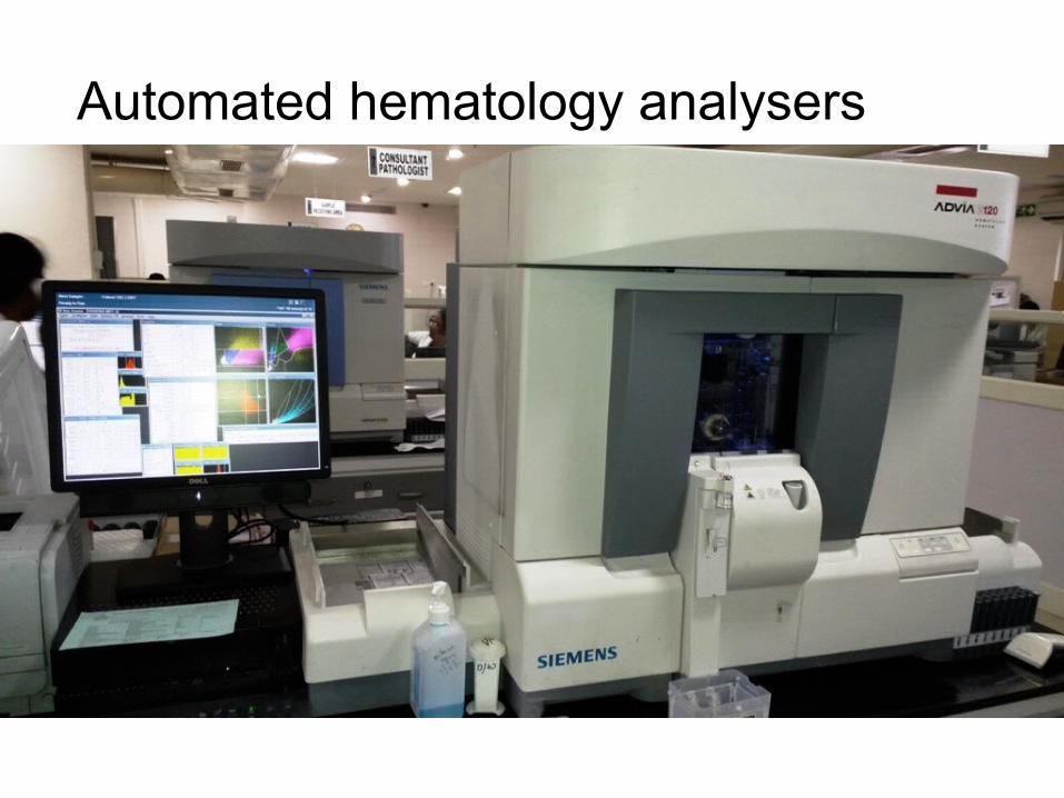
Automated hematology analysers

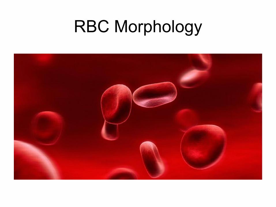
RBC Morphology

Anisocytosis
• abnormal erythropoiesis
• non-specific feature
• either macrocytes or microcytes or both

Poikilocytosis
• abnormal erythropoiesis
• non-specific feature• megaloblastic
anemia,• iron
deficiencyanemia,• thalassemia,• myelofibrosis,• MDS• Congenital
dyserythropoietic anemia

Microcytes
• defect in hemoglobin formation
• characteristic of iron deficiency anemia
• thalassemia• severe cases of
anemia of chronic disease
• sideroblastic anemias

Macrocytes
• classical in megaloblastic anemia
• aplastic anemia
• MDS• Alcoholics• Chronic liver disease• Hydroxyurea

Basophilic stippling
• thalassemias, • megaloblastic anemia• infections• liver disease• lead poisoning
• unstable hemoglobins• pyrimidine-5-
nucleotidase deficiency

Hypochromasia
• iron deficiency anemia
• sideroblastic anemia
• thalassemias

Dimorphic Red Cell Population
• two distinct populations
• development or resolution of IDA or anemia of chronic deficiency
• post blood transfusion• acquired sideroblastic
anemia

Spherocytes
• hereditary spherocytosis
• ABO hemolytic disease of the newborn
• autoimmune hemolytic anemia
• microspherocytes• sphero-echinocytes

Elliptocytes and Ovalocytes
• hereditary elliptocytosis
• hereditary pyropoikilocytosis
• southeast asian ovalocytosis

Schistocytes
• RBC fragments• thalassemias• CDA• Megaloblastic anemia• microangiopathic
hemolytic anemia• severe burns• hemolytic uremic
syndrome(HUS)• thrombotic
thrombocytopenic purpura(TTP)

Acanthocytes
• Abnormal phospholipid metabolism
• McLeod phenotype• Splenectomy• hyposplenism• Severe Liver disease
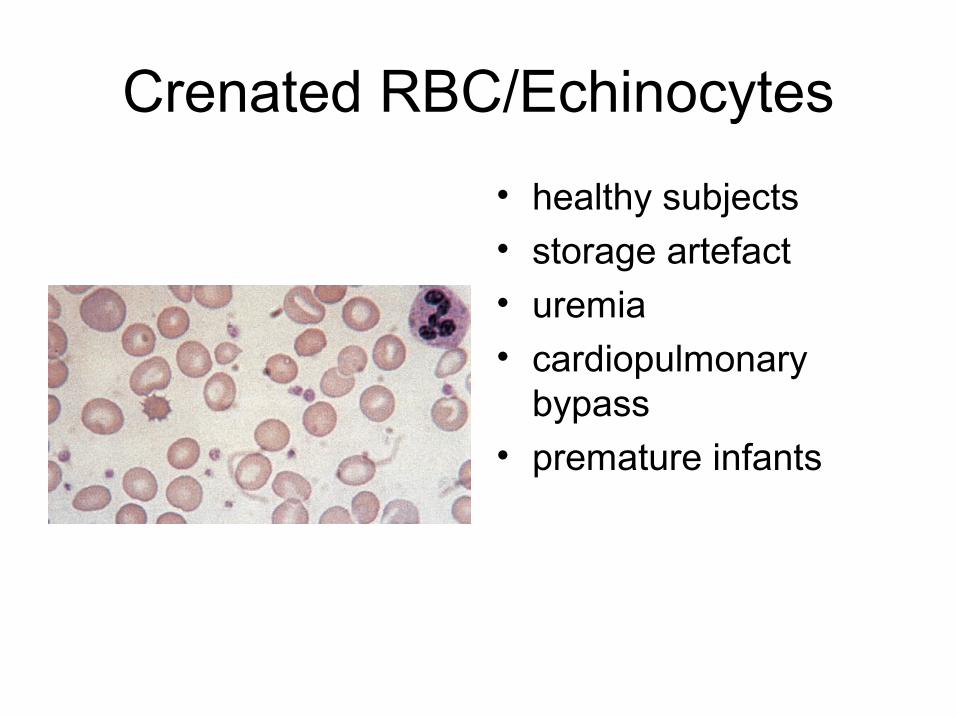
Crenated RBC/Echinocytes
• healthy subjects• storage artefact• uremia• cardiopulmonary
bypass• premature infants
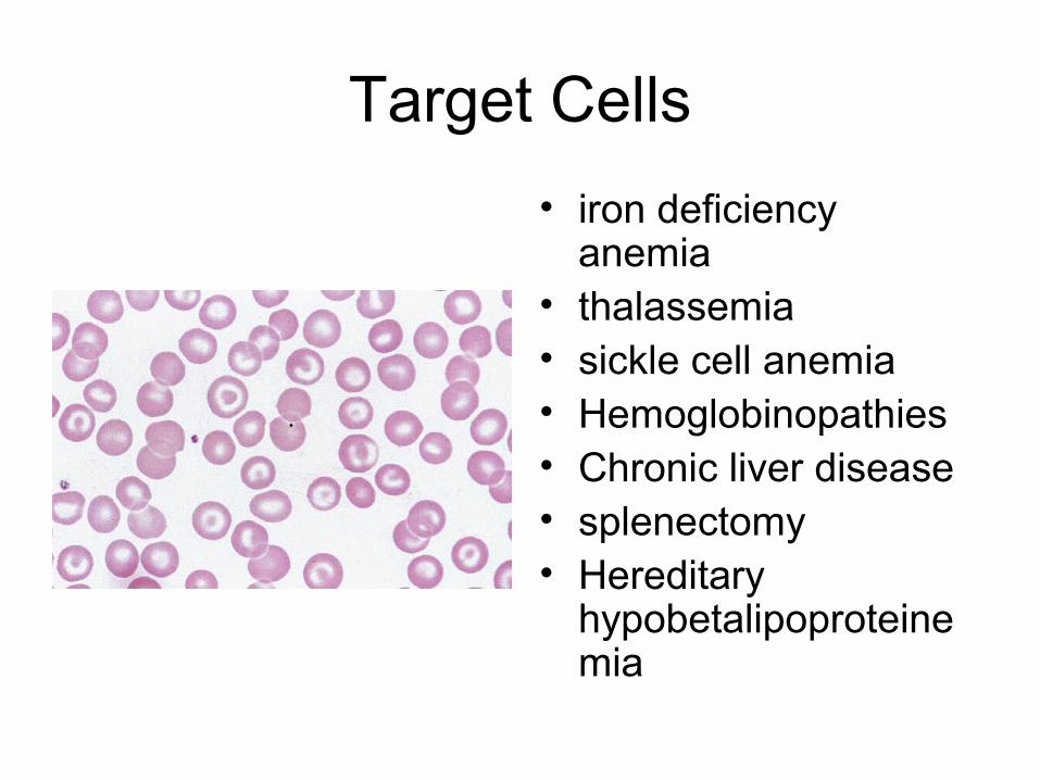
Target Cells
• iron deficiency anemia
• thalassemia• sickle cell anemia• Hemoglobinopathies• Chronic liver disease• splenectomy• Hereditary
hypobetalipoproteinemia

Stomatocytes
• Liver disease• alcoholics• MDS

Sickle Cells
• Sickling: blood subjected to anoxia
• Sickles and boat shaped forms
• Target cells – often a feature
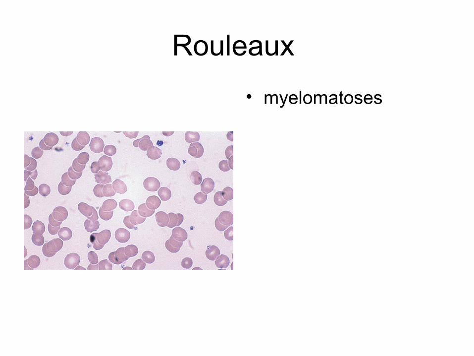
Rouleaux
• myelomatoses

Autoagglutination

Polychromasia
• Shades of bluish grey• Reticulocytes• Increased
erythropoiesis• Absence of
polychromasia -inadequate bone marrow response
Aplastic anemia Pure red cell aplasia

Nucleated RBCs
• More common in children• Severe anemia• Hemolytic disease of
newborn• Leucoerythroblastic
anemia:carcinomatoses and primary myelofibrosis
• Post splenectomy• Extramedullary
erythropoiesis• Sickle cell anemia • Septicemia• Cyanotic heart failure

Erythrocyte Inclusions: Howell Jolly Bodies
• Nuclear remnants• Small round, stain
purple• Post splenectomy• Splenic atrophy
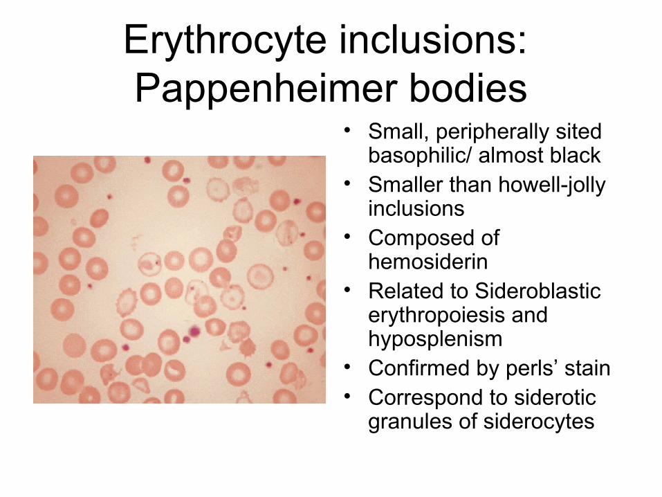
Erythrocyte inclusions: Pappenheimer bodies
• Small, peripherally sited basophilic/ almost black
• Smaller than howell-jolly inclusions
• Composed of hemosiderin
• Related to Sideroblastic erythropoiesis and hyposplenism
• Confirmed by perls’ stain• Correspond to siderotic
granules of siderocytes


Morphology of Leucocytes
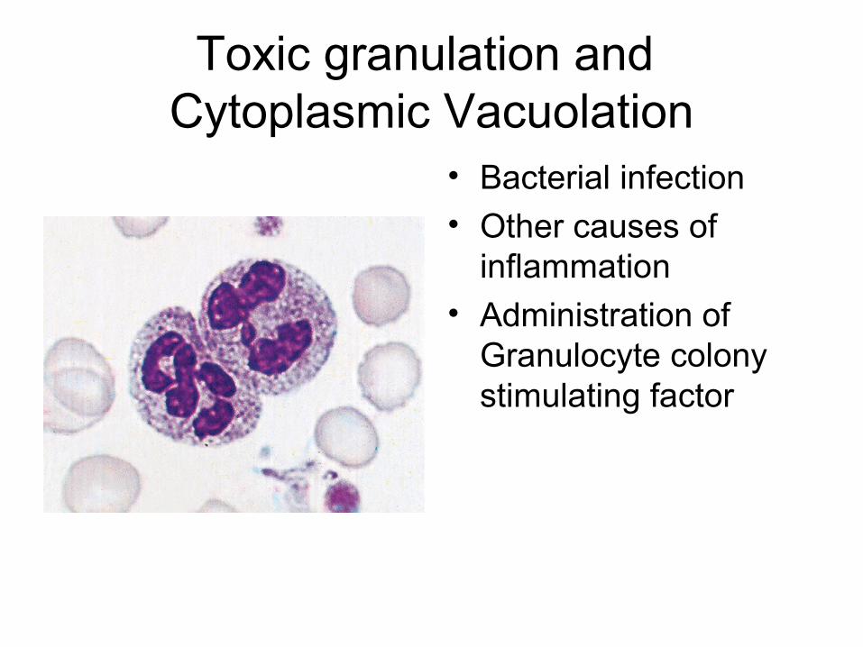
Toxic granulation and Cytoplasmic Vacuolation
• Bacterial infection• Other causes of
inflammation
• Administration of Granulocyte colony stimulating factor

• Poorly staining (hypogranular)
• Agranular neutrophils
• Myelodysplastic syndromes
• Some forms of myeloid leukemia

Chediak Higashi syndrome:
• Abnormal granules
• Giant but scanty azurophilic granules
• Functional defect• Susceptibility to
severe infection

Alder Reilly Anomaly:• Nucleus is obscured
by cytoplasmic granules
• Neutrophils function normally

Pelger-Huet Cells
• Benign inherited condition
• Pseudo pelger huet cells: MDS, AML with dysplastic maturation, chronic myeloid leukemia

Hypersegmented Nuclei
• Megaloblastic anemia• Uremia• Iron deficiency
anemia• Cytotoxic treatment –
methotrexate• Hydroxycarbamide

Left Shift

Basophilia
• Rarest (<1%)• Myeloproliferative
neoplasms
• CML: >10% impending accelerated phase/blast crisis

Eosinophilia• Eosinopenia: prolonged
steroid administration• Eosinophilia: - Allergic conditions, - parasitic infections, - reactive eosinophilias
( Lymphomas, ALL) - eosinophilic leukemia, - idiopathic hypereosinophilic
syndrome, - CML, - AML
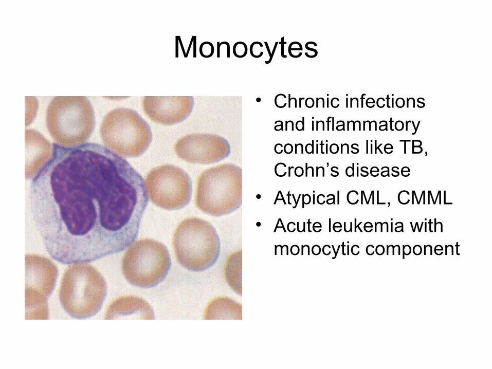
Monocytes
• Chronic infections and inflammatory conditions like TB, Crohn’s disease
• Atypical CML, CMML• Acute leukemia with
monocytic component

Lymphocytes
• Transformed lymphocytes: viral and bacterial lymphocytes
• Immunoblasts or turk cells
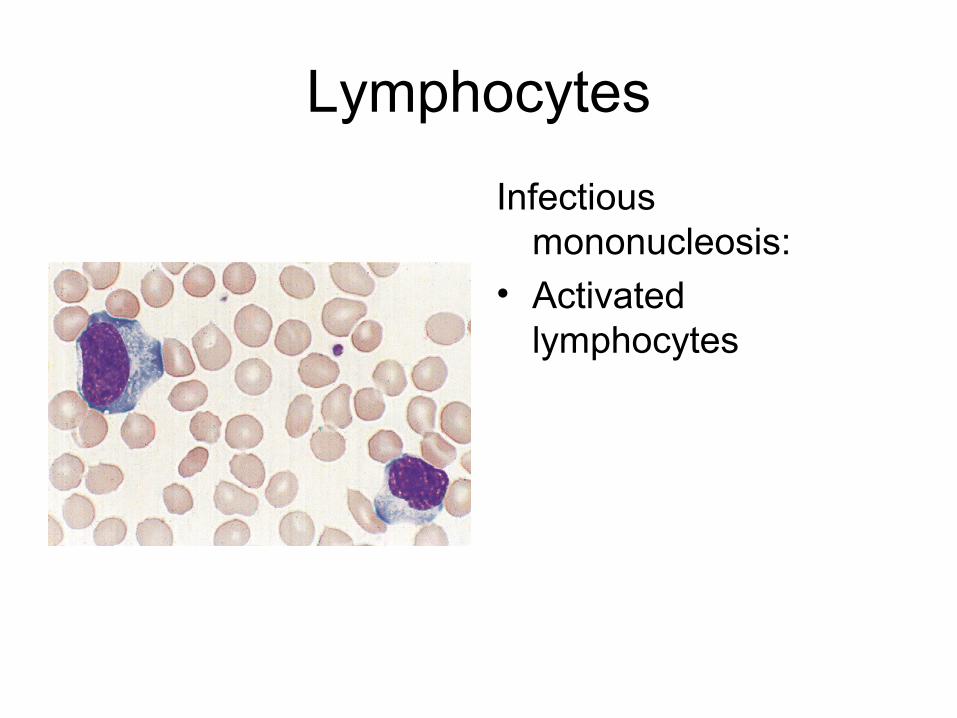
Lymphocytes
Infectious mononucleosis:
• Activated lymphocytes

Malignant Lymphoid Cells

Circulating blasts

Platelet Morphology

Large/Giant platelets
Large Platelets:• Increased platelet
production
• Hyposplenism• Severe immune
thrombocytopenia
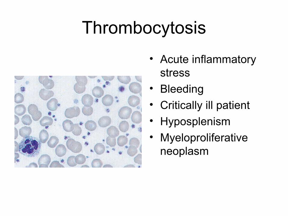
Thrombocytosis
• Acute inflammatory stress
• Bleeding
• Critically ill patient• Hyposplenism• Myeloproliferative
neoplasm

Grey platelet syndrome:• Hypogranular
platelets

Platelet satellitism
• antiplatelet autoantibodies
• Apparently healthy individuals
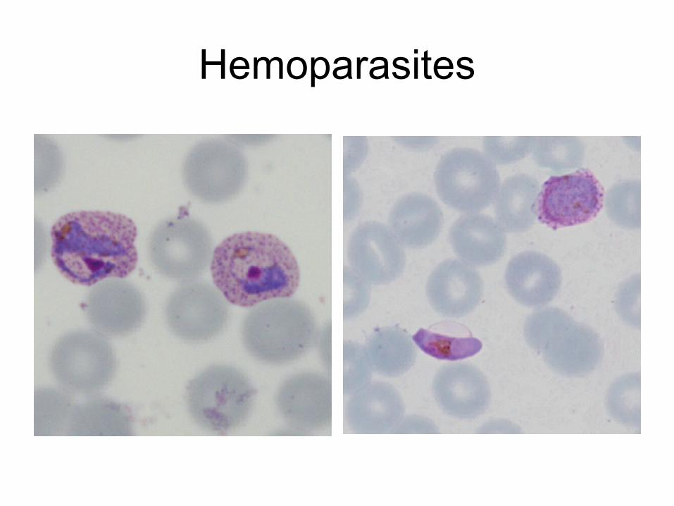
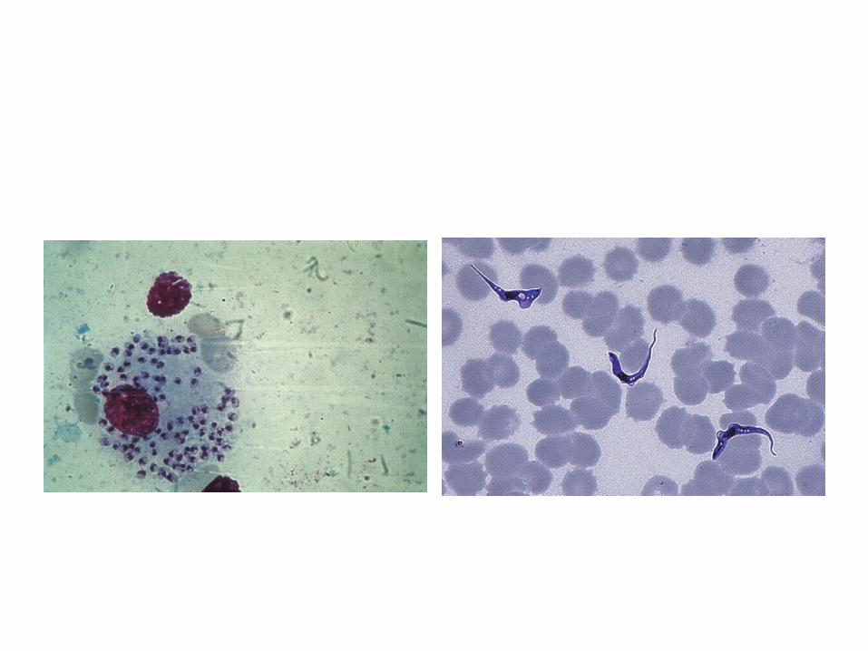
Hemoparasites


Summary
• Simple
• morphology
• Basic and foundational
• Further direction
• Patient management
References
• Dacie and Lewis practical hematology – 11th edition
• Wintrobe’s clinical hematology – 13th edition

Thank you