siglec-7 engagement by gbs β-protein suppresses pyroptotic...
TRANSCRIPT

Siglec-7 engagement by GBS β-protein suppressespyroptotic cell death of natural killer cellsJerry J. Fonga,b, Chih-Ming Tsaia,b, Sudeshna Sahaa,b, Victor Nizeta,c,d, Ajit Varkia,b,e,1, and Jack D. Buif,1
aGlycobiology Research and Training Center, School of Medicine, University of California, San Diego, La Jolla, CA 92093; bDepartment of Cellular andMolecular Medicine, School of Medicine, University of California, San Diego, La Jolla, CA 92093; cDepartment of Pediatrics, School of Medicine, University ofCalifornia, San Diego, La Jolla, CA 92093; dSkaggs School of Pharmacy and Pharmaceutical Sciences, School of Medicine, University of California, San Diego,La Jolla, CA 92093; eDepartment of Medicine, School of Medicine, University of California, San Diego, La Jolla, CA 92093; and fDepartment of Pathology,School of Medicine, University of California, San Diego, La Jolla, CA 92093
Edited by Lewis L. Lanier, University of California, San Francisco, CA, and approved August 31, 2018 (received for review March 21, 2018)
Natural killer (NK) cells are innate immune lymphocytes that recog-nize and destroy abnormal host cells, such as tumor cells or thoseinfected by viral pathogens. To safely accomplish these functions, NKcells display activating receptors that detect stress molecules or viralligands displayed at the cell surface, balanced by inhibitory receptorsthat bind to self-molecules. To date, such activating and inhibitoryreceptors on NK cells are not known to recognize bacterial determi-nants. Moreover, NK cell responses to direct interactions with extra-cellular bacteria are poorly explored. In this study, we observed thehuman neonatal pathogen group B Streptococcus (GBS) can directlyengage human NK cells. The interaction was mediated throughthe B6N segment of streptococcal β-protein, binding to the inhibi-tory receptor Siglec-7 via its amino-terminal V-set domain. Unlikeclassical Siglec binding, the interaction is also independent of its sialicacid recognition property. In contrast to WT GBS, mutants lackingβ-protein induced efficient pyroptosis of NK cells through the NLRP3inflammasome, with production and secretion of the proinflamma-tory cytokine IL-1β and dissemination of the cytotoxic molecule gran-zyme B. We postulate that GBS evolved β-protein engagement ofinhibitory human Siglec-7 to suppress the pyroptotic response of NKcells and thereby block recruitment of a broader innate immuneresponse, i.e., by “silencing the sentinel.”
natural killer cells | Siglec | group B Streptococcus | pyroptosis |inflammasome
Natural killer (NK) cells are lymphocytes of the innate im-mune system that recognize endogenous eukaryotic cells
under stress, such as tumor cells or cells infected by intracellularpathogens, modulating this process through an array of activat-ing and inhibitory receptors (1–3). Activating receptors on hu-man NK cells include NKG2D (4–6) and the natural cytotoxicityreceptor family consisting of NKp46, NKp44, and NKp30 (7).These receptors bind to a variety of ligands displayed on thesurface of eukaryotic cells during infection, or in response tostress or transformation (7–9). To avoid inadvertent destructionof healthy host cells, NK cells also express inhibitory receptorsthat bind to host molecules recognized as “self” (1), including theKIR (killer-cell Ig-like receptor) family, which recognizes HLAclass I molecules expressed on normal autologous cells. Thecombined landscape of activating and inhibitory ligands on atarget’s surface determines whether the NK cell becomes acti-vated, leading to cytokine secretion and release of cytotoxicmolecules such as perforin, granulysin, and granzymes (3). Theseactivating and inhibitory receptors are not known to recognizedeterminants on bacteria, and direct interactions or responsesagainst extracellular bacteria by NK cells are poorly explored (3,10). Here we report on the unexpected finding that the impor-tant human pathogen group B Streptococcus (GBS) engagesanother known inhibitory receptor on human NK cells, the sialicacid-recognizing Ig-like lectin-7 (Siglec-7).Siglec-7 is a member of the Siglec subfamily of CD33-related
Siglecs (CD33rSiglecs) (11), which are single-pass transmembranesialic acid-binding Ig-like lectins typically found on the surface of
leukocytes (12–14). The cytosolic domains of most CD33-relatedSiglecs harbor inhibitory intracellular ITIM motifs that induce animmunosuppressive signal, but some can instead recruit DAP-12with an activating intracellular domain, leading to augmentationof the immune response. Inhibitory Siglecs, which constitute themajority of CD33rSiglecs, can block cytokine secretion inducedthrough Toll-like receptor (TLR) signaling (14–18) and may haveevolved as a self-tolerance mechanism in which host leukocytesare inhibited when they recognize “self-associated molecularpatterns” (SAMPs) presented by sialic acids abundantly displayedon host cell surfaces (12, 19–23).Notably, certain bacterial pathogens have convergently evolved
diverse mechanisms for displaying Siglec ligands on their cellsurface, apparently to inhibit antipathogen immune responses viamolecular mimicry (24–26). For example, sialylated polysaccha-rides of GBS engage inhibitory CD33rSiglecs found on neutro-phils and myeloid lineage cells. Most such recognized microbialmimics of SAMPs for CD33rSiglec recognition are glycans.However, in at least one instance, Siglec-5 engagement also occursthrough the cell wall-anchored β-protein expressed by certain GBSstrains, with a similar suppression of the innate immune responseof myeloid lineage cells like neutrophils (27, 28). As Siglec-5 is notprominent on human lymphocytes (29), it is not clear whetherGBS β-protein can also inhibit this class of leukocytes.GBS induces a form of immunogenic cell death called pyroptosis,
mediated by an intracellular signaling complex called the
Significance
Direct interactions between natural killer (NK) cells and bac-teria are rarely observed, and the consequences of these in-teractions not well understood. We show that human NK cellsexposed to the bacterium group B Streptococcus (GBS) un-dergo pyroptosis, a type of inflammatory cell death. By re-leasing inflammatory mediators, pyroptosis is thought to be away for sentinels to die and alert the immune system of in-fection. Interestingly, we also found that GBS has a proteinthat binds to Siglec-7, an inhibitory molecule on NK cells.Siglec-7 inhibits pyroptosis and silences the sentinel activity ofNK cells by preventing the release of inflammatory molecules.These studies suggest that a human pathogen can silence thesentinel by specific interaction with a Siglec protein.
Author contributions: J.J.F., C.-M.T., V.N., A.V., and J.D.B. designed research; J.J.F., C.-M.T.,and S.S. performed research; J.J.F., V.N., A.V., and J.D.B. contributed new reagents/ana-lytic tools; J.J.F., C.-M.T., S.S., V.N., A.V., and J.D.B. analyzed data; and J.J.F., V.N., A.V., andJ.D.B. wrote the paper.
The authors declare no conflict of interest.
This article is a PNAS Direct Submission.
Published under the PNAS license.1To whom correspondence may be addressed. Email: [email protected] or [email protected].
This article contains supporting information online at www.pnas.org/lookup/suppl/doi:10.1073/pnas.1804108115/-/DCSupplemental.
Published online September 25, 2018.
10410–10415 | PNAS | October 9, 2018 | vol. 115 | no. 41 www.pnas.org/cgi/doi/10.1073/pnas.1804108115

inflammasome, which comprises several different signaling do-mains that multimerize upon binding of key ligands (30–33).Under canonical inflammasome activation, multimerization ofthe complex processes a proenzyme, such as caspase-1, into itsmature form, enabling it to enzymatically cleave proinflammatorycytokines such as IL-1β from its proform into its mature form (34,35). Cells activated for pyroptotic cell death ultimately swell andburst, allowing dissemination of the enzymatically cleaved proin-flammatory cytokines into the extracellular space for the recruit-ment and activation of nearby leukocytes such as neutrophils andmacrophages. Through this inflammatory pathway, pyroptosis actsas an “alarm signal” that promotes downstream host-protectiveantibacterial activities.In contrast to myeloid lineage cells, the predominant in-
hibitory CD33rSiglec on NK cells is Siglec-7, which engagessialylated ligands on tumor cells to suppress NK cell responses(12, 21, 36–41). However, as this outcome does not favor thehost, the natural role of Siglec-7 in controlling NK cell reactivityremains poorly explored. In this study, we report that GBSβ-protein binds Siglec-7. As direct interactions between extra-cellular bacteria and NK cells are rarely observed and in-completely understood (10), we examined the consequence ofthis engagement upon the NK cell innate immune response. Wefound that NK cells undergo pyroptosis after exposure to bacteria.However, when Siglec-7 was engaged by β-protein, pyroptosiswas inhibited, suggesting that GBS evolved this binding ability todown-regulate inflammasome activation and prevent recruitmentof other innate immune cells.
ResultsGBS β-Protein Engages NK Cells via Siglec-7. GBS sialylated poly-saccharide capsules of various serotypes engage the extracellulardomains of specific recombinant soluble human Siglec receptors,including Siglec-7 (24). We later found that certain GBSs canengage Siglec-5 via their β-protein, but it was unclear whetherother Siglec receptors also bind to this protein (27). A sub-sequent screen for binders to β-protein revealed that Siglec-7also bound. As Siglec-7 is expressed primarily on human NKcells, we used confocal microscopy to demonstrate direct contactbetween GBS labeled with FITC and the surface of NK cells(Fig. 1A). Although Siglec-7 is uniformly distributed across thesurface of uninfected NK cells, immunofluorescent labelingrevealed clear coclustering of receptors toward multiple FITC-
labeled WT GBSs on the surface of NK cells. Interestingly, basedon these confocal images, there may even be intracellular Siglec-7 receptors residing in endosomal compartments, but furthervalidation by costaining with endosomal markers will be requiredfor a more in-depth understanding.In contrast to the results with WT GBS, Siglec-7 did not
cluster around β-protein–deficient GBS (ΔBAC). It is worthnoting that there are no morphological differences between WTGBS vs. ΔBAC, but rather we believe there were more WT GBSscaptured in direct contact with the NK cell visualized. To com-plement these observations, flow cytometric analysis of WT GBSindeed showed a positive signal for binding to a Siglec-7 Fcchimera compared with total human IgG (negative control),whereas virtually no signal was seen for Siglec-7 Fc binding to theΔBAC mutant (Fig. 1B).The Siglec-5–binding region (B6N) of GBS β-protein spans
amino acid residues 1–153, but the adjacent IgA binding region(IgA-BR) spanning amino acid residues 154–225 is not recog-nized by Siglec-5 (28). Western blot analysis of purified B6N andIgA-BR peptides probed with Siglec-7 Fc also showed a directinteraction between Siglec-7 and B6N, but not IgA-BR (SI Ap-pendix, Fig. S1A). ELISA analysis with immobilized B6N or IgA-BR probed by Siglec-7 Fc supported these findings (SI Appendix,Fig. S1B). The Siglec-7–B6N interaction appeared to be rela-tively stronger than the previously recognized Siglec-5–B6Ninteraction. When Siglec-7 Fc was coincubated with the S7.7 anti–Siglec-7 monoclonal antibody, the binding signal was reduced tobackground levels (SI Appendix, Fig. S1C), but anti–Siglec-7 anti-body 194212 did not reduce the signal in a significant manner. Inview of these findings, subsequent experiments used clone S7.7 as ablocking antibody against Siglec-7 and β-protein interaction.To determine which extracellular domain on Siglec-7 might
contribute to B6N recognition, we probed immobilized B6N byusing previously constructed Siglec-Fc proteins with the V-setand underlying C2 domain swapped between Siglec-7 and Siglec-9 (26). By ELISA, we found that Siglec-7 V-set + Siglec-9 C2fusion Fc bound to B6N at nearly equal levels as Siglec-7 Fc(SI Appendix, Fig. S1D). However, the Siglec-9 V-set + Siglec-7C2 fusion Fc had no detectable binding to B6N, suggesting thatthe V-set domain of Siglec-7 is primarily responsible for B6Nengagement.
B
A
Fig. 1. GBS strain A909 (GBS A909) interacts withhuman NK cells through the cell wall-anchoredβ-protein and Siglec-7 receptor. (A) Images taken byconfocal microscopy show even distribution of Siglec-7 (red) across the cell surface of human NK cells un-der normal conditions. When NK cells are infectedwith FITC-labeled GBS (green), Siglec-7 on NK cellsclusters toward GBS. Infection with β-protein–deficientmutant GBS ΔBAC restores the even distribution ofSiglec-7 across the surface of NK cells. (B) Flow cytometryhistograms of WT GBS A909 and ΔBAC stained withrecombinant chimeric Siglec-7-Fc protein or IgG-Fccontrol. GBS A909 stained with Siglec-7-Fc showedpositive signals above IgG-Fc, but GBS ΔBAC did notdisplay any positive signals.
Fong et al. PNAS | October 9, 2018 | vol. 115 | no. 41 | 10411
IMMUNOLO
GYAND
INFLAMMATION

NK Cell Responses to GBS Are Suppressed by β-Protein Engagementof Siglec-7.Although NK cells are not typically studied as primaryresponders to extracellular bacteria, we determined the conse-quence of exposing NK cells to WT or ΔBAC GBS. We un-expectedly observed through flow cytometry analysis of forwardand side scatter that there were major morphological changes inNK cells incubated with the ΔBAC mutant compared with cellsincubated with WT GBS (Fig. 2A). Propidium iodide (PI) stainingshowed a much greater proportion of PI-positive NK cells whenexposed to the ΔBAC mutant vs. WT GBS (Fig. 2B). These dataindicate that GBS β-protein is cytoprotective for NK cells.GBS induces pyroptosis in cells that express inflammasome-
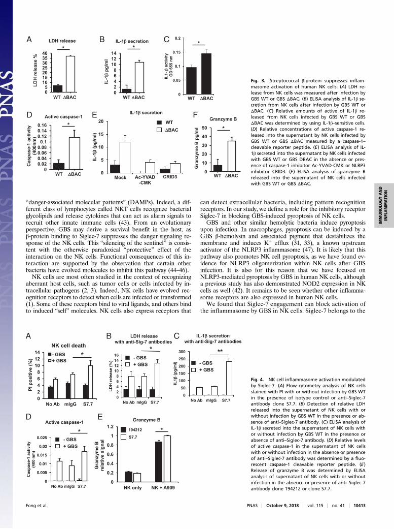
associated proteins, and the β-hemolysin/cytolysin (β-h/c) toxinhas been implicated as the principal activator of the NLRP3inflammasome complex (30, 31, 33). To explore inflammasomeactivation in human NK cells, we confirmed expression of NLRP3in freshly isolated, sterile human NK cells by flow cytometry inperipheral blood from healthy donors with an anti-NLRP3 antibody(Fig. 2C). These results support a previous study that also describedNLRP3 expression in human NK cells (42). We next examinedinflammasome-induced cell death events such as release of intra-cellular contents caused by cellular swelling and bursting, dissemi-nation of the proinflammatory cytokine IL-1β, and oligomerizationof inflammasome proteins into foci. NK cells were exposed to WTor ΔBAC GBS and then stained by using an anti-NLRP3 antibodyafter fixation and permeabilization. By using confocal micros-copy to visualize signals from the anti-NLRP3 antibody, we foundthat a greater percentage of NK cells contained NLRP3 foci whenexposed to the ΔBAC mutant compared with WT GBS (Fig. 2Dand SI Appendix, Fig. S2). Consistent with pyroptosis, release ofthe intracellular enzyme lactate dehydrogenase (LDH), a classicmarker of membrane-disrupted cell death, as well as IL-1β, wasmuch higher in the supernatant of NK cells exposed to ΔBAC vs.WT GBS (Fig. 3 A and B).Pro–IL-1β requires proteolytic cleavage by caspase-1 to be-
come active. To assess whether the IL-1β released from GBS-exposed NK cells is functionally active, supernatants from GBS-exposed NK cells were applied to an IL-1β reporter cell line.Supernatant from NK cells exposed to ΔBAC elicited a stron-ger signal from the IL-1β reporter cell line compared withsupernatants from WT GBS-exposed NK cells (Fig. 3C).
Furthermore, we found greater concentrations of active caspase-1 in the supernatant of ΔBAC-infected vs. WT-infected NK cells(Fig. 3D). IL-1β secretion was reduced in the presence of thecaspase-1 inhibitor Ac-YVAD-CMK and specific NLRP3 in-hibitor CRID3, indicating a crucial role of the NLRP3 inflam-masome for NK cell pyroptosis (Fig. 3E).NK cells possess cytolytic molecules and proteases including
granzymes (3). Granzyme B was released into the supernatantwith greater concentrations from NK cells exposed to ΔBACmutant compared with WT GBS (Fig. 3F). Collectively, thesedata suggest that the GBS β-protein inhibits formation of theinflammasome complex and its downstream effects such as celllysis and IL-1β secretion in NK cells.
Siglec-7 Engagement by β-Protein Suppresses Inflammasome Responsesin NK Cells. Α greater percentage of NK cells incubated with WTGBS in the presence of anti–Siglec-7 blocking antibodies were PI-positive compared with no antibody or isotype control antibody(Fig. 4A). LDH release was also increased from NK cells exposedto WT GBS in the presence of anti–Siglec-7 antibodies comparedwith isotype control antibodies (Fig. 4B). These observations wererecapitulated upon determination of the relative concentrations ofIL-1β, caspase-1, and granzyme B in the NK cell supernatant(Fig. 4 C–E). These data indicate that Siglec-7 can modulateinflammasome formation in NK cells, and our observation thatanti–Siglec-7 antibody blocks ligand engagement to enhanceinflammasome formation is consistent with the known inhibitoryproperties of Siglec-7.
DiscussionIn this work, we have explored a rarely studied direct interactionbetween NK cells and extracellular bacteria. Our experimentsuncovered three previously undescribed immune interactions:pyroptosis of NK cells in response to exposure to extracellularbacteria, Siglec-mediated inhibition of such pyroptosis, and NKcell Siglec interactions with β-protein on an important humanpathogen, GBS. Based on these findings, we propose that NK cellsmay function as early sentinels of bacterial infection via theirability to undergo pyroptosis, alerting the other components of theimmune response via production of the critical proinflammatorycytokine IL-1β and release of intracellular contents recognized as
A B
C D
Fig. 2. Streptococcal β-protein protects NK cellsfrom death after infection. (A) Forward-and side-scatter analysis of primary human NK cells in theuninfected state or infected with GBS WT or GBSΔBAC. (B) Flow cytometry analysis of primary CD56+
cells stained with PI to determine cell death withoutinfection or with infection by GBS WT or GBS ΔBAC.(C) Flow cytometry histograms of permeabilized pri-mary human NK cells stained with anti-NLRP3 anti-body from two donors. (D) Confocal microscopyimages of NK cells stained with anti-NLRP3 antibodywithout infection or with infection by GBS WT or GBSΔBAC. The graph quantifies the percentage of NKcells with NLRP3 foci per field of view for each in-cubation condition. Error bars represent SD.
10412 | www.pnas.org/cgi/doi/10.1073/pnas.1804108115 Fong et al.
“danger-associated molecular patterns” (DAMPs). Indeed, a dif-ferent class of lymphocytes called NKT cells recognize bacterialglycolipids and release cytokines that can act as alarm signals torecruit other innate immune cells (43). From an evolutionaryperspective, GBS may derive a survival benefit in the host, asβ-protein binding to Siglec-7 suppresses the danger signaling re-sponse of the NK cells. This “silencing of the sentinel” is consis-tent with the otherwise paradoxical “protective” effect of theinteraction on the NK cells. Functional consequences of this in-teraction are supported by the observation that certain otherbacteria have evolved molecules to inhibit this pathway (44–46).NK cells are most often studied in the context of recognizing
aberrant host cells, such as tumor cells or cells infected by in-tracellular pathogens (2, 3). Indeed, NK cells have evolved rec-ognition receptors to detect when cells are infected or transformed(1). Some of these receptors bind to viral ligands, and others bindto induced “self” molecules. NK cells also express receptors that
can detect extracellular bacteria, including pattern recognitionreceptors. In our study, we define a role for the inhibitory receptorSiglec-7 in blocking GBS-induced pyroptosis of NK cells.GBS and other similar hemolytic bacteria induce pyroptosis
upon infection. In macrophages, pyroptosis can be induced by aGBS β-hemolysin and associated pigment that destabilizes themembrane and induces K+ efflux (31, 33), a known upstreamactivator of the NLRP3 inflammasome (47). It is likely that thispathway also promotes NK cell pyroptosis, as we have found ev-idence for NLRP3 oligomerization within NK cells after GBSinfection. It is also for this reason that we have focused onNLRP3-mediated pyroptosis by GBS in human NK cells, althougha previous study has also demonstrated NOD2 expression in NKcells as well (42). It remains to be seen whether other inflamma-some receptors are also expressed in human NK cells.We found that Siglec-7 engagement can block activation of
the inflammasome by GBS in NK cells. Siglec-7 belongs to the
A B C
D E F
Fig. 3. Streptococcal β-protein suppresses inflam-masome activation of human NK cells. (A) LDH re-lease from NK cells was measured after infection byGBS WT or GBS ΔBAC. (B) ELISA analysis of IL-1β se-cretion from NK cells after infection by GBS WT orΔBAC. (C) Relative amounts of active of IL-1β re-leased from NK cells infected by GBS WT or GBSΔBAC was determined by using IL-1β–sensitive cells.(D) Relative concentrations of active caspase-1 re-leased into the supernatant by NK cells infected byGBS WT or GBS ΔBAC measured by a caspase-1–cleavable reporter peptide. (E) ELISA analysis of IL-1β secreted into the supernatant by NK cells infectedwith GBS WT or GBS DBAC in the absence or pres-ence of caspase-1 inhibitor Ac-YVAD-CMK or NLRP3inhibitor CRID3. (F) ELISA analysis of granzyme Breleased into the supernatant of NK cells infectedwith GBS WT or GBS ΔBAC.
A B C
D E
Fig. 4. NK cell inflammasome activation modulatedby Siglec-7. (A) Flow cytometry analysis of NK cellsstained with PI with or without infection by GBS WTin the presence of isotype control or anti–Siglec-7antibody clone S7.7. (B) Detection of relative LDHreleased into the supernatant of NK cells with orwithout infection by GBS WT in the presence or ab-sence of anti–Siglec-7 antibody. (C) ELISA analysis ofIL-1β secreted into the supernatant of NK cells withor without infection by GBS WT in the presence orabsence of anti–Siglec-7 antibody. (D) Relative levelsof active caspase-1 in the supernatant of NK cellswith or without infection in the absence or presenceof anti–Siglec-7 antibody was determined by a fluo-rescent caspase-1 cleavable reporter peptide. (E)Release of granzyme B was determined by ELISAanalysis of supernatant of NK cells with or withoutinfection in the absence or presence of anti–Siglec-7antibody clone 194212 or clone S7.7.
Fong et al. PNAS | October 9, 2018 | vol. 115 | no. 41 | 10413
IMMUNOLO
GYAND
INFLAMMATION
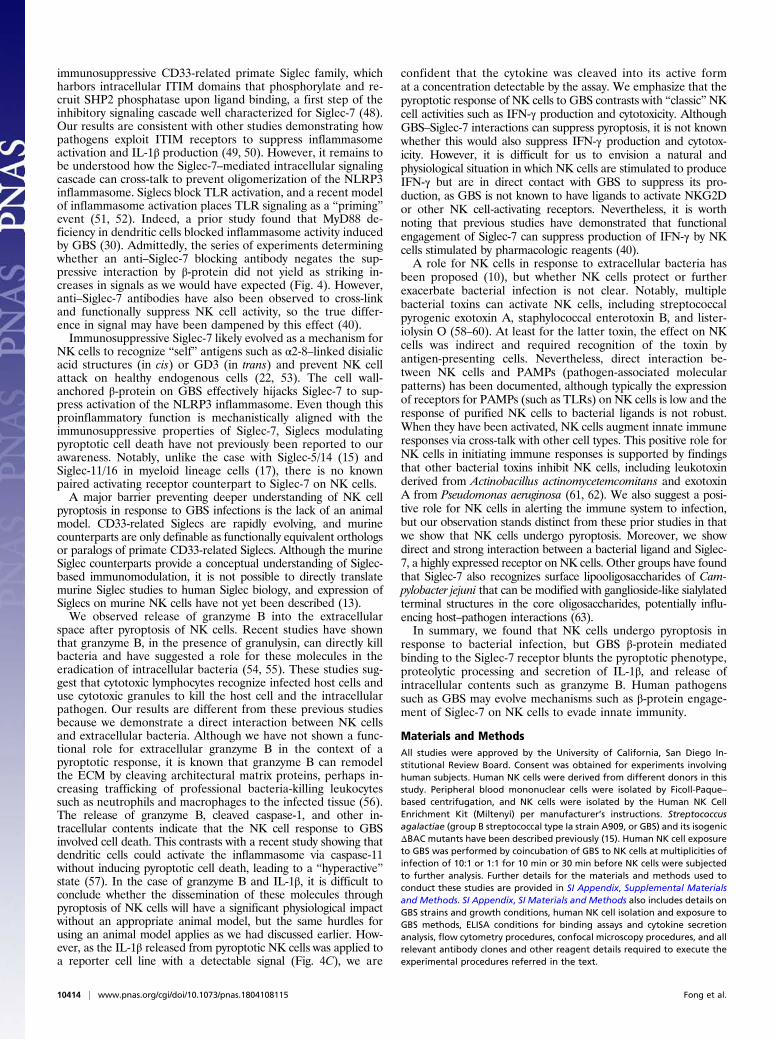
immunosuppressive CD33-related primate Siglec family, whichharbors intracellular ITIM domains that phosphorylate and re-cruit SHP2 phosphatase upon ligand binding, a first step of theinhibitory signaling cascade well characterized for Siglec-7 (48).Our results are consistent with other studies demonstrating howpathogens exploit ITIM receptors to suppress inflammasomeactivation and IL-1β production (49, 50). However, it remains tobe understood how the Siglec-7–mediated intracellular signalingcascade can cross-talk to prevent oligomerization of the NLRP3inflammasome. Siglecs block TLR activation, and a recent modelof inflammasome activation places TLR signaling as a “priming”event (51, 52). Indeed, a prior study found that MyD88 de-ficiency in dendritic cells blocked inflammasome activity inducedby GBS (30). Admittedly, the series of experiments determiningwhether an anti–Siglec-7 blocking antibody negates the sup-pressive interaction by β-protein did not yield as striking in-creases in signals as we would have expected (Fig. 4). However,anti–Siglec-7 antibodies have also been observed to cross-linkand functionally suppress NK cell activity, so the true differ-ence in signal may have been dampened by this effect (40).Immunosuppressive Siglec-7 likely evolved as a mechanism for
NK cells to recognize “self” antigens such as α2-8–linked disialicacid structures (in cis) or GD3 (in trans) and prevent NK cellattack on healthy endogenous cells (22, 53). The cell wall-anchored β-protein on GBS effectively hijacks Siglec-7 to sup-press activation of the NLRP3 inflammasome. Even though thisproinflammatory function is mechanistically aligned with theimmunosuppressive properties of Siglec-7, Siglecs modulatingpyroptotic cell death have not previously been reported to ourawareness. Notably, unlike the case with Siglec-5/14 (15) andSiglec-11/16 in myeloid lineage cells (17), there is no knownpaired activating receptor counterpart to Siglec-7 on NK cells.A major barrier preventing deeper understanding of NK cell
pyroptosis in response to GBS infections is the lack of an animalmodel. CD33-related Siglecs are rapidly evolving, and murinecounterparts are only definable as functionally equivalent orthologsor paralogs of primate CD33-related Siglecs. Although the murineSiglec counterparts provide a conceptual understanding of Siglec-based immunomodulation, it is not possible to directly translatemurine Siglec studies to human Siglec biology, and expression ofSiglecs on murine NK cells have not yet been described (13).We observed release of granzyme B into the extracellular
space after pyroptosis of NK cells. Recent studies have shownthat granzyme B, in the presence of granulysin, can directly killbacteria and have suggested a role for these molecules in theeradication of intracellular bacteria (54, 55). These studies sug-gest that cytotoxic lymphocytes recognize infected host cells anduse cytotoxic granules to kill the host cell and the intracellularpathogen. Our results are different from these previous studiesbecause we demonstrate a direct interaction between NK cellsand extracellular bacteria. Although we have not shown a func-tional role for extracellular granzyme B in the context of apyroptotic response, it is known that granzyme B can remodelthe ECM by cleaving architectural matrix proteins, perhaps in-creasing trafficking of professional bacteria-killing leukocytessuch as neutrophils and macrophages to the infected tissue (56).The release of granzyme B, cleaved caspase-1, and other in-tracellular contents indicate that the NK cell response to GBSinvolved cell death. This contrasts with a recent study showing thatdendritic cells could activate the inflammasome via caspase-11without inducing pyroptotic cell death, leading to a “hyperactive”state (57). In the case of granzyme B and IL-1β, it is difficult toconclude whether the dissemination of these molecules throughpyroptosis of NK cells will have a significant physiological impactwithout an appropriate animal model, but the same hurdles forusing an animal model applies as we had discussed earlier. How-ever, as the IL-1β released from pyroptotic NK cells was applied toa reporter cell line with a detectable signal (Fig. 4C), we are
confident that the cytokine was cleaved into its active format a concentration detectable by the assay. We emphasize that thepyroptotic response of NK cells to GBS contrasts with “classic”NKcell activities such as IFN-γ production and cytotoxicity. AlthoughGBS–Siglec-7 interactions can suppress pyroptosis, it is not knownwhether this would also suppress IFN-γ production and cytotox-icity. However, it is difficult for us to envision a natural andphysiological situation in which NK cells are stimulated to produceIFN-γ but are in direct contact with GBS to suppress its pro-duction, as GBS is not known to have ligands to activate NKG2Dor other NK cell-activating receptors. Nevertheless, it is worthnoting that previous studies have demonstrated that functionalengagement of Siglec-7 can suppress production of IFN-γ by NKcells stimulated by pharmacologic reagents (40).A role for NK cells in response to extracellular bacteria has
been proposed (10), but whether NK cells protect or furtherexacerbate bacterial infection is not clear. Notably, multiplebacterial toxins can activate NK cells, including streptococcalpyrogenic exotoxin A, staphylococcal enterotoxin B, and lister-iolysin O (58–60). At least for the latter toxin, the effect on NKcells was indirect and required recognition of the toxin byantigen-presenting cells. Nevertheless, direct interaction be-tween NK cells and PAMPs (pathogen-associated molecularpatterns) has been documented, although typically the expressionof receptors for PAMPs (such as TLRs) on NK cells is low and theresponse of purified NK cells to bacterial ligands is not robust.When they have been activated, NK cells augment innate immuneresponses via cross-talk with other cell types. This positive role forNK cells in initiating immune responses is supported by findingsthat other bacterial toxins inhibit NK cells, including leukotoxinderived from Actinobacillus actinomycetemcomitans and exotoxinA from Pseudomonas aeruginosa (61, 62). We also suggest a posi-tive role for NK cells in alerting the immune system to infection,but our observation stands distinct from these prior studies in thatwe show that NK cells undergo pyroptosis. Moreover, we showdirect and strong interaction between a bacterial ligand and Siglec-7, a highly expressed receptor on NK cells. Other groups have foundthat Siglec-7 also recognizes surface lipooligosaccharides of Cam-pylobacter jejuni that can be modified with ganglioside-like sialylatedterminal structures in the core oligosaccharides, potentially influ-encing host–pathogen interactions (63).In summary, we found that NK cells undergo pyroptosis in
response to bacterial infection, but GBS β-protein mediatedbinding to the Siglec-7 receptor blunts the pyroptotic phenotype,proteolytic processing and secretion of IL-1β, and release ofintracellular contents such as granzyme B. Human pathogenssuch as GBS may evolve mechanisms such as β-protein engage-ment of Siglec-7 on NK cells to evade innate immunity.
Materials and MethodsAll studies were approved by the University of California, San Diego In-stitutional Review Board. Consent was obtained for experiments involvinghuman subjects. Human NK cells were derived from different donors in thisstudy. Peripheral blood mononuclear cells were isolated by Ficoll-Paque–based centrifugation, and NK cells were isolated by the Human NK CellEnrichment Kit (Miltenyi) per manufacturer’s instructions. Streptococcusagalactiae (group B streptococcal type Ia strain A909, or GBS) and its isogenicΔBAC mutants have been described previously (15). Human NK cell exposureto GBS was performed by coincubation of GBS to NK cells at multiplicities ofinfection of 10:1 or 1:1 for 10 min or 30 min before NK cells were subjectedto further analysis. Further details for the materials and methods used toconduct these studies are provided in SI Appendix, Supplemental Materialsand Methods. SI Appendix, SI Materials and Methods also includes details onGBS strains and growth conditions, human NK cell isolation and exposure toGBS methods, ELISA conditions for binding assays and cytokine secretionanalysis, flow cytometry procedures, confocal microscopy procedures, and allrelevant antibody clones and other reagent details required to execute theexperimental procedures referred in the text.
10414 | www.pnas.org/cgi/doi/10.1073/pnas.1804108115 Fong et al.

ACKNOWLEDGMENTS. We thank members of the laboratories of J.D.B., A.V.,and V.N. for helpful discussions and suggestions. This study was supportedby a grant from The Hartwell Foundation (to J.D.B.), National Institutes of
Health (NIH) Grant CA157885 (to J.D.B.), NIH Grant 1P01HL107150/NationalHeart, Lung, and Blood Institute Program of Excellence in Glycosciences (toA.V. and V.N.), and NIH Grant R01GM032373 (to A.V.).
1. Pegram HJ, Andrews DM, Smyth MJ, Darcy PK, Kershaw MH (2011) Activating andinhibitory receptors of natural killer cells. Immunol Cell Biol 89:216–224.
2. Guillerey C, Huntington ND, Smyth MJ (2016) Targeting natural killer cells in cancerimmunotherapy. Nat Immunol 17:1025–1036.
3. Morvan MG, Lanier LL (2016) NK cells and cancer: You can teach innate cells newtricks. Nat Rev Cancer 16:7–19.
4. Bauer S, et al. (1999) Activation of NK cells and T cells by NKG2D, a receptor for stress-inducible MICA. Science 285:727–729.
5. Wu J, et al. (1999) An activating immunoreceptor complex formed by NKG2D andDAP10. Science 285:730–732.
6. Jamieson AM, et al. (2002) The role of the NKG2D immunoreceptor in immune cellactivation and natural killing. Immunity 17:19–29.
7. Biassoni R, Bottino C, Cantoni C, Moretta A (2002) Human natural killer receptors andtheir ligands. Curr Protoc Immunol Chapter 14:Unit 14.10.
8. Lanier LL (2015) NKG2D receptor and its ligands in host defense. Cancer Immunol Res3:575–582.
9. Groh V, Wu J, Yee C, Spies T (2002) Tumour-derived soluble MIC ligands impair ex-pression of NKG2D and T-cell activation. Nature 419:734–738.
10. Souza-Fonseca-Guimaraes F, Adib-Conquy M, Cavaillon JM (2012) Natural killer (NK)cells in antibacterial innate immunity: Angels or devils? Mol Med 18:270–285.
11. Angata T, Margulies EH, Green ED, Varki A (2004) Large-scale sequencing of theCD33-related Siglec gene cluster in five mammalian species reveals rapid evolution bymultiple mechanisms. Proc Natl Acad Sci USA 101:13251–13256.
12. Fraschilla I, Pillai S (2017) Viewing Siglecs through the lens of tumor immunology.Immunol Rev 276:178–191.
13. Schwarz F, Fong JJ, Varki A (2015) Human-specific evolutionary changes in the biologyof Siglecs. Adv Exp Med Biol 842:1–16.
14. Crocker PR, Paulson JC, Varki A (2007) Siglecs and their roles in the immune system.Nat Rev Immunol 7:255–266.
15. Ali SR, et al. (2014) Siglec-5 and Siglec-14 are polymorphic paired receptors thatmodulate neutrophil and amnion signaling responses to group B Streptococcus. J ExpMed 211:1231–1242.
16. Fong JJ, et al. (2015) Immunomodulatory activity of extracellular Hsp70 mediated viapaired receptors Siglec-5 and Siglec-14. EMBO J 34:2775–2788.
17. Schwarz F, et al. (2017) Paired Siglec receptors generate opposite inflammatory re-sponses to a human-specific pathogen. EMBO J 36:751–760.
18. Schwarz F, et al. (2015) Siglec receptors impact mammalian lifespan by modulatingoxidative stress. eLife 4:e06184.
19. Varki A (2011) Since there are PAMPs and DAMPs, there must be SAMPs? Glycan “self-associated molecular patterns” dampen innate immunity, but pathogens can mimicthem. Glycobiology 21:1121–1124.
20. Lizcano A, et al. (2017) Erythrocyte sialoglycoproteins engage Siglec-9 on neutrophilsto suppress activation. Blood 129:3100–3110.
21. Hudak JE, Canham SM, Bertozzi CR (2014) Glycocalyx engineering reveals a Siglec-based mechanism for NK cell immunoevasion. Nat Chem Biol 10:69–75.
22. Nicoll G, et al. (2003) Ganglioside GD3 expression on target cells can modulate NK cellcytotoxicity via Siglec-7-dependent and -independent mechanisms. Eur J Immunol 33:1642–1648.
23. Xiao H, Woods EC, Vukojicic P, Bertozzi CR (2016) Precision glycocalyx editing as astrategy for cancer immunotherapy. Proc Natl Acad Sci USA 113:10304–10309.
24. Carlin AF, Lewis AL, Varki A, Nizet V (2007) Group B streptococcal capsular sialic acidsinteract with Siglecs (immunoglobulin-like lectins) on human leukocytes. J Bacteriol189:1231–1237.
25. Carlin AF, et al. (2009) Molecular mimicry of host sialylated glycans allows a bacterialpathogen to engage neutrophil Siglec-9 and dampen the innate immune response.Blood 113:3333–3336.
26. Secundino I, et al. (2016) Host and pathogen hyaluronan signal through humanSiglec-9 to suppress neutrophil activation. J Mol Med (Berl) 94:219–233.
27. Carlin AF, et al. (2009) Group B Streptococcus suppression of phagocyte functions byprotein-mediated engagement of human Siglec-5. J Exp Med 206:1691–1699.
28. Nordström T, et al. (2011) Human Siglec-5 inhibitory receptor and immunoglobulin A(IgA) have separate binding sites in streptococcal beta protein. J Biol Chem 286:33981–33991.
29. Nguyen DH, Hurtado-Ziola N, Gagneux P, Varki A (2006) Loss of Siglec expression on Tlymphocytes during human evolution. Proc Natl Acad Sci USA 103:7765–7770.
30. Costa A, et al. (2012) Activation of the NLRP3 inflammasome by group B streptococci.J Immunol 188:1953–1960.
31. Gupta R, et al. (2014) RNA and β-hemolysin of group B Streptococcus induce in-terleukin-1β (IL-1β) by activating NLRP3 inflammasomes in mouse macrophages. J BiolChem 289:13701–13705.
32. Mohammadi N, et al. (2016) Neutrophils directly recognize group B streptococci andcontribute to interleukin-1β production during infection. PLoS One 11:e0160249.
33. Whidbey C, et al. (2015) A streptococcal lipid toxin induces membrane per-meabilization and pyroptosis leading to fetal injury. EMBO Mol Med 7:488–505.
34. Broz P, Dixit VM (2016) Inflammasomes: Mechanism of assembly, regulation andsignalling. Nat Rev Immunol 16:407–420.
35. Lamkanfi M, Dixit VM (2014) Mechanisms and functions of inflammasomes. Cell 157:1013–1022.
36. Nicoll G, et al. (1999) Identification and characterization of a novel Siglec, Siglec-7,expressed by human natural killer cells and monocytes. J Biol Chem 274:34089–34095.
37. Angata T, Varki A (2000) Siglec-7: A sialic acid-binding lectin of the immunoglobulinsuperfamily. Glycobiology 10:431–438.
38. Jandus C, et al. (2014) Interactions between Siglec-7/9 receptors and ligands influenceNK cell-dependent tumor immunosurveillance. J Clin Invest 124:1810–1820.
39. Kawasaki Y, et al. (2010) Ganglioside DSGb5, preferred ligand for Siglec-7, inhibits NKcell cytotoxicity against renal cell carcinoma cells. Glycobiology 20:1373–1379.
40. Shao JY, et al. (2016) Siglec-7 defines a highly functional natural killer cell subset andinhibits cell-mediated activities. Scand J Immunol 84:182–190.
41. Mikulak J, Di Vito C, Zaghi E, Mavilio D (2017) Host immune responses in HIV-1 in-fection: The emerging pathogenic role of Siglecs and their clinical correlates. FrontImmunol 8:314.
42. Qiu F, Maniar A, Diaz MQ, Chapoval AI, Medvedev AE (2011) Activation of cytokine-producing and antitumor activities of natural killer cells and macrophages by en-gagement of Toll-like and NOD-like receptors. Innate Immun 17:375–387.
43. Kinjo Y, et al. (2011) Invariant natural killer T cells recognize glycolipids from path-ogenic Gram-positive bacteria. Nat Immunol 12:966–974.
44. LaRock CN, Cookson BT (2012) The Yersinia virulence effector YopM binds caspase-1to arrest inflammasome assembly and processing. Cell Host Microbe 12:799–805.
45. Bergsbaken T, Cookson BT (2007) Macrophage activation redirects yersinia-infectedhost cell death from apoptosis to caspase-1-dependent pyroptosis. PLoS Pathog 3:e161.
46. Bergsbaken T, Fink SL, Cookson BT (2009) Pyroptosis: Host cell death and in-flammation. Nat Rev Microbiol 7:99–109.
47. Muñoz-Planillo R, et al. (2013) K+ efflux is the common trigger of NLRP3 in-flammasome activation by bacterial toxins and particulate matter. Immunity 38:1142–1153.
48. Yamaji T, Mitsuki M, Teranishi T, Hashimoto Y (2005) Characterization of inhibitorysignaling motifs of the natural killer cell receptor Siglec-7: Attenuated recruitment ofphosphatases by the receptor is attributed to two amino acids in the motifs.Glycobiology 15:667–676.
49. Nakayama M, et al. (2012) Inhibitory receptor paired Ig-like receptor B is exploited byStaphylococcus aureus for virulence. J Immunol 189:5903–5911.
50. Lu R, Pan H, Shively JE (2012) CEACAM1 negatively regulates IL-1β production in LPSactivated neutrophils by recruiting SHP-1 to a SYK-TLR4-CEACAM1 complex. PLoSPathog 8:e1002597.
51. Chen GY, et al. (2014) Broad and direct interaction between TLR and Siglec families ofpattern recognition receptors and its regulation by Neu1. eLife 3:e04066.
52. Latz E, Xiao TS, Stutz A (2013) Activation and regulation of the inflammasomes. NatRev Immunol 13:397–411.
53. Avril T, North SJ, Haslam SM, Willison HJ, Crocker PR (2006) Probing the cis interac-tions of the inhibitory receptor Siglec-7 with alpha2,8-disialylated ligands on naturalkiller cells and other leukocytes using glycan-specific antibodies and by analysis ofalpha2,8-sialyltransferase gene expression. J Leukoc Biol 80:787–796.
54. Dotiwala F, et al. (2017) Granzyme B disrupts central metabolism and protein syn-thesis in bacteria to promote an immune cell death program. Cell 171:1125–1137.e11.
55. Dotiwala F, et al. (2016) Killer lymphocytes use granulysin, perforin and granzymes tokill intracellular parasites. Nat Med 22:210–216.
56. Buzza MS, et al. (2005) Extracellular matrix remodeling by human granzyme B viacleavage of vitronectin, fibronectin, and laminin. J Biol Chem 280:23549–23558.
57. Aakhus AM, Stavem P, Hovig T, Pedersen TM, Solum NO (1990) Studies on a patientwith thrombocytopenia, giant platelets and a platelet membrane glycoprotein Ibwith reduced amount of sialic acid. Br J Haematol 74:320–329.
58. Sacks LV, et al. (1991) A streptococcal erythrogenic toxin preparation augmentsnatural killer activity of peripheral blood mononuclear cells. J Infect Dis 164:522–526.
59. D’Orazio JA, Burke GW, Stein-Streilein J (1995) Staphylococcal enterotoxin B activatespurified NK cells to secrete IFN-gamma but requires T lymphocytes to augment NKcytotoxicity. J Immunol 154:1014–1023.
60. Nomura T, et al. (2002) Essential role of interleukin-12 (IL-12) and IL-18 for gammainterferon production induced by listeriolysin O in mouse spleen cells. Infect Immun70:1049–1055.
61. Shenker BJ, et al. (1994) Flow cytometric analysis of the cytotoxic effects of Actino-bacillus actinomycetemcomitans leukotoxin on human natural killer cells. J LeukocBiol 55:153–160.
62. Michaels JE, Garfield SA, Hung JT, Cardell RRJ, Jr (1990) Labeling of hepatic glycogenafter short- and long-term stimulation of glycogen synthesis in rats injected with 3H-galactose. Am J Anat 188:419–428.
63. Avril T, Wagner ER, Willison HJ, Crocker PR (2006) Sialic acid-binding immunoglob-ulin-like lectin 7 mediates selective recognition of sialylated glycans expressed onCampylobacter jejuni lipooligosaccharides. Infect Immun 74:4133–4141.
Fong et al. PNAS | October 9, 2018 | vol. 115 | no. 41 | 10415
IMMUNOLO
GYAND
INFLAMMATION