sin título de diapositiva - jmordoh.com.ar · los factores de crecimiento se unen a sus receptores...
TRANSCRIPT
Oncogenes
Dra. María Marcela Barrio
Centro Investigaciones Oncológicas
FUNDACION CANCER - FUCA
Maestría Biología Molecular Médica - UBA
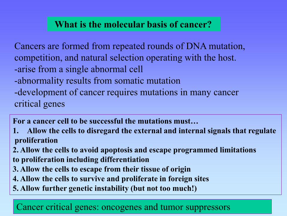
What is the molecular basis of cancer?
Cancers are formed from repeated rounds of DNA mutation,
competition, and natural selection operating with the host.
-arise from a single abnormal cell
-abnormality results from somatic mutation
-development of cancer requires mutations in many cancer
critical genes
For a cancer cell to be successful the mutations must…
1. Allow the cells to disregard the external and internal signals that regulate
proliferation
2. Allow the cells to avoid apoptosis and escape programmed limitations
to proliferation including differentiation
3. Allow the cells to escape from their tissue of origin
4. Allow the cells to survive and proliferate in foreign sites
5. Allow further genetic instability (but not too much!)
Cancer critical genes: oncogenes and tumor suppressors

Hallmarks of Cancer: The Next Generation. Douglas Hanahan and Robert A. Weinberg
Cell 144, 646-, March 4, 2011

Emerging Hallmarks and Enabling Characteristics: An increasing body of research suggests that two additional hallmarks of cancer are involved in the pathogenesis of some and perhaps all cancers. One involves the capability to modify, or reprogram, cellular metabolism in order to most effectively support neoplastic proliferation. The second allows cancer cells to evade immunological destruction, in particular by T and B lymphocytes, macrophages, and natural killer cells. Because neither capability is yet generalized and fully validated, they are labeled as emerging hallmarks. Additionally, two consequential characteristics of neoplasia facilitate acquisition of both core and emerging hallmarks. Genomic instability and thus mutability endow cancer cells with genetic alterations that drive tumor progression. Inflammation by innate immune cells designed to fight infections and heal wounds can instead result in their inadvertent support of multiple hallmark capabilities, thereby manifesting the now widely appreciated tumor-promoting consequences of inflammatory responses.
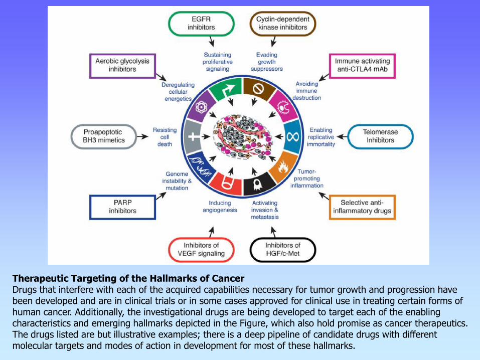
Therapeutic Targeting of the Hallmarks of Cancer Drugs that interfere with each of the acquired capabilities necessary for tumor growth and progression have been developed and are in clinical trials or in some cases approved for clinical use in treating certain forms of human cancer. Additionally, the investigational drugs are being developed to target each of the enabling characteristics and emerging hallmarks depicted in the Figure, which also hold promise as cancer therapeutics. The drugs listed are but illustrative examples; there is a deep pipeline of candidate drugs with different molecular targets and modes of action in development for most of these hallmarks.
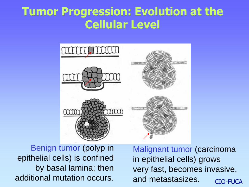
Tumor Progression: Evolution at the Cellular Level
Benign tumor (polyp in
epithelial cells) is confined
by basal lamina; then
additional mutation occurs.
Malignant tumor (carcinoma
in epithelial cells) grows
very fast, becomes invasive,
and metastasizes. CIO-FUCA
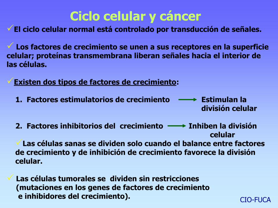
Ciclo celular y cáncer El ciclo celular normal está controlado por transducción de señales.
Los factores de crecimiento se unen a sus receptores en la superficie celular; proteínas transmembrana liberan señales hacia el interior de las células.
Existen dos tipos de factores de crecimiento:
1. Factores estimulatorios de crecimiento Estimulan la división celular
2. Factores inhibitorios del crecimiento Inhiben la división celular Las células sanas se dividen solo cuando el balance entre factores de crecimiento y de inhibición de crecimiento favorece la división celular.
Las células tumorales se dividen sin restricciones (mutaciones en los genes de factores de crecimiento e inhibidores del crecimiento).
CIO-FUCA


Growth of Normal and *Neoplastic
fibroblasts in culture
Growth Characteristics Normal Tumor
Density dependent inhibition of growth present absent
Growth factor requirements high low
Anchorage dependence present absent
Proliferative life span finite indefinite
Contact inhibition present absent
Adhesiveness high low
Morphology flat rounded
*Neoplastic: new shape; any
new or abnormal growth: specifically
a new growth of tissue in which the growth
is uncontrolled or aggressive.
Review of the neoplastic phenotype

Normal and transformed NIH3T3 cells
Normal NIH3T3
(immortal)
Transformed NIH3T3
Review of the neoplastic phenotype
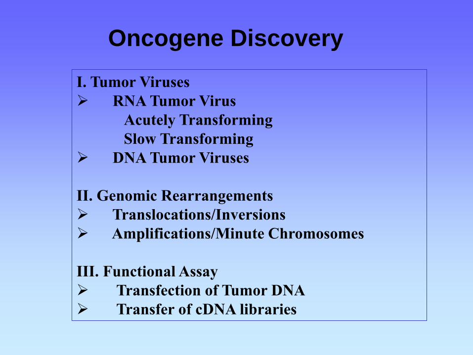
Oncogene Discovery
I. Tumor Viruses
RNA Tumor Virus
Acutely Transforming
Slow Transforming
DNA Tumor Viruses
II. Genomic Rearrangements
Translocations/Inversions
Amplifications/Minute Chromosomes
III. Functional Assay
Transfection of Tumor DNA
Transfer of cDNA libraries

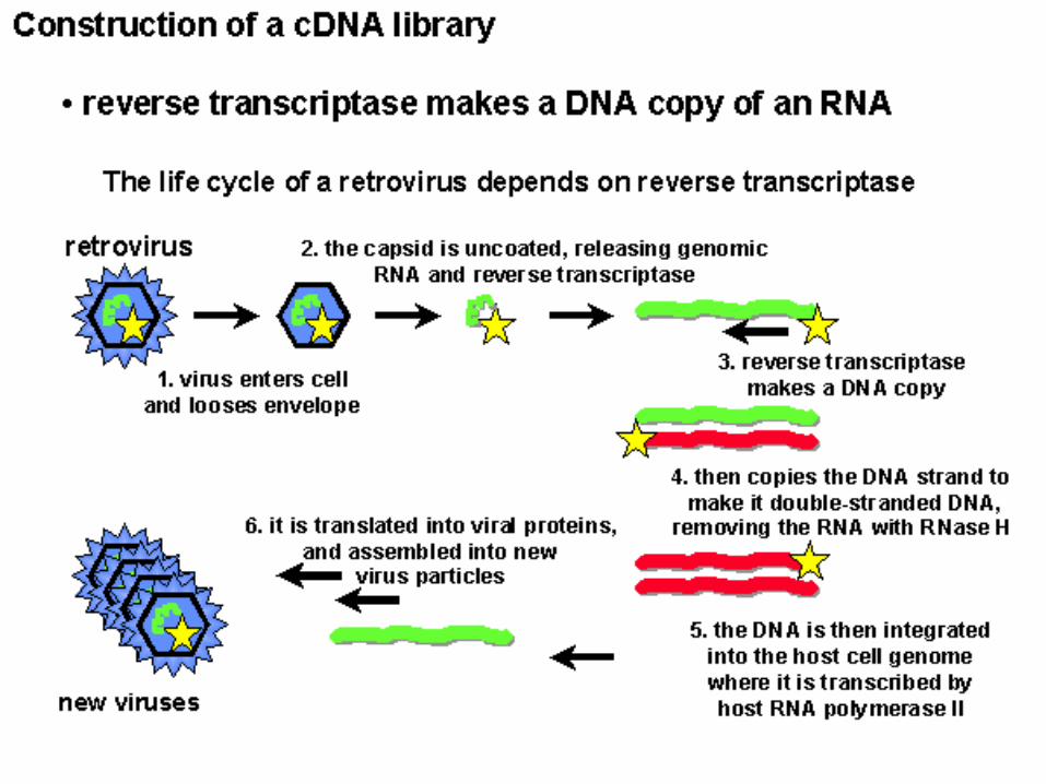
Discovery I. Tumor Viruses; RNA
Retrovirus: RNA genome reversed transcribed into proviral DNA
which integrates randomly into the host cell genome.
Productively infects only proliferating cells.
Peyton Rous:
1st evidence that viruses
could cause cancer (1911).
- Chickens
- fibrosarcoma
- Rous Sarcoma virus
- Nobel prize 1966
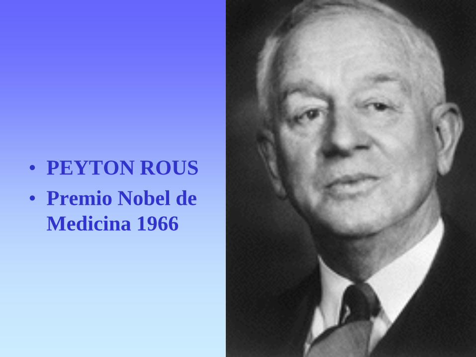
• PEYTON ROUS
• Premio Nobel de
Medicina 1966
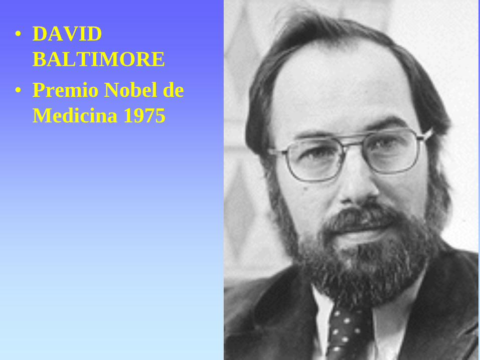
• DAVID
BALTIMORE
• Premio Nobel de
Medicina 1975
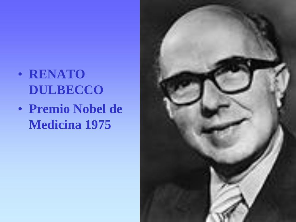
• RENATO
DULBECCO
• Premio Nobel de
Medicina 1975
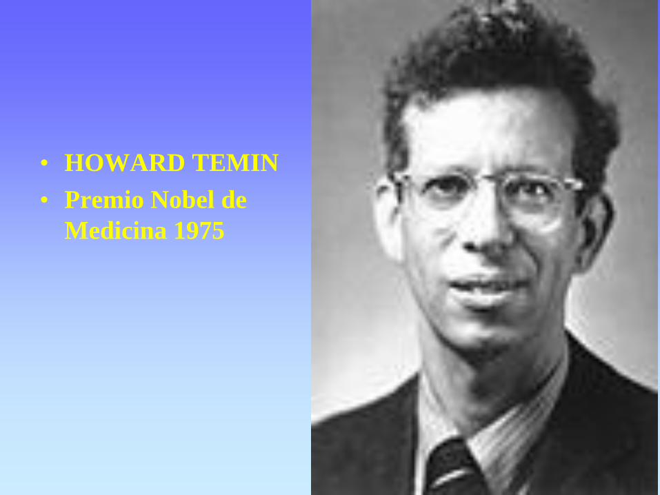
• HOWARD TEMIN
• Premio Nobel de
Medicina 1975

• HAROLD VARMUS
• Premio Nobel de
Medicina 1989
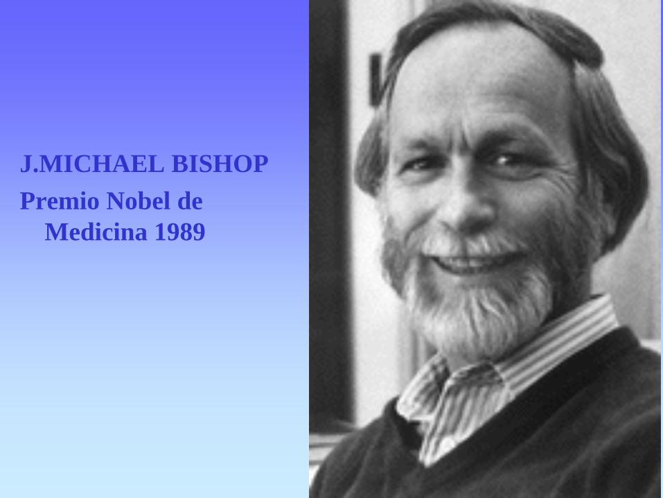
J.MICHAEL BISHOP
Premio Nobel de
Medicina 1989

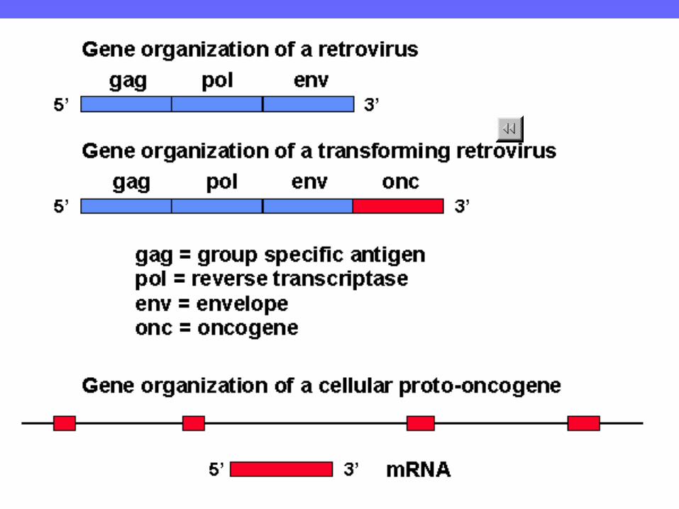
Retroviral Transduction
LTR LTR gag pol env
Acutely Transforming Retroviruses encode an onc gene.
LTR LTR gag pol env onc
pol
RSV has a env-onc fusion
Retrovirus normal
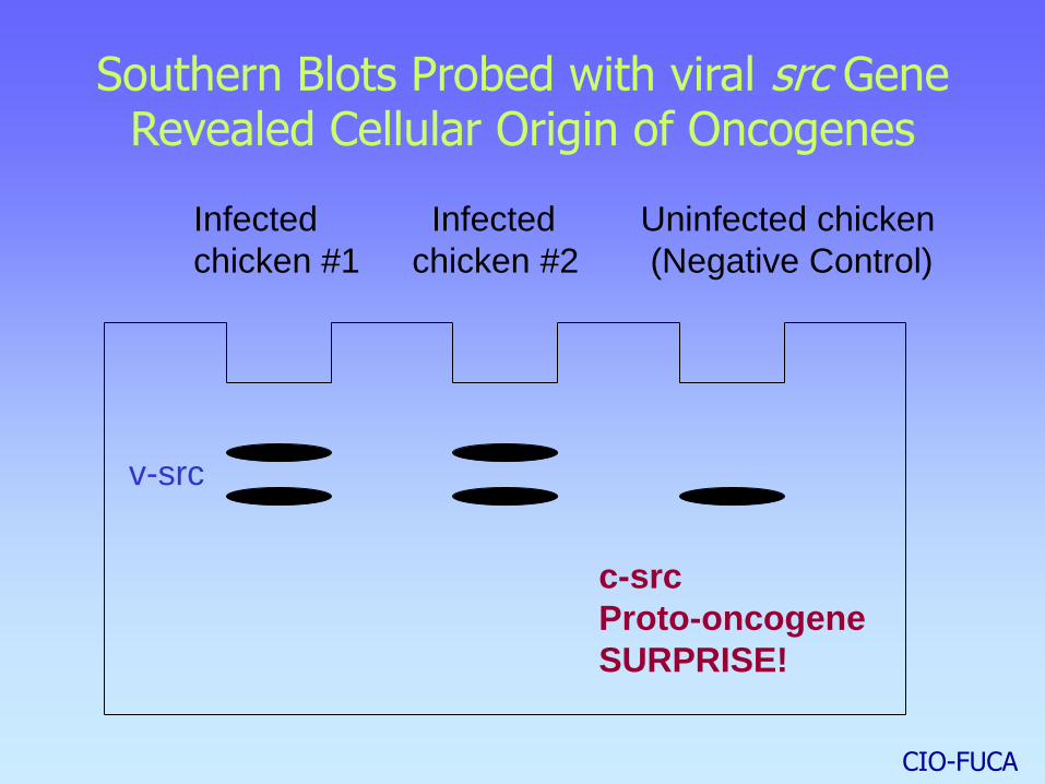
Southern Blots Probed with viral src Gene Revealed Cellular Origin of Oncogenes
Infected Infected Uninfected chicken
chicken #1 chicken #2 (Negative Control)
c-src
Proto-oncogene
SURPRISE!
v-src
CIO-FUCA

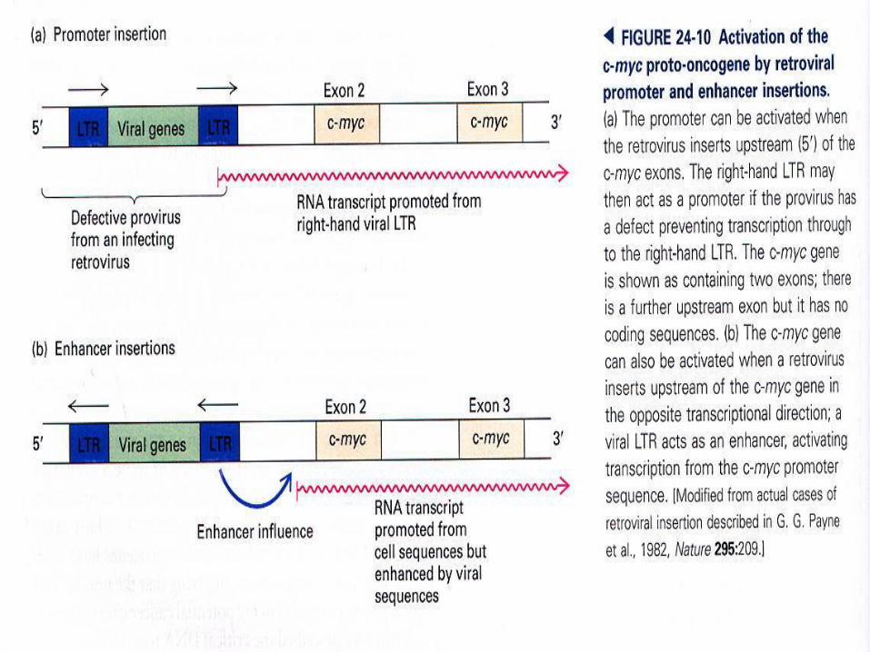
Slow transforming retroviruses
Enhancer insertion
May be 5’ or 3’ in either orientation.
gag pol env LTR LTR
gag pol env LTR LTR
Promoter insertion
Proto-oncogene
Slow transforming retroviruses activate proto-oncogenes by
insertional mutagenesis.
Dysregulated expression occurs after insertion of strong
promoters or enhancers into the genetic loci.
cellular gene
provirus
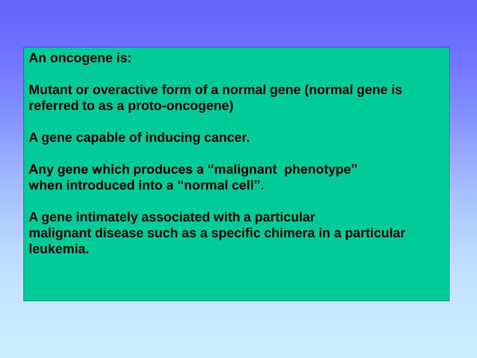
An oncogene is:
Mutant or overactive form of a normal gene (normal gene is
referred to as a proto-oncogene)
A gene capable of inducing cancer.
Any gene which produces a “malignant phenotype”
when introduced into a “normal cell”.
A gene intimately associated with a particular
malignant disease such as a specific chimera in a particular
leukemia.
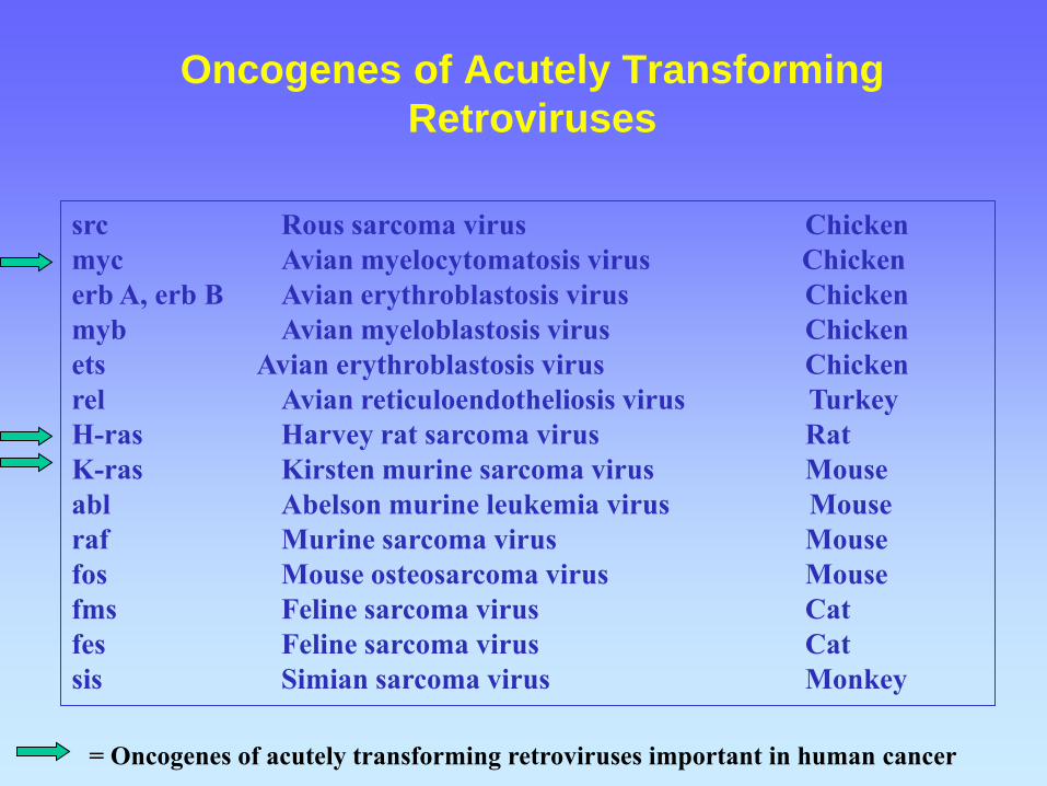
Oncogenes of Acutely Transforming
Retroviruses
src Rous sarcoma virus Chicken
myc Avian myelocytomatosis virus Chicken
erb A, erb B Avian erythroblastosis virus Chicken
myb Avian myeloblastosis virus Chicken
ets Avian erythroblastosis virus Chicken
rel Avian reticuloendotheliosis virus Turkey
H-ras Harvey rat sarcoma virus Rat
K-ras Kirsten murine sarcoma virus Mouse
abl Abelson murine leukemia virus Mouse
raf Murine sarcoma virus Mouse
fos Mouse osteosarcoma virus Mouse
fms Feline sarcoma virus Cat
fes Feline sarcoma virus Cat
sis Simian sarcoma virus Monkey
= Oncogenes of acutely transforming retroviruses important in human cancer

Robert Weinberg
Whitehead Institute-
MIT

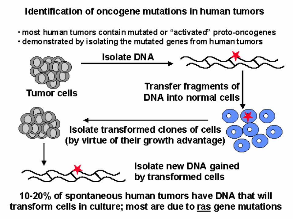
Discovery III. Identification of Oncogenes by
functional assays; *Transfection
DNA
Shear
mRNA cDNA
library
Transfect
Transfect
Transformed Phenotype
Tumor
Reporter Cell line
*transfection: putting foreign DNA
into mammalian cells.
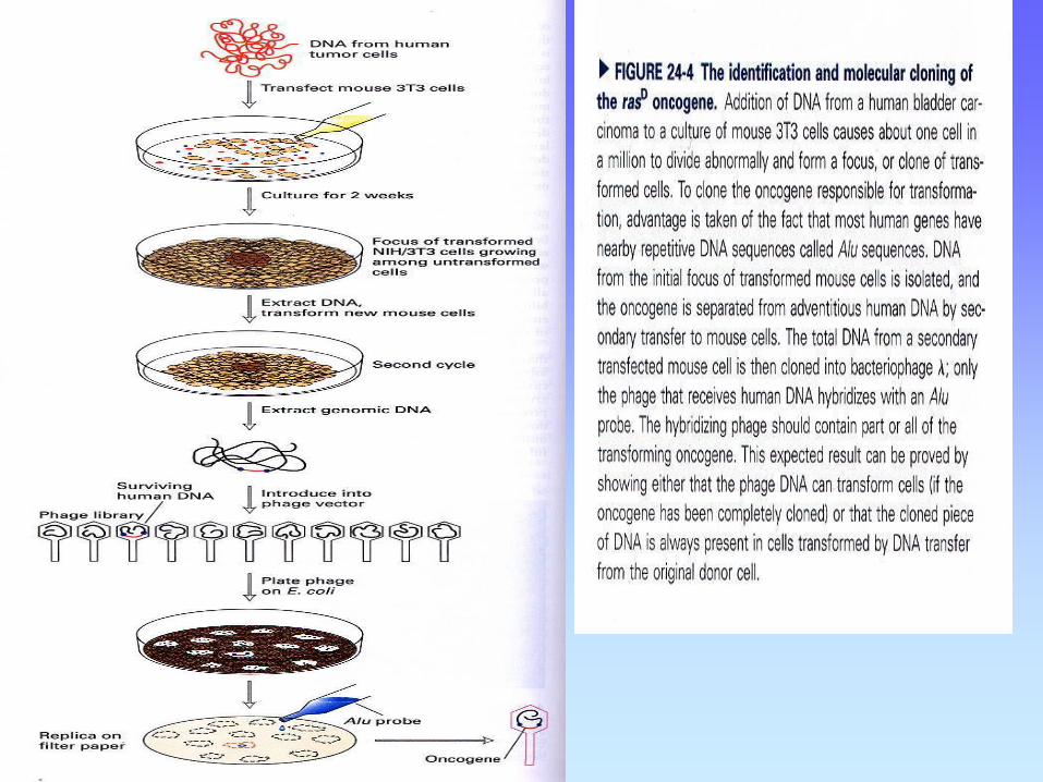
Some Oncogenes identified by Transfection
Weinberg- activated ras from bladder carcinoma.
Vande Woude- met oncogene which is hepatocyte growth factor receptor
from a chemically transformed cell line.
hst is a FGF-related gene identified from a human stomach carcinoma.
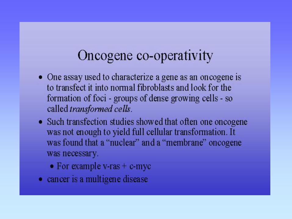

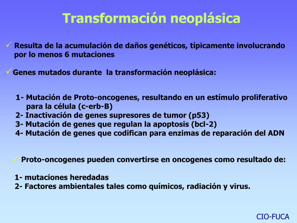
Transformación neoplásica
Resulta de la acumulación de daños genéticos, típicamente involucrando por lo menos 6 mutaciones
Genes mutados durante la transformación neoplásica:
1- Mutación de Proto-oncogenes, resultando en un estímulo proliferativo para la célula (c-erb-B) 2- Inactivación de genes supresores de tumor (p53) 3- Mutación de genes que regulan la apoptosis (bcl-2) 4- Mutación de genes que codifican para enzimas de reparación del ADN
Proto-oncogenes pueden convertirse en oncogenes como resultado de: 1- mutaciones heredadas 2- Factores ambientales tales como químicos, radiación y virus.
CIO-FUCA
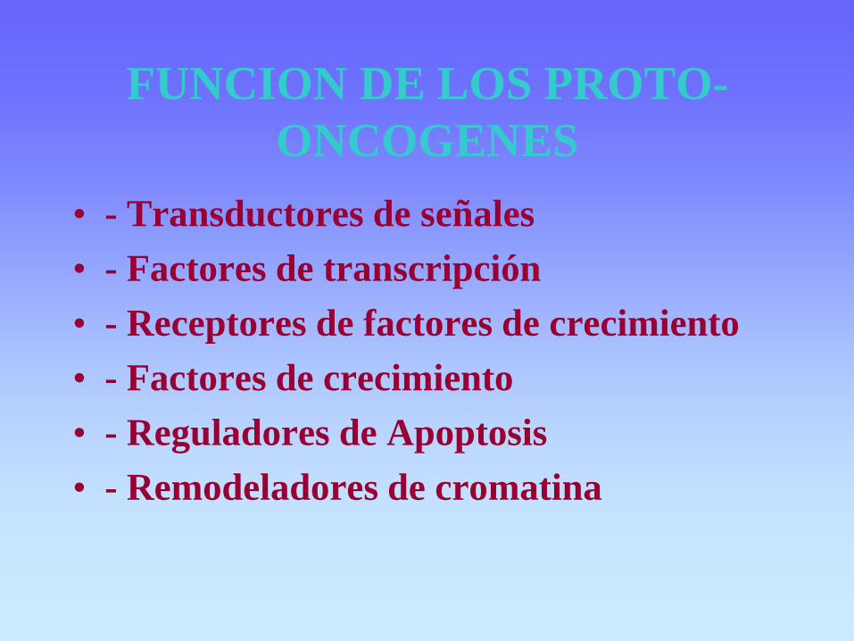
FUNCION DE LOS PROTO-
ONCOGENES
• - Transductores de señales
• - Factores de transcripción
• - Receptores de factores de crecimiento
• - Factores de crecimiento
• - Reguladores de Apoptosis
• - Remodeladores de cromatina

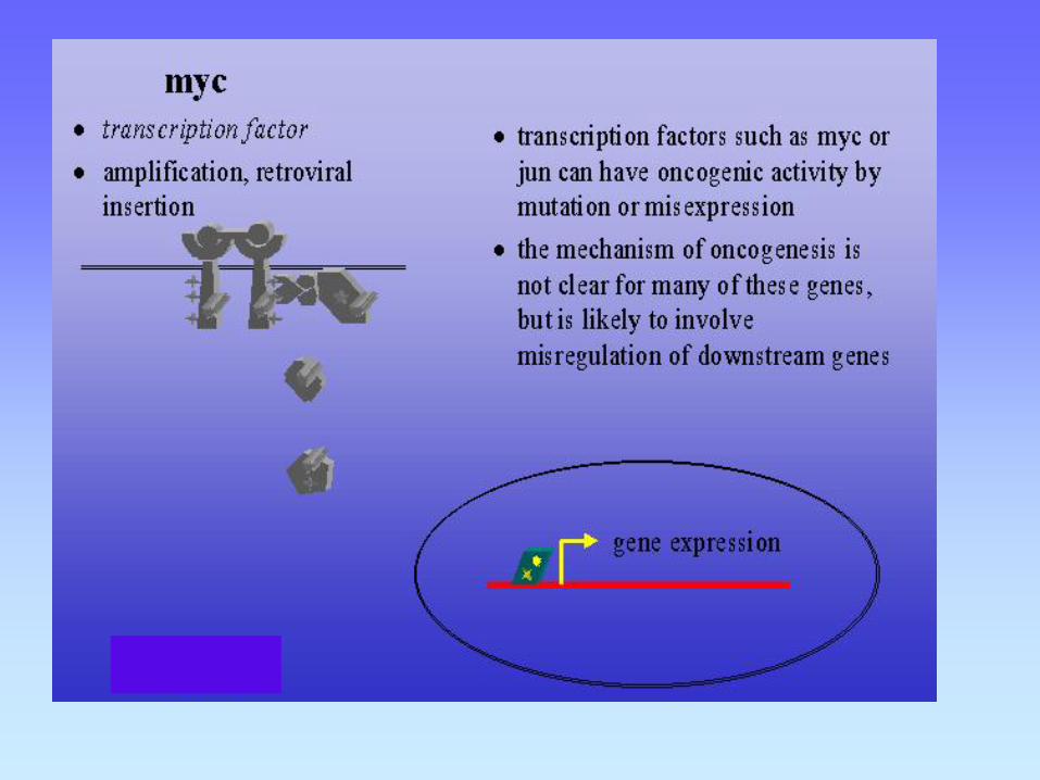
MECANISMOS DE ACTIVACION DE ONCOGENES
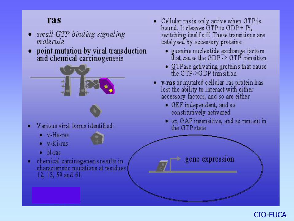
1- Mutaciones puntuales: Ejemplos: ras, erb-B, fms En ras (proteína G), un cambio en un único aminoácido inhibe la hidrólisis del GTP, prolongando el estado activado, independiente de factores de crecimiento, inhibiendo la interacción de ras con GAPs. Funciona como un interruptor molecular en la vía de transducción de señales que conecta los factores de crecimiento con la expresión de genes que controlan la proliferación celular. GF receptor Ras FT genes target división celular. Mutados en el 30% de todos los tumores CIO-FUCA
CIO-FUCA

BRAF oncogene
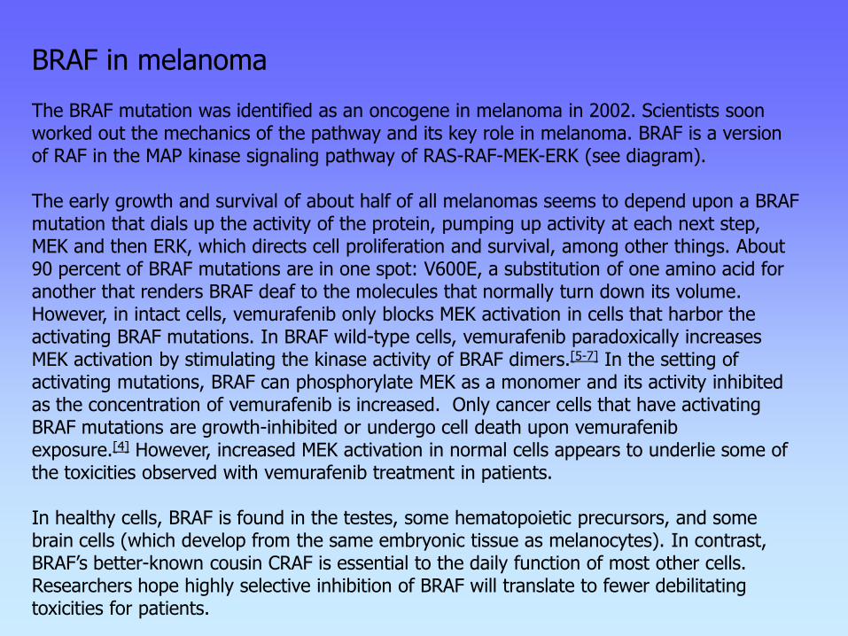
BRAF in melanoma The BRAF mutation was identified as an oncogene in melanoma in 2002. Scientists soon worked out the mechanics of the pathway and its key role in melanoma. BRAF is a version of RAF in the MAP kinase signaling pathway of RAS-RAF-MEK-ERK (see diagram). The early growth and survival of about half of all melanomas seems to depend upon a BRAF mutation that dials up the activity of the protein, pumping up activity at each next step, MEK and then ERK, which directs cell proliferation and survival, among other things. About 90 percent of BRAF mutations are in one spot: V600E, a substitution of one amino acid for another that renders BRAF deaf to the molecules that normally turn down its volume. However, in intact cells, vemurafenib only blocks MEK activation in cells that harbor the activating BRAF mutations. In BRAF wild-type cells, vemurafenib paradoxically increases MEK activation by stimulating the kinase activity of BRAF dimers.[5-7] In the setting of activating mutations, BRAF can phosphorylate MEK as a monomer and its activity inhibited as the concentration of vemurafenib is increased. Only cancer cells that have activating BRAF mutations are growth-inhibited or undergo cell death upon vemurafenib exposure.[4] However, increased MEK activation in normal cells appears to underlie some of the toxicities observed with vemurafenib treatment in patients. In healthy cells, BRAF is found in the testes, some hematopoietic precursors, and some brain cells (which develop from the same embryonic tissue as melanocytes). In contrast, BRAF’s better-known cousin CRAF is essential to the daily function of most other cells. Researchers hope highly selective inhibition of BRAF will translate to fewer debilitating toxicities for patients.
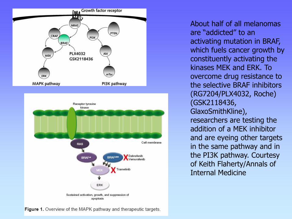
About half of all melanomas are “addicted” to an activating mutation in BRAF, which fuels cancer growth by constituently activating the kinases MEK and ERK. To overcome drug resistance to the selective BRAF inhibitors (RG7204/PLX4032, Roche) (GSK2118436, GlaxoSmithKline), researchers are testing the addition of a MEK inhibitor and are eyeing other targets in the same pathway and in the PI3K pathway. Courtesy of Keith Flaherty/Annals of Internal Medicine

Key components of the MAPK/ERK pathway. "P" represents phosphate, which communicates the signal. Top, epidermal growth factor (EGF) binds to the EGF receptor (EGFR) in the cell membrane, starting the cascade of signals. Further downstream, phosphate signal activates MAPK (also known as ERK). Bottom, signal enters the cell nucleus and causes transcription of DNA, which is then expressed as protein.
MAPK pathway
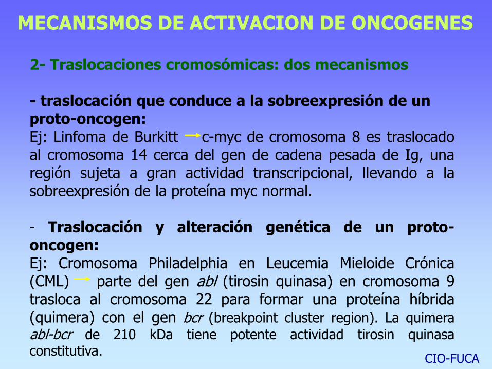
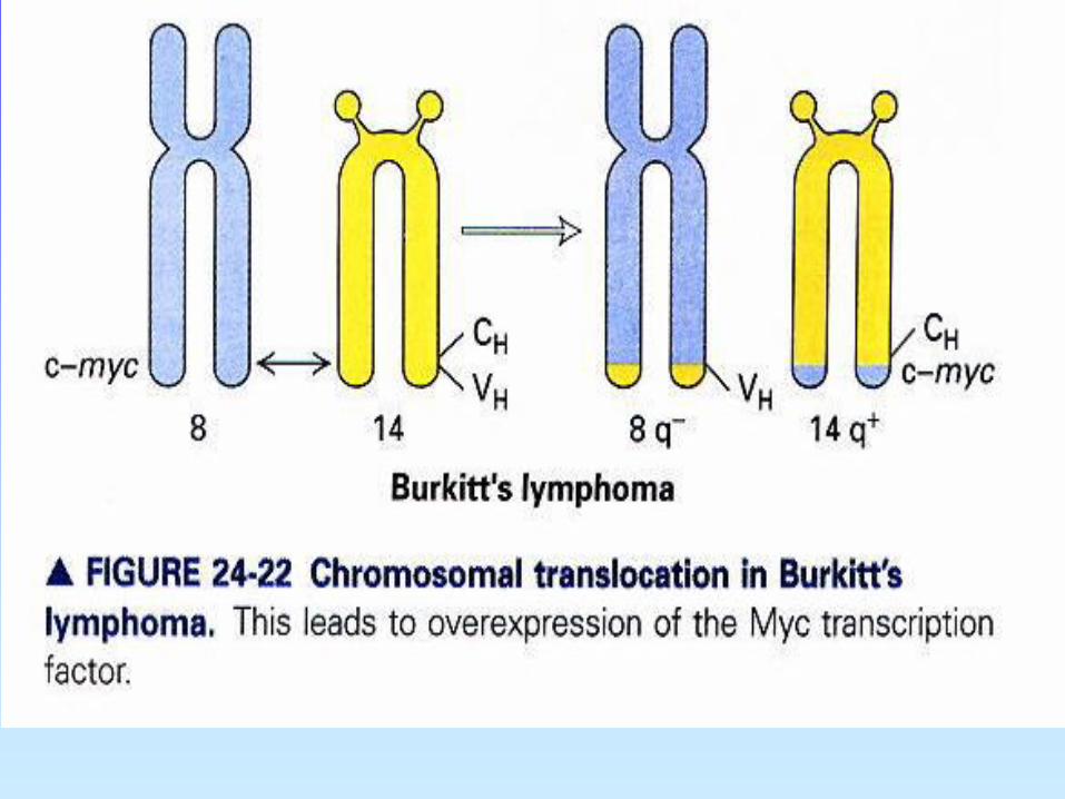
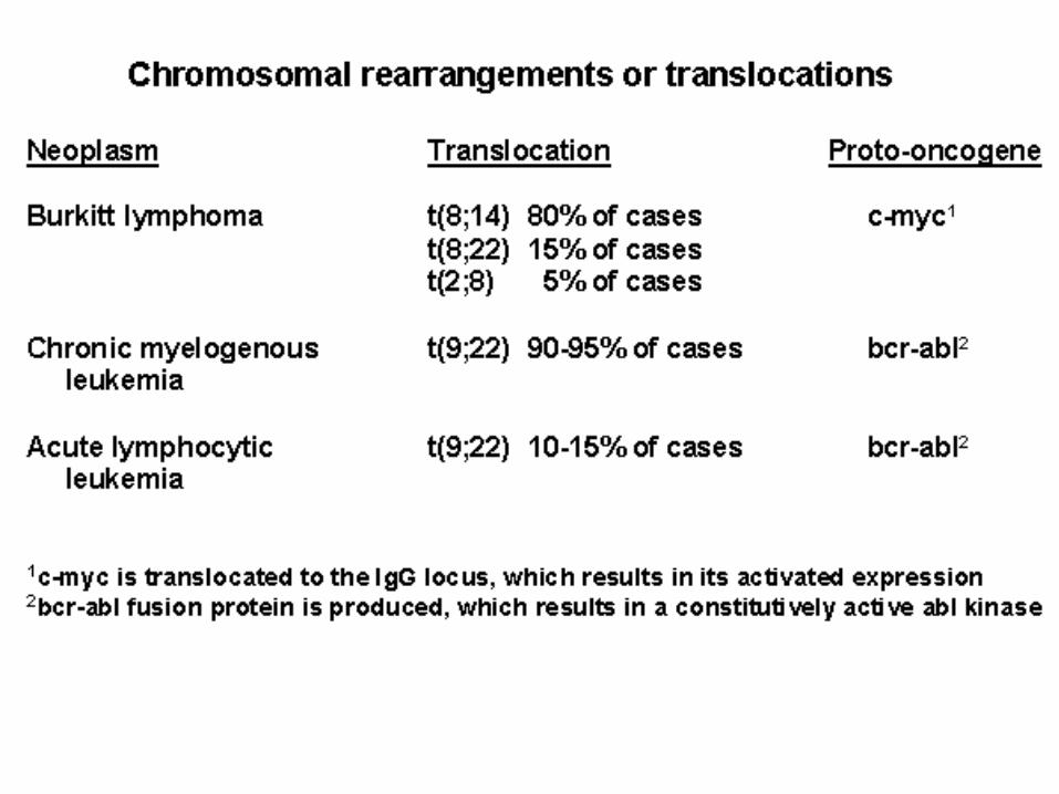
2- Traslocaciones cromosómicas: dos mecanismos - traslocación que conduce a la sobreexpresión de un proto-oncogen: Ej: Linfoma de Burkitt c-myc de cromosoma 8 es traslocado al cromosoma 14 cerca del gen de cadena pesada de Ig, una región sujeta a gran actividad transcripcional, llevando a la sobreexpresión de la proteína myc normal. - Traslocación y alteración genética de un proto-oncogen: Ej: Cromosoma Philadelphia en Leucemia Mieloide Crónica (CML) parte del gen abl (tirosin quinasa) en cromosoma 9 trasloca al cromosoma 22 para formar una proteína híbrida (quimera) con el gen bcr (breakpoint cluster region). La quimera
abl-bcr de 210 kDa tiene potente actividad tirosin quinasa constitutiva.
MECANISMOS DE ACTIVACION DE ONCOGENES
CIO-FUCA

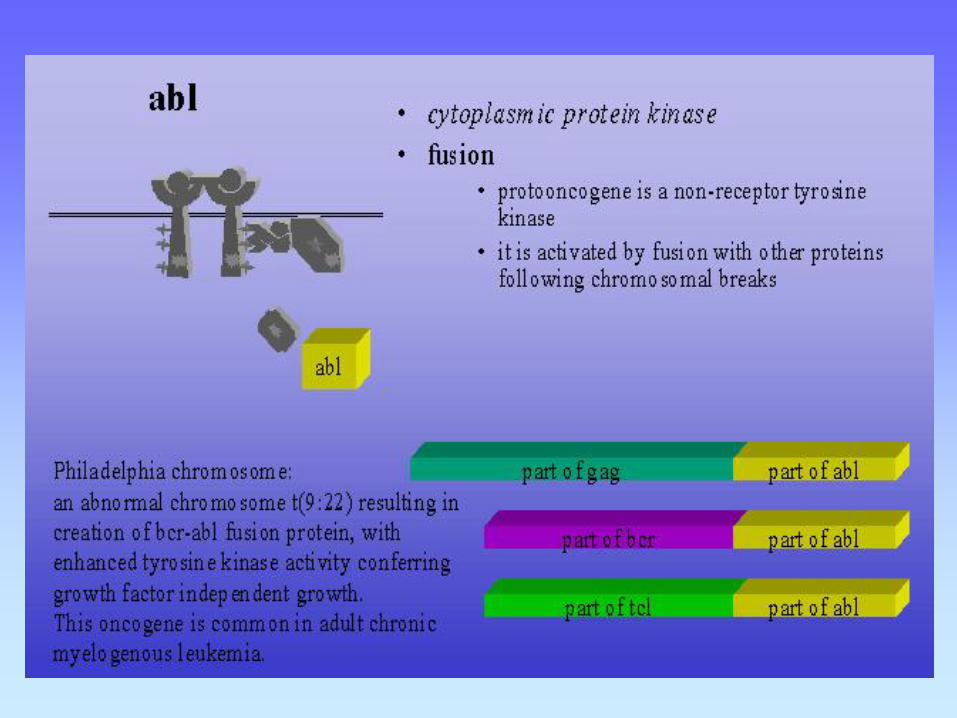

El protooncogen c-abl se fusiona con el gen bcr y el oncogen híbrido resultante bcr/c-abl es transcripcionalmente activo; el ciclo celular se desregula - se produce la leucemia mieloide crónica. Leucocitos únicos portadores del evento de translocación actuarían como origen
de la patología.
**-translocación recíproca entre crom. 9 y 22
CIO-FUCA
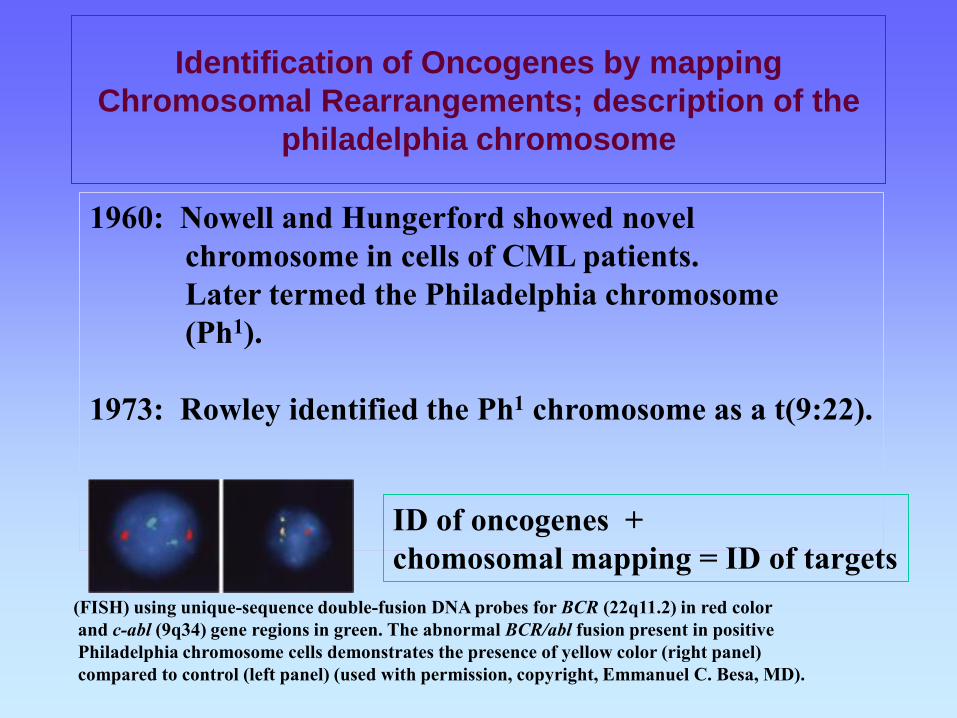
Identification of Oncogenes by mapping
Chromosomal Rearrangements; description of the
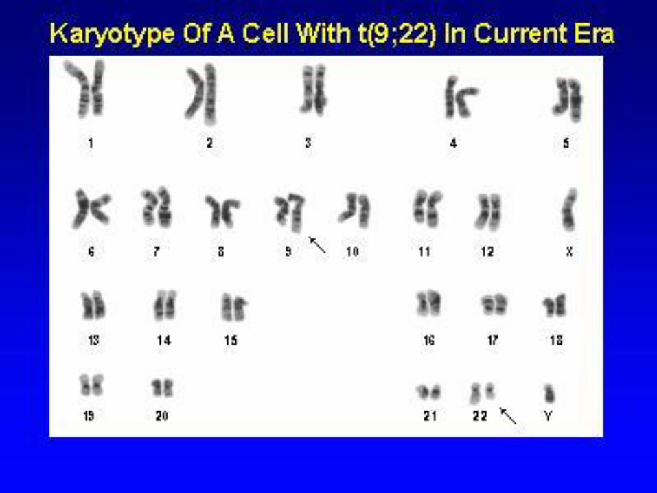
philadelphia chromosome
1960: Nowell and Hungerford showed novel
chromosome in cells of CML patients.
Later termed the Philadelphia chromosome
(Ph1).
1973: Rowley identified the Ph1 chromosome as a t(9:22).
ID of oncogenes +
chomosomal mapping = ID of targets
(FISH) using unique-sequence double-fusion DNA probes for BCR (22q11.2) in red color
and c-abl (9q34) gene regions in green. The abnormal BCR/abl fusion present in positive
Philadelphia chromosome cells demonstrates the presence of yellow color (right panel)
compared to control (left panel) (used with permission, copyright, Emmanuel C. Besa, MD).
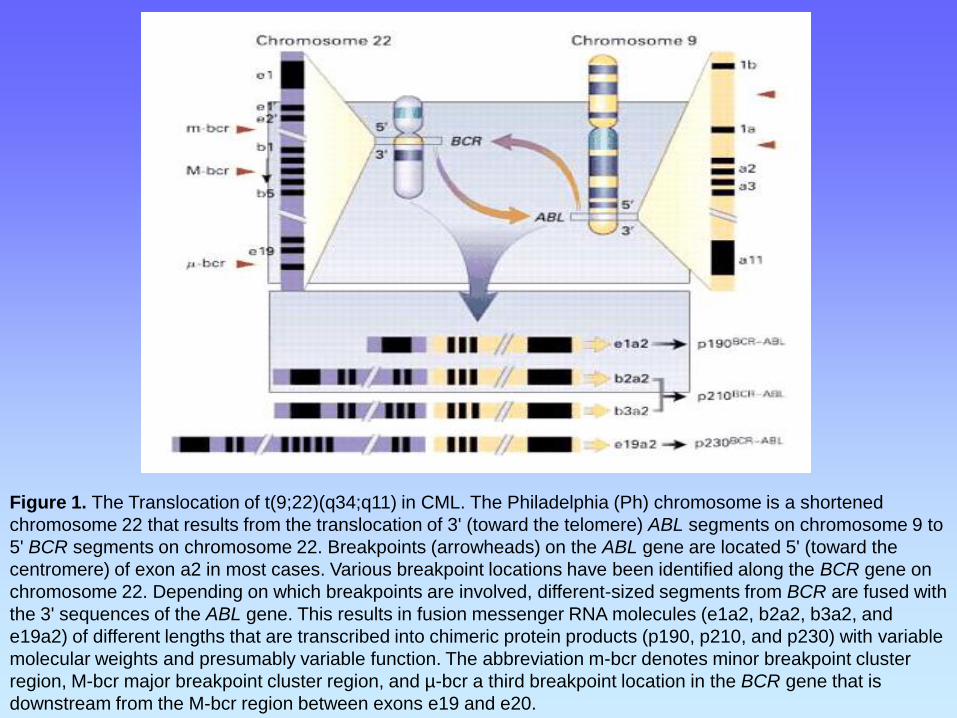
Figure 1. The Translocation of t(9;22)(q34;q11) in CML. The Philadelphia (Ph) chromosome is a shortened
chromosome 22 that results from the translocation of 3' (toward the telomere) ABL segments on chromosome 9 to
5' BCR segments on chromosome 22. Breakpoints (arrowheads) on the ABL gene are located 5' (toward the
centromere) of exon a2 in most cases. Various breakpoint locations have been identified along the BCR gene on
chromosome 22. Depending on which breakpoints are involved, different-sized segments from BCR are fused with
the 3' sequences of the ABL gene. This results in fusion messenger RNA molecules (e1a2, b2a2, b3a2, and
e19a2) of different lengths that are transcribed into chimeric protein products (p190, p210, and p230) with variable
molecular weights and presumably variable function. The abbreviation m-bcr denotes minor breakpoint cluster
region, M-bcr major breakpoint cluster region, and µ-bcr a third breakpoint location in the BCR gene that is
downstream from the M-bcr region between exons e19 and e20.
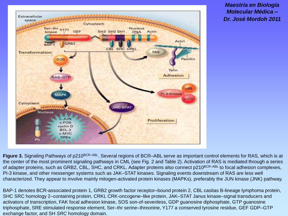
Figure 3. Signaling Pathways of p210BCR–ABL. Several regions of BCR–ABL serve as important control elements for RAS, which is at
the center of the most prominent signaling pathways in CML (see Fig. 2 and Table 2). Activation of RAS is mediated through a series
of adapter proteins, such as GRB2, CBL, SHC, and CRKL. Adapter proteins also connect p210BCR–ABL to focal adhesion complexes,
PI-3 kinase, and other messenger systems such as JAK–STAT kinases. Signaling events downstream of RAS are less well
characterized. They appear to involve mainly mitogen-activated protein kinases (MAPKs), preferably the JUN kinase (JNK) pathway.
BAP-1 denotes BCR-associated protein 1, GRB2 growth factor receptor–bound protein 2, CBL casitas B-lineage lymphoma protein,
SHC SRC homology 2–containing protein, CRKL CRK-oncogene–like protein, JAK–STAT Janus kinase–signal transducers and
activators of transcription, FAK focal adhesion kinase, SOS son-of-sevenless, GDP guanosine diphosphate, GTP guanosine
triphosphate, SRE stimulated response element, Ser–thr serine–threonine, Y177 a conserved tyrosine residue, GEF GDP–GTP
exchange factor, and SH SRC homology domain.
Maestría en Biología
Molecular Médica –
Dr. José Mordoh 2011
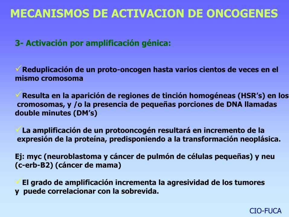
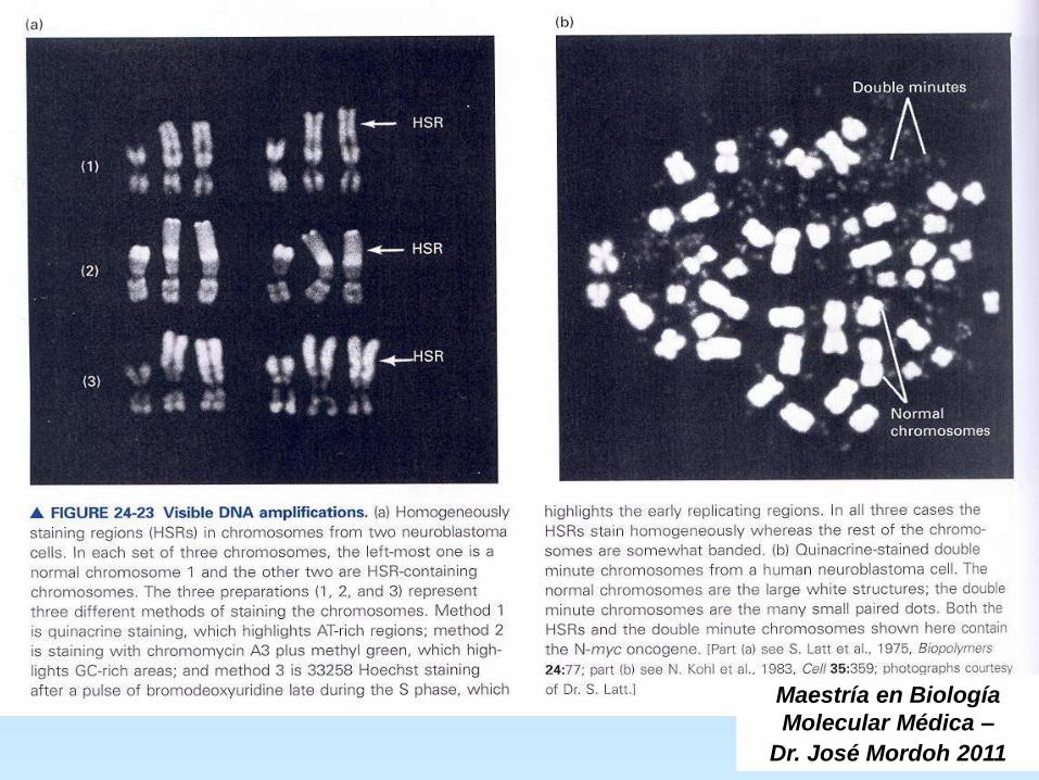
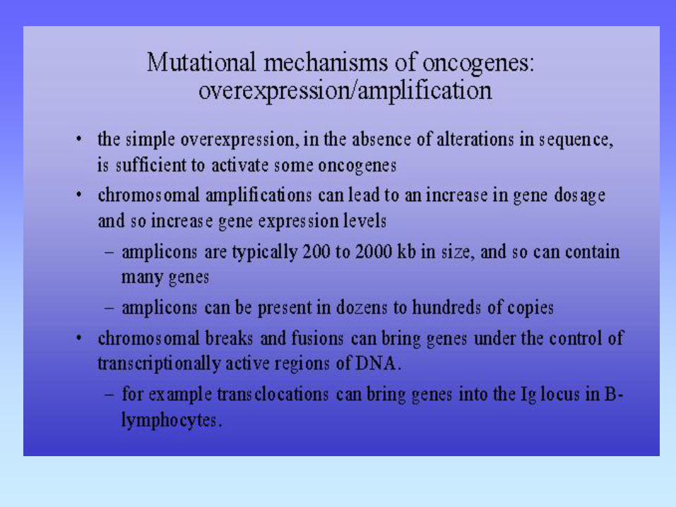
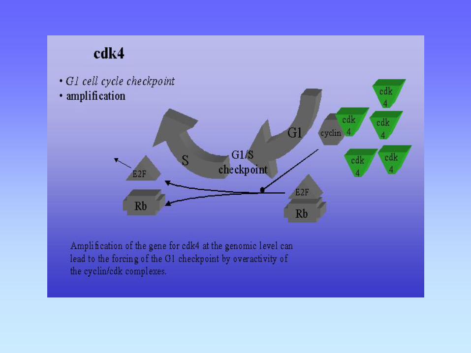
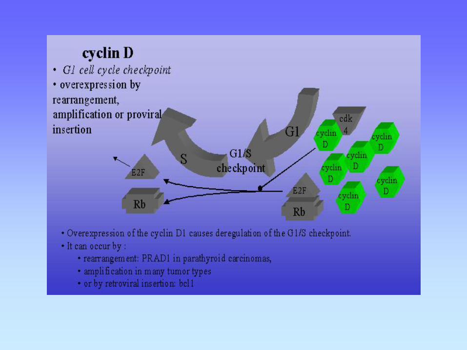
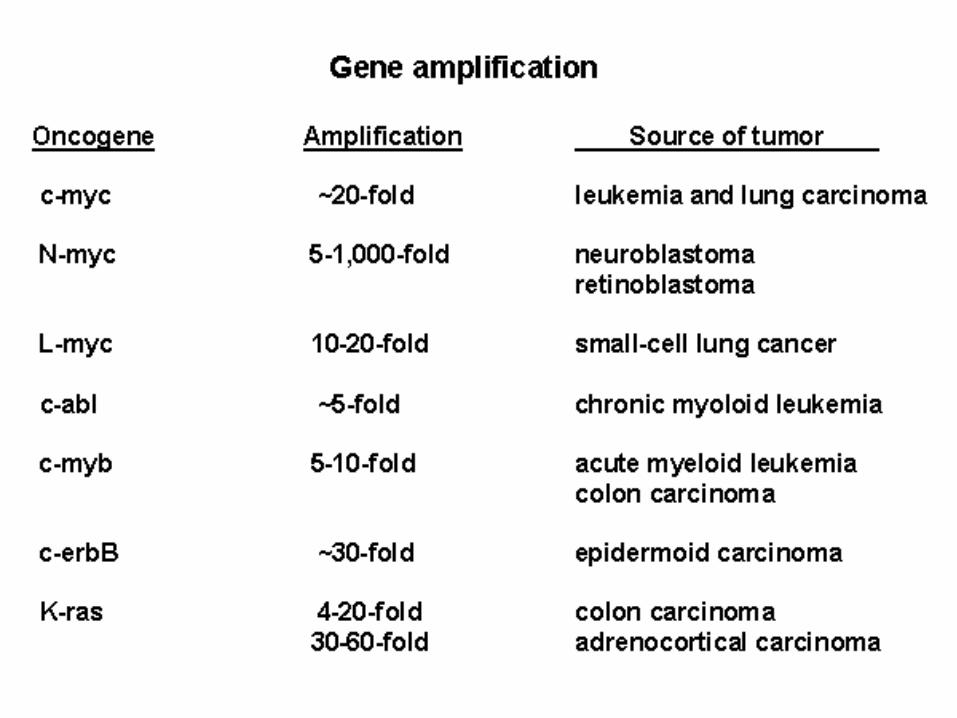
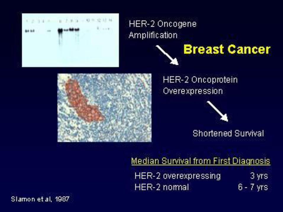
3- Activación por amplificación génica: Reduplicación de un proto-oncogen hasta varios cientos de veces en el mismo cromosoma Resulta en la aparición de regiones de tinción homogéneas (HSR’s) en los cromosomas, y /o la presencia de pequeñas porciones de DNA llamadas double minutes (DM’s) La amplificación de un protooncogén resultará en incremento de la expresión de la proteína, predisponiendo a la transformación neoplásica. Ej: myc (neuroblastoma y cáncer de pulmón de células pequeñas) y neu (c-erb-B2) (cáncer de mama) El grado de amplificación incrementa la agresividad de los tumores y puede correlacionar con la sobrevida.
MECANISMOS DE ACTIVACION DE ONCOGENES
CIO-FUCA
Maestría en Biología
Molecular Médica –
Dr. José Mordoh 2011

CIO-FUCA
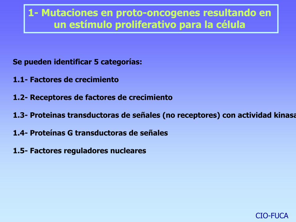
1- Mutaciones en proto-oncogenes resultando en un estímulo proliferativo para la célula
Se pueden identificar 5 categorías: 1.1- Factores de crecimiento 1.2- Receptores de factores de crecimiento 1.3- Proteinas transductoras de señales (no receptores) con actividad kinasa 1.4- Proteínas G transductoras de señales 1.5- Factores reguladores nucleares

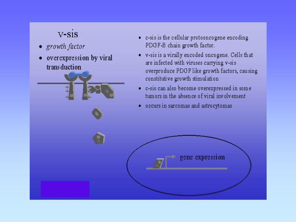
Factores de crecimiento
Por ejemplo PDGF, FGF . Sobreexpresión de PDGF, exceso de secreción por la célula, resulta en proliferación celular por mecanismo de feed-back autócrino. Asociado con astrocitomas y osteosarcomas humanos.
Similarmente hst-1 y hst-2 sobreexpresan FGF. Asociado con cáncer de estómago, vejiga y mama y con melanoma.
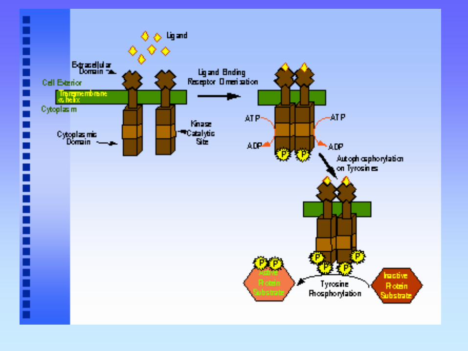
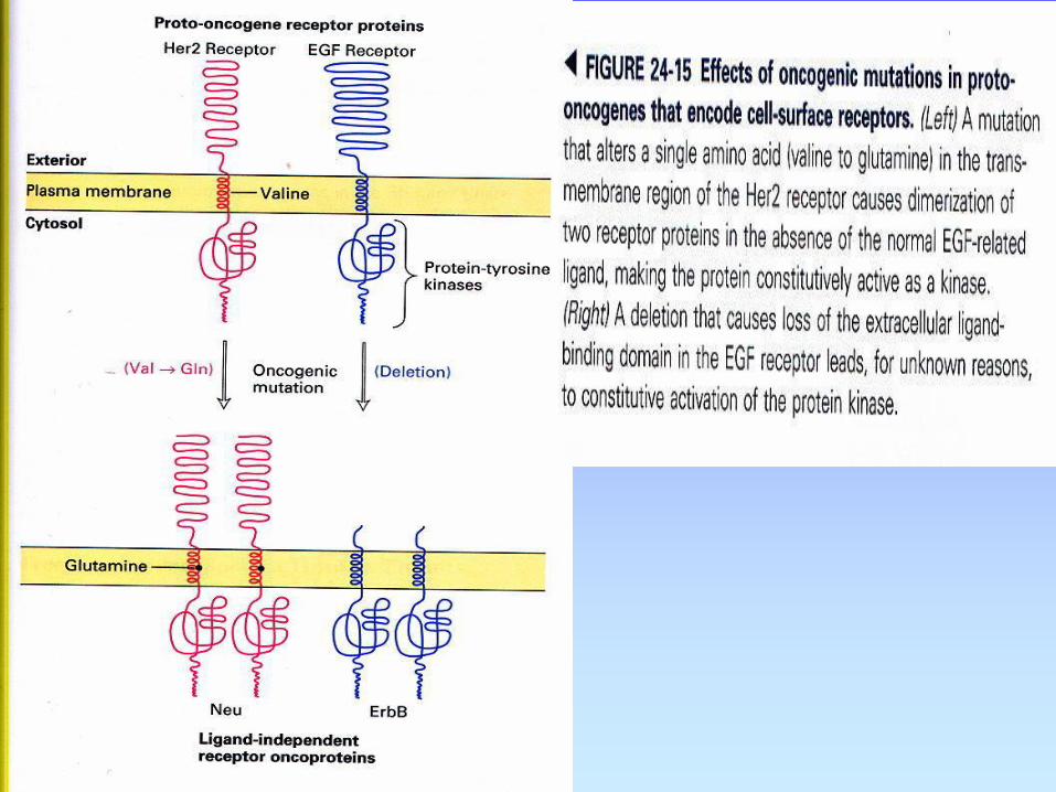
Receptores de Factores de crecimiento Receptores para EGF y CSF-1 han sido implicados en neoplasia.
Estos receptores son normalmente receptores de transmembrana y poseen una kinasa en su cara citoplasmática
CIO-FUCA

CIO-FUCA
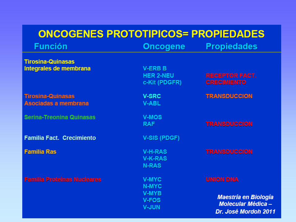
PROLIFERATIVE ONCOGENES
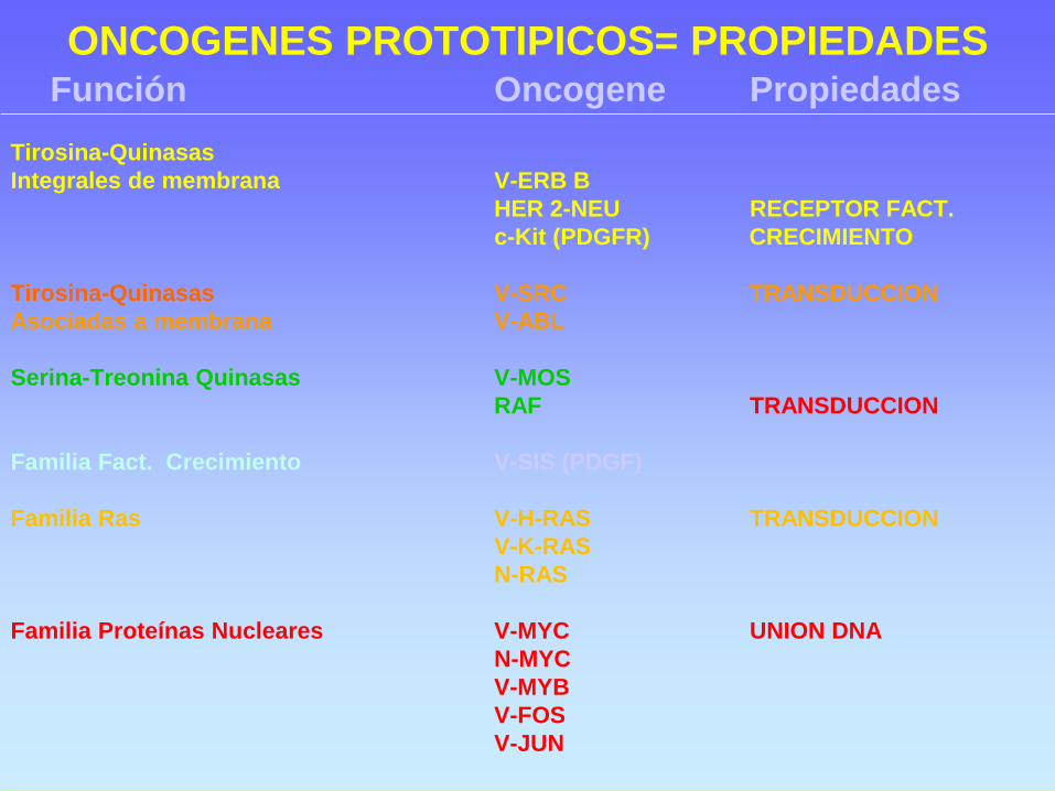
ONCOGENES PROTOTIPICOS= PROPIEDADES Función Oncogene Propiedades
Tirosina-Quinasas
Integrales de membrana V-ERB B
HER 2-NEU RECEPTOR FACT.
c-Kit (PDGFR) CRECIMIENTO
Tirosina-Quinasas V-SRC TRANSDUCCION
Asociadas a membrana V-ABL
Serina-Treonina Quinasas V-MOS
RAF TRANSDUCCION
Familia Fact. Crecimiento V-SIS (PDGF)
Familia Ras V-H-RAS TRANSDUCCION
V-K-RAS
N-RAS
Familia Proteínas Nucleares V-MYC UNION DNA
N-MYC
V-MYB
V-FOS
V-JUN
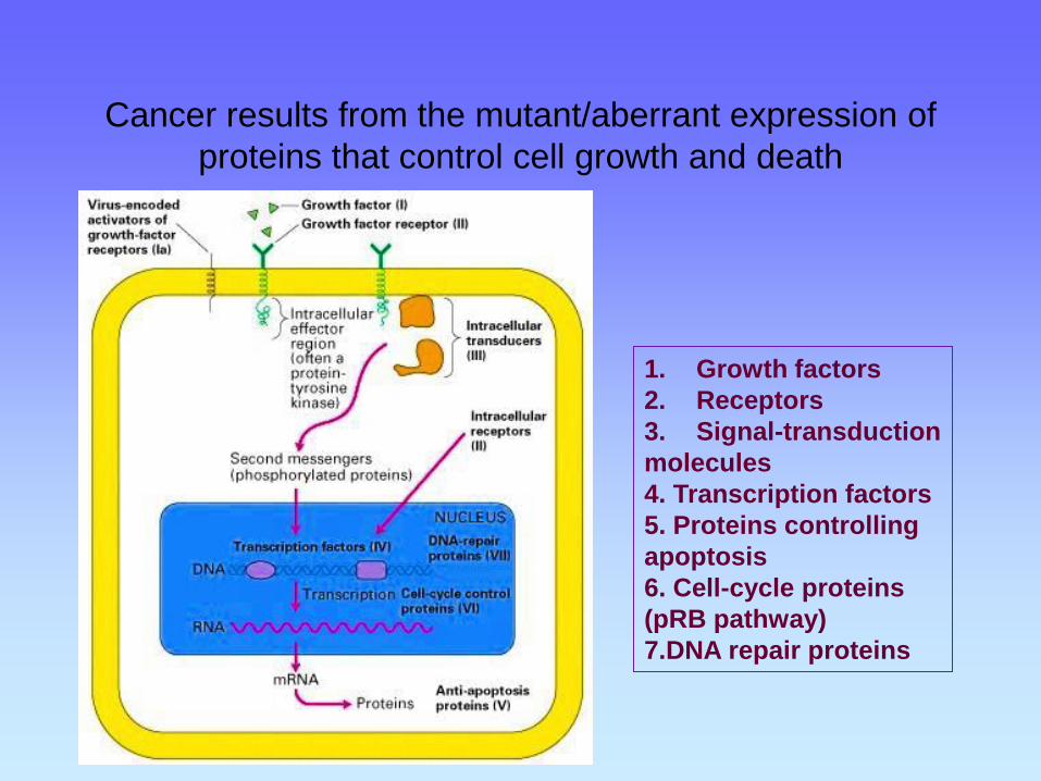
Cancer results from the mutant/aberrant expression of
proteins that control cell growth and death
1. Growth factors
2. Receptors
3. Signal-transduction
molecules
4. Transcription factors
5. Proteins controlling
apoptosis
6. Cell-cycle proteins
(pRB pathway)
7.DNA repair proteins
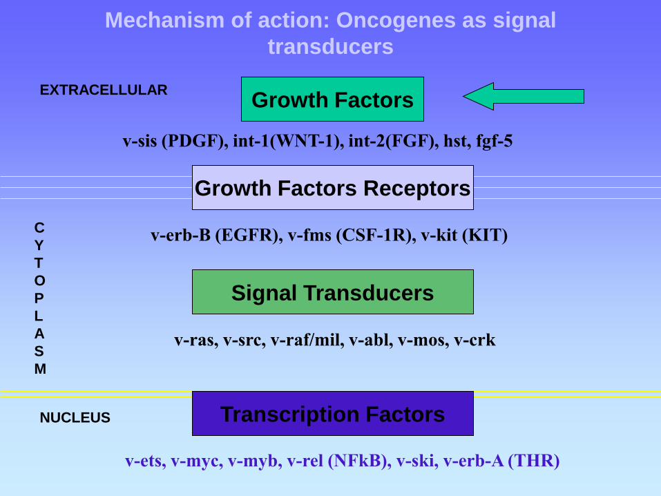
Mechanism of action: Oncogenes as signal
transducers
Growth Factors
v-sis (PDGF), int-1(WNT-1), int-2(FGF), hst, fgf-5
Growth Factors Receptors
v-erb-B (EGFR), v-fms (CSF-1R), v-kit (KIT)
Signal Transducers
v-ras, v-src, v-raf/mil, v-abl, v-mos, v-crk
Transcription Factors
v-ets, v-myc, v-myb, v-rel (NFkB), v-ski, v-erb-A (THR)
C
Y
T
O
P
L
A
S
M
EXTRACELLULAR
NUCLEUS
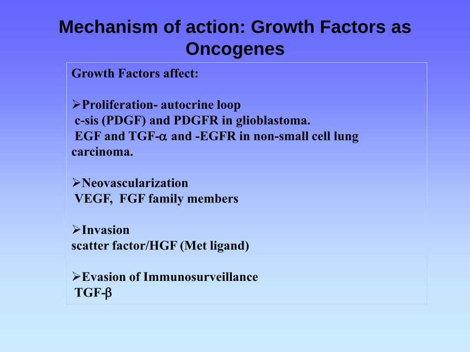
Mechanism of action: Growth Factors as
Oncogenes
Growth Factors affect:
Proliferation- autocrine loop
c-sis (PDGF) and PDGFR in glioblastoma.
EGF and TGF-a and -EGFR in non-small cell lung
carcinoma.
Neovascularization
VEGF, FGF family members
Invasion
scatter factor/HGF (Met ligand)
Evasion of Immunosurveillance
TGF-b

Oncogenes as signal transducers
Growth Factors
v-sis (PDGF), int-1(WNT-1), int-2(FGF), hst, fgf-5
Growth Factors Receptors
v-erb-B (EGFR), v-fms (CSF-1R), c-kit (KIT)
Signal Transducers
v-ras, v-src, v-raf/mil, v-abl, v-mos, v-crk
Transcription Factors
v-ets, v-myc, v-myb, v-rel (NFkB), v-ski, v-erb-A (THR)
C
Y
T
O
P
L
A
S
M
EXTRACELLULAR
NUCLEUS

Epidermal growth factor receptor (EGFR)
EGFR inhibition
MAP kinase (MAPK) signal pathway
Cell proliferation / cell survival
EGFR tyrosine kinase
inhibitor (EGFR-TKI)
EGFR EGFR CerbB2 / HER2
EGF EGF EGF
CIO-FUCA
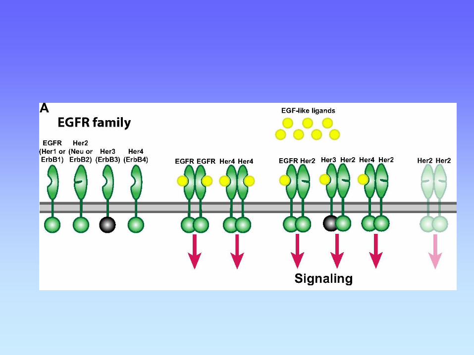
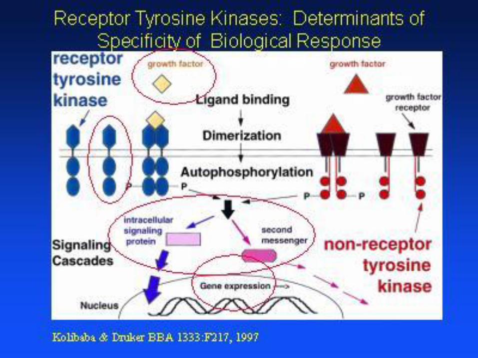
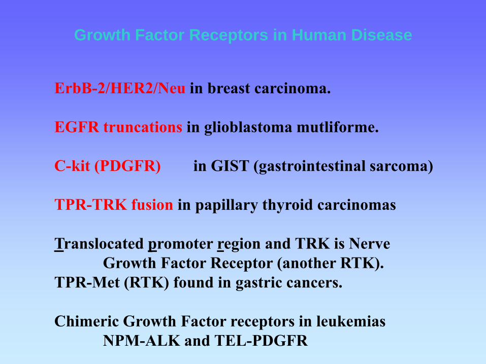
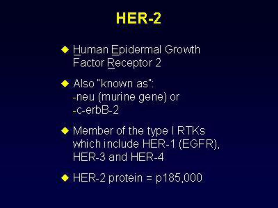
Growth Factor Receptors in Human Disease
ErbB-2/HER2/Neu in breast carcinoma.
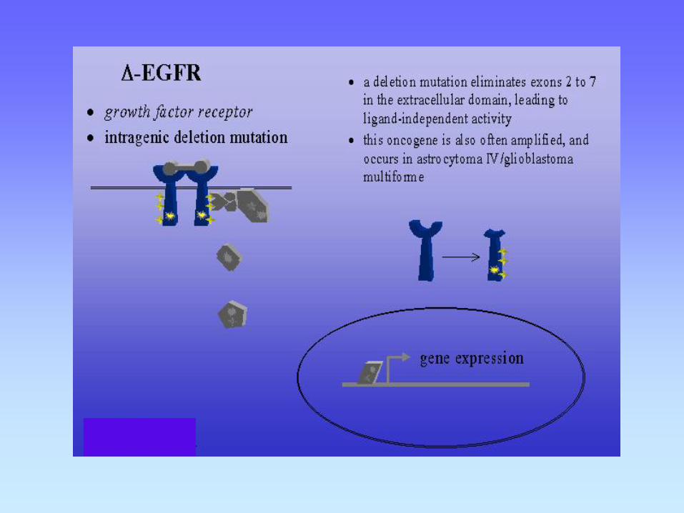
EGFR truncations in glioblastoma mutliforme.
C-kit (PDGFR) in GIST (gastrointestinal sarcoma)
TPR-TRK fusion in papillary thyroid carcinomas
Translocated promoter region and TRK is Nerve
Growth Factor Receptor (another RTK).
TPR-Met (RTK) found in gastric cancers.
Chimeric Growth Factor receptors in leukemias
NPM-ALK and TEL-PDGFR
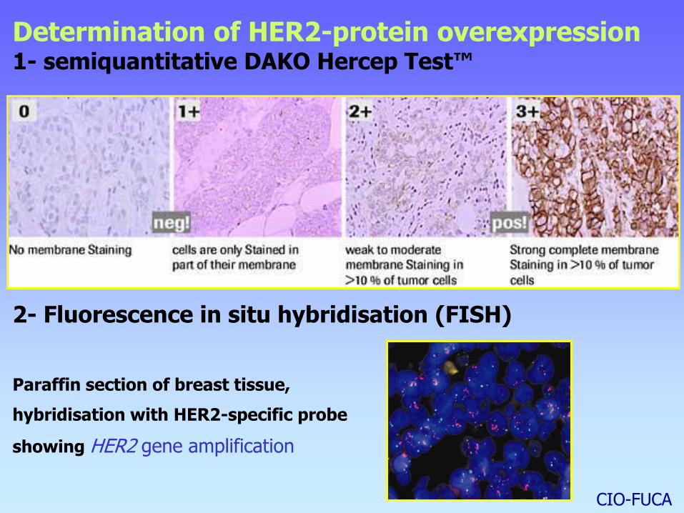
Determination of HER2-protein overexpression 1- semiquantitative DAKO Hercep Test™
CIO-FUCA
2- Fluorescence in situ hybridisation (FISH)
Paraffin section of breast tissue,
hybridisation with HER2-specific probe
showing HER2 gene amplification
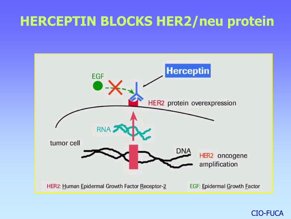
HERCEPTIN BLOCKS HER2/neu protein
CIO-FUCA
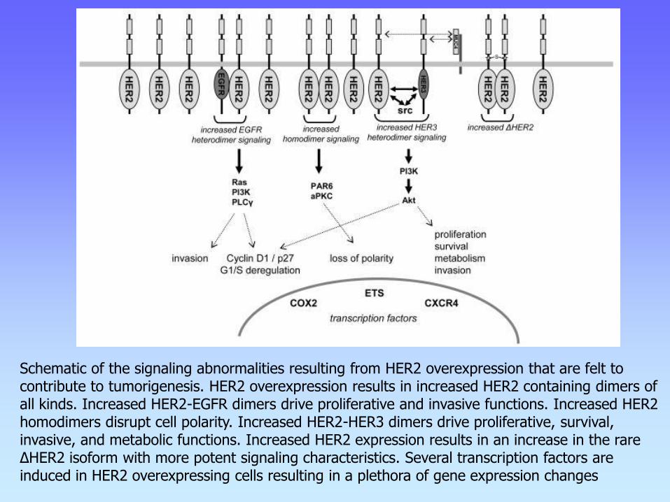
Schematic of the signaling abnormalities resulting from HER2 overexpression that are felt to contribute to tumorigenesis. HER2 overexpression results in increased HER2 containing dimers of all kinds. Increased HER2-EGFR dimers drive proliferative and invasive functions. Increased HER2 homodimers disrupt cell polarity. Increased HER2-HER3 dimers drive proliferative, survival, invasive, and metabolic functions. Increased HER2 expression results in an increase in the rare ΔHER2 isoform with more potent signaling characteristics. Several transcription factors are induced in HER2 overexpressing cells resulting in a plethora of gene expression changes
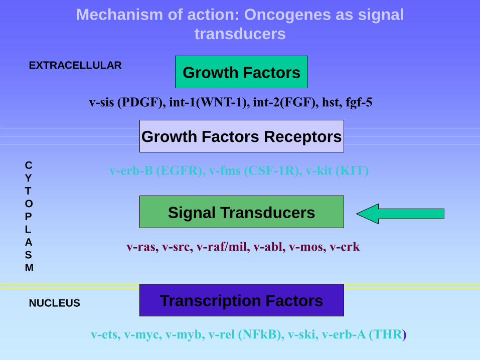
Mechanism of action: Oncogenes as signal
transducers
Growth Factors
v-sis (PDGF), int-1(WNT-1), int-2(FGF), hst, fgf-5
Growth Factors Receptors
v-erb-B (EGFR), v-fms (CSF-1R), v-kit (KIT)
Signal Transducers
v-ras, v-src, v-raf/mil, v-abl, v-mos, v-crk
Transcription Factors
v-ets, v-myc, v-myb, v-rel (NFkB), v-ski, v-erb-A (THR)
C
Y
T
O
P
L
A
S
M
EXTRACELLULAR
NUCLEUS

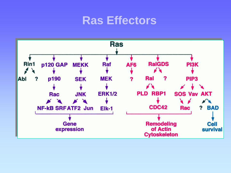
Ras Effectors
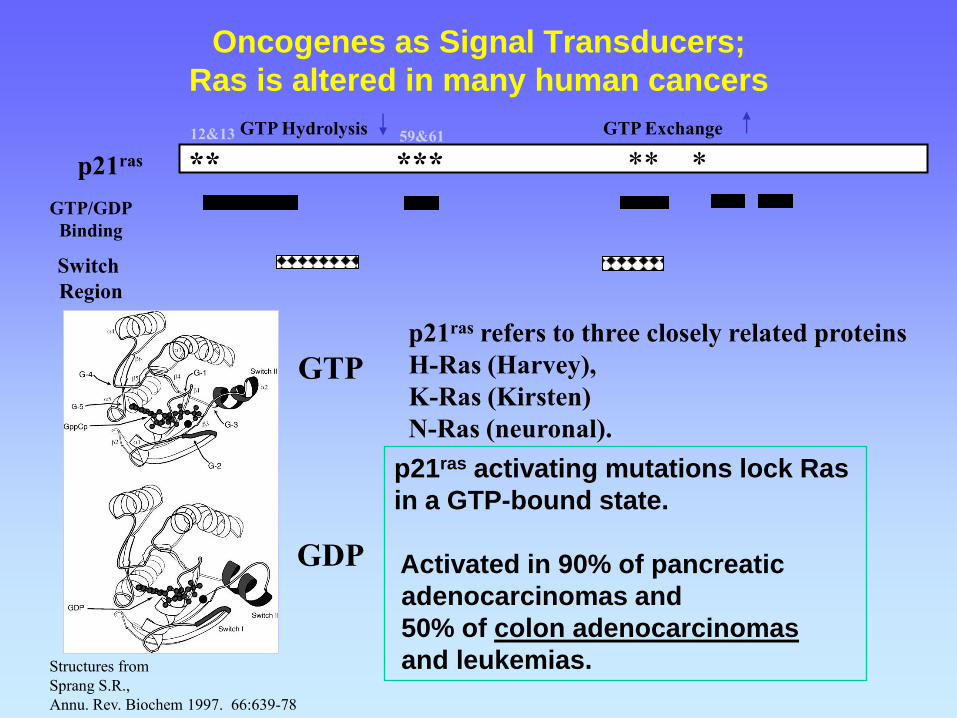
Oncogenes as Signal Transducers;
Ras is altered in many human cancers
p21ras activating mutations lock Ras
in a GTP-bound state.
Activated in 90% of pancreatic
adenocarcinomas and
50% of colon adenocarcinomas
and leukemias.
p21ras refers to three closely related proteins
H-Ras (Harvey),
K-Ras (Kirsten)
N-Ras (neuronal).
** *** ** * p21ras
12&13 59&61
GTP/GDP
Binding
Switch
Region
GTP Exchange GTP Hydrolysis
GTP
GDP
Structures from
Sprang S.R.,
Annu. Rev. Biochem 1997. 66:639-78
Proteínas G transductoras de señales
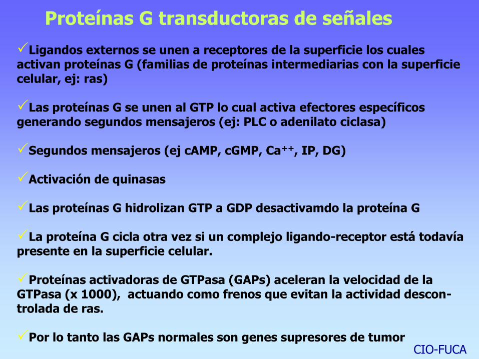
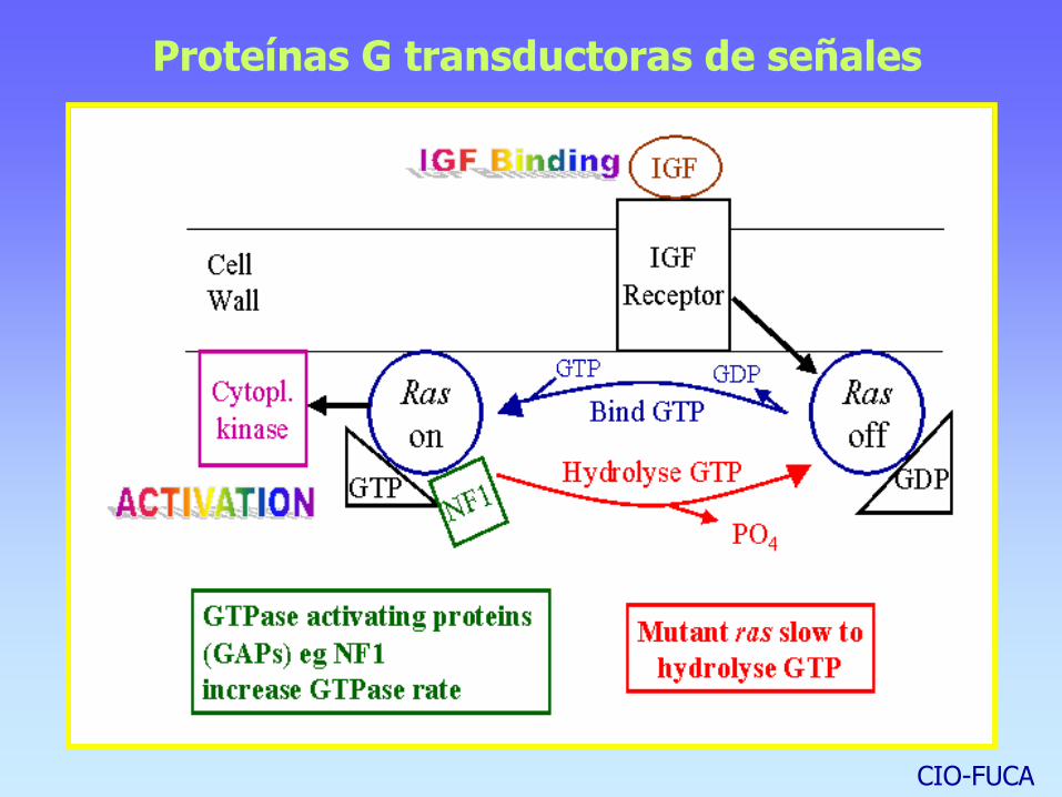
Ligandos externos se unen a receptores de la superficie los cuales activan proteínas G (familias de proteínas intermediarias con la superficie celular, ej: ras) Las proteínas G se unen al GTP lo cual activa efectores específicos generando segundos mensajeros (ej: PLC o adenilato ciclasa) Segundos mensajeros (ej cAMP, cGMP, Ca++, IP, DG) Activación de quinasas Las proteínas G hidrolizan GTP a GDP desactivamdo la proteína G La proteína G cicla otra vez si un complejo ligando-receptor está todavía presente en la superficie celular. Proteínas activadoras de GTPasa (GAPs) aceleran la velocidad de la GTPasa (x 1000), actuando como frenos que evitan la actividad descon-trolada de ras.
Por lo tanto las GAPs normales son genes supresores de tumor
CIO-FUCA
Proteínas G transductoras de señales
CIO-FUCA

Mechanism of action: Oncogenes as signal
transducers
Growth Factors
v-sis (PDGF), int-1(WNT-1), int-2(FGF), hst, fgf-5
Growth Factors Receptors
v-erb-B (EGFR), v-fms (CSF-1R), v-kit (KIT)
Signal Transducers
v-ras, v-src, v-raf/mil, v-abl, v-mos, v-crk
Transcription Factors
v-ets, v-myc, v-myb, v-rel (NFkB), v-ski, v-erb-A (THR)
C
Y
T
O
P
L
A
S
M
EXTRACELLULAR
NUCLEUS
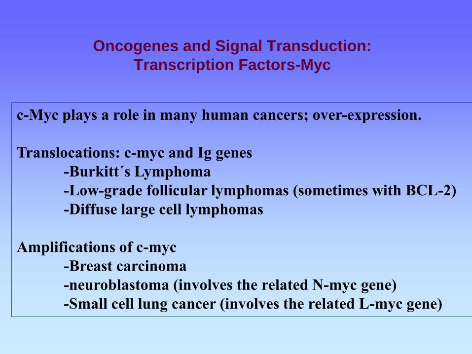
Oncogenes and Signal Transduction:
Transcription Factors-Myc
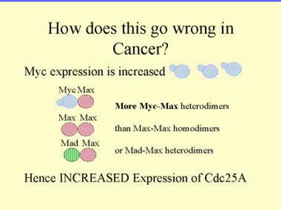
c-Myc plays a role in many human cancers; over-expression.
Translocations: c-myc and Ig genes
-Burkitt´s Lymphoma
-Low-grade follicular lymphomas (sometimes with BCL-2)
-Diffuse large cell lymphomas
Amplifications of c-myc
-Breast carcinoma
-neuroblastoma (involves the related N-myc gene)
-Small cell lung cancer (involves the related L-myc gene)
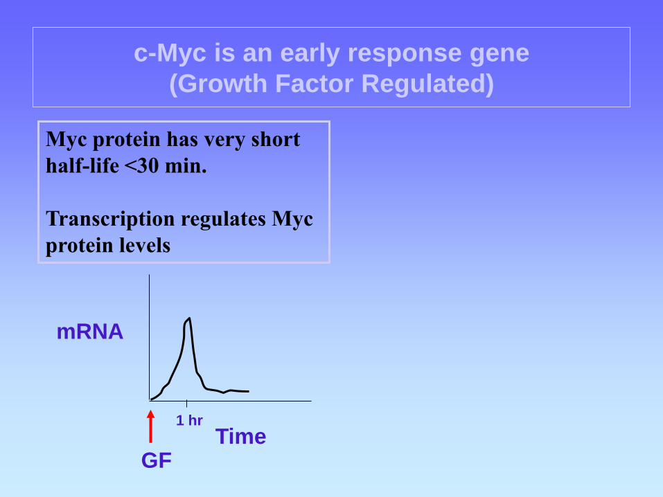
c-Myc is an early response gene
(Growth Factor Regulated)
GF
mRNA
Time 1 hr
Myc protein has very short
half-life <30 min.
Transcription regulates Myc
protein levels
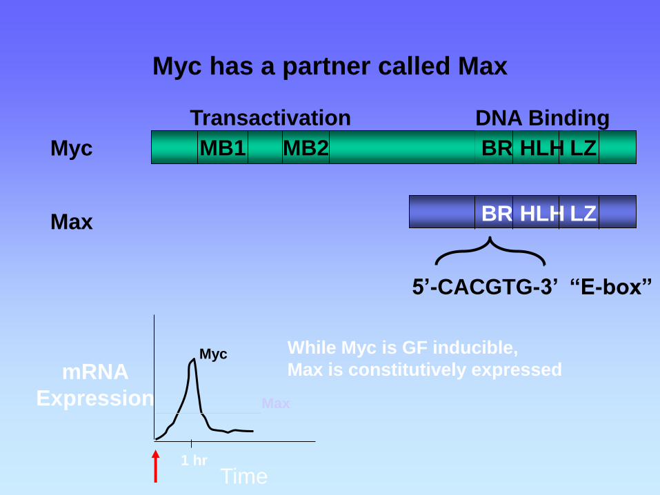
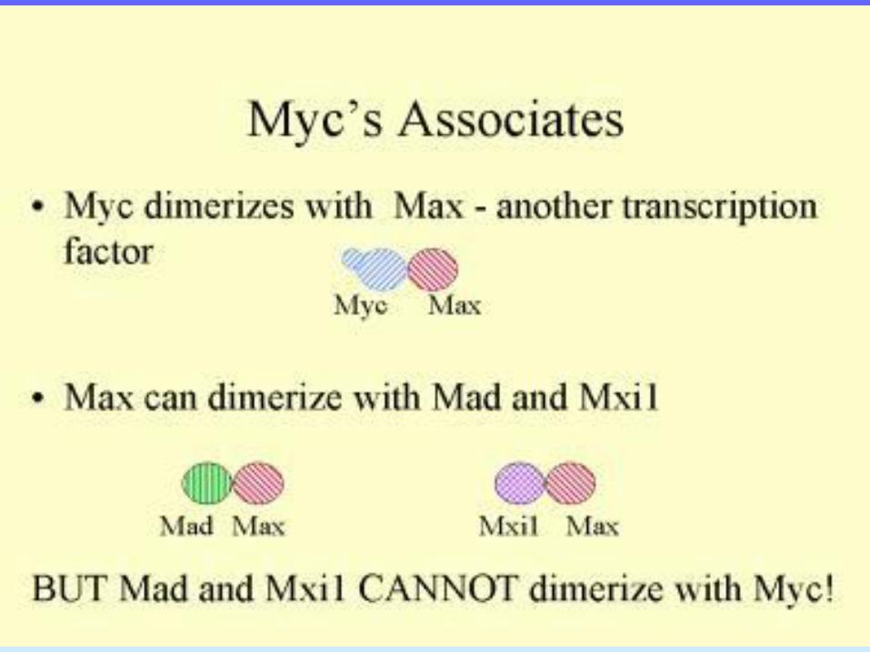
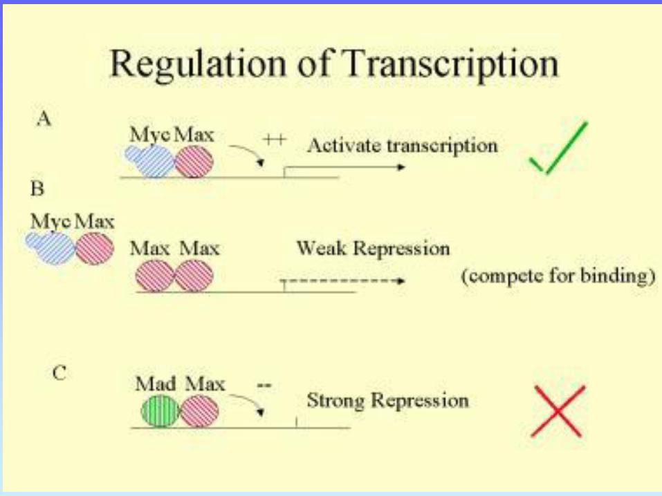
Myc has a partner called Max
BR HLH LZ MB1 MB2
BR HLH LZ
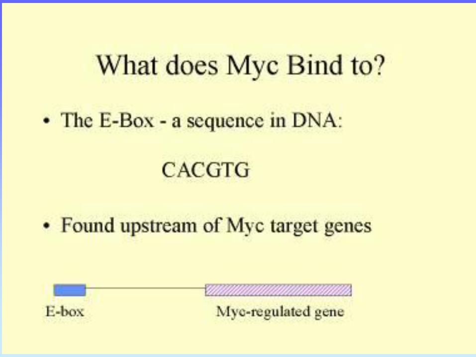
5’-CACGTG-3’ “E-box”
Transactivation DNA Binding
Myc
Max
mRNA
Expression
Time 1 hr
Myc
Max
While Myc is GF inducible,
Max is constitutively expressed
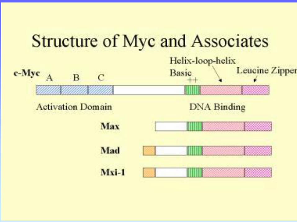
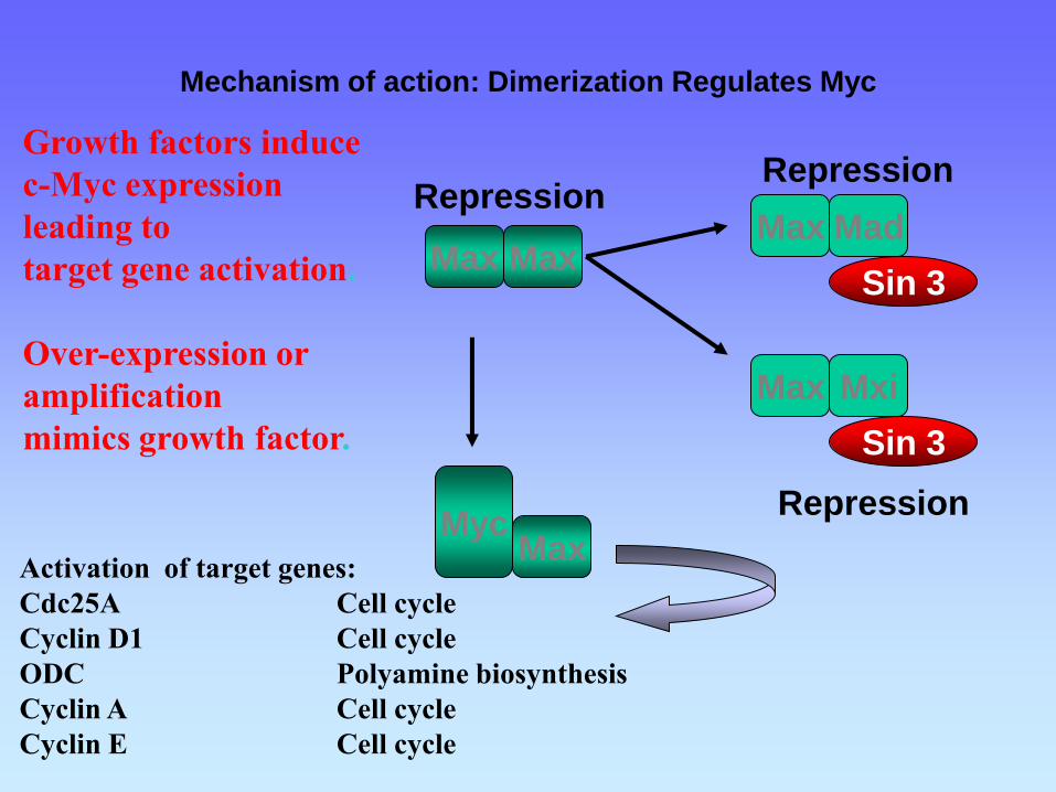
Mechanism of action: Dimerization Regulates Myc
Myc Max
Max Max Max Mad
Max Mxi
Sin 3
Sin 3
Repression Repression
Repression
Activation of target genes:
Cdc25A Cell cycle
Cyclin D1 Cell cycle
ODC Polyamine biosynthesis
Cyclin A Cell cycle
Cyclin E Cell cycle
Growth factors induce
c-Myc expression
leading to
target gene activation.
Over-expression or
amplification
mimics growth factor.
Apoptosis Regulators • The BCL2 gene, which is involved in the initiation of almost all follicular lymphomas and some diffuse large B-cell lymphomas encodes a cytoplasmic protein that localizes to mitochondria and increases cell survival by inhibiting apoptosis. • BCL2 is also important in chronic lymphocytic leukemia and lung cancer. •The BCL2 family members BCL-XL and BCL2 inhibit apoptosis and are up-regulated in many cancers. • Two main pathways lead to apoptosis: 1- the stress pathway: triggered by proteins that contain the BCL2 homology 3 domain; this domain inactivates BCL2 and BCL-XL (which normally inhibit apoptosis) and thereby activates the caspases that induce apoptosis. Drugs that mimic the BCL2 homology 3 domain and can bind to BCL-XL or BCL2 (peptides or small organic molecules that bind in a groove of these proteins) are under development. This approach has attracted considerable attention because many tumors overexpress BCL2 or related proteins
2- the death-receptor pathway: is activated by the binding of Fas ligand, TRAIL, and tumor necrosis factor α, to their corresponding (death) receptors on the cell surface. Activation of death receptors activates caspases that cause cell death