spreading of α-synuclein in the face of axonal transport deficits in parkinson's disease: a...
TRANSCRIPT

Neurobiology of Disease xxx (2014) xxx–xxx
YNBDI-03267; No. of pages: 8; 4C: 2, 4, 5, 6
Contents lists available at ScienceDirect
Neurobiology of Disease
j ourna l homepage: www.e lsev ie r .com/ locate /ynbd i
Review
Spreading of α-synuclein in the face of axonal transport deficits inParkinson's disease: A speculative synthesis
Jennifer T. Lamberts a, Erin N. Hildebrandt a, Patrik Brundin a,b,⁎a Laboratory for Translational Parkinson's Disease Research, Center for Neurodegenerative Science, Van Andel Research Institute, Grand Rapids, MI, USAb Neuronal Survival Unit, Wallenberg Neuroscience Center, Lund University, Lund, Sweden
⁎ Corresponding author at: Van Andel Institute, 333 Bos49503, USA.
E-mail address: [email protected] (P. Brundin).Available online on ScienceDirect (www.sciencedir
http://dx.doi.org/10.1016/j.nbd.2014.07.0020969-9961/© 2014 Elsevier Inc. All rights reserved.
Please cite this article as: Lamberts, J.T., et al.,synthesis, Neurobiol. Dis. (2014), http://dx.d
a b s t r a c t
a r t i c l e i n f oArticle history:Received 11 April 2014Accepted 2 July 2014Available online xxxx
Keywords:Braak stagingPrion-likeMitochondriaMicrogliaOligodendrocytesAlphaherpesvirus
Parkinson's disease (PD) ismainly attributed to degeneration of dopamine neurons in the substantia nigra, but itsetiopathogenesis also includes impaired protein clearance and axonal transport dysfunction, among others. Thespread ofα-synuclein (α-syn) aggregates from one neuron to another, in a prion-likemanner, is hypothesized tocontribute to PD progression. Axonal transport is likely to play a crucial role in this movement of α-synaggregates between brain regions. At the same time, deficits in axonal transport are suggested to contribute toneuronal failure in PD. In this review, we discuss the apparent contradiction that axonal transport might beessential for disease progression, while dysfunction of axonal transport could simultaneously be a cornerstoneof PD pathogenesis.We speculate aroundmodels that reconcile how axonal transport can play such a paradoxicalrole.
twick Ave NE, Grand Rapids, MI
ect.com).
Spreading ofα-synuclein in the face of axonal toi.org/10.1016/j.nbd.2014.07.002
© 2014 Elsevier Inc. All rights reserved.
Contents
Introduction . . . . . . . . . . . . . . . . . . . . . . . . . . . . . . . . . . . . . . . . . . . . . . . . . . . . . . . . . . . . . . . . . 0Fundamentals of axonal transport . . . . . . . . . . . . . . . . . . . . . . . . . . . . . . . . . . . . . . . . . . . . . . . . . . . . . . . 0Functional axonal transport contributes to PD pathogenesis . . . . . . . . . . . . . . . . . . . . . . . . . . . . . . . . . . . . . . . . . . . 0
. Braak staging of PD pathology . . . . . . . . . . . . . . . . . . . . . . . . . . . . . . . . . . . . . . . . . . . . . . . . . . . 0
. Axonal transport of α-syn . . . . . . . . . . . . . . . . . . . . . . . . . . . . . . . . . . . . . . . . . . . . . . . . . . . . . 0Dysfunctional axonal transport contributes to PD pathogenesis . . . . . . . . . . . . . . . . . . . . . . . . . . . . . . . . . . . . . . . . . . 0
. Mitochondrial damage . . . . . . . . . . . . . . . . . . . . . . . . . . . . . . . . . . . . . . . . . . . . . . . . . . . . . . . 0
. Glial cell activation . . . . . . . . . . . . . . . . . . . . . . . . . . . . . . . . . . . . . . . . . . . . . . . . . . . . . . . . 0Dynamic involvement of axonal transport in PD: a speculative synthesis . . . . . . . . . . . . . . . . . . . . . . . . . . . . . . . . . . . . . . 0
. Phases of axonal transport functionality . . . . . . . . . . . . . . . . . . . . . . . . . . . . . . . . . . . . . . . . . . . . . . . 0
. Axonal transport imbalance . . . . . . . . . . . . . . . . . . . . . . . . . . . . . . . . . . . . . . . . . . . . . . . . . . . . 0
. A speculative synthesis . . . . . . . . . . . . . . . . . . . . . . . . . . . . . . . . . . . . . . . . . . . . . . . . . . . . . . 0Therapeutic implications . . . . . . . . . . . . . . . . . . . . . . . . . . . . . . . . . . . . . . . . . . . . . . . . . . . . . . . . . . . 0Conclusions and future directions . . . . . . . . . . . . . . . . . . . . . . . . . . . . . . . . . . . . . . . . . . . . . . . . . . . . . . . 0Acknowledgments . . . . . . . . . . . . . . . . . . . . . . . . . . . . . . . . . . . . . . . . . . . . . . . . . . . . . . . . . . . . . . 0References . . . . . . . . . . . . . . . . . . . . . . . . . . . . . . . . . . . . . . . . . . . . . . . . . . . . . . . . . . . . . . . . . . 0
Introduction
Parkinson's disease (PD) is the second most common adult-onsetneurodegenerative disease, for which there is still no cure. The diseaseis characterized primarily by motor disturbances that are due to theloss of dopamine neurons in the substantia nigra, although non-motor
ransport deficits in Parkinson's disease: A speculative

Fig. 1. Basic mechanisms of axonal transport. In healthy neurons, anterograde axonal transport (toward the nerve terminal) is performed by the microtubule plus-end-directed motorkinesin, while the minus-end-directed motor dynein is responsible for retrograde transport (toward the cell body). In general, newly synthesized proteins and synaptic vesicles aretransported in the anterograde direction, recycling organelles such as endosomes and lysosomes in the retrograde direction, and mitochondria in both directions. As a synaptic protein,α-synuclein (α-syn) is normally transported by anterograde axonal transport to the nerve terminal where it plays a role in synapse homeostasis.
2 J.T. Lamberts et al. / Neurobiology of Disease xxx (2014) xxx–xxx
symptoms such as depression are experienced by most patients (Fahn,2003). While a variety of mechanisms are suggested to contribute tothe etiopathogenesis of PD, including impaired protein clearance, axo-nal transport dysfunction, mitochondrial damage, and inflammation,the specific cause of the disease remains unknown.
Almost all PD patients develop intraneuronal protein aggregates.These proteinaceous inclusions, named Lewy bodies (or Lewyneurites) after their discoverer (Lewy, 1912), are composed primar-ily of misfolded α-synuclein (α-syn) protein (Goedert et al., 2013;Spillantini et al., 1997). In addition, mutations (A30P, E46K, A53T)or multiplications in the α-syn gene cause autosomal-dominant PD(Hardy et al., 2006). These findings underscore the importance of α-syn in the pathogenesis of PD.
After thorough investigation of α-syn aggregation in post-mortembrains, Braak and colleagues proposed that Lewy pathology propagatesthroughout the brain in a stereotypic manner (Braak et al., 2003a). Sev-eral lines of evidence now suggest that this propagation occurs via aprion-like mechanism, in which misfolded α-syn is responsible for itsown pathologic accumulation (Olanow and Brundin, 2013). In orderfor α-syn pathology to spread over such long distances, it is thoughtthat α-syn aggregates undergo axonal transport (George et al., 2013;Ubeda-Bañon et al., 2013). At the same time, it is proposed that axonaltransport is dysfunctional in PD (De Vos et al., 2008; Millecamps andJulien, 2013). How can transport of α-syn aggregates be occurring inthe face of general axonal transport dysfunction? In this review, we ad-dress this apparent contradiction, and we speculate about the dynamiccontribution of axonal transport to PD pathogenesis. We focus ourdiscussion primarily on PD, as the role of axonal transport in otherneurodegenerative diseases such as Alzheimer's disease (AD), amyotro-phic lateral sclerosis (ALS), and Huntington's disease (HD) has beenreviewed extensively elsewhere (De Vos et al., 2008; Millecamps andJulien, 2013; Morfini et al., 2009; Roy et al., 2005).
Fundamentals of axonal transport
Owing to their extreme polarity and long processes, neurons musttransport proteins over very long distances. In fact, human neuronscan possess axons in excess of 1 m in length. Axonal transport allowsneurons to supply essential components to the distal axon and nerveterminals and recycle proteins back to the cell body. Thus, newlysynthesized proteins and synaptic vesicles are transported along
Please cite this article as: Lamberts, J.T., et al., Spreading ofα-synuclein in thsynthesis, Neurobiol. Dis. (2014), http://dx.doi.org/10.1016/j.nbd.2014.07
axons in the anterograde direction (toward the nerve terminal),while recycling organelles such as endosomes and lysosomes aretransported retrogradely (toward the cell body) (Fig. 1). Mitochondriaare transported in both directions (Millecamps and Julien, 2013;Nakata et al., 1998).
Protein and organelle cargos are transported in the axon bymolecu-lar motor proteins that “walk” along the length of microtubule tracks(Hirokawa and Takemura, 2005). The microtubule plus-end-directedmotor kinesin is primarily responsible for anterograde transport,whereas the minus-end-directed motor dynein moves cargo in the ret-rograde direction (Fig. 1). Axonal transport can be divided into twocomponents: fast and slow (Lasek et al., 1984; Tytell et al., 1981). Mem-branous organelles such as mitochondria and vesicles are rapidlytransported in the fast component of axonal transport, while solubleproteins and cytoskeletal fragments are transported in the slow compo-nent (Brown, 2003). Slow component cargoes move quickly but makeextended pauses, and therefore they appear to have a slower overalltransport velocity (Brown, 2000; Roy et al., 2000; Wang et al., 2000).Despite differences in the average rate of transport between the fastand slow component (Millecamps and Julien, 2013), both sets of cargoare transported by kinesin and dynein motors (Brown, 2003; Shahand Cleveland, 2002). It is unclear what causes slow component pro-teins to make these frequent and extended pauses, but recent evidencesuggests that the phosphorylation of kinesin and dynein subunits byaxonal kinases may play an important role (Morfini et al., 2009).
Functional axonal transport contributes to PD pathogenesis
Braak staging of PD pathology
In the early 2000s, Braak and colleagues proposed thatα-syn pathol-ogy spreads through the nervous system in a characteristic fashion asPD progresses (Braak et al., 2003a). These researchers even suggestedthat thefirstα-syn aggregates are formed before the affected person ex-periences any motor symptoms, and therefore before being diagnosedwith PD. Specifically, Braak classified idiopathic PD into stages 1–6according to the presence of Lewy pathology in the brain. In stages 1and 2, which represent “pre-motor” PD, pathology is found first in theolfactory bulb and the dorsal motor nucleus of the vagus nerve, andlater in the brainstem. Individuals with either stage 1 or stage 2 PD aresuggested to exhibit anosmia and constipation. It is not until stage 3,
e face of axonal transport deficits in Parkinson's disease: A speculative.002

3J.T. Lamberts et al. / Neurobiology of Disease xxx (2014) xxx–xxx
when Lewy pathology is found in the substantia nigra, that the charac-teristic motor disturbances are observed. In stages 4, 5, and 6, Lewypathology is observed in progressively rostral regions, including thestriatum and cortex. These later neuropathological stages correspondin time to points in the clinical progression when patients may beginto exhibit dementia.
Following this neuropathological investigation, Braak et al. went onto suggest that PD pathology originates in the gut and spreads towardthe brain via the vagus nerve through the specific retrograde transportof an unknown pathogen, which they speculated might be a neuro-tropic virus (Braak et al., 2003b). Recent evidence suggests thatthis “pathogen” is actually α-syn. In particular, human grafting stud-ies demonstrated that Lewy pathology appears in grafted neuronsafter only 10 years, suggesting that misfolded α-syn had transferredfrom diseased host neurons to grafted neurons during that timeperiod (Kordower et al., 2008; Li et al., 2008). This hypothesis is sup-ported by studies showing that α-syn can directly transfer betweencells both in vitro and in vivo (Angot et al., 2012; Desplats et al., 2009;Hansen et al., 2011; Kordower et al., 2011). Moreover, inoculation ofmice with either recombinant α-syn fibrils or brain homogenates fromdiseased α-syn transgenic mice resulted in the progressive spreadingof α-syn pathology (Luk et al., 2012a, 2012b). Together, these resultssuggest that α-syn behaves in a prion-like manner to propagate itsown pathologic accumulation (Frost and Diamond, 2010; Krammeret al., 2009; Olanow and Brundin, 2013). An emerging hypothesis positsthat this spreading of PD pathology involves the axonal transport of α-syn aggregates between synaptically-connected brain regions (Georgeet al., 2013; Ubeda-Bañon et al., 2013).
Axonal transport of α-syn
The exact physiological function of α-syn is still poorly understood.However, we do know that α-syn is primarily a presynaptic proteinwith possible roles in lipid binding and synaptic vesicle dynamics(Bendor et al., 2013). As such, α-syn is believed to be synthesized inthe cell body and transferred to the distal axon and nerve terminals byaxonal transport (Fig. 1) (Kahle et al., 2000; Roy, 2009). In particular, na-tive α-syn is thought to undergo mainly slow axonal transport (Jensenet al., 1999; Tang et al., 2012), which is consistent with the notion thatslow axonal transport is responsible for the movement of cytoskeletalproteins and macromolecular protein complexes. Nevertheless, a com-plete understanding of how native α-syn is transported in neurons isstill lacking, and relatively little is known about how mutations and α-syn misfolding influence its movement inside neurons.
Studies in vitro have shown thatα-syn aggregates are transported inneurons. In mouse primary cortical neurons grown in microfluidicdevices, fibrillarα-syn traveled anterogradely by slow axonal transport(Freundt et al., 2012). On the other hand, α-syn aggregates weretransported bidirectionally (both anterograde and retrograde) inmouse primary cortical neurons cultured in a similarmicrofluidic cham-ber (Danzer et al., 2011; Volpicelli-Daley et al., 2011).
Intra-axonal transport of α-syn has also been demonstrated in sym-pathetic neurons, which are located along anatomical pathways that areimplicated in Braak staging (Braak et al., 2003a). In a cell culture system,sympathetic nerve terminals took up extracellular secreted α-syn andtransported it in the retrograde direction toward the cell body, whereit then accumulated (Pan-Montojo et al., 2012). In vivo, overexpressionof humanα-syn via direct injection of viral vectors into the vagus nerveof the rat induced progressive caudo-rostral spreading of human α-synimmunostaining from the medulla to the pons, midbrain, and forebrain(Ulusoy et al., 2013). Partial resection of the vagus nerve resulted in re-ducedα-syn pathology in the dorsalmotor nucleus (Pan-Montojo et al.,2012), suggesting that propagation of PD pathology is halted if the axonis damaged. These results support the hypothesis that PD pathologyspreads to the brain through the retrograde transport of α-syn in thevagus nerve. However, it was not clear from these studies whether the
Please cite this article as: Lamberts, J.T., et al., Spreading ofα-synuclein in thsynthesis, Neurobiol. Dis. (2014), http://dx.doi.org/10.1016/j.nbd.2014.07
transported species was α-syn aggregates or monomeric α-syn thatlater accumulated in cell soma.
Early pathologic accumulation of α-syn in the olfactory bulb issuggested to contribute to anosmia in a condition coined “pre-motor”PD (Braak et al., 2003a). In a recent study in mice, researchers foundthat α-syn oligomers injected directly into the olfactory bulb were rap-idly transported—within minutes to hours—to brain regions that areconnected to the olfactory bulb (Rey et al., 2013). In contrast, fibrillarα-syn was not transferred between interconnected structures to anysignificant extent (Rey et al., 2013). These results suggest that smallaggregates are the primary species of α-syn that spread betweenbrain regions as PD pathology progresses. At the same time, because itcan be difficult to detect very low numbers of fibrils inside neurons,the findings by Rey et al. do not exclude the possibility thatα-syn fibrilsthat occasionally transferred from one brain region to another could actas seeds that trigger the formation of Lewy pathology at their destina-tion. In such amodel, one could hypothesize that eitherα-syn oligomersor monomers provide the building blocks that allow single or low num-bers of transported α-syn fibrils to grow intoα-syn aggregates that canthen be observed by immunohistochemical staining. Alternatively, thehighly mobile α-syn oligomers might be sufficient to act as seeds forfurther α-syn aggregation. In either case, the recent work of Rey et al.has clearly established that α-syn oligomers and monomers readilymove between brain regions and can even cross synapses, suggestingthat they could contribute to long distance spreading of PD pathology(Rey et al., 2013). Additional studies in brains of experimental animalsare needed to clarify which molecular species of α-syn acts as the seedin vivo, andwhich formsmost efficiently template the formation of largeraggregates. Due to the well-mapped anatomy of olfactory pathways andthe putative relevance of olfactory pathology to “pre-motor” PD, defininghow α-syn spreads from the olfactory bulb will be highly relevant toour understanding of how α-syn aggregates affect progressively distalstructures within the PD brain.
The studies described above, which take place over a relatively shorttime course, raise another important question: how canα-syn be trans-ferred within hours to days in experimental models, when PD requiresyears or even decades to develop in humans? For example,α-syn trans-fer in vivo was observed as early as 20 min post-injection of recombi-nant α-syn protein (Rey et al., 2013), or 2 weeks post-infection withα-syn virus (Ulusoy et al., 2013). One hypothesis to reconcile these ob-servations is that the transport of α-syn aggregates occurs in periodic“bursts”. Though each individual burst would not itself be pathogenic,the cumulative effect of several of these events over time could eventu-ally lead to the formation of large-scale aggregates that are neurotoxic.Indeed, a recent hypothesis in the context of AD suggests that the initialpropagation of misfolded protein seeds might occur early on in diseaseprogression, whereas the neuropathology associated with theseproteopathic seeds develops gradually over time (Domert et al., 2014).
It should be noted that although widespread α-syn aggregation isdetected in brains of people with PD, it is still not clear whether α-synpathology is directly responsible for neurodegeneration. Indeed, thereis sometimes a mismatch between the presence of α-syn pathologyand the manifestation of clinical symptoms (Burke et al., 2008). Itcannot be ruled out that small (or even large) α-syn aggregates act ina neuroprotective manner by sequestering toxic α-syn species, akin towhat has been suggested for mutant huntingtin aggregates in HD(Arrasate and Finkbeiner, 2012). Even if α-syn aggregation is found toplay a key role in the neurodegenerative process, it is also currentlynot clear at what point the aggregated α-syn starts to impair the func-tion of the host neuron and send it irrevocably on a path towarddeath. Thus, it is conceivable that phosphorylated α-syn aggregatesare dynamic structures, and that neurons exhibiting Lewy pathologycould degrade α-syn aggregates even at later stages. Regardless of theeffects of α-syn aggregates in PD pathogenesis, a deeper understandingof the intraneuronal transport of these aggregates will likely contributeto a better understanding of disease progression.
e face of axonal transport deficits in Parkinson's disease: A speculative.002
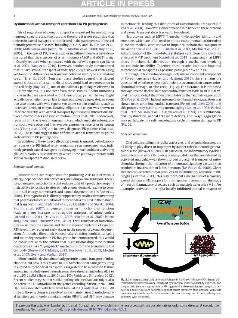
Fig. 2. Self-perpetuating cycle of neuron damage in Parkinson's disease (PD). Strong bidi-rectional links between axonal transport dysfunction, mitochondrial dysfunction, andα-synuclein (α-syn) aggregation in PD suggests that these mechanisms might partici-pate in a deleterious feed-forward loop that causes extensive axon damage. While thepoint of entry into this cycle is not known, it is clear that any one of these pathways canbe linked with the others.
4 J.T. Lamberts et al. / Neurobiology of Disease xxx (2014) xxx–xxx
Dysfunctional axonal transport contributes to PD pathogenesis
Strict regulation of axonal transport is important for maintainingneuronal structure and function, and therefore it is not surprising thatdefects in axonal transport are implicated in the pathogenesis of severalneurodegenerative diseases, including AD, ALS, and HD (De Vos et al.,2008; Millecamps and Julien, 2013; Morfini et al., 2009; Roy et al.,2005). In the case of PD, several studies in cultured neurons have dem-onstrated that the transport of α-syn mutants (A30P and A53T) is sig-nificantly reduced when compared with that of wild type α-syn (Sahaet al., 2004; Yang et al., 2010). However, another study demonstratedthat in vivo axonal transport of wild type α-syn slowed with agingyet found no differences in transport between wild type and mutantα-syn (Li et al., 2004). Together, these studies suggest that slowedaxonal transport of α-syn to distal sites could lead to aggregation inthe cell body (Roy, 2009), one of the hallmark pathologies observed inPD. Nevertheless, it is not clear from these studies if point mutationsin α-syn that are associated with PD are crucial per se. The key mightactually be whether α-syn has formed pathologic aggregates, an eventthat also occurs with wild type α-syn under certain conditions such asincreased levels of α-syn. Notably, oligomeric α-syn was shown tointerfere directly with axonal transport by disrupting interactions be-tween microtubules and kinesin motors (Prots et al., 2013). Moreover,reductions in the levels of kinesin motors, which mediate anterogradetransport, were observed inα-syn overexpressingmice prior to neuronloss (Chung et al., 2009) and in newly diagnosed PD patients (Chu et al.,2012). These data suggest that deficits in axonal transport might beearly events in PD pathogenesis.
In addition to these direct effects on axonal transport, pathologic α-syn species (i.e. PD-linked α-syn mutants, α-syn aggregates) may indi-rectly perturb axonal transport by damagingmitochondria or activatingglial cells. Various mechanisms by which these pathways interact withaxonal transport are discussed below.
Mitochondrial damage
Mitochondria are responsible for producing ATP to fuel variousenergy-dependent cellular processes, including axonal transport. There-fore, damage tomitochondria that reduces total ATP production impairstheir ability to localize to sites of high energy demand, leading to com-promised energy homeostasis and axonal degeneration (De Vos et al.,2008). This hypothesis is directly supported by studies demonstratingthat pharmacological inhibition ofmitochondria resulted in their abnor-mal transport in axons (Arnold et al., 2011; Miller and Sheetz, 2004;Morfini et al., 2007). In general, impairing mitochondrial functionleads to a net increase in retrograde transport of mitochondria(Arnold et al., 2011; De Vos et al., 2007; Morfini et al., 2007; Perrotand Julien, 2009; Shirendeb et al., 2012). Thus, transport of mitochon-dria away from the synapse and the subsequent depletion of synapticATP levels may represent early stages in the process of axonal degener-ation. Although a direct link between altered mitochondrial transportand neurodegeneration in PD has yet to be demonstrated, this wouldbe consistent with the notion that nigrostriatal dopamine neurondeath occurs via a “dying-back” mechanism from the terminals to thecell body (Burke and O'Malley, 2013; Kordower et al., 2013; Morfiniet al., 2007; Overk and Masliah, 2014).
Mitochondrial dysfunction clearly perturbs axonal transport ofmito-chondria, but how is this related to PD?Mitochondrial damage resultingin altered mitochondrial transport is suggested to be a common featureamongmany adult-onset neurodegenerative diseases, including AD (Yeet al., 2012), ALS (Shi et al., 2010), andHD (Reddy and Shirendeb, 2012).Recent studies suggest that similar pathogenic mechanisms might alsobe active in PD. Mutations in the genes encoding parkin, PINK1, andDJ-1 are associated with late-onset familial PD (Hardy et al., 2006). Allthree of these proteins are involved in themaintenance of mitochondri-al function, and therefore mutant parkin, PINK1, and DJ-1 may damage
Please cite this article as: Lamberts, J.T., et al., Spreading ofα-synuclein in thsynthesis, Neurobiol. Dis. (2014), http://dx.doi.org/10.1016/j.nbd.2014.07
mitochondria, leading to a disruption of mitochondrial transport (DeVos et al., 2008). However, a direct relationship between these proteinsand axonal transport defects is yet to be defined.
Neurotoxins such as MPTP (1-methyl-4-4phenylpyridinium) androtenone, which are often used to induce experimental parkinsonismin rodent models, were shown to impair mitochondrial transport inthe axon (Arnold et al., 2011; Cartelli et al., 2013; Morfini et al., 2007).Administration of the microtubule stabilizer epothilone D rescued theeffects of MPTP treatment (Cartelli et al., 2013), suggesting that MPTPalters mitochondrial distribution through a mechanism involvingmicrotubule instability. Together, these results implicate impairedmitochondrial transport as a possible pathogenic event in PD.
Although mitochondrial damage is clearly an important componentof PD pathogenesis (Hauser and Hastings, 2013), there remains thequestion of whether α-syn dysfunction or accumulation causes mito-chondrial damage, or vice versa (Fig. 2). For instance, it is proposedthat age-related decline in mitochondrial function leads to an initial ax-onal transport deficit that then precipitates further axonal degeneration(De Vos et al., 2008). Indeed, intermediate filament reorganization wasshown to disruptmitochondrial transport (Perrot and Julien, 2009), andthis process may occur during normal aging (Gou et al., 1995; Parhadet al., 1995; Soussan et al., 1994; Vickers et al., 1994). Thus, mitochon-drial dysfunction, axonal transport deficits, and α-syn aggregationmay participate in a self-perpetuating cycle of neuron damage in PD(Fig. 2).
Glial cell activation
Glial cells, includingmicroglia, astrocytes, and oligodendrocytes, areall likely to play direct or important bystander roles in neurodegenera-tive disease (Ilieva et al., 2009). In particular, the inflammatory cytokinetumor necrosis factor (TNF)—oneofmany cytokines that are released byactivated microglia—was shown to perturb axonal transport of mito-chondria through the initiation of a neuronal signaling cascade thatresulted in inactivation of kinesin motors (De Vos et al., 2000). Giventhat neuron-secreted α-syn produces an inflammatory response in mi-croglia (Kim et al., 2013), this may represent a mechanism of secondaryaxonal damage in PD. Support for this hypothesis comes from the studyof neuroinflammatory diseases such as multiple sclerosis (MS). Forexample, activated microglia locally inhibited axonal transport of
e face of axonal transport deficits in Parkinson's disease: A speculative.002

5J.T. Lamberts et al. / Neurobiology of Disease xxx (2014) xxx–xxx
the synaptic vesicle protein synaptophysin in a nitric-oxide depen-dent manner (Stagi et al., 2005). Moreover, axonal damage in MSpatients was found to correlate with the number of activatedmicrog-lia (Neumann, 2003). Together, this suggests that inflammatorymol-ecules released by activated microglia can directly perturb axonaltransport in neurons through the activation of cytokine receptor sig-naling pathways.
Oligodendrocytes have also been shown to modulate axonal trans-port within the axons that they myelinate (Witt and Brady, 2000). Forexample, oligodendrocytes providemetabolic support to axons throughthe delivery of lactate, and disruption of this function resulted in axonaldamage (Lee et al., 2012). In mice that overexpress α-syn in oligoden-drocytes, a mouse model of multiple system atrophy (MSA), protea-some inhibition resulted in mitochondrial stress and axonal swellingin affected neurons (Stefanova et al., 2012). Together, these data suggestthat dysregulation of oligodendrocyte function may contribute to thepathogenesis of synucleinopathies such as MSA.
Dynamic involvement of axonal transport in PD: a speculativesynthesis
Thus far, we have reviewed literature evidence suggesting thataxonal transport is both responsible for the progression ofα-syn pathol-ogy in PD and commonly dysfunctional in PD. How can axonal transportbreak down over the course of the disease while at the same time con-tribute to the transfer of α-syn between interconnected brain regions,for which functional transport is presumably required? Althoughfurther study is required to address this contradiction, we offer specula-tion as to how these results may be reconciled, given the currentevidence.
Phases of axonal transport functionality
One potential explanation for this dual contribution of axonal trans-port to PD pathogenesis is that it occurs in two (ormore) phases (Fig. 3).In an individual neuron, it is possible that there are distinct early and
Fig. 3. Proposedmodel of phases of axonal transport functionality in Parkinson's disease. (A) Invesicles). At the same time, small α-synuclein (α-syn) aggregates are being transported withinphase, more α-syn begins to accumulate. These α-syn microaggregates could disrupt microtub
Please cite this article as: Lamberts, J.T., et al., Spreading ofα-synuclein in thsynthesis, Neurobiol. Dis. (2014), http://dx.doi.org/10.1016/j.nbd.2014.07
late phases of the disease, in which axonal transport is differentiallyaffected. In the early phase, axonal transport would still be functional,and therefore small α-syn aggregates could be transported within theneuron (Fig. 3a). In the late phase, larger α-syn aggregates may beginto form, contributing to gross axonal transport defects (Fig. 3b). Thesedefects could arise from either physical blockade of axonal transport(e.g. formation of Lewy neurites) or secondary effects caused by damageto other pathways (e.g.mitochondria, glial cells), as described above. In-deed, a recent study by Chu et al. demonstrated that kinesin and dyneinlevels are differentially altered in human PD patients depending upondisease severity (Chu et al., 2012). These data support the existence ofdistinct phases of axonal transport functionality in PD, although thephysiological significance of these findings remains to be determined.
When examining a population of neurons, it must be taken into ac-count that each neuron may be in a different state of axonal transportfunction at any given time point (Fig. 4). Thus, the proposed early andlate phases might only be apparent when axonal transport is evaluatedin individual neurons. It is also possible that the transport of α-syn ag-gregates in one neuron could precipitate this same behavior within aninterconnected neuron through the release and subsequent uptake ofthe transported α-syn species (Fig. 4). In this way, changes in axonaltransport function would appear to “migrate” from one neuron to an-other over time. If this is indeed the case, one might expect the patternof this migration to mirror the spread of PD pathology as described byBraak and colleagues (Braak et al., 2003a).
To investigate this model, axonal transport function should be eval-uated longitudinally in experimentalmodels of PD. If the contribution ofaxonal transport to PD pathogenesis indeed occurs in phases, the effectsseen in early stages of the disease may disappear or change over time,and this can only be ascertained by repeatedly evaluating the same sam-ple or group of samples over an extended period. For instance live cellimaging would be useful in an in vitro setting, as cells would remain in-tact after each time point. On the other hand, longitudinal studies in vivowill require the development of robust α-syn imaging technologieswith PET or MRI that allow repeated tracking of α-syn in live animals(Eberling et al., 2013).
an early phase, axonal transport is functional in terms of normal cargoes (i.e.mitochondria,the axon, which might facilitate the formation of α-syn oligomers over time. (B) In a lateules and molecular motors, resulting in the deficits in axonal transport.
e face of axonal transport deficits in Parkinson's disease: A speculative.002

Fig. 4. Phases of axonal transport functionality over time. Distinct early and late phases ofaxonal transport, distinguished by the extent of transport of α-synuclein (α-syn) aggre-gates, could occur in different neurons at different times. In a given neuron, axonal trans-port dysfunction might only be apparent once the late phase begins, and therefore apopulation of neurons may or may not exhibit axonal transport deficits depending uponthe time at which they are analyzed. Changes in axonal transport function might also ap-pear tomigrate fromoneneuron to another over time,which couldmimic the progressionof Parkinson's disease pathology outlined by Braak and colleagues.
6 J.T. Lamberts et al. / Neurobiology of Disease xxx (2014) xxx–xxx
Axonal transport imbalance
Another potential explanation for the paradoxical involvement ofaxonal transport in PD pathogenesis is that axonal transport becomesunbalanced rather than simply defective overall. For example, axonaltransport deficits observed in PD might preferentially affect one direc-tion of transport, leaving the unaffected direction functionally able totransport α-syn aggregates. Although this hypothesis has not yet beendirectly tested, several studies have hinted at the presence of axonaltransport imbalance in PD. In one study, anterograde transport was re-duced relative to retrograde transport in MPTP-treated squid axoplasm(Morfini et al., 2007). Moreover, kinesin levels were decreased early oninmousemodels of PD (Chu et al., 2012; Chung et al., 2009), suggestingthat anterograde transport, which utilizes kinesin motors, may bepreferentially reduced. As mentioned above, Braak staging suggests
Fig. 5. Proposed model of axonal transport imbalance in Parkinson's disease. The selective intedirection. Thus, the localization of synaptic proteins such as α-synuclein (α-syn) would bewhere they might eventually form Lewy neurites and Lewy bodies.
Please cite this article as: Lamberts, J.T., et al., Spreading ofα-synuclein in thsynthesis, Neurobiol. Dis. (2014), http://dx.doi.org/10.1016/j.nbd.2014.07
that retrograde transport mechanisms are responsible for the propaga-tion of PD pathology from one brain region to another (Braak et al.,2003b). Thus, preferential destruction of anterograde axonal transportcould be responsible for the transport defects commonly observed inPD models, whereas retrograde transport might remain available forthe transfer ofα-syn aggregates (Fig. 5). To test this model, future stud-ies in experimental models of PD should carefully examine both direc-tions of axonal transport.
A speculative synthesis
Regardless of themechanism bywhich axonal transport contributesto PD pathogenesis, a clear pattern emerges when considering theabovemodels. In either case, axonal transport of smallα-syn aggregateswould be predicted to occur under physiological conditions (i.e. theearly phase in the first model and balanced transport in the secondmodel). However, under these conditions α-syn aggregates wouldalso be efficiently degraded by the intact proteostatic system, and there-fore their transport would not be detrimental to the neuron. At a latertime, somepathological insult such as impaired protein clearance,mito-chondrial damage, or inflammation might then change this situation.Under these pathological conditions (i.e. the late phase in the firstmodel and unbalanced transport in the secondmodel), the transportedα-syn aggregates may not be degraded as efficiently, and thereforemultiple rounds of transport could result in the formation of large-scale aggregates that then begin to perturb axonal transport as well asother cellular functions. Moreover, the pathological insult itself maycause axonal transport deficits at this stage.
Intriguingly, this situation is analogous to the process by whichalphaherpesviruses such as herpes simplex virus type 1 andpseudorabiesvirus are thought to propagate within neurons. Once alphaherpesvirusenters a neuron, it hijacks the molecular motor kinesin to facilitateits movement within the cell. The virus does this by enhancing neu-ronal activity, which subsequently halts mitochondrial transport andfrees kinesin from its mitochondrial cargo (Kramer and Enquist, 2012).In PD, perhaps a pathological insult akin to the enhanced neuronal ac-tivity elicited by alphaherpesvirus disrupts mitochondrial transport,resulting in the increased availability of molecular motors. Smallaggregates of α-syn might then commandeer these cargo-less motors,and this could lead to its excessive accumulation in non-physiologiccellular compartments such as the cell body. Though this connectionis indeed speculative, it does present an interesting perspective from
rruption of anterograde axonal transport could lead to biased transport in the retrogradedisrupted, leaving retrograde motors available to transport α-syn toward the cell body
e face of axonal transport deficits in Parkinson's disease: A speculative.002

7J.T. Lamberts et al. / Neurobiology of Disease xxx (2014) xxx–xxx
which to design future experiments on the role of axonal transport inPD pathogenesis.
Therapeutic implications
The notion that axonal transport is dynamically involved in PD path-ogenesis presents several new therapeutic possibilities. For instance anagent that blocks the transport of α-syn aggregates may prevent thespread of PD pathology (George et al., 2013), while amolecule that pre-serves functional axonal transport might slow disease progression(Brunden et al., 2013; Roy et al., 2005). Davunetide, a small moleculethat promotes microtubule assembly, restored axonal transport in neu-ronal cells derived from PD patients and could potentially be used as adisease-modifying therapeutic for PD (Esteves et al., 2014). However,as we illustrate here, the involvement of axonal transport in PD patho-genesis is dynamic, and therefore several important points should beconsidered when proposing new PD therapies.
If the contribution of axonal transport to PD pathogenesis indeedoccurs in phases, interventions that target the transport ofα-syn aggre-gates in early phases of the disease may not be effective in later phaseswhen α-syn aggregates are no longer being transported and axonaltransport in general has started to break down. On the other hand,agents that enhance axonal transport may only work in the late phasewhen axonal transport is defective.
If the contribution of axonal transport to PD pathogenesis involves achange in the balance between anterograde and retrograde transport, itmay be more effective to develop therapeutics that restore the balanceof axonal transport rather than those that enhance all transport mecha-nisms equally. A potentially promising therapeutic intervention given atthe wrong time or in the wrong situation could actually do more harmthan good. Taken together, when designing new therapeutic strategiesit will be important to consider the possibility that axonal transportmight be dynamically involved in the etiopathogenesis of PD.
Conclusions and future directions
Despite extensive study of axonal transport in various experimentalmodels of PD, several unanswered questions remain. First, thoughaxonal transport probably plays an important role in PD pathogenesis,it does not help to explain why certain neuronal populations such asdopamine neurons of the substantia nigra are especially vulnerable todegeneration in PD. Given that all neurons perform axonal transport,why might axonal transport be altered in some neuronal populationsbut not others? To answer this question, future research must focuson defining both physiological and pathological mechanisms of axonaltransport in several neuron types.
Second, while axonal transport of α-syn aggregates has been dem-onstrated experimentally, it is not known what initiates the transportof these protein complexes. Sinceα-syn ismainly a synaptic protein, ax-onal transport is presumably important for the delivery of newly syn-thesized α-syn to its primary site of action at the nerve terminal(Kahle et al., 2000; Roy, 2009). Thus, it is not immediately clear howtransport of α-syn aggregates might be relevant. Are α-syn aggregatestransported to intracellular sites where they might be more efficientlydegraded? Alternatively, has axonal transport of α-syn aggregatesevolved as a strategy to move the aggregates from cellular locationswhere they cause the most harm to sites where their presence is lesstoxic?
Finally, it is unclear whether α-syn aggregates form before they canbe transported, or if excessive delivery of the native protein to certaincompartments of the neuron (i.e. terminals, dendrites, or soma) in-creases the risk of aggregates forming simply because the local α-synconcentration becomes too high and overrides normal proteostaticmechanisms. These important questions must be addressed so thatwe may better understand how axonal transport contributes to PDpathogenesis.
Please cite this article as: Lamberts, J.T., et al., Spreading ofα-synuclein in thsynthesis, Neurobiol. Dis. (2014), http://dx.doi.org/10.1016/j.nbd.2014.07
Acknowledgments
JTL is a postdoctoral fellow supported by the Peter C. and EmajeanCook Foundation. ENH is supported by the VanAndel Institute GraduateSchool. The authors gratefully acknowledge Van Andel Institute for thefinancial support for their own research in this area.
References
Angot, E., Steiner, J.A., Lema Tomé, C.M., Ekström, P., Mattsson, B., Björklund, A., Brundin,P., 2012. α-Synuclein cell-to-cell transfer and seeding in grafted dopaminergicneurons in vivo. PLoS One 7, e39465.
Arnold, B., Cassady, S.J., VanLaar, V.S., Berman, S.B., 2011. Integrating multiple aspects ofmitochondrial dynamics in neurons: age-related differences and dynamic changesin a chronic rotenone model. Neurobiol. Dis. 41, 189–200.
Arrasate, M., Finkbeiner, S., 2012. Protein aggregates in Huntington's disease. Exp. Neurol.238, 1–11.
Bendor, J.T., Logan, T.P., Edwards, R.H., 2013. The function of α-synuclein. Neuron 79,1044–1066.
Braak, H., Del Tredici, K., Rüb, U., de Vos, R.A.I., Jansen Steur, E.N.H., Braak, E., 2003a.Staging of brain pathology related to sporadic Parkinson's disease. Neurobiol. Aging24, 197–211.
Braak, H., Rub, U., Gai, W.P., Del Tredici, K., 2003b. Idiopathic Parkinson's disease: possibleroutes by which vulnerable neuronal types may be subject to neuroinvasion by anunknown pathogen. J. Neural Transm. 110, 517–536.
Brown, A., 2000. Slow axonal transport: stop and go traffic in the axon. Nat. Rev. Mol. CellBiol. 1, 153–156.
Brown, A., 2003. Axonal transport of membranous and nonmembranous cargoes: aunified perspective. J. Cell Biol. 160, 817–821.
Brunden, K.R., Trojanowski, J.Q., Smith, A.B., Lee, V.M.-Y., Ballatore, C., 2013. Microtubule-stabilizing agents as potential therapeutics for neurodegenerative disease. Bioorg.Med. Chem. http://dx.doi.org/10.1016/j.bmc.2013.12.046.
Burke, R.E., Dauer, W.T., Vonsattel, J.P.G., 2008. A critical evaluation of the Braak stagingscheme for Parkinson's disease. Ann. Neurol. 64, 485–491. http://dx.doi.org/10.1002/ana.21541.
Burke, R.E., O'Malley, K., 2013. Axon degeneration in Parkinson's disease. Exp. Neurol. 246,72–83.
Cartelli, D., Casagrande, F., Busceti, C.L., Bucci, D., Molinaro, G., Traficante, A., Passarella, D.,Giavini, E., Pezzoli, G., Battaglia, G., Cappelletti, G., 2013. Microtubule alterations occurearly in experimental parkinsonism and the microtubule stabilizer epothilone D isneuroprotective. Sci. Rep. 3, 1837.
Chu, Y., Morfini, G.A., Langhamer, L.B., He, Y., Brady, S.T., Kordower, J.H., 2012. Alterationsin axonal transport motor proteins in sporadic and experimental Parkinson's disease.Brain 135, 2058–2073.
Chung, C.Y., Koprich, J.B., Siddiqi, H., Isacson, O., 2009. Dynamic changes in presynapticand axonal transport proteins combined with striatal neuroinflammation precededopaminergic neuronal loss in a rat model of AAV α-synucleinopathy. J. Neurosci.29, 3365–3373.
Danzer, K.M., Ruf, W.P., Putcha, P., Joyner, D., Hashimoto, T., Glabe, C., Hyman, B.T.,McLean, P.J., 2011. Heat-shock protein 70 modulates toxic extracellular α-synucleinoligomers and rescues trans-synaptic toxicity. FASEB J. 25, 326–336.
De Vos, K., Severin, F., Van Herreweghe, F., Vancompernolle, K., Goossens, V., Hyman, A.,Grooten, J., 2000. Tumor necrosis factor induces hyperphosphorylation of kinesinlight chain and inhibits kinesin-mediated transport of mitochondria. J. Cell Biol.149, 1207–1214.
De Vos, K.J., Chapman, A.L., Tennant, M.E., Manser, C., Tudor, E.L., Lau, K.-F., Brownlees, J.,Ackerley, S., Shaw, P.J., McLoughlin, D.M., Shaw, C.E., Leigh, P.N., Miller, C.C.J.,Grierson, A.J., 2007. Familial amyotrophic lateral sclerosis-linked SOD1 mutants per-turb fast axonal transport to reduce axonal mitochondria content. Hum. Mol. Genet.16, 2720–2728.
De Vos, K.J., Grierson, A.J., Ackerley, S., Miller, C.C.J., 2008. Role of axonal transport in neu-rodegenerative diseases. Annu. Rev. Neurosci. 31, 151–173.
Desplats, P., Lee, H.-J., Bae, E.-J., Patrick, C., Rockenstein, E., Crews, L., Spencer, B.,Masliah, E., Lee, S.-J., 2009. Inclusion formation and neuronal cell death throughneuron-to-neuron transmission of α-synuclein. Proc. Natl. Acad. Sci. U. S. A.106, 13010–13015.
Domert, J., Rao, S.B., Agholme, L., Brorsson, A.-C., Marcusson, J., Hallbeck, M., Nath, S., 2014.Spreading of amyloid-β peptides via neuritic cell-to-cell transfer is dependent on in-sufficient cellular clearance. Neurobiol. Dis. 65, 82–92.
Eberling, J.L., Dave, K.D., Frasier, M.A., 2013. α-Synuclein imaging: a critical need forParkinson's disease research. J. Park. Dis. 3, 565–567.
Esteves, A.R., Gozes, I., Cardoso, S.M., 2014. The rescue of microtubule-dependent trafficrecovers mitochondrial function in Parkinson's disease. Biochim. Biophys. Acta1842, 7–21.
Fahn, S., 2003. Description of Parkinson's disease as a clinical syndrome. Ann. N. Y. Acad.Sci. 991, 1–14.
Freundt, E.C., Maynard, N., Clancy, E.K., Roy, S., Bousset, L., Sourigues, Y., Covert, M., Melki,R., Kirkegaard, K., Brahic, M., 2012. Neuron-to-neuron transmission of α-synuclein fi-brils through axonal transport. Ann. Neurol. 72, 517–524.
Frost, B., Diamond, M.I., 2010. Prion-like mechanisms in neurodegenerative diseases. Nat.Rev. Neurosci. 11, 155–159.
George, S., Rey, N.L., Reichenbach, N., Steiner, J.A., Brundin, P., 2013.α-Synuclein: the longdistance runner. Brain Pathol. 23, 350–357.
e face of axonal transport deficits in Parkinson's disease: A speculative.002

8 J.T. Lamberts et al. / Neurobiology of Disease xxx (2014) xxx–xxx
Goedert, M., Spillantini, M.G., Del Tredici, K., Braak, H., 2013. 100 years of Lewy pathology.Nat. Rev. Neurol. 9, 13–24.
Gou, J.P., Eyer, J., Leterrier, J.F., 1995. Progressive hyperphosphorylation of neurofilamentheavy subunits with aging: possible involvement in the mechanism of neurofilamentaccumulation. Biochem. Biophys. Res. Commun. 215, 368–376.
Hansen, C., Angot, E., Bergström, A.-L., Steiner, J.A., Pieri, L., Paul, G., Outeiro, T.F., Melki, R.,Kallunki, P., Fog, K., Li, J.Y., Brundin, P., 2011. α-Synuclein propagates from mousebrain to grafted dopaminergic neurons and seeds aggregation in cultured humancells. J. Clin. Invest. 121, 715–725.
Hardy, J., Cai, H., Cookson, M.R., Gwinn-Hardy, K., Singleton, A., 2006. Genetics ofParkinson's disease and parkinsonism. Ann. Neurol. 60, 389–398.
Hauser, D.N., Hastings, T.G., 2013. Mitochondrial dysfunction and oxidative stress inParkinson's disease and monogenic parkinsonism. Neurobiol. Dis. 51, 35–42.
Hirokawa, N., Takemura, R., 2005. Molecular motors and mechanisms of directionaltransport in neurons. Nat. Rev. Neurosci. 6, 201–214.
Ilieva, H., Polymenidou, M., Cleveland, D.W., 2009. Non-cell autonomous toxicity inneurodegenerative disorders: ALS and beyond. J. Cell Biol. 187, 761–772.
Jensen, P.H., Li, J.Y., Dahlström, A., Dotti, C.G., 1999. Axonal transport of synucleins ismediated by all rate components. Eur. J. Neurosci. 11, 3369–3376.
Kahle, P.J., Neumann, M., Ozmen, L., Muller, V., Jacobsen, H., Schindzielorz, A., Okochi, M.,Leimer, U., van Der Putten, H., Probst, A., Kremmer, E., Kretzschmar, H.A., Haass, C.,2000. Subcellular localization of wild-type and Parkinson's disease-associatedmutantα-synuclein in human and transgenic mouse brain. J. Neurosci. 20, 6365–6373.
Kim, C., Ho, D.-H., Suk, J.-E., You, S., Michael, S., Kang, J., Joong Lee, S., Masliah, E.,Hwang, D., Lee, H.-J., Lee, S.-J., 2013. Neuron-released oligomeric α-synuclein isan endogenous agonist of TLR2 for paracrine activation of microglia. Nat.Commun. 4, 1562.
Kordower, J.H., Chu, Y., Hauser, R.A., Freeman, T.B., Olanow, C.W., 2008. Lewy body-likepathology in long-term embryonic nigral transplants in Parkinson's disease. Nat.Med. 14, 504–506.
Kordower, J.H., Dodiya, H.B., Kordower, A.M., Terpstra, B., Paumier, K., Madhavan, L.,Sortwell, C., Steece-Collier, K., Collier, T.J., 2011. Transfer of host-derived α-synuclein to grafted dopaminergic neurons in rat. Neurobiol. Dis. 43, 552–557.
Kordower, J.H., Olanow, C.W., Dodiya, H.B., Chu, Y., Beach, T.G., Adler, C.H., Halliday, G.M.,Bartus, R.T., 2013. Disease duration and the integrity of the nigrostriatal system inParkinson's disease. Brain 136, 2419–2431.
Kramer, T., Enquist, L.W., 2012. Alphaherpesvirus infection disrupts mitochondrialtransport in neurons. Cell Host Microbe 11, 504–514.
Krammer, C., Schatzl, H.M., Vorberg, I., 2009. Prion-like propagation of cytosolic proteinaggregates: insights from cell culture models. Prion 3, 206–212.
Lasek, R.J., Garner, J.A., Brady, S.T., 1984. Axonal transport of the cytoplasmic matrix. J. CellBiol. 99, 212s–221s.
Lee, Y., Morrison, B.M., Li, Y., Lengacher, S., Farah, M.H., Hoffman, P.N., Liu, Y., Tsingalia, A.,Jin, L., Zhang, P.-W., Pellerin, L., Magistretti, P.J., Rothstein, J.D., 2012. Oligodendrogliametabolically support axons and contribute to neurodegeneration. Nature 487,443–448.
Lewy, F.H., 1912. Paralysis agitans. I. Pathologische anatomie. In: Lewandowsky, M.,Abelsdorff, G. (Eds.), Handbuch Der Neurologie. Springer-Verlag, Berlin, pp. 920–933.
Li, W., Hoffman, P.N., Stirling, W., Price, D.L., Lee, M.K., 2004. Axonal transport of humanα-synuclein slows with aging but is not affected by familial Parkinson's disease-linked mutations. J. Neurochem. 88, 401–410.
Li, J.Y., Englund, E., Holton, J.L., Soulet, D., Hagell, P., Lees, A.J., Lashley, T., Quinn, N.P.,Rehncrona, S., Björklund, A., Widner, H., Revesz, T., Lindvall, O., Brundin, P., 2008.Lewy bodies in grafted neurons in subjects with Parkinson's disease suggest host-to-graft disease propagation. Nat. Med. 14, 501–503.
Luk, K.C., Kehm, V., Carroll, J., Zhang, B., O'Brien, P., Trojanowski, J.Q., Lee, V.M.-Y., 2012a.Pathological α-synuclein transmission initiates Parkinson-like neurodegeneration innontransgenic mice. Science 338, 949–953.
Luk, K.C., Kehm, V.M., Zhang, B., O'Brien, P., Trojanowski, J.Q., Lee, V.M.-Y., 2012b. Intrace-rebral inoculation of pathological α-synuclein initiates a rapidly progressive neuro-degenerative α-synucleinopathy in mice. J. Exp. Med. 209, 975–986.
Millecamps, S., Julien, J.-P., 2013. Axonal transport deficits and neurodegenerative dis-eases. Nat. Rev. Neurosci. 14, 161–176.
Miller, K.E., Sheetz, M.P., 2004. Axonal mitochondrial transport and potential are correlat-ed. J. Cell Sci. 117, 2791–2804.
Morfini, G., Pigino, G., Opalach, K., Serulle, Y., Moreira, J.E., Sugimori, M., Llinas, R.R., Brady,S.T., 2007. 1-Methyl-4-phenylpyridinium affects fast axonal transport by activation ofcaspase and protein kinase C. Proc. Natl. Acad. Sci. U. S. A. 104, 2442–2447.
Morfini, G.A., Burns, M., Binder, L.I., Kanaan, N.M., LaPointe, N., Bosco, D.A., Brown, R.H.,Brown, H., Tiwari, A., Hayward, L., Edgar, J., Nave, K.-A., Garberrn, J., Atagi, Y., Song,Y., Pigino, G., Brady, S.T., 2009. Axonal transport defects in neurodegenerative dis-eases. J. Neurosci. 29, 12776–12786.
Nakata, T., Terada, S., Hirokawa, N., 1998. Visualization of the dynamics of synaptic vesicleand plasma membrane proteins in living axons. J. Cell Biol. 140, 659–674.
Neumann, H., 2003. Molecular mechanisms of axonal damage in inflammatory centralnervous system diseases. Curr. Opin. Neurol. 16, 267–273.
Olanow, C.W., Brundin, P., 2013. Parkinson's disease and alpha synuclein: is Parkinson'sdisease a prion-like disorder? Mov. Disord. 28, 31–40.
Overk, C.R., Masliah, E., 2014. Pathogenesis of synaptic degeneration in Alzheimer'sdisease and Lewy body disease. Biochem. Pharmacol. 88, 508–516.
Please cite this article as: Lamberts, J.T., et al., Spreading ofα-synuclein in thsynthesis, Neurobiol. Dis. (2014), http://dx.doi.org/10.1016/j.nbd.2014.07
Pan-Montojo, F., Schwarz, M., Winkler, C., Arnhold, M., O'Sullivan, G.A., Pal, A., Said, J.,Marsico, G., Verbavatz, J.-M., Rodrigo-Angulo, M., Gille, G., Funk, R.H.W., Reichmann,H., 2012. Environmental toxins trigger PD-like progression via increased α-synuclein release from enteric neurons in mice. Sci. Rep. 2, 898.
Parhad, I.M., Scott, J.N., Cellars, L.A., Bains, J.S., Krekoski, C.A., Clark, A.W., 1995. Axonalatrophy in aging is associated with a decline in neurofilament gene expression. J.Neurosci. Res. 41, 355–366.
Perrot, R., Julien, J.-P., 2009. Real-time imaging reveals defects of fast axonal transportinduced by disorganization of intermediate filaments. FASEB J. 23, 3213–3225.
Prots, I., Veber, V., Brey, S., Campioni, S., Buder, K., Riek, R., Bohm, K.J., Winner, B., 2013. α-Synuclein oligomers impair neuronal microtubule-kinesin interplay. J. Biol. Chem.288, 21742–21754.
Reddy, P.H., Shirendeb, U.P., 2012. Mutant huntingtin, abnormal mitochondrial dynamics,defective axonal transport of mitochondria, and selective synaptic degeneration inHuntington's disease. Biochim. Biophys. Acta 1822, 101–110.
Rey, N.L., Petit, G.H., Bousset, L., Melki, R., Brundin, P., 2013. Transfer of human α-synuclein from the olfactory bulb to interconnected brain regions in mice. ActaNeuropathol. 126, 555–573.
Roy, S., 2009. The paradoxical cell biology of α-synuclein. Results Probl. Cell Differ. 48,159–172.
Roy, S., Coffee, P., Smith, G., Liem, R.K., Brady, S.T., Black, M.M., 2000. Neurofilaments aretransported rapidly but intermittently in axons: implications for slow axonal trans-port. J. Neurosci. 20, 6849–6861.
Roy, S., Zhang, B., Lee, V.M.-Y., Trojanowski, J.Q., 2005. Axonal transport defects: a com-mon theme in neurodegenerative diseases. Acta Neuropathol. 109, 5–13.
Saha, A.R., Hill, J., Utton, M.A., Asuni, A.A., Ackerley, S., Grierson, A.J., Miller, C.C., Davies,A.M., Buchman, V.L., Anderton, B.H., Hanger, D.P., 2004. Parkinson's disease α-synuclein mutations exhibit defective axonal transport in cultured neurons. J. CellSci. 117, 1017–1024.
Shah, J.V., Cleveland, D.W., 2002. Slow axonal transport: fast motors in the slow lane. Curr.Opin. Cell Biol. 14, 58–62.
Shi, P., Strom, A.-L., Gal, J., Zhu, H., 2010. Effects of ALS-related SOD1 mutants on dynein-and KIF5-mediated retrograde and anterograde axonal transport. Biochim. Biophys.Acta 1802, 707–716.
Shirendeb, U.P., Calkins, M.J., Manczak, M., Anekonda, V., Dufour, B., McBride, J.L.,Mao, P., Reddy, P.H., 2012. Mutant huntingtin's interaction with mitochondrialprotein Drp1 impairs mitochondrial biogenesis and causes defective axonaltransport and synaptic degeneration in Huntington's disease. Hum. Mol. Genet.21, 406–420.
Soussan, L., Tchernakov, K., Bachar-Lavi, O., Yuvan, T., Wertman, E., Michaelson, D.M.,1994. Antibodies to different isoforms of the heavy neurofilament protein (NF-H)in normal aging and Alzheimer's disease. Mol. Neurobiol. 9, 83–91.
Spillantini, M.G., Schmidt, M.L., Lee, V.M., Trojanowski, J.Q., Jakes, R., Goedert, M., 1997. α-Synuclein in Lewy bodies. Nature 388, 839–840.
Stagi, M., Dittrich, P.S., Frank, N., Iliev, A.I., Schwille, P., Neumann, H., 2005. Breakdown ofaxonal synaptic vesicle precursor transport by microglial nitric oxide. J. Neurosci. 25,352–362.
Stefanova, N., Kaufmann, W.A., Humpel, C., Poewe, W., Wenning, G.K., 2012. Systemicproteasome inhibition triggers neurodegeneration in a transgenic mouse modelexpressing human α-synuclein under oligodendrocyte promoter: implications formultiple system atrophy. Acta Neuropathol. 124, 51–65.
Tang, Y., Das, U., Scott, D.A., Roy, S., 2012. The slow axonal transport ofα-synuclein-mech-anistic commonalities amongst diverse cytosolic cargoes. Cytoskeleton (Hoboken)69, 506–513.
Tytell, M., Black, M.M., Garner, J.A., Lasek, R.J., 1981. Axonal transport: each major ratecomponent reflects the movement of distinct macromolecular complexes. Science214, 179–181.
Ubeda-Bañon, I., Saiz-Sanchez, D., la Rosa-Prieto, de, C., Martinez-Marcos, A., 2013. α-Synuclein in the olfactory system in Parkinson's disease: role of neural connectionson spreading pathology. Brain Struct. Funct. http://dx.doi.org/10.1007/s00429-013-0651-2.
Ulusoy, A., Rusconi, R., Pérez-Revuelta, B.I., Musgrove, R.E., Helwig, M., Winzen-Reichert,B., Di Monte, D.A., 2013. Caudo-rostral brain spreading of α-synuclein throughvagal connections. EMBO Mol. Med. 5, 1051–1059.
Vickers, J.C., Riederer, B.M., Marugg, R.A., Buee-Scherrer, V., Buee, L., Delacourte, A.,Morrison, J.H., 1994. Alterations in neurofilament protein immunoreactivity inhuman hippocampal neurons related to normal aging and Alzheimer's disease.Neuroscience 62, 1–13.
Volpicelli-Daley, L.A., Luk, K.C., Patel, T.P., Tanik, S.A., Riddle, D.M., Stieber, A., Meaney, D.F.,Trojanowski, J.Q., Lee, V.M.-Y., 2011. Exogenous α-synuclein fibrils induce Lewy bodypathology leading to synaptic dysfunction and neuron death. Neuron 72, 57–71.
Wang, L., Ho, C.L., Sun, D., Liem, R.K., Brown, A., 2000. Rapid movement of axonalneurofilaments interrupted by prolonged pauses. Nat. Cell Biol. 2, 137–141.
Witt, A., Brady, S.T., 2000. Unwrapping new layers of complexity in axon/glial relation-ships. Glia 29, 112–117.
Yang, M.-L., Hasadsri, L., Woods, W.S., George, J.M., 2010. Dynamic transport and localiza-tion of α-synuclein in primary hippocampal neurons. Mol. Neurodegener. 5, 9.
Ye, X., Tai, W., Zhang, D., 2012. The early events of Alzheimer's disease pathology: frommitochondrial dysfunction to BDNF axonal transport deficits. Neurobiol. Aging 33,1122.e1–1122.e10.
e face of axonal transport deficits in Parkinson's disease: A speculative.002