structureandbiosynthesisoftwoexopolysaccharides … ·...
TRANSCRIPT

Structure and Biosynthesis of Two ExopolysaccharidesProduced by Lactobacillus johnsonii FI9785*
Received for publication, August 2, 2013, and in revised form, September 4, 2013 Published, JBC Papers in Press, September 9, 2013, DOI 10.1074/jbc.M113.507418
Enes Dertli‡§1, Ian J. Colquhoun¶, A. Patrick Gunning�, Roy J. Bongaerts¶, Gwénaëlle Le Gall¶, Boyan B. Bonev**2,Melinda J. Mayer‡3, and Arjan Narbad‡
From the ‡Gut Health and Food Safety Programme, Institute of Food Research, Colney, Norwich NR4 7UA, United Kingdom, the§Department of Food Engineering, Faculty of Engineering, Bayburt University, Bayburt 69000, Turkey, and the ¶Analytical SciencesUnit, Institute of Food Research, the �Food and Health Programme, Institute of Food Research, and the **School of BiomedicalSciences, University of Nottingham, Nottingham NG7 2UH, United Kingdom
Background: Bacterial cell surface polysaccharides are important in pathogenesis, cell adhesion, and protection againstharsh environments.Results: Two novel exopolysaccharide (EPS) structures were identified in Lactobacillus johnsonii.Conclusion: The eps cluster is essential for production of both EPS, but epsE is required only for the heteropolymer.Significance: This study will guide functional analysis of EPS in survival and colonization of gut commensals.
Exopolysaccharides were isolated and purified from Lactoba-cillus johnsonii FI9785, which has previously been shown to actas a competitive exclusion agent to controlClostridium perfrin-gens in poultry. Structural analysis by NMR spectroscopyrevealed that L. johnsonii FI9785 can produce two types ofexopolysaccharide: EPS-1 is a branched dextran with theunusual feature that every backbone residue is substituted witha2-linkedglucoseunit, andEPS-2was shown tohave a repeatingunit with the following structure: -6)-�-Glcp-(1–3)-�-Glcp-(1–5)-�-Galf-(1–6)-�-Glcp-(1–4)-�-Galp-(1–4)-�-Glcp-(1-. Siteson both polysaccharides were partially occupied by substituentgroups: 1-phosphoglycerol and O-acetyl groups in EPS-1 and asingle O-acetyl group in EPS-2. Analysis of a deletion mutant(�epsE) lacking the putative priming glycosyltransferase genelocated within a predicted eps gene cluster revealed that themutant could produceEPS-1 but not EPS-2, indicating that epsEis essential for the biosynthesis of EPS-2. Atomic force micros-copy confirmed the localization of galactose residues on theexterior ofwild type cells and their absence in the�epsEmutant.EPS2was found to adopt a randomcoil structural conformation.Deletion of the entire 14-kb eps cluster resulted in an acapsularmutant phenotype that was not able to produce either EPS-2 orEPS-1. Alterations in the cell surface properties of the EPS-spe-cific mutants were demonstrated by differences in binding of ananti-wild type L. johnsonii antibody. These findings provideinsights into the biosynthesis and structures of novel exopoly-saccharides produced by L. johnsonii FI9785, which are likely toplay an important role in biofilm formation, protection againstharsh environment of the gut, and colonization of the host.
Exopolysaccharides (EPS)4 encapsulate some bacteria, eitherremaining bound to the cell or being released into the environ-ment (1, 2). They have been shown to be important for thegenus Lactobacillus for their putative roles in colonization,adhesion, stress resistance, host-bacteria interactions, and alsoimmunomodulation, which are all important properties relatedto their probiotic functions (3). EPS are also of considerableinterest to the food industry, due to their rheological propertiesand GRAS (generally regarded as safe) status (1). The structureof bacterial EPS has a wide diversity among different speciesdue to the different sugarmonomers and glycosidic linkages pres-ent in their repeating units. Those containing only one type ofsugar molecule are described as homopolysaccharides, whereasheteropolysaccharides are composedofdifferent sugarmonomers(2, 3).Thestructuraldifferencesof thecapsularEPS influence theirfunctional characteristics in relation to colonization and regula-tion of host response (3–5). Therefore, identification of the pri-mary structure of capsular polysaccharides produced bymembersof the genus Lactobacillus may provide valuable information onthe functional properties of EPS.Lactobacillus johnsonii FI9785 is a poultry-derived isolate
that is being investigated as a potential probiotic that may begiven to poultry for use as a competitive exclusion agent tocontrol Clostridium perfringens (6). C. perfringens is a cause ofhuman food poisoning, but some strains are also responsible fornecrotic enteritis in poultry, causing problems of animal wel-fare as well as huge economic losses to the poultry industryworldwide. L. johnsonii FI9785 has been shown to adhere wellto tissue culture and chick gut explant tissues, out-competingpathogenic bacteria in challengemodels. However, themode ofaction by which L. johnsonii FI9785 achieves this protectiveeffect is unknown.
* This work was supported in part by Biotechnology and Biological ScienceResearch Council (BBSRC) Strategic Core Grants BB/J004529/1 andBB/J004545/1 and the European Union FP7 TORNADO program.Author’s Choice—Final version full access.
1 Supported by the Turkish Ministry of Education.2 Recipient of instrument funding from BBSRC Grant BB/C510924 and a con-
tribution from Varian Inc. (Agilent Technologies).3 To whom correspondence should be addressed: Gut Health and Food Safety
Programme, Institute of Food Research, Colney, Norwich, NR4 7UA, UnitedKingdom. Tel.: 44-1603-255284; Fax: 44-1603-507723; E-mail: [email protected].
4 The abbreviations used are: EPS, exopolysaccharide(s); AFM, atomic forcemicroscopy; TEM, transmission electron microscopy; pN, piconewtons;TOCSY, total correlation spectroscopy; ROESY, rotating frame NOE spec-troscopy; HMBC, heteronuclear multiple-bond correlation spectroscopy;HSQC, heteronuclear single quantum correlation; MAS, magic anglespinning.
THE JOURNAL OF BIOLOGICAL CHEMISTRY VOL. 288, NO. 44, pp. 31938 –31951, November 1, 2013Author’s Choice © 2013 by The American Society for Biochemistry and Molecular Biology, Inc. Published in the U.S.A.
31938 JOURNAL OF BIOLOGICAL CHEMISTRY VOLUME 288 • NUMBER 44 • NOVEMBER 1, 2013
by guest on July 8, 2018http://w
ww
.jbc.org/D
ownloaded from

L. johnsonii 142 and L. johnsonii NCC533 have also beenshown to produce capsular EPS, and deletion of the eps clusterin the strain NCC533 resulted in an acapsular phenotype andaffected residence time in the murine gut (7, 8). Little is knownabout the function of the capsular EPS and the mechanism ofthe biosynthesis for the genus Lactobacillus. Previously, thegenome of L. johnsonii FI9785 was shown to include a 14.9-kbregion that harbors 14 putative genes that may be responsiblefor the EPS biosynthesis in this strain (Fig. 1) (9). The predictedroles of these genes include regulation of sugar biosynthesis,chain length determination, biosynthesis of the repeating unit,polymerization, and export. This cluster has six putative genesencoding glycosyltransferases, which transfer a sugar moiety tothe activated acceptor molecule (2, 10). On the basis of homol-ogy to conserved domains, the product of the first glycosyl-transferase gene, epsE, was predicted to initiate the capsularEPS biosynthesis by adding the first sugar to the undecaprenyl-phosphate, whereas another gene in this cluster, epsC, was pre-dicted to encode a tyrosine-protein kinase involved in regula-tion of capsular EPS biosynthesis (Fig. 1). Changes in the epscluster resulted in alterations in the accumulation level of EPSin derivatives of L. johnsonii FI9785; a �epsE deletion mutantwas still able to produce EPS but in lower quantities, whereas anincrease in EPS production was observed for a spontaneousepsCD88Nmutant (9). In order to understand the changes in EPSproduction after these mutations, knowledge of the primarystructure of the EPS produced by the wild type and derivativestrains is a prerequisite.In the present study, we identified the structure of two dif-
ferent capsular EPS produced by L. johnsonii FI9785. We alsoinvestigated strains with mutations in specific genes of the epscluster to examine effects on the structure and biosynthesis ofthese EPS polymers as well as on the cell surface structure ofL. johnsonii FI9785. Moreover, we confirmed the localization
of specific sugar residues in situ. These characterizations mayhelp us to identify the importance of the structure of the cap-sular EPS to the bacterial cell surface, which may have animpact on colonization and pathogen exclusion by commensalresident gut bacteria.
EXPERIMENTAL PROCEDURES
Bacterial Strains and Culture Conditions—L. johnsonii FI9785wild type strain and its derivatives, described previously (9) orproduced in this study, are listed in Table 1. All strains weregrown under static conditions at 37 °C in MRS broth (9) with2% filter sterilized glucose as the carbon source. To select andmaintain plasmids, chloramphenicol (Roche Applied Science)was added at 7.5 �g/ml.Deletion of the eps Gene Cluster—The entire eps cluster was
deleted using a previously described method with some modi-fications (8). The chloramphenicol resistance gene from plas-mid pUK200 (11) was amplified using Phusion polymerase(Finnzymes) with primers CAT_XHOF (5�-AACTCGAG-CACCCATTAGTTC-3�) and CATR_SPLICE1170 (5�-AG-TACTGTCCTTTACTAACGGGGCAGGT-3�), introducing aXhoI restriction site and a tail for splice overlap extension PCRwith sequence from the FI9785_1170 gene (altered nucleo-tides underlined throughout). The first 390 bp of the epsAgene and some upstream sequence was amplified using primers5epsA_KpnF (5�-AAAGGTACCAAATTAAATAACAAGAG-3�) and epsA_R1 (5�-CGGTAAGTTAACTTTCATATCTCG-3�). The partial epsA product was then restricted and ligatedinto KpnI/XhoI-restricted pG�host9 (12) using Fastlink DNAligase (Epicenter). The ligation product was transformed intoelectrocompetent Escherichia coli MC1022, and positive colo-nies were selected with erythromycin (400 �g/ml) and con-firmed by colony PCR using GoTaq polymerase (Promega) andprimers pGhost1 (5�- AGTCACGACGTTGTAAAACGACG-
FIGURE 1. Molecular organization of the eps cluster of L. johnsonii FI9785. The cluster has 14 genes that are predicted to encode a transcriptional regulator(epsA), a polymerization and chain length determination protein (epsB), a tyrosine-protein kinase (epsC), a protein-tyrosine phosphate phosphohydrolase(epsD), the priming glycosyltransferase UDP-phosphate galactosephosphotransferase (epsE) and five glycosyltransferases (1178 –1174), an oligosacchariderepeat unit polymerase (1173), a mutase (glf), an oligosaccharide translocase (epsU), and an EPS biosynthesis protein (1170) (9).
TABLE 1Bacterial strains used in this study and their EPS content
Strain Genotype Description EPS contenta Source
L. johnsonii FI9785 Wild type Wild type strain 832 � 36 Ref. 9L. johnsonii FI10386 epsCD88N one bp change in epsC gene 968 � 34 Ref. 9L. johnsonii FI10844 �epsE epsE gene deleted 638 � 41 Ref. 9L. johnsonii FI10773 epsCD88N::pepsC FI10386 with wild type epsC in expression plasmid pFI2560 1082 � 47 Ref. 9L. johnsonii FI10878 �epsE::pepsE FI10844 with epsE in sense orientation in plasmid pFI2560 920 � 53 Ref. 9L. johnsonii FI10879 �epsE::pepsEA/S FI10844 with epsE in antisense orientation in plasmid pFI2560 638 � 64 Ref. 9L. johnsonii FI10754 �eps_cluster eps gene cluster deleted This study
a �g/109 cells measured by GC, mean of triplicate samples � S.D. (9).
Two EPS Cover the Cell Surface of L. johnsonii FI9785
NOVEMBER 1, 2013 • VOLUME 288 • NUMBER 44 JOURNAL OF BIOLOGICAL CHEMISTRY 31939
by guest on July 8, 2018http://w
ww
.jbc.org/D
ownloaded from

3�) and pGhostR (5�-TACTACTGACAGCTTCCAAGG-3�).Plasmids were extracted using a plasmid minikit (Qiagen) andsequenced to confirm the partial epsA gene insertion. The finalconstruct was named pG�host9epsAp. To amplify the partialFI9785_1170 gene with 280 bp of non-coding region, primers1170F_SPLICECAT (5�-ACCTGCCCCGTTAGTAAAGGA-CAGTACT-3�) and 1170_ncR (5�-TATTAAGCTTTCCAT-TTCCTGC-3�) were used, introducing a tail for splice overlapextension PCRwith the chloramphenicol resistance gene prod-uct and incorporating a HindIII restriction site, respectively.The products from these two reactions were then used as tem-plates for splice overlap extension PCR together with theprimer pair CAT_XHOF and 1170_ncR to produce a 1585-bpproduct. This was then digested with XhoI and HindIII andsubcloned as before into pG�host9epsAp. The deletion plas-mid was transformed into L. johnsonii FI9785 by electropora-tion (13), and the method of gene replacement was performedas described by Denou et al. (8). The transformants were se-lected on MRS plates supplemented with chloramphenicol at30 °C as the permissive temperature for plasmid replication fol-lowed by inoculation in MRS broth supplemented with chlor-amphenicol (7.5�g/ml) at 42 °C as the non-permissive temper-ature for five serial passages. The culture was diluted and platedon MRS containing chloramphenicol at 42 °C to obtain singlecolonies that were replica-plated ontoMRS agar with chloram-phenicol and MRS with erythromycin to identify EryS, CmR
clones. A positive clonewas selected, and the deletion of the epscluster was confirmed by PCR (L. johnsonii �eps_cluster).Transmission Electron Microscopy (TEM)—100 �l of 25%
glutaraldehyde was added to a 1-ml bacterial suspension in anEppendorf tube and left to fix for 1.5 h. The suspensions werecentrifuged and washed three times in 0.05 M sodium cacody-late buffer. After the final wash, the cell pellets were mixed 1:1with molten 2% low melting point agarose (Type VII; Sigma),which was solidified by chilling and chopped into small pieces(�1 mm3). The sample pieces were left overnight in 2.5%glutaraldehyde, 0.05 M sodium cacodylate buffer (pH 7.2). Thesamples were transferred to a Leica EM TP tissue processor(Leica Microsystems UK Ltd., Milton Keynes) where they werewashed; postfixed in 1%osmium tetroxide, 0.05M sodium caco-dylate for 2 h; washed; and dehydrated through an ethanolseries (30, 50, 70, 90, and 100% � 2) with 1 h between eachchange. The sampleswere infiltratedwith a 1:1mix of LRWhitemedium grade resin (London Resin Company Ltd.) to 100%ethanol, followed by a 2:1 and a 3:1 mix and finally 100% resin,with 1 h between each change. This was followed by two morechanges into fresh 100% resin, with periods of 8 h between. Sixtissue blocks from each sample were placed into gelatin cap-sules with fresh resin and polymerized overnight at 60 °C. Sec-tions�90 nm thickwere cut using an ultramicrotome (UltracutE, Reichert-Jung) collected on film/carbon-coated coppergrids, and stained sequentially with uranyl acetate (saturated in50% ethanol) and Reynold’s lead citrate. Sections were exam-ined and imaged in an FEI Tecnai G2 20 Twin transmissionelectron microscope at 200 kV.Isolation of Capsular Exopolysaccharides—Exopolysaccha-
rides were isolated from500-ml cultures of bacteria grown for 2days at 37 °C inMRS broth as described previously (9). In addi-
tion to the capsular EPS isolated from the bacterial cell pellets,the capsular EPS that was retained in the supernatant duringthe centrifugation steps was also harvested and processed sep-arately. These fractions were designated as pellet and superna-tant EPS preparations.Atomic Force Microscopy (AFM); Immobilization of Lectins
on AFM Tips—Silicon nitride AFM tips (PNP-TR, NanoworldAG) were functionalized using a four-step procedure (carriedout at 21 °C). The first step involved incubation of the tips in a2% solution of (3-mercaptopropyl)trimethoxysilane (Sigma-Al-drich) in toluene (dried over a 4-Å molecular sieve) for 1 h,followed by washing with toluene and then chloroform. In thesecond step, the silanized tips were incubated for 1 h in a 0.1%solution of a heterobifunctional linker, MAL-PEG-SCM, 2 kDa(Creative PEGWorks) in chloroform. Unbound linker waswashed off with chloroform, and the tips were driedwith argon.The third step involved covalent attachment of a lectin fromPseudomonas aeruginosa (PA1; Sigma-Aldrich) by incubationof the tips in 1 mg/ml solutions of the lectin in phosphate-buffered saline (PBS) at pH 7.4 for 1 h at 21 °C, followed by aPBS washing step. The fourth step involved incubation of thelectin-functionalized cantilevers in a 10 mg/ml solution of gly-cine in PBS to “amine”-cap any unreacted succinimide groups,followed by washing in PBS. Lectin-functionalized tips werestored under PBS at 4 °C overnight before use.Immobilization of EPS on Glass Slides—Extracted EPS sam-
pleswere covalently attached to glass slides using the proceduredescribed above but with a different intermediate linker. Theglass was initially functionalized with (3-mercaptopropyl)tri-methoxysilane, and then a 2mMsolutionof a carbohydrate-bind-ing heterobifunctional linker �-maleimidophenylbutyric acidhydrazide hydrochloride in methanol was incubated on theslide for 1 h at 21 °C, followed by a methanol rinsing step. Next,solutions of the extracted EPS samples (0.1% in PBS) were incu-bated on the slides for 1 h at 21 °C and then rinsed with PBS.Finally, slides were incubated in 10 mg/ml solutions of glucose inPBS to sugar-cap any remaining unreacted hydrazide groups.Forcemappingmeasurements on the EPS-coated slides were car-ried out as below.Force Mapping Measurements—Bacterial cells were electro-
statically attached to glass slides to enable force mapping to becarried out in aqueous buffer. Freshly washed glass slides wereincubated in a 0.01% solution of poly-L-lysine (Sigma-Aldrich)for 5min at 20 °C. Treated slides were drained and dried for 1 hat 60 °C and then allowed to cool to room temperature. Bacte-rial cell suspensions (�108 cells/ml) in distilled water wereincubated on the treated slides for 1 h. The slides were rinsedwith distilled water to remove any non-adherent cells, andexcess liquid was removed before insertion into the liquid cellof the atomic force microscope, where they were immersed inPBS. All binding measurements on cell surfaces were carriedout under PBS using a MFP-3D BIO atomic force microscope(Asylum Research Inc.). The experimental data were capturedin “force-volume” mode (at a rate of 2 �m/s in the z directionand at a scan rate of 1 Hz and a pixel density of 32 � 32). In thismode, the instrument ramps the z piezo element of the scannerby a predetermined amount at each sample point over aselected scan area and records the subsequent deflection of the
Two EPS Cover the Cell Surface of L. johnsonii FI9785
31940 JOURNAL OF BIOLOGICAL CHEMISTRY VOLUME 288 • NUMBER 44 • NOVEMBER 1, 2013
by guest on July 8, 2018http://w
ww
.jbc.org/D
ownloaded from

cantilever as it is pushed into (maximum load force, 300 pN)and then retracted away from the sample surface. This pro-duces a matrix of 1024 force versus distance curves relating tothe image coordinates. The spring constant, k, of the cantileverswas determined by fitting the thermal noise spectra (14), yield-ing typical values in the range 0.01–0.04 newtons/m. Adhesionin force spectra was quantified using a bespoke Excel macro(15), which fits a straight line to the base line of the retractportion of the force-distance data, andwormlike chain fitting ofthe adhesion peaks was performed using a routine in the instru-ment’s software.Production of Anti-wild Type Antibodies—L. johnsonii FI9785
was grown in MRS, and the cells were inactivated with 1% for-malin and incubated for 30 min at room temperature. Inacti-vated cells were dialyzed against PBS. Polyclonal anti-wild typeantibodies were raised in rabbits by BioGenes (Germany) to atiter of �1:200,000. The specificity of the antibody was testedby ELISA (16).Immunodetection of Bacterial Surface Changes by Flow
Cytometry—Wild type and derivative strainswere grown to sta-tionary phase, washed twice in PBS, and resuspended in PBS toan optical density (A600) of 1.0. Cells were transferred (100�l/well) onto a normal binding microtiter plate (Greiner Bio-One); BSA (1mg/ml in PBS) was included as a negative control.25 �l of diluted antibody (1:200 in PBS) was added per well andincubated at room temperature for 30 min. 175 �l of PBS wasadded to each well, the plate was centrifuged at 4000 � g for 15min, and the pellet was resuspended in 100 �l of fluorescein-conjugated goat anti-rabbit IgG (Sigma-Aldrich) (1:750 in PBS)solution. The antibody-bacteria complexes were then incu-bated at room temperature for 15 min. PBS (200 �l) was addedto each well, and the antibody responses to the strains weremeasured as the median fluorescence from the green fluores-cein, detected via PMT sensors in channel FL1 (530/30) at 568–583 nm in a FC500 cytometer (Beckman Coulter). A total of20,000 events/sample were acquired at a low rate. Flow cytom-etry data were analyzed using FlowJo (TreeStar).NMR Spectroscopy Analysis—NMR samples were prepared
by adding 600 �l of D2O to �1 mg of each lyophilized polysac-charide, followed by vigorous mixing and centrifugation.Supernatants (550 �l) were transferred to 5-mm NMR tubes.Spectra were measured at 600 MHz (1H) and 150 MHz (13C)using a Bruker Avance 600NMR spectrometer equipped with aTCI cryoprobe. Sample temperature was set at 300 K for aninitial 1H NMR screening of all samples and at 338 K for subse-quent two-dimensional and 13C NMR studies of the wild type,epsCD88N, and �epsE samples. The 90° pulses were 9.1 �s (1H)and 10 �s (13C), and spectra were acquired with presaturationof the residual HDO signal using standard Bruker methods andparameters (name of the pulse sequence is shown in italic type,followed by the number of scans for each experiment (NS)): 1H(noesygppr1d, NS � 64); 13C (zgpg30, NS � 20,000); COSY(cosygpmfqfpr, NS� 32); TOCSY (mlevphpr.2, NS� 32,mixingtime � 100 ms); ROESY (roesyphpr, NS � 24, mixing time �400 ms); HSQC (hsqcetgpprsisp2.2, NS � 64); HMBC (hmbcg-plpndprqf, NS � 64); HSQC-TOCSY (hsqcdietgpsisp.2, NS �128, mixing time � 150 ms).
Homonuclear experiments were run with spectral widths of12 ppm in both dimensions (or 3.5 ppm for higher resolution inTOCSY and ROESY); heteronuclear experiments were runwith spectral widths of 12 ppm (1H) � 166 ppm (13C HSQC,HSQC-TOCSY) or 250 ppm (13C HMBC) acquired into 2048(TD) � 256 matrices and Fourier transformed with zero fillinginto 2048 � 1024 matrices. Spectra were referenced to themethyl signal of DSS (�1H � 0 ppm, �13C � 0 ppm) via themethyl signal of ethanol (present as an impurity in all samples)at �1H � 1.18 ppm and �13C � 19.59 ppm with respect to DSS.Note that on this scale, the chemical shifts of acetone are (�1H�2.208 ppm, �13C � 32.69 ppm) and will be different from the val-ues used bymany authors in carbohydrate NMR (17).Solid State NMR Spectroscopy—EPS samples were hydrated
and loaded in 4-mmMASNMRrotors. Solid-stateNMRexper-iments were carried out on a Varian 400-MHz VNMRS directdrive spectrometer with a 4-mm T3 MAS NMR probe (VarianInc.). Temperaturewas regulated using balanced heated/vortextube-cooled gas flow (18). All 31P spectra were referencedexternally to 10% H3PO4 at 0 ppm. Spectra were acquired at2 °C under 12-kHz MAS following 104-kHz direct excitation31P pulse (�/2 � 2.4 �s) without proton decoupling, and 8192transients were averaged in acquisition. The interpulse delaywas set to 5 s, but in some experiments, it was extended to 30 sto ensure uniform excitation, including putative long T1 spe-cies. Longitudinal relaxation times were determined forassigned resonances using inversion recovery with 104-kHzpulses and relaxation delays of 0.001, 0.01, 0.1, 1, 3, and 5 s, andthe repeat time was set at 15 s. Spectra were processed andanalyzed using ACD/Labs (Advanced Chemistry DevelopmentInc.). Individual resonances were approximated by simultane-ous fitting to Gauss-Lorentzian line shapes.
RESULTS
Structural Analysis of EPS by NMR Spectroscopy—To inves-tigate the role of specific genes of the eps cluster in capsular EPSbiosynthesis and production level, we compared the structureof capsular EPS isolated from the wild type, the �epsE deletionmutant, and the epsC single base pairmutant and their comple-mented strains as well as the �eps_cluster, where the entire14.6-kb gene cluster was removed. None of the changes in theeps cluster affected the growth rate of L. johnsonii strains (datanot shown). Two types of EPS extracts were prepared, cell sur-face-associated (“pellet”) and EPS extracted from the superna-tant (“supernatant”). EPS was harvested from all strains; EPSextractions from the �eps_cluster strain gave a much loweryield of the final freeze-dried product, but the sample wastreated in the sameway and subjected toNMRanalysis with theother samples.An initial screening of all pellet and supernatant EPS samples
by 1H NMR at 300 K showed that two anomeric signals at 5.17and 5.11 ppmwere a major feature of all cell surface-associated(pellet) EPS preparations. These signals were also present in thesupernatant series, although in most cases, they were no longerthe major ones in the anomeric region. The polysaccharidesugar rings were partially acetylated because a cluster of at leastsix methyl singlet signals was observed between 1.98 and 2.08ppm plus, in some samples, an isolated singlet at 2.16 ppm.
Two EPS Cover the Cell Surface of L. johnsonii FI9785
NOVEMBER 1, 2013 • VOLUME 288 • NUMBER 44 JOURNAL OF BIOLOGICAL CHEMISTRY 31941
by guest on July 8, 2018http://w
ww
.jbc.org/D
ownloaded from

Representative samples were selected for detailed NMR stud-ies, and for these, the temperature was increased to 338 K as asignificant sharpening of 1H signals was obtained (Fig. 2A) (e.g.the apparent singlets at 5.17 (labeled b1) and 5.11 ppm (c1)wererevealed as doublets); also, the residual HDO signal (4.41 ppm)did not interfere with any other peaks at this temperature.The 1H and 13C NMR spectra of the representative samples
(anomeric regions shown in Fig. 2,A andB) also confirmed thatL. johnsonii FI9785 produced a mixture of two exopolysaccha-rides; in particular, the pattern of intensities found in the dif-ferent samples suggested that the two signals labeled b1 and c1
belonged to one polysaccharide (EPS-1), whereas the six signalslabeled a1 and d1–h1 belonged to a second one (EPS-2). Thesignals were labeled a–h in descending order of 1H chemicalshift, as shown in Fig. 2A; the correlation between the directlylinked 1H and 13C atoms was established using the HSQC spec-trum and was used to label the 13C anomeric signals (Fig. 2B).Integration of the 1H and 13C anomeric regions showed that theEPS-1 repeating unit wasmade up of two sugar units, present inequal amounts (the 13C signal of b1 is slightly broader than thatof c1, accounting for the difference in signal heights); the EPS-2repeating unit contained six different sugar units. Signals
FIGURE 2. NMR analysis shows two novel exopolysaccharides. A, 600-MHz 1H NMR spectra (anomeric region, 338 K, D2O) of exopolysaccharides producedby L. johnsonii FI9785 and two mutant strains. Sugar units b and c are from EPS-1, and units a and d– h are from EPS-2. Peaks labeled m are from the growthmedium, those labeled S are from the supernatant fraction, and those labeled P are from the pellet fraction. B, 150-MHz 13C NMR spectra (anomeric region, 338K, D2O) of exopolysaccharides produced by L. johnsonii FI9785 and a mutant strain. Sugar units b and c are from EPS-1, and units a and d– h are from EPS-2. Peakslabeled m are from the growth medium. C, 600-MHz two-dimensional NMR spectra (338 K, D2O) of exopolysaccharides from L. johnsonii epsCD88N (S). Left, TOCSYspectrum showing coupling networks associated with each anomeric signal; right, ROESY spectrum. Labels indicate hydrogens brought into proximity acrossglycosidic linkages (a1–f3, c1– b2, etc.).
Two EPS Cover the Cell Surface of L. johnsonii FI9785
31942 JOURNAL OF BIOLOGICAL CHEMISTRY VOLUME 288 • NUMBER 44 • NOVEMBER 1, 2013
by guest on July 8, 2018http://w
ww
.jbc.org/D
ownloaded from

labeled m were found in control samples prepared frommedium that had not been inoculatedwith bacteria andwill notbe discussed further. The structures of EPS-1 and EPS-2 weredetermined using a combination of two-dimensional NMRmethods: COSY, TOCSY, HSQC, and HSQC-TOCSY, toassign the 1H and 13C chemical shifts within each sugar ring andROESY and HMBC to determine the sequence of the sugarsand their linkage positions. Results of the ROESY and HMBCexperiments are summarized inTable 2, and the chemical shiftsof the two polysaccharides are reported in Table 3 (EPS-1) andTable 4 (EPS-2).EPS-1—The structure of EPS-1 was determined mainly from
experiments on the wild type (WT-bacterial pellet) sample.Rings b and c were found to be both �-Glcp; b1 and c1 had3J12 � 3.5Hz, consistent with� configuration. In both rings, H1was linked to H5 through all intermediate protons in theTOCSY experiment, and the shapes of the cross-peaks indi-cated substantial couplings throughout, as expected for Glcp.The HSQC-TOCSY experiment linked H1 for each ring to allcarbons of the same ring, including C6. In particular, b1 and c1
were linked to C6 signals at 68.66 and 63.42 ppm, respectively.Chemical shifts of EPS-1 are reported in Table 3. The connec-tivities (Table 2) showed that EPS-1 consists of a chain of�-(1,6)-linked Glcp residues (ring b), all of which are addition-ally substituted at position 2 with a single �-Glcp (ring c), asshown in Fig. 3. The chemical shifts of rings b and c are close tothose reported for (1,2,6)�-Glcp and t-�-Glcp in adextran isolated
FIGURE 3. Structure of exopolysaccharides EPS-1 and EPS-2. The sugarrings in EPS-1 and EPS-2 are labeled A–H, and these letters correspond withthe (lowercase) labeling of the NMR signals in Fig. 2.
TABLE 2Connectivities between the anomeric 1H signal of each ring and other resonances revealed by ROESY and HMBC experimentsBoldface numbers indicate �1H (or �13C) of atoms involved in glycosidic linkages.
AnomericROE, �1H (label) HMBC, �13C (label)Label �1H
ppm ppm ppma1 5.31 3.46 (f2), 3.66 (f3) 85.67 (f3)b1 5.17 3.78 (b6), 5.11 (c1) 68.66 (b6)c1 5.11 3.56 (c2), 3.71 (b2), 5.17 (b1) 78.62 (b2)d1 5.01 3.73 (e6), 3.96 (e6�) 69.26 (e6)e1 4.93 3.58 (e2), 3.85/3.92 (h6/6�), 4.04 (h4) 80.38 (h4)f1 4.66 3.48 (f5), 3.66 (f3), 3.83 (d6), 4.06 (d5), 4.13 (d4) 80.55 (d5)g1 4.53 3.60 (g5), 3.66 (g3), 3.90 (a6), 4.14 (a6�) 71.22 (a6)h1 4.51 3.60 (h2), 3.66 (g4), 3.74 (h3), 3.79 (h5), 3.84/3.92 (h6/6�) 81.80 (g4)
TABLE 31H and 13C chemical shifts of EPS-1 repeating unit
Label UnitChemical shift
1 2 3 4 5 6
ppmb (1,2,6)�Glcp36 H 5.17 3.71 3.86 3.62 3.89 3.78, 4.03
C 98.42 78.62 74.55 72.38 73.09 68.66c t-�Glcp32 H 5.11 3.56 3.77 3.45 3.92 3.78, 3.86
C 99.21 74.22 75.83 72.38 74.79 63.421-Phosphoglycerola H 3.90, 3.97 3.91 3.62, 3.69
C 69.11 73.54 65.11a Partial substituent on unit c. Substituted unit c: H5/C5 � 4.02/73.77 ppm; H6/C6 � 4.11/67.0 ppm
TABLE 41H and 13C chemical shifts of EPS-2 repeating unitRows follow the same order as sugars in EPS-2 repeating unit with g linked to a.
Label UnitChemical shift
1 2 3 4 5 6
ppma (1,6)�Glcp33 H 5.31 3.59 3.75 3.54 4.16 3.89, 4.14
C 101.95 74.43 75.74 72.18 73.80 71.22f (1,3)�Glcp35 H 4.66 3.46 3.66 3.66 3.48 3.76, 3.93
C 104.83 74.78 85.67 72.78 78.27 63.55d (1,5)�Galf36 H 5.01 4.13 4.27 4.12 4.05 3.83
C 110.42 83.62 79.03 84.42 80.55 63.98e (1,6)�Glcp34 H 4.93 3.58 3.74 3.49 4.22 3.73, 3.96
C 102.86 74.50 75.53 72.38 73.80 69.26h (1,4)�Galp34 H 4.51 3.59 3.74 4.04 3.79 3.84, 3.92
C 106.09 73.80 75.04 80.38 78.11 63.07g (1,4)�Glcp36 H 4.53 3.39 3.66 3.66 3.60 3.84, 4.00
C 105.33 75.53 77.16 81.80 77.54 63.04
Two EPS Cover the Cell Surface of L. johnsonii FI9785
NOVEMBER 1, 2013 • VOLUME 288 • NUMBER 44 JOURNAL OF BIOLOGICAL CHEMISTRY 31943
by guest on July 8, 2018http://w
ww
.jbc.org/D
ownloaded from

from Leuconostoc citreum E497 (19); however, EPS-1 containednone of the unbranched (1,6)�-Glcp residues that were themajorconstituents of the L. citreum E497 dextran backbone.EPS-2—The structure was determined mainly from the
epsCD88N (supernatant) sample. Four of the six sugar units werereadily identified as Glcp on the basis of the TOCSY spectrum(Fig. 2C), in which all four had coupling networks extendingfromH1 toH5 and,moreweakly, toH6 (in some rings, only oneH6 was visible, the other being obscured by overlap). The twoanomeric signals a1 and e1 (both 3J12 � 3.5 Hz) were associatedwith�-Glcp, whereas f1 and g1 (both 3J12 � 7.9 Hz) belonged to�-Glcp units. All 13C chemical shifts within eachGlc ring couldbe determined byHSQC-TOCSY, including C6. The downfieldshifts of two C6 resonances (a6 � 71.22 ppm and e6 � 69.26ppm, relative to f6 � 63.55 ppm and g6 � 63.04 ppm) indicatedthat the two �-Glcp units were 6-linked. Similarly, the down-field shifts of C3 in ring f (f3� 85.67 ppm) andC4 in ring g (g4�81.80 ppm) indicated that the �-Glcp units, f and g, were 3- and4-linked, respectively. A fifth sugar unitwith anomeric signalh1(3J12 � 7.3 Hz) was identified as �-Galp because the TOCSYcoupling network from h1 terminatedwith a narrow cross-peak(3J34 small and 3J45 � 0 Hz) at h4 � 4.04 ppm. The remainingchemical shifts (h5, h6/6�) were determined from the ROESYspectrum.The 13C shift ofh4� 80.38 ppmpointed to a 4-linked�-Galp unit. Chemical shifts of the sixth sugar unit (d1 � 4.93ppm, 3J12 � 2 Hz) could be assigned from the combined two-dimensional NMR experiments; the presence of six 13C signalsin the HSQC-TOCSY spectrum indicated that ring d was ahexose. However, the anomeric carbon (d1 � 110.42 ppm) aswell as the d2–d4 1H and 13C chemical shifts were found con-siderably downfield of the typical values expected for pyranoserings (excluding linkage positions), suggesting that dwas prob-ably a furanose residue. The EPS produced by L. johnsonii 142was reported to contain a (1,5)-�-Galf (galactofuranose) resi-due (7), and it had NMR parameters similar to those of d inTable 4. We also found from the ROESY and HMBC experi-ments that d was 5-linked (Table 2), so we conclude that d inEPS-2 is a (1,5)-�-Galf unit. The proposed linkage positions (x)in all rings were confirmed by the detection in ROESY andHMBC spectra of H1
�C1�OCxHx and H1�C1�OCx interresiduecross-peaks that were not present in the TOCSY or HSQC-TOCSY spectra (see Fig. 2C for TOCSY and ROESY spectra ofepsCD88N). These additional connectivities also allowed thesequence of sugar residues in the hexasaccharide repeating unitof EPS-2 to be determined as shown in Fig. 3 and Table 4.The composition of the EPS mixtures produced by the wild
type, the epsCD88N and �epsE mutants, and their comple-mented strains could be readily assessed from the anomericregion of the 1H NMR spectra following the unequivocalassignment of signals to EPS-1 and EPS-2. The wild type,epsCD88N, and its complemented strain produced both EPS-1and EPS-2, whereas �epsE and its derivative strain containingthe wild type gene in the antisense orientation produced onlythe dextran, EPS-1. However, the ability to produce EPS-2 aswell as EPS-1 was restored in the �epsE strain complementedwith the wild type epsE gene. Importantly, the �eps_clustermutant was unable to produce either EPS-1 or EPS-2 (data notshown).
Substituent Groups—We did not attempt to determine thelocations of all of the acetyl groups; themajor signals arise fromnon-acetylated sugar rings, and the reported chemical shifts inTables 3 and 4 correspond to these. However, we found that theacetyl group that gave rise to the isolated 1H signal at 2.16 ppmwas lost upon extended storage of the epsCD88N (supernatant)sample. It showed that this acetyl group was present in EPS-2because its loss was accompanied by minor changes elsewherein the EPS-2 spectrum (e.g. a1, which appears as two unequalintensity doublets in Fig. 2A, becomes a simple doublet afterloss of the acetyl group). The �epsE mutant that lacked EPS-2still had the cluster of signals at 1.98–2.08 ppm (not the 2.16ppm signal), and therefore the acetyl groups that give rise tothat cluster must be associated with EPS-1. Integration of the1H spectrum of �epsE showed that the total level of O-acetylgroup substitution in EPS-1 amounted to about 0.3 of one -OHgroup, with substituents distributed unequally across the sevenavailable -OH groups.WT and �epsE mutant samples were also investigated by
high resolution 31P MAS solid state NMR; WT (�75% EPS-1,25% EPS-2) and �epsE (�100% EPS-1) showed multiple peaks(Fig. 4), most of which were common to both spectra (Table 5).Given that 1H and 13C spectra showed the presence of impuri-ties, we cannot exclude the possibility that impurities are alsoresponsible for some of the 31P signals. However, 13C andHSQC spectra of the�epsEmutant revealed the presence of the1-phosphoglycerol substituent (20) at a level of about 0.2 of one-OH group in EPS-1. Characteristic 13C signals for the 1-phos-phoglycerol group were C1 69.1 ppm (CH2, d, 2JPC � 5.7 Hz);C2 73.5 ppm (CH, d, 3JPC � 7.4 Hz); C3 65.1 ppm (CH2, s) withassociated 1H signals (Table 4) in excellent agreement with
FIGURE 4. Solid state 31P MAS NMR spectra of EPS from wild type L. john-sonii and from the �epsE mutant. Peak numbering corresponds to listing inTable 5.
Two EPS Cover the Cell Surface of L. johnsonii FI9785
31944 JOURNAL OF BIOLOGICAL CHEMISTRY VOLUME 288 • NUMBER 44 • NOVEMBER 1, 2013
by guest on July 8, 2018http://w
ww
.jbc.org/D
ownloaded from

those reported for the 1-phosphoglycerol unit reported in theEPS produced by Lactobacillus paracasei 34-1; also, the major31P signal (0.6 ppm) in �epsE is close to that reported (0.88ppm) in L. paracasei 34-1 EPS, which is stated to be typical of aphosphodiester (20). 13C signals from the 1-phosphoglycerolgroup were also present in the WT (predominantly EPS-1)spectrum but were not found in the spectrum of anothermutant (data not shown), which produced essentially onlyEPS-2. Therefore, the 1-phosphoglycerol substituent is associ-ated only with EPS-1; the low level of substitution makes a fullassignment of the substituted sugar units difficult, but plausibleassignments of minor peaks in the 13C and two-dimensionalspectra of �epsE suggest that the substituent is located on thet-�Glc side chain of EPS-1. The TOCSY spectrum of �epsEreveals two signals at 4.11 and 4.02 ppm linked to an anomericsignal at 5.10 ppm; the corresponding 13C signals from theHSQC/DEPT spectra are at 67.0 ppm (CH2, broad s) and 73.77ppm (CH, d, 3JPC � 7.4 Hz) and were assigned as C6 and C5,respectively, of unit c carrying a substituent. These signals arenot present in the main unsubstituted t-�Glc unit c with theanomeric signal at 5.11 ppm (Table 3). The minor peaks areconsistent with the location of the 1-phosphoglycerol group atC6 of the t-�Glc, c, producing expected (20) downfield shifts ofH6/C6 (4.11/67.0 ppm), an upfield shift of the neighboring C5(73.77 ppm), and downfield shift of H5 (4.02 ppm). Thesechemical shifts may be compared with the corresponding val-ues for the unsubstituted unit c given in Table 3.Transmission Electron Microscopy—TEM showed the accu-
mulation of the EPS to the cell surface, where they formed acapsule as an outer cell surface layer in L. johnsonii FI9785 (Fig.5). An EPS layer still accumulated at the cell surface of the�epsEmutant, consisting solely of EPS-1, whereas the EPS layerwas absent from the �eps_clustermutant (Fig. 5). We analyzedall strains using TEM, but the observed differences in the thick-ness of the EPS layer did not match the yields of EPS measuredin previous work, suggesting that the preparation procedureresulted in the loss of some EPS from the cell surface (Fig. 5)into the culture medium. Washing with buffers that have noEPS cross-linking potential has been reported to remove cap-sular EPS (21); in particular, the epsCD88N mutant shown pre-viously to have an increased accumulation of EPS (9) appearedto have a similar or slightly reduced capsule thickness com-pared with the wild type strain, and this may have implicationsfor the nature of the interactions of the EPS within the capsuleand with the cell wall.
Antibody Responses Measured by Flow Cytometry—Flowcytometry has recently become an important tool to detect theantibody responses against live bacteria (22). To investigate thecell surface changes after eps mutations, responses to an anti-body raised against the whole cells of the wild type FI9785 weredetected by using flow cytometry. Themedian value of the fluo-rescent signal showed the specific binding of the antibody toeach strain. The non-EPS producing strain, the �eps_clustermutant, showed a significantly higher response to this poly-clonal antibody compared with the wild type and the othermutants (Fig. 6). The increase of the antibody response in thisdeletion strain was around 3 times higher than the antibodyresponse to wild type cells, suggesting the exposure of the cellsurface epitopes after loss of the EPS layer. Similarly, the anti-body response to the �epsEmutant was higher than that to thewild type and the other strains except the �eps_clustermutant.An increased antibody response was also seen in the�epsE::pepsEA/S strain, although to a lesser extent than the�epsE mutant, whereas the �epsE strain complemented withthe wild type gene showed a similar antibody response to thewild type (Fig. 6). This indicates that although the �epsE
FIGURE 5. Accumulation of exopolysaccharides on the cell surface ofL. johnsonii FI9785 and derivative strains. TEM analysis of L. johnsoniiFI9785 and mutant strains in MRS medium showing the variation in the EPSlayer. Bar, 100 nm.
TABLE 5Solid state 31P MAS NMR spectroscopic parameters of hydrated wild type and �epsE mutant EPS from L. johnsoniiAll chemical shifts in ppm and integrals were obtained from simultaneous Gauss-Lorentzian fitting of the entire spectra using ADC-Labs. Chemical shifts were externallyreferenced to 0 ppm for H3PO4. Longitudinal relaxation times (s) were obtained by inversion recovery. ND, not determined.
WT (EPS-1 and EPS-2) �epsE (EPS-1)CSiso Integral Fraction Peak T1 CSiso Integral Fraction Peak T1
ppm s ppm s0.68 8 2 4 ND 0.85 47 11 4 1.450.26 9 3 3 1.36 0.16 27 6 3 0.920.69 100 33 2 1.02 0.58 100 25 2 1.280.96 68 22 1 0.67 0.85 23 5 1 0.872.96 6 2 3.01 4 1
3.27 5 13.46 3 1 3.34 7 13.57 6 2 3.60 5 1
Two EPS Cover the Cell Surface of L. johnsonii FI9785
NOVEMBER 1, 2013 • VOLUME 288 • NUMBER 44 JOURNAL OF BIOLOGICAL CHEMISTRY 31945
by guest on July 8, 2018http://w
ww
.jbc.org/D
ownloaded from

mutant retains an EPS layer, the inability to produce EPS-2 as acapsular material at the cell surface may have resulted in anincreased availability of the cell surface epitopes for antibodybinding. Despite the increased levels of EPS production in theepsCD88Nmutant and its complemented derivative, the levels ofantibody response were similar to the wild type, suggesting thatEPS-2 is not highly immunogenic.Atomic ForceMicroscopy—Probing the cell surfaces of two of
the L. johnsonii strains with a D-galactose-specific lectin (PA1)-functionalized AFM tip allowed an in situ discrimination of thedifferent EPS produced, given that EPS-2 has galactose residues
that are absent in EPS-1 (Fig. 3). Fig. 7 shows comparative force-volume images of the wild type and �epsE mutant strains,allowing the topography of the cells to be compared with theadhesive interactions detected. The left-hand panels depicttopography, and the right-hand panelsdepict the levels of adhe-sion encountered by the PA-1-functionalized AFM tip at eachimaging point. A close-packed cluster ofwild type cells (Fig. 7A)can be seen, and a single �epsE mutant cell is visualized (Fig.7C). The adhesion maps reveal that a larger number of the pix-els displayed adhesion above the base-line level (�50 pN) forthe wild type sample (Fig. 7B) than the �epsE mutant sample(Fig. 7D). Analysis of the adhesion data captured on the twosamples allowed a quantitative comparison to be made. Themodal value for both samples occurs between 50 and 55 pN(Fig. 8A). Although the base-line level of adhesion appears sim-ilar for both samples, the wild type data set has a greater pro-portion of adhesion events in the higher value categories thanthe �epsE data set (inset), indicating a higher degree of specificinteractions.The lower base-line adhesion values surrounding the mode
in both sets may well be due to nonspecific adhesion betweenthe AFM tip and the cell surfaces. This can arise from severalsources; one is electrostatic interaction between the tip and cell,although in the current experiment, this should beminimal dueto the screening action of the buffer solution used. Anotherpossible source can be penetration of the AFM tip apex into thebacterial cell wall during the approach phase of the measure-ment. This causes capillary adhesion as the tip is pulled awayfrom the cell surface. In order to minimize this, the maximumloading force was kept to a moderately low value (300 pN), butsomepenetration or deformation of the cell surface is inevitable
FIGURE 6. Anti-wild type antibody responses to the wild type and derivativestrains measured by flow cytometry. Results are the mean of duplicate exper-iments � S.D. (error bars) Significant differences were determined by an inde-pendent t test compared with the wild type. *, p 0.05; **, p 0.005.
FIGURE 7. Force-volume images obtained with a PA1-functionalized AFM tip. Shown are L. johnsonii (wild type) topography (A) and adhesion (B) as well asL. johnsonii (�epsE mutant) topography (C) and adhesion (D).
Two EPS Cover the Cell Surface of L. johnsonii FI9785
31946 JOURNAL OF BIOLOGICAL CHEMISTRY VOLUME 288 • NUMBER 44 • NOVEMBER 1, 2013
by guest on July 8, 2018http://w
ww
.jbc.org/D
ownloaded from
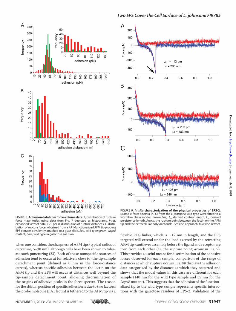
when one considers the sharpness ofAFM tips (typical radius ofcurvature, 5–30 nm), although cells have been shown to toler-ate such puncturing (23). Both of these nonspecific sources ofadhesion tend to occur at (or relatively close to) the tip-sampledetachment point (defined as 0 nm in the force-distancecurves), whereas specific adhesion between the lectin on theAFM tip and the EPS will occur at distances well beyond thetip-sample detachment point, allowing discrimination ofthe origins of adhesive peaks in the force spectra. The reasonfor the shift in position of specific adhesion is due to two factors;the probemolecule (PA1 lectin) is tethered to the AFM tip via a
flexible PEG linker, which is �12 nm in length, and the EPStargeted will extend under the load exerted by the retractingAFM tip-cantilever assembly before the ligand and receptor aretorn from each other (i.e. the rupture point; arrow in Fig. 9).This provides a useful means for discrimination of the adhesiveforces observed for each sample, comparison of the range ofdistances at which rupture occurs. Fig. 8B displays the adhesiondata categorized by the distance at which they occurred andshows that the modal values in this case are different for eachsample (140 nm for the wild type sample and 35 nm for the�epsEmutant). This suggests that the adhesion of the function-alized tip to the wild type sample represents specific interac-tions with the galactose residues of EPS-2. Validation of the
350
300
250
200
150
100
50
0
45
40
35
30
25
20
15
0
frequ
ency
10
5
45
40
35
30
25
20
15
0
10
5
frequ
ency
frequ
ency
adhesion (pN)
4050
302010
0fre
quen
cy
8070 90 100
110
120
130
2510 40 55 70 85 100
130
115
145
160
175
190
205
220
adhesion (pN)
700
140
210
280
350
420
560
490
630
700
770
840
910
adhesion distance (nm)
400 80 120
160
200
240
320
280
360
400
440
480
520
600
640
680
720
adhesion (pN)
FIGURE 8. Adhesion data from force-volume data. A, distribution of ruptureforce magnitudes using data from Fig. 7 depicted as histograms. Inset,expanded view of data �70 pN. B, distribution of rupture distances. C, distri-bution of rupture forces obtained from a PA1-functionalized AFM tip probingEPS extracts covalently attached to a glass slide. Red, wild type; green, �epsEmutant; blue, wild type in galactose solution.
FIGURE 9. In situ characterization of the physical properties of EPS-2.Example force spectra (A–C) from the L. johnsonii wild type were fitted to awormlike chain model (brown line). Lc, derived contour length; Lp, derivedpersistence length. Arrow, the rupture point between the lectin on the AFMtip and the extracellular polysaccharide. Red line, approach; blue line, retract.
Two EPS Cover the Cell Surface of L. johnsonii FI9785
NOVEMBER 1, 2013 • VOLUME 288 • NUMBER 44 JOURNAL OF BIOLOGICAL CHEMISTRY 31947
by guest on July 8, 2018http://w
ww
.jbc.org/D
ownloaded from

lectin-functionalized tip binding to extracted EPS from thewildtype and the �epsE deletion mutant (both covalently attachedto glass slides) confirmed that the PA1 lectin bound only to EPSfrom thewild type. The frequency of bindingwas reduced in thepresence of free galactose, confirming that it was due to lectin-carbohydrate association (Fig. 8C).Fig. 9 shows three example force spectra obtained on thewild
type sample that exhibit well resolved specific adhesive interac-tions on the retract (blue) portion of the force versus distancecurves that occur well beyond the tip-bacterial surface detach-ment point. These can be fitted to a wormlike chain polymerscaling model (24, 25) to derive two principal characteristicparameters, persistence length, Lp, and contour length, Lc. Per-sistence length is a measure of the flexibility of the polymerchain, and contour length provides a direct measure of themolecular size.
DISCUSSION
The capsular EPS is thought to be involved in the functionalproperties of colonization and persistence of both commensaland pathogenic bacteria (26, 27). In pathogens, the productionof a capsule can be a major virulence factor, yet many of thebiosynthetic mechanisms for EPS production are similarbetween pathogens and commensals. There are few reports onthe structure determination and identification of biosyntheticmechanisms of capsular EPS produced by commensal gut bac-teria, such as L. johnsonii FI9785. In this study, we determinedthe structure of two different EPS produced in situ by thisbacterium. We assessed the effects on EPS resulting from thedeletion of the epsE gene (predicted to encode a UDP-phos-phate galactose phosphotransferase that initiates EPS biosyn-thesis), a spontaneous mutation in the epsC gene (epsCD88N)(described as a putative tyrosine protein kinase) that has a rolein the regulation of EPS biosynthesis, and amutation where theentire eps gene cluster had been removed (9).It was interesting to find that L. johnsonii FI9785 was capable
of producing two different types of capsular EPS: EPS-1 andEPS-2. EPS-1 is a novel dextran with the unusual feature thatevery �-(1,6)-linked Glcp backbone residue was substituted atO2 with a terminal �-Glcp unit. EPS-2 is a heteropolysaccha-ride that has a unique hexasaccharide repeating unit composedof four glucose and two galactose residues. To our knowledge,the structures of the two exopolysaccharides are unique amongEPS produced by any bacteria. The production of�-glucanwithdifferent linkages is quite common for the genus Lactobacillus,and glucosyltransferases encoded by genes designated as gtf arecommonly responsible for the production of these dextran-typeexopolysaccharides (28–31). The L. johnsonii FI9785 genomedoes not contain any annotated genes with clear homology toglucansucrases. The production of more than one EPS has alsobeen demonstrated in other lactic acid bacteria; Lactobacillusplantarum EP56 expressed two heteropolysaccharides, onecell-bound and one released (32), whereas the two EPS pro-duced by Leuconostoc pseudomesenteroidesR2were both lineardextrans with different characteristics (33).EPS phosphorylation has been shown to affect interactions
with the host; phosphate groups associated with EPS from Lac-tobacillus delbrueckii subsp. bulgaricus have been shown to be
required for lymphocyte activation (34), whereas artificialphosphorylation of a dextran from Leuconostoc mesenteroidesincreased its immunostimulatory potential (35). EPS-1 wasfound to be partly substituted with the 1-phosphoglycerol moi-ety. Such substitution increases the net charge of the EPS,which could play an important role as determinant of interac-tions between cells, with host surfaces and with ions and pep-tides in the environment (32, 36), as well as modulating EPSpacking and permeability. Different degrees of phosphorylationand unique phosphorylation patterns may influence theobserved differences in cellular adhesion between the wild typeand the �epsE mutant. We found evidence for partial acetyla-tion of both EPS-1 (atmultiple sites) and EPS-2 (at a single site),although we did not establish the precise location of the sub-stituents.O-Acetylation of bacterial EPS is frequently reportedin both lactic acid bacteria (37–40) and others, including Kleb-siella aerogenes, E. coli O8:K27, and the plant pathogen Pseu-domonas flavescens (41–43). Acetylation can alter the physicalproperties of the EPS, giving, for example, increased viscosity insolution. In the context of the gut environment, we speculatethat acetylation provides protection of the EPS from manytypes of hydrolases produced by gut bacteria.AFM was used to investigate cell surface differences using a
D-galactose-specific lectin-functionalized tip. The adhesionmaps obtained for the wild type (which produces EPS-1 andEPS-2) and the �epsE mutant (which only produces EPS-1)reveal a clear difference in the frequency and magnitude ofadhesive events captured, showing higher adhesion in the wildtype, agreeing with the loss of a galactose-rich EPS in thismutant. In addition to detecting and spatially locating thegalactose-bearing EPS-2 on the wild type sample, further anal-ysis of the force spectra yielded information about the physicalproperties of the polysaccharide. Force spectra obtained on thewild type sample fitted the wormlike chainmodel (24, 25), indi-cating that EPS-2 adopts a semiflexible random coil conforma-tion. The fact that this information can be obtained in situwith-out the need to isolate the polysaccharide illustrates the powerof AFM to measure important intrinsic properties of bacterialcell surfaces (44).Recently, Fanning et al. (45) showed that the putative prim-
ing glycosyltransferase Bbr_0430 was essential for the biosyn-thesis of EPS in Bifidobacterium breve UCC2003. In contrast,we found that the �epsEmutant was still producing EPS-1; thissuggested that the production of EPS-1 could be independentfrom the eps gene cluster of L. johnsonii FI9785. But deletingthis entire eps cluster from the genome of L. johnsonii FI9785resulted in the loss of both EPS-1 and EPS-2 production, sug-gesting that at least one of the genes in this cluster is requiredfor the production of EPS-1. These results are consistent withprevious reports where the deletion of the eps gene cluster inL. johnsoniiNCC533 resulted in an acapsular strain (8). The epsgene cluster of L. johnsonii FI9785 has a genetic organizationsimilar to those of identified gene clusters for the biosynthesisof capsular or extracellular heteropolysaccharides (45–47).Wesuggest that this gene cluster, which harbors six putative glyco-syltransferase genes, might be responsible for the biosynthesisof heteropolysaccharide EPS-2; in addition, one of these glyco-syltransferases may have a bifunctional role to produce the
Two EPS Cover the Cell Surface of L. johnsonii FI9785
31948 JOURNAL OF BIOLOGICAL CHEMISTRY VOLUME 288 • NUMBER 44 • NOVEMBER 1, 2013
by guest on July 8, 2018http://w
ww
.jbc.org/D
ownloaded from

homopolymer EPS-1 (48). Alternatively, a novel gene from thegenome of L. johnsonii FI9785 may be involved in EPS-1 pro-duction in conjunction with a gene(s) in the eps cluster. Poten-tially, the sixmonosaccharide units in the heteropolysaccharideEPS-2 might be added by each glycosyltransferase to form thelong-chain capsular EPS-2 initiated by the priming glycosyl-transferase epsE. Another gene supporting the role of the epscluster in EPS-2 production is the glf gene, which putativelyencodes the UDP-galactopyranose mutase (9). This has beenpredicted to convert UDP-galactopyranose to UDP-galacto-furanose in Lactobacillus rhamnosus GG (47) and may beresponsible for the presence of the galactofuranose residue inthe repeating unit structure of EPS-2.Based on our findings, we propose that EpsE is the first gly-
cosyltransferase responsible for attachment of the first sugarmonomer to a lipid carrier because the �epsE mutant was notable to produce EPS-2. The role of this glycosyltransferase hasbeen demonstrated in both Gram-positive and Gram-negativebacteria (46, 47, 49–51). Previously, it was shown that the inac-tivation of the priming glycosyltransferase of L. rhamnosusGGresulted in the absence of the galactose-rich EPS layer on thecell surface, whereas a glucose-rich polysaccharide was stilldetectable attached to the cell surface (47). Similarly, it wasshown that deletion of the cpsIaE gene, which initiates the poly-saccharide biosynthesis in streptococci, resulted in a non-cap-sular phenotype (49). In the current study, we showed that afterinactivation of the epsE gene, a second capsular EPS that wasformed by glucose monomers only was still detectable inL. johnsonii FI9785. These results demonstrate the essentialrole of the epsE gene in EPS-2 accumulation on the cell surfaceof lactobacilli, and further work to investigate the L. johnsoniiFI9785 EpsE proteinmay confirm its proposed role as the prim-ing glycosyltransferase and identify the first monosaccharide ofthe chain.Our previous work on the epsCD88N mutant showed that
there was an increase in the production of EPS in this strain (9).This mutant could produce both EPS-1 and EPS-2, and thealteration of EPS accumulation level was not related to struc-tural changes in the EPS. The increase in EPS content was pos-sibly due to the production of a higher level of EPS-2 than thewild type, related to the putative role of EpsC in the regulationof EPS-2 biosynthesis (49, 52). The characterization of the roleof capsular EPS and investigation of the potential genes forEPS-1 biosynthesis is currently in progress.The structure of capsular EPS has been shown to have an
impact on the immunomodulation, biofilm formation, and col-onization properties of producing bacteria (4, 45, 53, 54). Interms of the lifestyle of the poultry gastrointestinal tract-de-rived commensal L. johnsonii FI9785, these two EPS could havea protective effect, improving the survival of the bacteria in theexternal environment and during transit through the gut. Pre-viously, we have reported that differences in the cell surface-associated EPS caused bymutations in the eps cluster affect theadhesion and aggregation properties of L. johnsonii FI9785 (9).Both of these characteristics can have an impact upon intra-and interspecies interactions as well as interactions with thehost gastrointestinal tract. Here we have detected the cell sur-face changes after mutations in the eps gene cluster using anti-
L. johnsonii FI9785 antibody responses. Górska and co-workers(7) found that the heteropolysaccharide from L. johnsonii 142,isolated from the murine gut, reacted to a whole cell antibody.Interestingly, the �epsEmutant, which could only produce the�-glucan as a capsular EPS, showed a higher antibody responseto the L. johnsonii antibody than thewild type, and this increasewas intensified in the acapsular �eps_cluster mutant, whereasstrains producing higher levels of EPS did not show anincreased response. The inability to produce EPS-2 or the EPS-1/EPS-2 mixture as a capsular material at the cell surface mayhave resulted in the exposure and presentation of cell surfaceepitopes like surface proteins for antibody binding in�eps_cluster and �epsE mutants. Another explanation forincreased antibody response in �epsE might be that glucose-containing epitopes could be more antigenic than galactose-containing epitopes, as noted previously (55). Deletion of a geneproducing a levan EPS from Lactobacillus reuteri prevented theinduction of regulatory T cells caused by colonization with thewild type strain (54), whereas EPS-deficient strains of B. breveelicited a stronger immune response than the wild type (45).EPS layers in these two examples were shown to have a positiveeffect on persistence and colonization during in vivo studies(45, 54). Our findings suggest that the gastrointestinal coloni-zation and recognition of the wild type L. johnsonii FI9785, the�eps_cluster and the �epsE strains by the immune systemwould be different because of the described structural differ-ences and imply a further biological role for the EPS in protect-ing the bacteria against an immune response.In conclusion, this study has revealed simultaneous synthesis
of two novel polysaccharide structures by L. johnsonii FI9785.Synthesis of both polymers is dependent on the identified epsgene cluster; however, the precise regulation of the biosynthesisof individual EPS has yet to be identified. Further structuralfunctional characterization using the isolated mutants willallow us to elucidate the physiological importance of these cellsurface structures in bacterial survival, host colonization, andpathogen exclusion.
Acknowledgments—We thank Kathryn Cross (Imaging Platform,Institute of Food Research) for TEM analysis and Dr. E. Maguin(Institut National de la Recherche Agronomique) for provision of thepG�host vector.
REFERENCES1. Badel, S., Bernardi, T., and Michaud, P. (2011) New perspectives for lac-
tobacilli exopolysaccharides. Biotechnol. Adv. 29, 54–662. De Vuyst, L., and Degeest, B. (1999) Heteropolysaccharides from lactic
acid bacteria. FEMS Microbiol. Rev. 23, 153–1773. Delcour, J., Ferain, T., Deghorain,M., Palumbo, E., andHols, P. (1999) The
biosynthesis and functionality of the cell-wall of lactic acid bacteria. An-tonie Van Leeuwenhoek 76, 159–184
4. Hidalgo-Cantabrana, C., López, P., Gueimonde,M., los Reyes-Gavilán, C.,Suárez, A., Margolles, A., and Ruas-Madiedo, P. (2012) Immune modula-tion capability of exopolysaccharides synthesised by lactic acid bacteriaand Bifidobacteria. Probiotics Antimicrob. Proteins 4, 227–237
5. Lin,M.H., Yang, Y. L., Chen, Y. P., Hua, K. F., Lu, C. P., Sheu, F., Lin, G. H.,Tsay, S. S., Liang, S. M., and Wu, S. H. (2011) A novel exopolysaccharidefrom the biofilm of Thermus aquaticus YT-1 induces the immune re-sponse through Toll-like receptor 2. J. Biol. Chem. 286, 17736–17745
6. La Ragione, R. M., Narbad, A., Gasson, M. J., andWoodward, M. J. (2004)
Two EPS Cover the Cell Surface of L. johnsonii FI9785
NOVEMBER 1, 2013 • VOLUME 288 • NUMBER 44 JOURNAL OF BIOLOGICAL CHEMISTRY 31949
by guest on July 8, 2018http://w
ww
.jbc.org/D
ownloaded from

In vivo characterization of Lactobacillus johnsonii FI9785 for use as adefined competitive exclusion agent against bacterial pathogens in poul-try. Lett. Appl. Microbiol. 38, 197–205
7. Górska, S., Jachymek, W., Rybka, J., Strus, M., Heczko, P. B., and Gamian,A. (2010) Structural and immunochemical studies of neutral exopolysac-charide produced by Lactobacillus johnsonii 142. Carbohydr. Res. 345,108–114
8. Denou, E., Pridmore, R. D., Berger, B., Panoff, J. M., Arigoni, F., and Brus-sow, H. (2008) Identification of genes associated with the long-gut-persis-tence phenotype of the probiotic Lactobacillus johnsonii strain NCC533using a combination of genomics and transcriptome analysis. J. Bacteriol.190, 3161–3168
9. Horn, N.,Wegmann, U., Dertli, E., Mulholland, F., Collins, S. R.,Waldron,K. W., Bongaerts, R. J., Mayer, M. J., and Narbad, A. (2013) Spontaneousmutations reveals influence of exopolysaccharide on Lactobacillus john-sonii surface characteristics. PloS One 8, e59957
10. Jolly, L., Newell, J., Porcelli, I., Vincent, S. J., and Stingele, F. (2002) Lacto-bacillus helveticus glycosyltransferases. From genes to carbohydrate syn-thesis. Glycobiology 12, 319–327
11. Wegmann, U., Klein, J. R., Drumm, I., Kuipers, O. P., and Henrich, B.(1999) Introduction of peptidase genes from Lactobacillus delbrueckiisubsp. lactis into Lactococcus lactis and controlled expression. Appl. En-viron. Microbiol. 65, 4729–4733
12. Maguin, E., Prevost, H., Ehrlich, S. D., and Gruss, A. (1996) Efficient in-sertional mutagenesis in lactococci and other Gram-positive bacteria. J.Bacteriol. 178, 931–935
13. Horn, N., Wegmann, U., Narbad, A., and Gasson, M. J. (2005) Character-isation of a novel plasmid p9785S from Lactobacillus johnsonii FI9785.Plasmid 54, 176–183
14. Hutter, J. L., and Bechhoefer, J. (1993) Calibration of atomic force micro-scope tips. Rev. Sci. Instrum. 64, 1868–1873
15. Gunning, A. P., Chambers, S., Pin, C., Man, A. L., Morris, V. J., and Nico-letti, C. (2008) Mapping specific adhesive interactions on living humanintestinal epithelial cells with atomic force microscopy. FASEB J. 22,2331–2339
16. Mackenzie, D. A., Jeffers, F., Parker, M. L., Vibert-Vallet, A., Bongaerts,R. J., Roos, S., Walter, J., and Juge, N. (2010) Strain-specific diversity ofmucus-binding proteins in the adhesion and aggregation properties ofLactobacillus reuteri.Microbiology 156, 3368–3378
17. van de Velde, F., Pereira, L., and Rollema, H. S. (2004) The revised NMRchemical shift data of carrageenans. Carbohydr. Res. 339, 2309–2313
18. Ciesielski, F., Griffin, D. C., Rittig, M., and Bonev, B. B. (2009) High-reso-lution J-coupled C-13 MAS NMR spectroscopy of lipid membranes.Chem. Phys. Lipids 161, 77–85
19. Maina, N. H., Tenkanen, M., Maaheimo, H., Juvonen, R., and Virkki, L.(2008) NMR spectroscopic analysis of exopolysaccharides produced byLeuconostoc citreum and Weissella confusa. Carbohydr. Res. 343, 1446–1455
20. Robijn, G. W., Wienk, H. L., van den Berg, D. J., Haas, H., Kamerling, J. P.,and Vliegenthart, J. F. (1996) Structural studies of the exopolysaccharideproduced by Lactobacillus paracasei 34–1.Carbohydr. Res. 285, 129–139
21. Suo, Z., Yang, X., Avci, R., Kellerman, L., Pascual, D. W., Fries, M., andSteele, A. (2007) HEPES-stabilized encapsulation of Salmonella typhimu-rium. Langmuir 23, 1365–1374
22. Haas, A., Zimmermann, K., Graw, F., Slack, E., Rusert, P., Ledergerber, B.,Bossart, W., Weber, R., Thurnheer, M. C., Battegay, M., Hirschel, B., Ver-nazza, P., Patuto, N., Macpherson, A. J., Gunthard, H. F., Oxenius, A., andSwiss HIV Cohort Study (2011) Systemic antibody responses to gut com-mensal bacteria during chronic HIV-1 infection. Gut 60, 1506–1519
23. Suo, Z., Avci, R., Deliorman, M., Yang, X., and Pascual, D. W. (2009)Bacteria survive multiple puncturings of their cell walls. Langmuir 25,4588–4594
24. Kratky, O., and Porod, G. (1949) Röntgenuntersuchung gelöster Faden-moleküle. Recueil des Travaux Chimiques des Pays-Bas 68, 1106–1122
25. Flory, P. (1998) Statistical Mechanics of Chain Molecules, Hanser,Munchen
26. Deutsch, S. M., Parayre, S., Bouchoux, A., Guyomarc’h, F., Dewulf, J.,Dols-Lafargue, M., Bagliniere, F., Cousin, F. J., Falentin, H., Jan, G., and
Foligne, B. (2012) Contribution of surface �-glucan polysaccharide tophysicochemical and immunomodulatory properties of Propionibacte-rium freudenreichii. Appl. Environ. Microbiol. 78, 1765–1775
27. Alp, G., Aslim, B., Suludere, Z., and Akca, G. (2010) The role of hemag-glutination and effect of exopolysaccharide production on bifidobacteriaadhesion to Caco-2 cells in vitro.Microbiol. Immunol. 54, 658–665
28. Kralj, S., van Geel-Schutten, G. H., Dondorff, M.M., Kirsanovs, S., van derMaarel, M. J., and Dijkhuizen, L. (2004) Glucan synthesis in the genusLactobacillus. Isolation and characterization of glucansucrase genes, en-zymes and glucan products from six different strains. Microbiology 150,3681–3690
29. Kralj, S., van Geel-Schutten, G. H., Rahaoui, H., Leer, R. J., Faber, E. J., vander Maarel, M. J., and Dijkhuizen, L. (2002) Molecular characterization ofa novel glucosyltransferase from Lactobacillus reuteri strain 121 synthe-sizing a unique, highly branched glucan with �-(134) and �-(136) glu-cosidic bonds. Appl. Environ. Microbiol. 68, 4283–4291
30. van Leeuwen, S. S., Kralj, S., van Geel-Schutten, I. H., Gerwig, G. J., Di-jkhuizen, L., and Kamerling, J. P. (2008) Structural analysis of the �-D-glucan (EPS180) produced by the Lactobacillus reuteri strain 180 glucan-sucrase GTF180 enzyme. Carbohydr. Res. 343, 1237–1250
31. van Hijum, S. A., Kralj, S., Ozimek, L. K., Dijkhuizen, L., and van Geel-Schutten, I. G. (2006) Structure-function relationships of glucansucraseand fructansucrase enzymes from lactic acid bacteria. Microbiol. Mol.Biol. Rev. 70, 157–176
32. Tallon, R., Bressollier, P., and Urdaci, M. C. (2003) Isolation and charac-terization of two exopolysaccharides produced by Lactobacillus planta-rum EP56. Res. Microbiol. 154, 705–712
33. Paulo, E. M., Boffo, E. F., Branco, A., Valente, A. M., Melo, I. S., Ferreira,A. G., Roque, M. R., and Assis, S. A. (2012) Production, extraction andcharacterization of exopolysaccharides produced by the native Leuconos-toc pseudomesenteroides R2 strain. An. Acad. Bras. Cienc. 84, 495–508
34. Kitazawa, H., Harata, T., Uemura, J., Saito, T., Kaneko, T., and Itoh, T.(1998) Phosphate group requirement for mitogenic activation of lympho-cytes by an extracellular phosphopolysaccharide from Lactobacillus del-brueckii ssp. bulgaricus. Int. J. Food Microbiol. 40, 169–175
35. Sato, T., Nishimura-Uemura, J., Shimosato, T., Kawai, Y., Kitazawa, H.,and Saito, T. (2004) Dextran from Leuconostoc mesenteroides augmentsimmunostimulatory effects by the introduction of phosphate groups. J.Food Prot. 67, 1719–1724
36. Looijesteijn, P. J., Trapet, L., de Vries, E., Abee, T., and Hugenholtz, J.(2001) Physiological function of exopolysaccharides produced by Lacto-coccus lactis. Int. J. Food Microbiol. 64, 71–80
37. Faber, E. J., van den Haak, M. J., Kamerling, J. P., and Vliegenthart, J. F.(2001) Structure of the exopolysaccharide produced by Streptococcusthermophilus S3. Carbohydr. Res. 331, 173–182
38. Robijn, G. W., van den Berg, D. J., Haas, H., Kamerling, J. P., and Vliegen-thart, J. F. (1995) Determination of the structure of the exopolysaccharideproduced by Lactobacillus sake 0–1. Carbohydr. Res. 276, 117–136
39. Rodrıguez-Carvajal, M. A., Sanchez, J. I., Campelo, A. B., Martınez, B.,Rodrıguez, A., andGil-Serrano, A.M. (2008) Structure of the high-molec-ular weight exopolysaccharide isolated from Lactobacillus pentosusLPS26. Carbohydr. Res. 343, 3066–3070
40. Staaf, M., Yang, Z., Huttunen, E., and Widmalm, G. (2000) Structuralelucidation of the viscous exopolysaccharide produced by Lactobacillushelveticus Lb161. Carbohydr. Res. 326, 113–119
41. Cescutti, P., Toffanin, R., Fett, W. F., Osman, S. F., Pollesello, P., andPaoletti, S. (1998) Structural investigation of the exopolysaccharide pro-duced by Pseudomonas flavescens strain B62. Degradation by a fungalcellulase and isolation of the oligosaccharide repeating unit. Eur.J. Biochem. 251, 971–979
42. Sutherland, I. W., Jann, K., and Jann, B. (1970) The isolation of O-acetylated fragments from the K antigen of Escherichia coli 08:K27(A):H by the action of phage-induced enzymes from Klebsiella aero-genes. Eur. J. Biochem. 12, 285–288
43. Sutherland, I. W., and Wilkinson, J. F. (1968) The exopolysaccharide ofKlebsiella aerogens A3 (S1) (type 54). The isolation of O-acetylated octa-saccharide, tetrasaccharide and trisaccharide. Biochem. J. 110, 749–754
44. Dupres, V., Menozzi, F. D., Locht, C., Clare, B. H., Abbott, N. L., Cuenot,
Two EPS Cover the Cell Surface of L. johnsonii FI9785
31950 JOURNAL OF BIOLOGICAL CHEMISTRY VOLUME 288 • NUMBER 44 • NOVEMBER 1, 2013
by guest on July 8, 2018http://w
ww
.jbc.org/D
ownloaded from

S., Bompard, C., Raze, D., and Dufrene, Y. F. (2005) Nanoscale mappingand functional analysis of individual adhesins on living bacteria. Nat.Methods 2, 515–520
45. Fanning, S., Hall, L. J., Cronin, M., Zomer, A., MacSharry, J., Goulding, D.,Motherway, M. O., Shanahan, F., Nally, K., Dougan, G., and van Sinderen,D. (2012) Bifidobacterial surface-exopolysaccharide facilitates commen-sal-host interaction through immune modulation and pathogen protec-tion. Proc. Natl. Acad. Sci. U.S.A. 109, 2108–2113
46. Bentley, S. D., Aanensen, D. M., Mavroidi, A., Saunders, D., Rabbinow-itsch, E., Collins, M., Donohoe, K., Harris, D., Murphy, L., Quail, M. A.,Samuel, G., Skovsted, I. C., Kaltoft, M. S., Barrell, B., Reeves, P. R., Parkhill,J., and Spratt, B. G. (2006) Genetic analysis of the capsular biosyntheticlocus from all 90 pneumococcal serotypes. PLoS Genet. 2, e31
47. Lebeer, S., Verhoeven, T. L., Francius, G., Schoofs, G., Lambrichts, I.,Dufrene, Y., Vanderleyden, J., and De Keersmaecker, S. C. (2009) Identi-fication of a gene cluster for the biosynthesis of a long, galactose-richexopolysaccharide inLactobacillus rhamnosusGGand functional analysisof the priming glycosyltransferase. Appl. Environ. Microbiol. 75, 3554–3563
48. Luzhetskyy, A., and Bechthold, A. (2008) Features and applications ofbacterial glycosyltransferases. Current state and prospects. Appl. Micro-biol. Biotechnol. 80, 945–952
49. Cieslewicz,M. J., Kasper, D. L.,Wang, Y., andWessels, M. R. (2001) Func-tional analysis in type Ia group B Streptococcus of a cluster of genes in-volved in extracellular polysaccharide production by diverse species of
streptococci. J. Biol. Chem. 276, 139–14650. Minic, Z., Marie, C., Delorme, C., Faurie, J. M., Mercier, G., Ehrlich, D.,
and Renault, P. (2007) Control of EpsE, the phosphoglycosyltransferaseinitiating exopolysaccharide synthesis in Streptococcus thermophilus, byEpsD tyrosine kinase. J. Bacteriol. 189, 1351–1357
51. van Kranenburg, R., Marugg, J. D., van Swam, II,Willem, N. J., and de Vos,W.M. (1997)Molecular characterization of the plasmid-encoded eps genecluster essential for exopolysaccharide biosynthesis in Lactococcus lactis.Mol. Microbiol. 24, 387–397
52. Morona, J. K.,Morona, R.,Miller, D. C., and Paton, J. C. (2003)Mutationalanalysis of the carboxy-terminal (YGX)4 repeat domain of CpsD, an auto-phosphorylating tyrosine kinase required for capsule biosynthesis inStreptococcus pneumoniae. J. Bacteriol. 185, 3009–3019
53. Lebeer, S., Verhoeven, T. L., Perea Velez, M., Vanderleyden, J., and DeKeersmaecker, S. C. (2007) Impact of environmental and genetic factorson biofilm formation by the probiotic strain Lactobacillus rhamnosusGG.Appl. Environ. Microbiol. 73, 6768–6775
54. Sims, I. M., Frese, S. A., Walter, J., Loach, D., Wilson, M., Appleyard, K.,Eason, J., Livingston, M., Baird, M., Cook, G., and Tannock, G. W. (2011)Structure and functions of exopolysaccharide produced by gut commen-sal Lactobacillus reuteri 100–23. ISME J. 5, 1115–1124
55. Iwamori, M., Shibagaki, T., Nakata, Y., Adachi, S., and Nomura, T. (2009)Distribution of receptor glycolipids for Lactobacilli in murine digestivetract and production of antibodies cross-reactive with them by immuni-zation of rabbits with lactobacilli. J. Biochem. 146, 185–191
Two EPS Cover the Cell Surface of L. johnsonii FI9785
NOVEMBER 1, 2013 • VOLUME 288 • NUMBER 44 JOURNAL OF BIOLOGICAL CHEMISTRY 31951
by guest on July 8, 2018http://w
ww
.jbc.org/D
ownloaded from

Gall, Boyan B. Bonev, Melinda J. Mayer and Arjan NarbadEnes Dertli, Ian J. Colquhoun, A. Patrick Gunning, Roy J. Bongaerts, Gwénaëlle Le
FI9785johnsoniiLactobacillusStructure and Biosynthesis of Two Exopolysaccharides Produced by
doi: 10.1074/jbc.M113.507418 originally published online September 9, 20132013, 288:31938-31951.J. Biol. Chem.
10.1074/jbc.M113.507418Access the most updated version of this article at doi:
Alerts:
When a correction for this article is posted•
When this article is cited•
to choose from all of JBC's e-mail alertsClick here
http://www.jbc.org/content/288/44/31938.full.html#ref-list-1
This article cites 54 references, 16 of which can be accessed free at
by guest on July 8, 2018http://w
ww
.jbc.org/D
ownloaded from