survival, proliferation and cell cycle of swine fibroblast ... · salmonella enterica, fibroblast,...
TRANSCRIPT
Advances in Microbiology, 2016, 6, 942-952 http://www.scirp.org/journal/aim
ISSN Online: 2165-3410 ISSN Print: 2165-3402
DOI: 10.4236/aim.2016.613088 November 9, 2016
Survival, Proliferation and Cell Cycle of Swine Fibroblast after Infection with Salmonella enterica
Mizuki Masuda1, Yijie Guo1,2, Kengo Kuroda1, Jun Xu1, Hiroshi Yoneyama1, Tomokazu Fukuda3, Bernard Mudenda Hang’ombe4, Katsuya Okuno1, Junko Nishimura5*, Emiko Isogai1*
1Laboratory of Animal Microbiology, Graduate School of Agricultural Science, Tohoku University, Sendai, Japan 2Department of Immunobiology and Pathogenic Biology, Medical School of Xi’an Jiaotong University, Xi’an, China 3Laboratory of Cell Engineering and Molecular Genetics, Faculty of Science and Engineering, Iwate University, Morioka, Japan 4Department of Clinical Microbiology, School of Veterinary Medicine, University of Zambia, Lusaka, Zambia 5Laboratory of Nutrition and Life Sciences, Department of Biotechnology and Environmental Engineering, Faculty of Engineering, Hachinohe Institute of Technology, Hachinohe, Japan
Abstract Food-borne salmonellosis continues to be a major health concern worldwide. Carry- contamination of Salmonella frequently occurs in meat production. We focused on cell dynamics of swine fibroblasts after infection with Salmonella enterica serovar Enteritidis and Typhimurium, because fibroblast can be a target cell for Salmonella latent infection. It was found that both S. Enteritidis and S. Typhimurium were able to adhere and invade to swine fibroblasts. The proliferations in fibroblasts were dif-ferent between each serovar. S. Enteritidis reached to the maximum at 24 hr after in-fection while S. Typhimurium did not. In addition, the decrease in the G0/G1 phase cells and increase in G2/M phase cells on the fibroblast were observed by both Sal-monella infection. Cell death including apoptosis in the cells was inhibited by the in-fection of Salmonella. These results suggest that nontyphoidal Salmonella can survive for the long term by modifying bacterial cell proliferation and preventing cell death of host cells.
Keywords Salmonella enterica, Fibroblast, Swine, Cell Death, Cell Cycle
1. Introduction
Salmonella enterica causes various diseases, particularly, nontyphoidal salmonellae are very important in reportable food-borne infections. Salmonella is an intracellular pa-thogen that can invade eukaryotic cells and manage to survive in the living host cell [1]
How to cite this paper: Masuda, M., Guo, Y.J., Kuroda, K., Xu, J., Yoneyama, H., Fu-kuda, T., Hang’ombe, B.M., Okuno, K., Nishimura, J. and Isogai, E. (2016) Survival, Proliferation and Cell Cycle of Swine Fibrob-last after Infection with Salmonella enterica. Advances in Microbiology, 6, 942-952. http://dx.doi.org/10.4236/aim.2016.613088 Received: August 11, 2016 Accepted: November 6, 2016 Published: November 9, 2016 Copyright © 2016 by authors and Scientific Research Publishing Inc. This work is licensed under the Creative Commons Attribution International License (CC BY 4.0). http://creativecommons.org/licenses/by/4.0/
Open Access
M. Masuda et al.
943
[2] [3]. It is well known that this bacteria is able to invade via Type Three Secretion System (TTSS) encoded in Salmonella Pathogenicity Island 1 (SPI-1) [4]. When Sal-monella invades to host cells, the SPI-1 effector protein of the TTSS is injected into ep-ithelial cells, thereby causing rearrangement of actin cytoskeleton [5] [6] [7], membrane ruffling and formation of micropinosomes [8]. As a case of cytoskeletal rearrangement, it is revealed that SPI-1 proteins SipABCD, SopE and SopE2 are involved [9].
In the present, it has been found that the infection route of Salmonella Typhimurium against fibroblast cells was quite different from that of epithelial cells, in which the SPI-1 effectors SipB and SipC were unnecessary. And this strain is able to suppress cell growth by stopping cell division in fibroblast cells after invasion [10] [11] [12]. Based on these findings, it is considered that S. Typhimurium could alter the routes into fi-broblasts, and persistent infection and asymptomatic carrier are caused.
Although it has been known the importance of salmonellosis to public health, the mechanism of the Salmonella carrier state hasn’t been well known still now. We ex-amined the invasion of Salmonella into fibroblasts, because there was a risk of giving rise to the persistent infection. In addition, we show here invasion and proliferation of S. Typhimurium in the fibroblasts differs from that of S. Enteritidis.
2. Materials and Methods 2.1. Bacterial Strains and Swine Fibroblast Cell Line
Salmonella enterica serovar Enteritidis strain zSE1 isolated in Zambia and Typhimu-rium wild type strain st1wt were cultured properly in Trypticase Soy Broth (TSB) at 37˚C for 18 hr [13]. Pig embryonic fibroblasts (PEFs), which infected with simian va-cuolating virus 40 large T fragment (PEFs-SV40) to achieve immortalization, used in this study [14]. Cells were cultured in Dulbecco’s modified Eagle’s medium (DMEM) (Nacalai Tesque Inc., Kyoto, Japan) with 10% PBS and 1% antibiotic-antimycotic mixed stock solution (Nacalai Tesque Inc.) at 37˚C in 5% CO2. Cells were maintained under exponential growth condition and used as the host cells in further experiments.
2.2. Bacterial Infection Assays
PEFs were seeded in a 24-well-plate to reach a density of 1.0 × 10⁵ cells/well at the time of infection. The medium was changed to DMEM with 10% FBS (without antibiotics) 2 hr before bacterial infection to eliminate any potential effects of the antibiotics. PEFs were infected with overnight cultured bacteria at a multiplicity of infection (MOI) of 5:1 (bacteria to eukaryotic cells). To count the adhesive bacteria at 0, 20, 60, and 100 min after the infection, wells were washed with PBS containing 0.1% (wt/vol) sodium dodecyl sulfate (SDS) (Wako Pure Chemical Industries Ltd., Osaka, Japan) and 1% (vol/vol) Triton X-100 (Wako Pure Chemical Industries Ltd.) as lysis buffer. The inva-sive bacteria were counted, and cells were washed with PBS repeatedly. Fresh culture medium containing 100 µg/ml gentamicin was added for 2 hr post-infection, and then the cultured cells were lysed with lysis buffer.

M. Masuda et al.
944
2.3. Immunofluorescence Microscopy
Extracellular (adherent) and intracellular bacteria were stained by immunofluorescent microscopy by the methods of Aiastui et al. [10]. Briefly, extracellular bacteria were stained in nonpermeabilized cells with polyclonal rabbit anti-Salmonella lipopolysac-charide (LPS) antibodies (S. Enteritidis O4 and S. Typhimurium O9, Denka Seiken Co., Ltd, Tokyo, Japan), followed by anti-rabbit Alexa Fluor 594 F(ab’)2 fragment antibody (Invitrogen, CA, USA). Upon permiabilization by treatment with 0.2% Triton X-100, intracellular bacteria were stained with anti-Salmonella LPS antibody above, followed by anti-rabbit Alxa-Fluor 488 F(ab’)2 fragment antibody (Invitrogen) as secondary an-tibodies.Cells were observed with a Fluorescent microscope (FSX 100, Olympus, Tokyo, Japan).
2.4. Intracellular Proliferation
Fresh DMEM medium containing 100 µg/ml gentamicin was added into 2 hr infected fibroblast. After cultivation for 24 hr, PEFs were dissolved in lysis buffer and intracel-lular bacterial numbers were counted.
2.5. Live Cell Count
Bacteria-infected PEFs (MOI = 5:1 as described above) were treated at 0, 20, 60, 100 min after the infection with 0.05% Triton X-100 and cells were collected. Cells were mixed with trypan blue (Invitrogen) and living cells were counted using a CSTI Coun-ter (Cell Science & Technology Institute, Inc., Miyagi, Japan). In order to assess the ef-fects of intracellular pathogens and long-term infection, the medium was changed to fresh medium containing 100 µg/ml gentamicin 2 hr after infection. After gentamicin treatment for 24 hr, live PEFs were counted according to mentioned.
2.6. MTT Assay
PEFs were seeded in a 96-well-plate and incubated for 24 hr to reach a density of 3.0 × 10³ cells/well at the time of infection. The medium was changed to DMEM with 10% FBS (without antibiotics) 2 hr before bacterial infection to eliminate any antibiotic ef-fects. Cells were infected with Salmonella (MOI = 5:1), and then 10 μl of MTT reagent from the MTT Cell Proliferation Assay kit (Funakoshi Co., Ltd., Tokyo, Japan) was added immediately after infection. After post-infection for 2 hr, the medium was changed to DMEM containing 100 µg/ml gentamicin, and MTT reagent was added to each samples at 0, 2, and 24 hr.
2.7. Apoptosis and Cell Cycle Assay
Apoptosis of infected cell was analyzed with MuseTM Cell Analyzer (Merck Millipore Inc., Darmstadt, Germany).PEFs were inoculated 0.1 × 10⁵ cells per well. S. Enteritidis or S. Typhimurium was added into well in the ratio of 5:1 (MOI) and incubated for 2 hr. After infection, PEFs were washed with PBS and collected with 0.05% trypsin. And then PEFs were centrifuged (800 × g) and washed with PBS. Infected PEFs were col-

M. Masuda et al.
945
lected with 0.05% trypsin and added Muse Annexin & Dead Cell Reagent and standing for 30 min at room temperature in a dark place. Apoptotic cells were detected with the analyzer. Furthermore, apoptosis profile of PEFs infected with S. Enteritidis zSE1 after infection for 24 hr was monitored using the flow cytometry.
The cell cycle of PEFs was also analyzed by using the MuseTM Cell Cycle Assay Kit (Merck Millipore Inc.), according to the procedure described by the manufacturer. In-fected PEFs were collected with 0.05% trypsin, centrifuged (800 × g), and washed with PBS. Collected cells were suspended in cold 70% ethanol and incubated for over 3 hr at −20˚C for fixation. Fixed cells were washed with PBS and stained with the cell cycle reagent for 30 min. Samples were measured by using the above analyzer.
2.8. Statistical Test
In this study, all of the experiments were carried out with at least triplicated samples. Mean and standard deviations were calculated from the multiple data. The statistical significance was evaluated unpaired t-test. After statistical analysis, p values of less than 0.05 were considered statistically significant.
3. Results and Discussion 3.1. Infection to Swine Fibroblasts
Although it has been revealed that fibroblasts are highly involved in the persistence of pathogenic Salmonella [15], the mechanisms of long-term and persistent infection of Salmonella enterica are still unknown. In this study, we investigated Salmonella-fi- broblast interactions to clarify the survival strategy of Salmonella in the host fibroblasts.
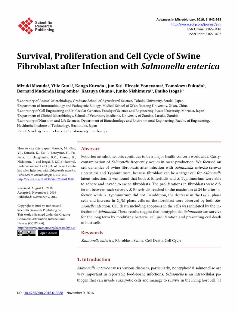
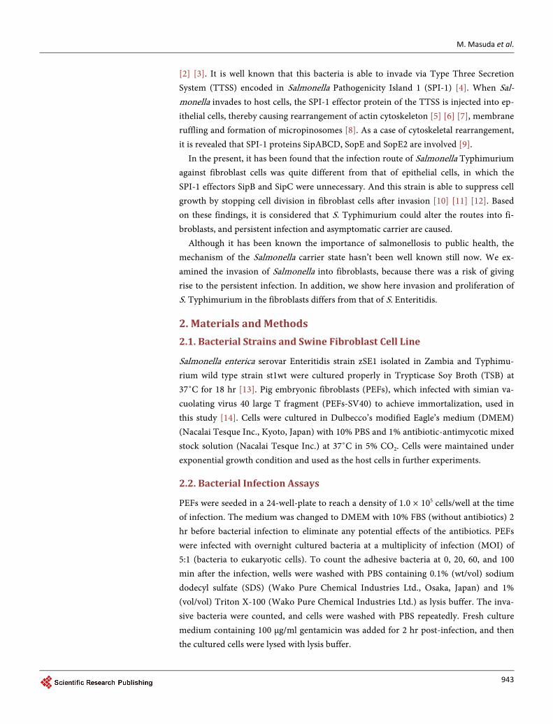
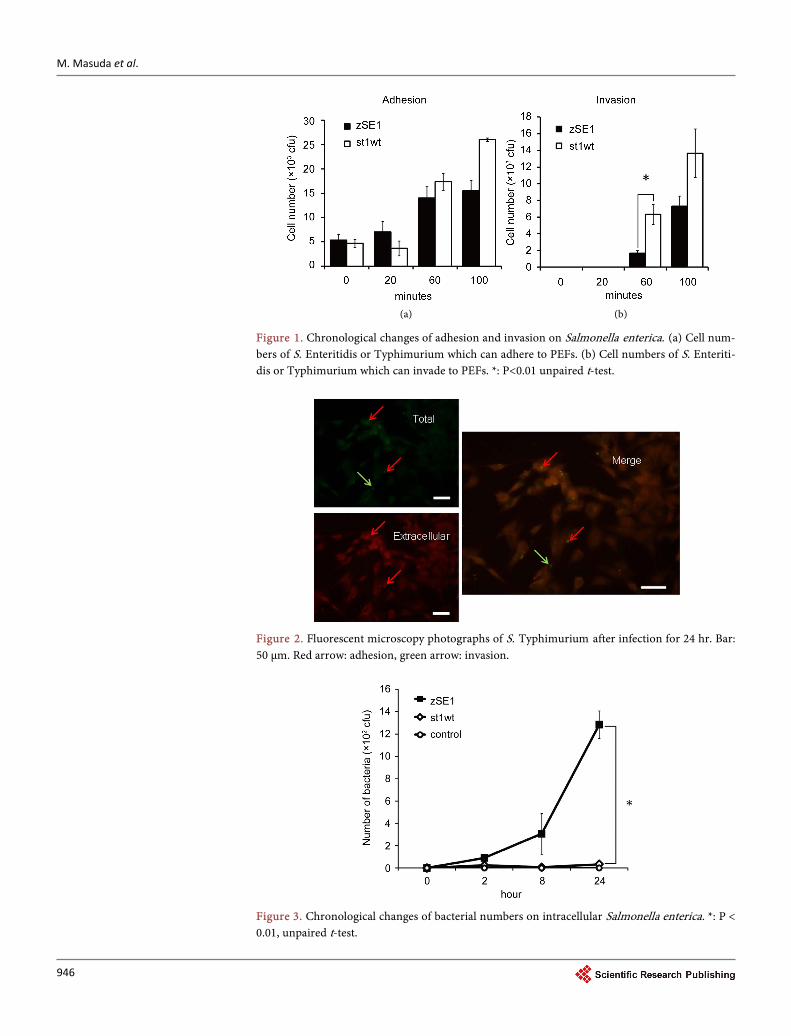
We first confirmed the infection of Salmonella to PEFs by conducting infection assay and immunofluorescence. Each cell of S. Enteritidis and Typhimurium adhered ap-proximately 1% for a start, and adherent cell number was increased time-dependently (Figure 1(a)). At 100 min, adherent number of S. Typhimurium was significantly higher than that of S. Enteritidis (P < 0.01). Cell invasion was observed after 60 min of the infection. S. Typhimurium invaded cells more aggressively than S. Enteritidis (Figure 1(b), P < 0.01 at 60 min after the infection). The states infected with S. Enteri-ditis or S. Typhimurium were photographed with a fluorescent microscope and typical photo-images at 24 hr after S. Typhimurium infection were showed (Figure 2). Fur-thermore, the intracellular proliferation of Salmonella cells was different between S. Enteritidis and S. Typhimurium, i.e., S. Enteritidis reproduced sharply after invasion to PEFs, on the other hand, S. Typhimurium was almost not (Figure 3, P < 0.01). Namely, S. Enteritidis zSE1 reached 1.28 × 10⁴ cfu after infection for 24 hr, while S. Typhimu-rium st1wt couldn’t proliferate in host cells and only reached 3.3 × 10² cfu.
3.2. Influences to Viability and Lifespan of Host Fibroblast
In order to assess the influence of host cells infected with pathogenic Salmonella, we analyzed the viability and proliferation of infected PEFs. The number of living infected
M. Masuda et al.
946
(a) (b)
Figure 1. Chronological changes of adhesion and invasion on Salmonella enterica. (a) Cell num-bers of S. Enteritidis or Typhimurium which can adhere to PEFs. (b) Cell numbers of S. Enteriti-dis or Typhimurium which can invade to PEFs. *: P<0.01 unpaired t-test.
Figure 2. Fluorescent microscopy photographs of S. Typhimurium after infection for 24 hr. Bar: 50 μm. Red arrow: adhesion, green arrow: invasion.
Figure 3. Chronological changes of bacterial numbers on intracellular Salmonella enterica. *: P < 0.01, unpaired t-test.

M. Masuda et al.
947
PEFs was counted after trypan blue staining. As a result, it was found that living cells were increased after infection with both Salmonella strains compared to non-infected cells (Figure 4). The proliferation of living PEFs was measured by MTT assay after 0, 2 and 24 hr infection (Figure 5). The proliferation of PEFs infected for 24 hr was re-markably increased, particularly, a significant difference was revealed on S. Typhimu-rium. These results indicate that Salmonella is able to survive within host fibroblasts and enhance the proliferation of host cells. It is found that S. Typhimurium led to en-hance the proliferation in epithelial cells [16] and suppress it in dendritic cells and fi-broblasts [11] [17]. Furthermore, S. Enteritidis into human fibroblasts increased during 1 day after infection and could survive until 14 to 28 days [18]. Based on these findings, it is considered that Salmonella grows rapidly in fibroblast and gradually controls
Figure 4. Proliferation of PEFs infected with zSE1 or st1wt. (a) The number of PEFs infected with S. Enteritidis or Typhimurium and non-infected-PEFs were counted using the trypan blue method. 0, 20, 60, 100 min after infection, cell number of PEFs infected with bacteria tended to be larger that of non-infected PEFs. (b) 24 hr after infection, PEFs infected with zSE1 or st1wt were about 1.52 fold or 1.47 fold respectively compared with non-infected PEFs. **: P < 0.05, un-paired t-test.

M. Masuda et al.
948
Figure 5. Time-lapse changes of cell proliferation on host PEFs. Cell viabil-ity was measured by MTT cell proliferation assay. Colorimetrical formazan produced from live cell metabolism were detected as absorbance at 570 nm. *: P < 0.01 unpaired t-test.
them after that, and Salmonella is possible long-term survival by repressing the prolife-ration in restricted host cells.
3.3. Effect to Viability of Fibroblast
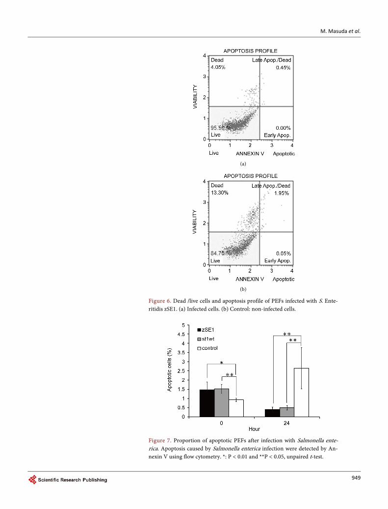
We demonstrated the cell death pattern of host cells after Salmonella infection as shown in Figure 6. As shown in Figure 7, apoptotic cells counted was 1% - 1.5% (sig-nificantly higher than control) at the beginning of the infection. After 24 hr infection, the percentage of apoptotic cells significantly increased in non-infected cells; in con-trast, the percentage of apoptotic cells was approximately 0.5% on Salmonella infected cells. Total dead cell number was also decreased by the Salmonella infection (data not shown).
To further explain such phenomenon, cell cycle of infected cells was analyzed by us-ing a Cell Analyzer. The cell cycle of infected or non-infected PEFs was measured by flow cytometry 24 hr after addition of bacteria. The results revealed that G0/G1 phases of infected cells were reduced and the G2/M phases were prolonged compared with non-infected cells, respectively (Figure 8). It has been found that enteropathogenic Escherichia coli and enterohaemorrhagic E. coli infused effector protein Cif to euka-ryotic host cells and led to arrest cell cycle, and the barrier of the epithelium cell be-came weak by stopping a cell cycle, and bacteria became easy to infect cells [19]. And the G0 phase was longer to repair DNA in cell cycle [20], the apoptosis was caused when DNA damage could not repair [21] [22]. In addition, it has been known that Salmonella led to induce apoptosis of host chicken fibroblasts [23], and the ratio of apoptotic PEFs infected with Salmonella Enteritidis or Typhimurium might be increased. Based on these findings, it seems that Salmonella infection induces the alteration of the cell cycle on host PEFs, and interrupts apoptosis of the host cell to survive in host cells. Namely, Salmonella of the intracellular parasitism may lead to repress cell cycle after invasion.
M. Masuda et al.
949
(a)
(b)
Figure 6. Dead /live cells and apoptosis profile of PEFs infected with S. Ente-ritidis zSE1. (a) Infected cells. (b) Control: non-infected cells.
Figure 7. Proportion of apoptotic PEFs after infection with Salmonella ente-rica. Apoptosis caused by Salmonella enterica infection were detected by An-nexin V using flow cytometry. *: P < 0.01 and **P < 0.05, unpaired t-test.

M. Masuda et al.
950
Figure 8. The ratio of cell cycle phase in PEFs after infection with Salmonella enterica. *: P < 0.01 and **: P < 0.05, unpaired t-test.
It has been known that bacteria are able to suit host cell environment by promoting
the most successful conditions for infection [24]. In this study, we demonstrated that Salmonella was able to survive in fibroblasts by manipulating both lifespan and apopto-sis of host cell. This phenomenon is intended to optimize the circumstances of Salmo-nella survival and promote the persistent infection of Salmonella in livestock. This se-ries of strategy could be a notable fact that highlights new concepts of Salmonella infec-tion of fibroblasts in domestic animals, and encourages people to reconsider the hidden issue in food safety.
References [1] Hohmann, E.L. (2001) Nontyphoidal Salmonellosis. Clinical Infectious Diseases, 32, 263-
269. http://dx.doi.org/10.1086/318457
[2] Wood, R.L. and Rose, R. (1992) Populations of Salmonella Typhimurium in Internal Or-gans of Experimentally Infected Carrier Swine. American Journal of Veterinary Research, 53, 653-658.
[3] Fedorka-Cray, P.J., Whipp, S.C., Isaacson, R.E., Nord, N. and Lager, K. (1994) Transmis-sion of Salmonella Typhimurium to Swine. Veterinary Microbiology, 41, 333-344. http://dx.doi.org/10.1016/0378-1135(94)90029-9
M. Masuda et al.
951
[4] Galán, J.E. and Wolf-Watz, H. (2006) Protein Delivery into Eukaryotic Cells by Type III Secretion Machines. Nature, 444, 567-573. http://dx.doi.org/10.1038/nature05272
[5] Finlay, B.B., Ruschkowski, S. and Dedhar, S. (1991) Cytoskeletal Rearrangements Accom-panying Salmonella Entry into Epithelial Cells. Journal of Cell Science, 99, 283-296.
[6] Patel, J.C. and Galán, J.E. (2005) Manipulation of the Host Actin Cytoskeleton by Salmo-nella—All in the Name of Entry. Current Opinion in Microbiology, 8, 10-15. http://dx.doi.org/10.1016/j.mib.2004.09.001
[7] Zhou, D. and Galán, J. (2001) Salmonella Entry into Host Cells: The Work in Concert of Type III Secreted Effector Proteins. Microbes and Infection, 3, 1293-1298. http://dx.doi.org/10.1016/j.mib.2004.09.001
[8] Garcia-del Portillo, F. and Finlay, B.B. (1994) Salmonella Invasion of Nonphagocytic Cells Induces Formation of Macropinosomes in the Host Cell. Infection and Immunity, 62, 4641-4645.
[9] Hansen-Wester, I. and Hensel, M. (2001) Salmonella Pathogenicity Islands Encoding Type III Secretion Systems. Microbes and Infection, 3, 549-559. http://dx.doi.org/10.1016/S1286-4579(01)01411-3
[10] Aiastui, A., Pucciarelli, M.G. and García-del Portillo, F. (2010) Salmonella enterica serovar Typhimurium Invades Fibroblasts by Multiple Routes Differing from the Entry into Epi-thelial Cells. Infection and Immunity, 78, 2700-2713. http://dx.doi.org/10.1128/IAI.01389-09
[11] García-del Portillo, F. (2001) Salmonella Intracellular Proliferation: Where, When and How? Microbes and Infection, 3, 1305-1311. http://dx.doi.org/10.1016/S1286-4579(01)01491-5
[12] Knodler, L.A., Finlay, B.B. and Steele-Mortimer, O. (2005) The Salmonella Effector Protein SopB Protects Epithelial Cells from Apoptosis by Sustained Activation of Akt. The Journal of Biological Chemistry, 280, 9058-9064. http://dx.doi.org/10.1074/jbc.M412588200
[13] Isogai, E., Makungu, C., Yabe, J., Sinkala, P., Nambota, A., Isogai, H., Fukushi, H., Si-lungwe, M., Mubita, C., Syakalima, M., Hang’ombe, B.M., Kozaki, S. and Yasuda, J. (2005) Detection of Salmonella invA by Isothermal and Chimeric Primer-Initiated Amplification of Nucleic Acids (ICAN) in Zambia. Comparative Immunology, Microbiology, and Infec-tious Diseases, 28, 363-370. http://dx.doi.org/10.1016/j.cimid.2005.10.001
[14] Donai, K., Kiyono, T., Eitsuka, T., Guo, Y., Kuroda, K., Sone, H., Isogai, E. and Fukuda, T. (2014) Bovine and Porcine Fibroblasts Can Be Immortalized with Intact Karyotype by the Expression of Mutant Cyclin Dependent Kinase 4, Cyclin D, and Telomerase. Journal of Biotechnology, 176, 50-57. http://dx.doi.org/10.1016/j.jbiotec.2014.02.017
[15] Lawley, T.D., Bouley, D.M., Hoy, Y.E., Gerke, C., Relman, D.A. and Monack, D.M. (2008) Host Transmission of Salmonella enterica serovar Typhimurium Is Controlled by Virulence Factors and Indigenous Intestinal Microbiota. Infection and Immunity, 76, 403-416. http://dx.doi.org/10.1128/IAI.01189-07
[16] Steele-Mortimer, O., Brumell, J.H., Knodler, L.A., Méresse, S., Lopez, A. and Finlay, B.B. (2002) The Invasion-associated Type III Secretion System of Salmonella enterica serovar Typhimurium Is Necessary for Intracellular Proliferation and Vacuole Biogenesis in Epi-thelial Cells. Cellular Microbiology, 4, 43-54. http://dx.doi.org/10.1046/j.1462-5822.2002.00170.x
[17] Jantsch, J., Cheminay, C., Chakravortty, D., Lindig, T., Hein, J. and Hensel, M. (2003) Intracellular Activities of Salmonella enterica in Murine Dendritic Cells. Cellular Microbi-ology, 5, 933-945. http://dx.doi.org/10.1046/j.1462-5822.2003.00334.x

M. Masuda et al.
952
[18] Huppertz, H. and Heesemann, J. (1997) Invasion and Persistence of Salmonella in Human Fibroblasts Positive or Negative for Endogenous HLA B27. Annals of the Rheumatic Dis-eases, 56, 671-676. http://dx.doi.org/10.1136/ard.56.11.671
[19] Oswald, E., Nougayrède, J.P., Taieb, F. and Sugai, M. (2005) Bacterial Toxins That Mod-ulate Host Cell-Cycle Progression. Current Opinion in Microbiology, 8, 83-91. http://dx.doi.org/10.1016/j.mib.2004.12.011
[20] Schafer, K.A. (1998) The Cell Cycle: A Review. Veterinary Pathology, 35, 461-478. http://dx.doi.org/10.1177/030098589803500601
[21] Pietenpol, J.A. and Stewart, Z.A. (2002) Cell Cycle Checkpoint Signaling: Cell Cycle Arrest versus Apoptosis. Toxicology, 181-182, 475-481. http://dx.doi.org/10.1016/S0300-483X(02)00460-2
[22] Walworth, N.C. (2000) Cell-Cycle Checkpoint Kinases: Checking in on the Cell Cycle. Current Opinion in Cell Biology, 12, 697-704. http://dx.doi.org/10.1016/S0955-0674(00)00154-X
[23] Jagadish, B.H. and Saxena, M.K. (2008) Salmonella Typhimurium Invasion Induces Apop-tosis in Chicken Embryo Fibroblast. Current Science, 95, 512-514.
[24] Edelblum, K.L., Yan, F., Yamaoka, T. and Polk, D.B. (2006) Regulation of Apoptosis during Homeostasis and Disease in the Intestinal Epithelium. Inflammatory Bowel Disease, 12, 413-424. http://dx.doi.org/10.1097/01.MIB.0000217334.30689.3e
Submit or recommend next manuscript to SCIRP and we will provide best service for you:
Accepting pre-submission inquiries through Email, Facebook, LinkedIn, Twitter, etc. A wide selection of journals (inclusive of 9 subjects, more than 200 journals) Providing 24-hour high-quality service User-friendly online submission system Fair and swift peer-review system Efficient typesetting and proofreading procedure Display of the result of downloads and visits, as well as the number of cited articles Maximum dissemination of your research work
Submit your manuscript at: http://papersubmission.scirp.org/ Or contact [email protected]