tarsal-metatarsal joint - des moines university - iowa ...€¦ · tarsal-metatarsal joint primary...
TRANSCRIPT

Tarsal-Metatarsal Joint Primary & Revision Arthrodesis
Paul Dayton, DPM, MS, FACFAS
Disclosure: Speaker for Orthofix and Biomet

Trauma Patterns
• Classifications
– Lots of them
– Do they really help ?
• Must Evaluate
– Angulation & Translocation
– Transverse & Sagittal displacement
• Spontaneous reduction may hide the “true deformity”
– Does it match the other side?
– Careful attention to the “key-stone”

The subtle ones are the toughest
• Often missed in the ED
• History and physical exam is key
• Alignment of the second metatarsal base medially is an important radiographic finding

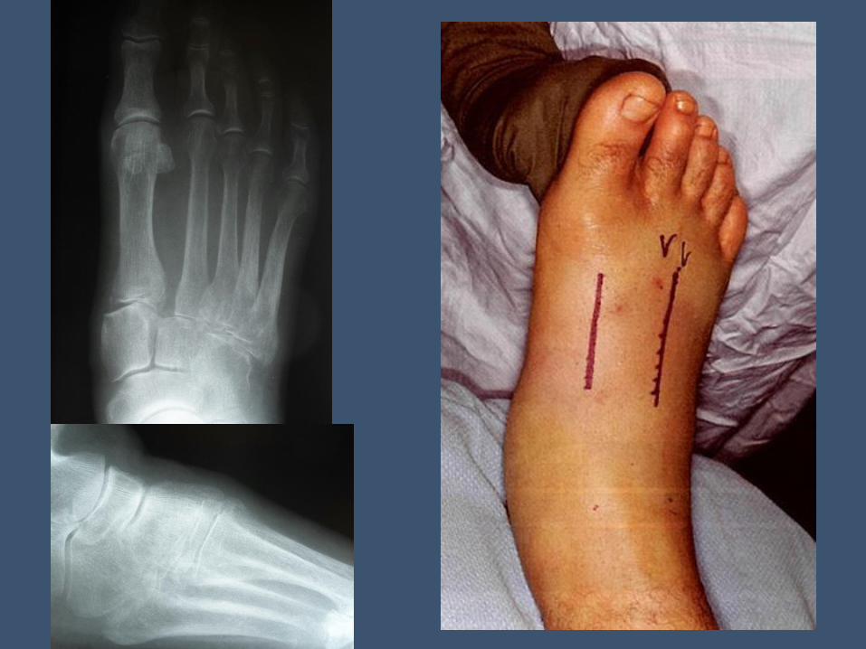
Always take a picture of the
other foot!!
Do CT and MRI Help?
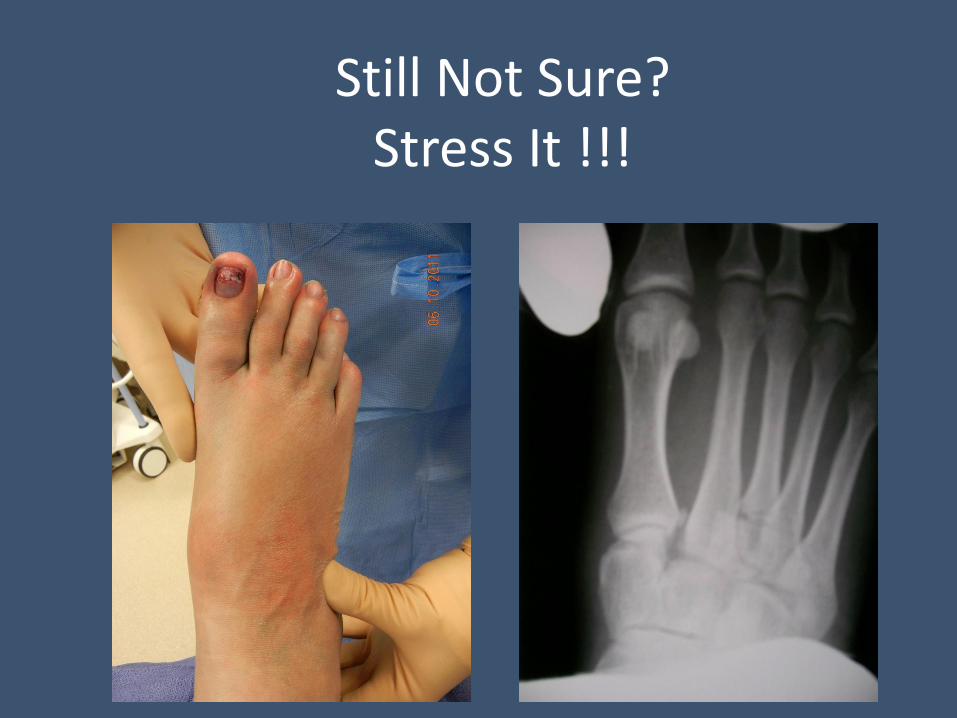
Still Not Sure? Stress It !!!

Small Shift Equals Big Deformity

Clinical Questions for Treatment of TMTJ Dislocation
Tradition vs. Evidence
• Reduction vs. No Reduction
• Closed vs. Open
• Wires vs. Screws
• ORIF vs. Fusion
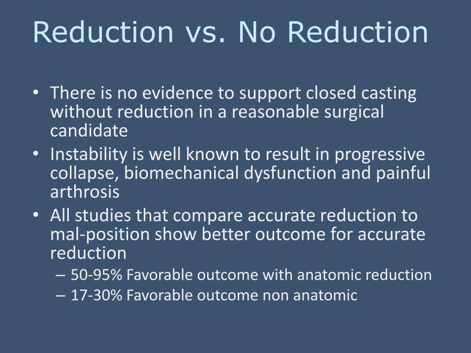
Reduction vs. No Reduction
• There is no evidence to support closed casting without reduction in a reasonable surgical candidate
• Instability is well known to result in progressive collapse, biomechanical dysfunction and painful arthrosis
• All studies that compare accurate reduction to mal-position show better outcome for accurate reduction – 50-95% Favorable outcome with anatomic reduction – 17-30% Favorable outcome non anatomic

Positional changes from unstable
Lisfranc’s causes severe progressive
functional abnormalities

Biomechanics
• Elevated first ray • Reduced first ray
weight-bearing
• Hallux Limitus
• Midfoot over-pronation • Forefoot abduction
• Internal leg rotation
• STJ, MTJ and ankle compensatory changes
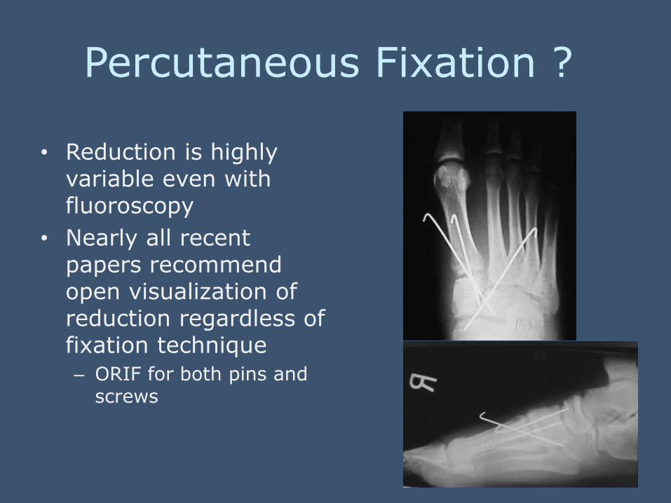
Percutaneous Fixation ?
• Reduction is highly variable even with fluoroscopy
• Nearly all recent papers recommend open visualization of reduction regardless of fixation technique
– ORIF for both pins and screws

Inadequate Reduction With Closed Pinning
• Wilson 1972
• Arntz 1988
• Myerson 1989
– ORIF gave superior result to closed pinning
– 100% of patients had DJD of tarsal-metatarsal joints regardless of reduction

ORIF Kuo 2000, Coetzee 2008
• Incomplete stability after ORIF
• Late collapse
• High incidence of DJD
• High rate of revision surgery

Late Instability and DJD

Arthrodesis vs. ORIF Coetzee JBJS 2006
• ORIF – 15 of 20 had loss of correction, arthrosis and or
pain – 16 of 20 needed hardware removal (2nd Surgery) – 7 needed revision within 4 year period (3rd
Surgery) – AOFAS score 57.1
• Primary Arthrodesis of 1-2-3 TMTJ – AOFAS score 86.9 – No revision – Rare 2nd surgery (hardware removal)

Primary Ligamentous Injury Coetzee 2008
• Ligamentous injury – Dorsal Instability
• Much more stable • ORIF acceptable
– Medial second metatarsal base fracture – ORIF
– Plantar Instability • Less reliable • Arthrodesis
– Pure ligamentous instability • Arthrodesis
ORIF vs. Primary Arthrodesis for Lisfranc Injuries
Henning FAI 2009
• Prospective, randomized study of patients presenting with Lisfranc’s fracture dislocation
– ORIF vs. primary arthrodesis 1-2-3 TMTJ
– Follow-up to one year
– Had 94% fusion rate in the arthrodesis group
– Found similar short term functional results between groups however showed that Arthrodesis resulted in a statistically significant decrease in the number of follow-up surgeries

ORIF vs Complete TMTJ Arthrodesis Mulier 2002
• ORIF did better functionally than complete TMTJ arthrodesis
• 94% of ORIF had arthrosis at 36 months
• Fuse only 1 – 2 - 3
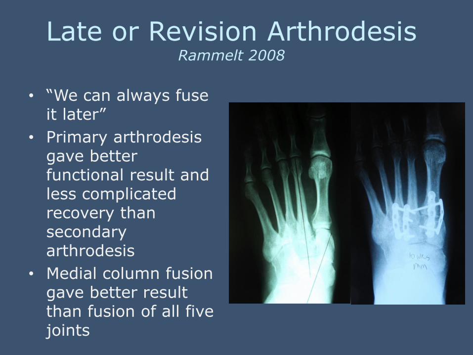
Late or Revision Arthrodesis Rammelt 2008
• “We can always fuse it later”
• Primary arthrodesis gave better functional result and less complicated recovery than secondary arthrodesis
• Medial column fusion gave better result than fusion of all five joints

ORIF
• High rate of second and third surgeries
• High rate of DJD in first 5 years
• No post operative advantages
• Progressive deformity and adjacent joint damage
• Not a more conservative operation
Arthrodesis
• One and done
• No more complicated than ORIF
• Better patient function
• Excellent union rates
• Superior to late arthrodesis
• Stability is the key to the TMTJ
• Most conservative option

• It is well documented closed pinning and ORIF have high failure rates and high revision rates
• We have to consider adjacent joint damage from years of walking on a progressively deformed foot
• Lisfranc’s joint is a stability segment not a mobility segment
• There is no good functional or surgical argument against fusion

Surgical Technique
• Incision – 2 or 3 incisions
• Bone preparation – Remove all subchondral bone – Correct the deformity
• Fixation – Has to be contourable to the anatomy – Low profile
• Post operative course – NWB for 4-6 weeks


Control The Variables for Consistent Healing
• Vascular Preservation • Periosteal, Endosteal, Surrounding soft tissues
• Minimal soft tissue disruption
• NO tissue plane separation
• Limit heat generated with saws and burrs
• Mechanical stability • Directly affects cellular activity
• Inappropriate motion interferes with healing
• Absolute rigidity may also slow healing
• Stable, non rigid fixation with controlled micro-motion can stimulate healing

Bridge Fixation “Internal Fixator”
• Plate and screws form a “single beam” load bearing construct
• Multi-planar stability
• Bone segments are stable but not compressed therefore low gap strain
• Controlled micro motion based on the mechanical characteristics of the fixator • Mono vs bi-planar construct
• Stiff vs flexible plates
• Number of screws per plate
• Stimulates callus healing and the callus strengthens the fixation construct
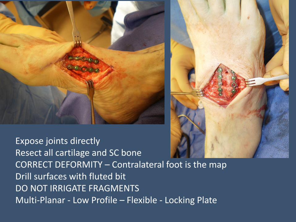
Expose joints directly Resect all cartilage and SC bone CORRECT DEFORMITY – Contralateral foot is the map Drill surfaces with fluted bit DO NOT IRRIGATE FRAGMENTS Multi-Planar - Low Profile – Flexible - Locking Plate

8 weeks

TMTJ Fusion is Excellent for Deformity Correction


Key Points
• TMTJ stability and alignment is important for function
• Arthrodesis is the most stable option for repair with the lowest revision rate
• There is no good argument from a surgical or functional standpoint for “Joint Preservation” in the TMTJ
• Locking plates provide a stable, load bearing construct that tolerates external stresses better than traditional AO techniques
• Must prioritize deformity correction in both acute and late reconstructions