the absence of ige antibody-mediated augmentation of immune responses in cd23-deficient mice
TRANSCRIPT
Proc. Natd. Acad. Sci. USAVol. 91, pp. 6835-6839, July 1994Immunology
The absence of IgE antibody-mediated augmentation of immuneresponses in CD23-deficient mice
(ow-ffty gE /rept/g
HIROSHI FUJIWARA*t1, HITOSHI KIKUTANIt§, SACHIKO SUEMATSU*, TETSUJI NAKAtt, KANJI YOSHIDAt,KENJI YOSHIDAt, TAKASHI TANAKA*tt, MASAKI SUEMURAt, NAOKI MATSUMOTO¶, SOMEI KoJIMA¶,TADAMITSU KISHIMOTO*, AND NOBUAKI YOSHIDA**Rsearch Institute, Osaka Medical Center for Maternal and Child Health, Izumi, Osaka 590-02, Japan; tInstitute for Molecular and Cellular Biology, OsakaUniversity, Suita, Osaka 565, Japan; IDepartment of Parasitology, Institute of Medical Science, University of Tokyo, Minato-ku, Tokyo 108, Japan; and*Department of Medicine III, Osaka University Medical School, Suita, Osaka 565, Japan
Contributed by Tadamitsu Kishimoto, March 10, 1994
ABSTRACT The CD23 antigen, a low-affinity receptor forIgE (FceRfl), is a type I membrane-bound glycoproteinexprs on various cells, particularly mature B cells. Anumber of fnts have been ascribed to CD23, includingspecific regulation of IgE production, IgE-medlated yotoxic-Ity and release of mediators, IgE-dependent anten fang,promotion of B-cell growth, prevention of genal center Bcells from apoptoss, p ife of myelold precursors, andmaturation of early thymocytes. It is not clear whether theseactivities represent invivo fons. To explore us vivo func-tim of CD23, we have produced CD23-deflcient mice. Thesemice displayed normal lymphocyte differentiation and couldmount normal antibody iespuding gE responsesupon Immnization with T-dependent antgs and infectionWith Npeuoslrongrus brwsensis. Germinal center formationafter immunization and in vitro proliferative response B cellswere not afected in mutant mice. However, antigen-pecificIgE-medlated enhancement of antibody responses was severelyhpiced
CD23 is a type II membrane-bound glycoprotein expressedmainly on mature B cells (1, 2). The extracellular carboxyl-terminal of this molecule is released from cells as solubleCD23 (3). This molecule has been originally identified as alow-affinity Fc receptor for IgE (FcERII) expressed on hu-man peripheral blood B cells and some lymphoblastoid celllines by Spiegelberg and his colleagues (4, 5). Independently,CD23 has been described as a B-cell activation antigenexpressed on Epstein-Barr virus (EBV)-transformed B cells(6, 7). Molecular cloning of FceRII has demonstrated theidentity between FceRII and CD23 (2, 8).The IgE binding activity ofCD23 implies its role in effector
functions of IgE and regulation of IgE synthesis (9). In fact,CD23 on monocytes, macrophages, and eosinophils has beenreported to be involved in IgE-dependent cytotoxicity andmediator release (10, 11). Some anti-CD23 antibodies andsoluble CD23 have been shown to modify in vitro IgEsecretion of human B cells (12-14). Recently, it has beenreported that an interaction of CD23 with CD21, a moleculeknown as an EBV receptor or a complement receptor 2, isinvolved in IgE production in humans and rats (15, 16).However, a role of CD23 in regulation of IgE production isstill controversial since most of these functions cannot bereproduced in the mouse system (17).A tight correlation between CD23 expression and EBV
transformation ofB cells suggests a possible role of CD23 inthe transformation and growth of B cells. Affinity-purifiedsoluble CD23 promotes the proliferation of both activated
normal B cells and EBV-transformed B cells (18). Gordon etal. (19) have reported that purified native and recombinantsoluble CD23 induced the proliferation of activated human Bcells. However, experiments by other groups, including ours,have failed to reproduce this activity in human and mousesystems (17, 20).
In addition, CD23 has been shown to induce the prolifer-ation ofhuman myeloid precursors (21) and the maturation ofhuman early thymocytes (22). A question may be raisedwhether all of these activities really represent in vivo func-tions of CD23.To reevaluate the functions of CD23 in vivo, we have
produced CD23-deficient mice. These mice displayed normallymphocyte differentiation and could mount normal antibodyresponses; however, antigen-specific IgE-mediated enhance-ment of antibody responses was severely impaired.
MATERIAL AND METHODSGene Targeting. The targeting vector contained a 4.1-kb
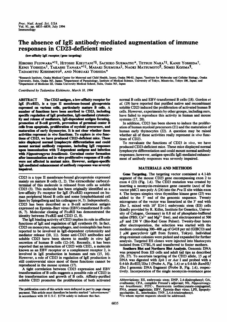
segment of the mouse CD23 gene encompassing exon 2 toexon 4 (23) (Fig. 1A). The CD23 mutation was created byinserting a neomycin-resistance gene cassette (neo) of thevector pMC1 neo-poly A (24) into the Pvu II site within exon4. The herpes simplex virus thymidine kinase gene (25) wasadded to the 5' end of the genomic sequence. Twentymicrograms of the vector was linearized at the 3' end withXho I, mixed with 107 E14-1 embryonic stem (ES) cells(kindly provided by R. Kdhn, Institute for Genetics, Univer-sity of Cologne, Germany) in 0.8 ml of phosphate-bufferedsaline (PBS; Ca2+ and Mg2+ free), and electroporated at 500juF and 230 V (Bio-Rad Gene Pulser). Twenty-four hoursafter electroporation, the selection was started with themedium containing 300-400 pAg of G418 per ml (GIBCO) and2 pM gancyclovir (gift from Syntex, Tokyo). Individualdrug-resistant colonies were picked and expanded for furtheranalysis. Targeted ES clones were injected into blastocystsisolated from C57BL/6 and transferred to foster mothers.
Southern Blot and Northern Blot Analysis. Genomic DNAwas prepared from ES cells and adult tail tips as described(26, 27). To ascertain targeting of the CD23 allele, 15 ptg ofDNA was digested with Sph I or Aat I and probed with a0.4-kb BstEII/Xba I (Probe A, Fig. 1A) or a 0.6-kb BamHI/Xba I genomic DNA fragment (Probe B, Fig. 1A), respec-tively. Incorporation of the single neomycin-resistance gene
Abbreviations: ES, embryonic stem; DNP, 2,4-dinitrophenyl; OA,ovalbumin; CFA, complete Freund's adjuvant; Nb, Nippostrongy-rus brasiliensis; FITC-, fluorescein isothiocyanate-conjugated;PNA, peanut agglutinin; EBV, Epstein-Barr virus; LPS, lipopoly-saccharide; IL, interleukin; TNP, 2,4,6-trinitrophenyl.§To whom reprint requests should be addressed.
6835
Tle publication costs of this article were defrayed in part by page chargepayment. This article must therefore be hereby marked "advertisement"in accordance with 18 U.S.C. §1734 solely to indicate this fact.
6836 Immunology: Fujiwara et al.
was further confirmed using a neo probe. For analysis ofCD23 mRNA, total RNA was prepared from splenic B cellsstimulated with lipopolysaccharide (LPS; 10 ug/ml) in thepresence of interleukin 4 (IL-4; 60 units/ml, Genzyme) for 36hr as described (28), electrophoresed, transferred to Hy-bond-N (Amersham), and hybridized with a CD23 cDNAfragment or a jactin probe.
Immunization and ELISAs. Seven-week-old mice wereimmunized i.p. with 50 Ag of 2,4-dinitrophenyl ovalbumin(DNP-OA) in complete Freund's adjuvant (CFA) to induceIgM, IgG, and IgA, or in alum to induce IgE, at day 0 andgiven booster immunizations on day 14. DNP-specific anti-body responses after immunization were measured byELISA with DNP-bovine serum albumin-coated plates ex-cept for the IgE class. Alkaline phosphatase-labeled isotype-specific antibodies (Southern Biotechnology Associates)were used for detection. For the DNP-specific IgE antibodyassay, plates were coated with anti-IgE monoclonal antibody77, diluted sample and standard antibody SPE-7 (29) (anti-DNP monoclonal IgE antibody) (both antibodies were kindlyprovided by Y. Hattori, Kowa Research Institute, Tsukuba,Japan) were added, and bound DNP-specific IgE was de-tected by biotinylated DNP-human serum albumin followedby avidin-horseradish peroxidase.
RESULTSCD23 Gene Targeting. The CD23 mutation was created by
inserting a neomycin-resistance gene cassette (neo) withinexon 4 (Fig. 1A). The ES cells were transfected with thelinearized vector by electroporation. After selection withG418 and gancyclovir, six of the doubly resistant clones werefound to carry the CD23 mutation on a single allele, asexamined by PCR and Southern blot analysis. A singleintegration event was further confirmed in these clones bySouthern blot analysis using the neo probe (data not shown).Chimeric mice derived from three of six ES clones transmit-ted the mutated genotype to their offspring. Mice heterozy-gous for the mutated allele were then intercrossed to producehomozygous mice. Southern blot analysis of the resultingoffspring is shown in Fig. 1B. Hybridization with probe Ayielded a 2.7-kb Sph I fragment for the mutated allele and a5.5-kb fragment for the wild-type allele. In addition, hybrid-ization with probe B ofAat I-digested DNA displayed 4.5-kband 7.3-kb bands for the mutated and wild-type alleles,respectively. To determine the CD23 mRNA expression,RNA from spleen cells stimulated with LPS and IL-4 wasprepared and analyzed by Northern blotting. As shown inFig. 1C, a 2.2-kb CD23 transcript was observed in RNA fromwild-type mice, whereas no hybridizing RNA band wasdetectable from CD23-targeted mice, confirming the func-tional disruption of the CD23 gene. In the following experi-ments, mice homozygous for the mutated allele or the wild-type allele in the background of(129/Ola x CS7BL/6)F2 wereexamined. Three independent mutant lines displayed indis-tinguishable phenotypes.
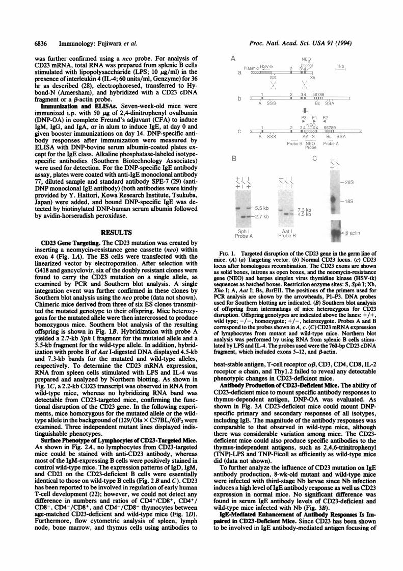
Surface Phenotype of Lymphocytes of CD23-Targeted Mice.As shown in Fig. 2A, no lymphocytes from CD23-targetedmice could be stained with anti-CD23 antibody, whereasmost of the IgM-expressing B cells were positively stained incontrol wild-type mice. The expression patterns of IgD, IgM,and CD21 on the CD23-deficient B cells were essentiallyidentical to those on wild-type B cells (Fig. 2 B and C). CD23has been reported to be involved in regulation of early humanT-cell development (22); however, we could not detect anydifference in numbers and ratios of CD4+/CD8+, CD4+/CD8-, CD4-/CD8+, and CD4-/CD8- thymocytes betweenage-matched CD23-deficient and wild-type mice (Fig. ID).Furthermore, flow cytometric analysis of spleen, lymphnode, bone marrow, and thymus cells using antibodies to
:I
I El
i II E I I[fil
ai
:0 w --r
1.:.1I 0 I
11111
B
_ 55- fi
I
Sph 'Probe .A
FIG. 1. Targeted disruption of the CD23 gene in the germ line ofmice. (A) (a) Targeting vector. (b) Normal CD23 locus. (c) CD23locus after homologous recombination. The CD23 exons are shownas solid boxes, introns as open boxes, and the neomycin-resistancegene (NEO) and herpes simplex virus thymidine kinase (HSV-tk)sequences as hatched boxes. Restriction enzyme sites: S, Sph I; Xh,Xho I; A, Aat I; Bs, BstEII. The positions of the primers used forPCR analysis are shown by the arrowheads, P1-P3. DNA probesused for Southern blotting are indicated. (B) Southern blot analysisof offspring from intermatings of mice heterozygous for CD23disruption. Offspring genotypes are indicated above the lanes: +/+,wild type; -/-, homozygote; +/-, heterozygote. Probes A and Bcorrespond to the probes shown inA, c. (C) CD23 mRNA expressionof lymphocytes from mutant and wild-type mice. Northern blotanalysis was performed by using RNA from splenic B cells stimu-lated by LPS and IL-4. The probes used were the 760-bp CD23 cDNAfragment, which included exons 5-12, and 3-actin.
heat-stable antigen, T-cell receptor aB, CD3, CD4, CD8, IL-2receptor a chain, and Thyl.2 failed to reveal any detectablephenotypic changes in CD23-deficient mice.
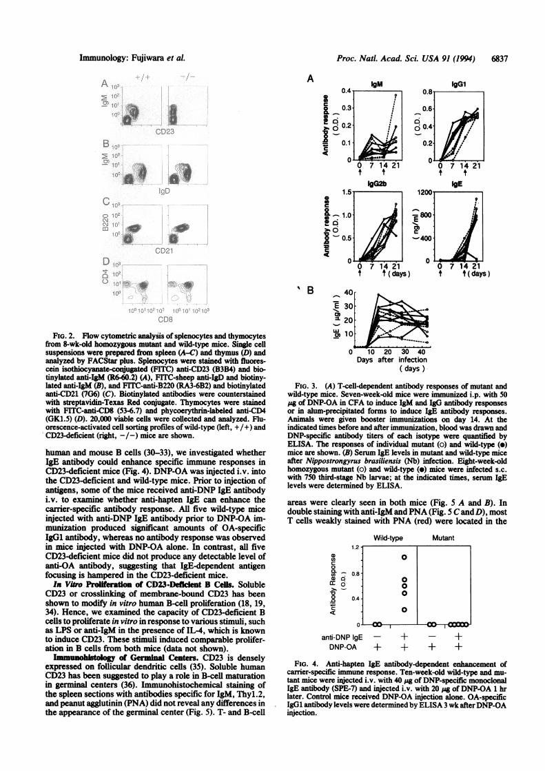
Antibody Production of CD23-Deficient Mice. The ability ofCD23-deficient mice to mount specific antibody responses tothymus-dependent antigen, DNP-OA was evaluated. Asshown in Fig. 3A CD23-deficient mice could mount DNP-specific primary and secondary responses of all isotypes,including IgE. The magnitude of the antibody responses wascomparable to that observed in wild-type mice, althoughthere was considerable variation among mice. The CD23-deficient mice could also produce specific antibodies to thethymus-independent antigens, such as 2,4,6-trinitrophenyl(TNP)-LPS and TNP-Ficoll as efficiently as wild-type micedid (data not shown).To further analyze the influence of CD23 mutation on IgE
antibody production, 8-wk-old mutant and wild-type micewere infected with third-stage Nb larvae since Nb infectioninduces a high level ofIgE antibody response as well as CD23expression in normal mice. No significant difference wasfound in serum IgE antibody levels of CD23-deficient andwild-type mice infected with Nb (Fig. 3B).
IgE-Mediated Enhancement of Antibody Responses Is Im-paired in CD23-Deficient Mice. Since CD23 has been shownto be involved in IgE antibody-mediated antigen focusing of
Proc. Natl. Acad Sci. USA 91 (1994)
Proc. Natd. Acad. Sci. USA 91 (1994) 6837
A
a~1C"..U
10. BCD23
B ro10-
J. *: i
ILD
mD8.D
*_
[gm IgGI
0.40 0.8
0.1 0.2AI1
IIG~a
1.57
&@1.0C
0- 0.5C
0 1
CD21
r'n 9
' B
0DB
FiG. 2. Flow cytometric analysis of splenocytes and thymocytesfrom 8-wk-old homozygous mutant and wild-type mice. Single cellsuspensions were prepared from spleen (A-C) and thymus (D) andanalyzed by FACStar plus. Splenocytes were stained with fluores-cein isothiocyanate-conjugated (FfTC) anti-CD23 (B3B4) and bio-tinylated anti-IgM (R6-60.2) (A), FITC-sheep anti-IgD and biotiny-lated anti-IgM (B), and FITC-anti-B220 (RA3-6B2) and biotinylatedanti-CD21 (7G6) (C). Biotinylated antibodies were counterstainedwith streptavidin-Texas Red conjugate. Thymocytes were stainedwith FITC-anti-CD8 (53-6.7) and phycoerythrin-labeled anti-CD4(GK1.5) (D). 20,000 viable cells were collected and analyzed. Flu-orescence-activated cell sorting profiles of wild-type (left, +/+) andCD23-deficient (right, -/-) mice are shown.
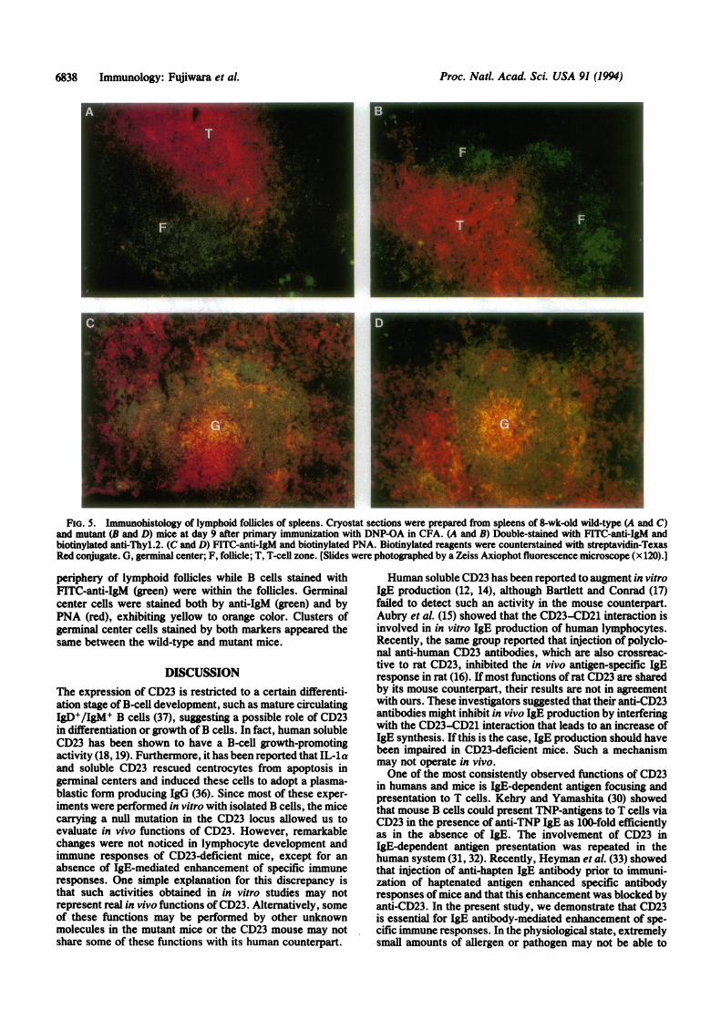
human and mouse B cells (30-33), we investigated whetherIgE antibody could enhance specific immune responses inCD23-deficient mice (Fig. 4). DNP-OA was injected i.v. intothe CD23-deficient and wild-type mice. Prior to injection ofantigens, some of the mice received anti-DNP IgE antibodyi.v. to examine whether anti-hapten IgE can enhance thecarrier-specific antibody response. All five wild-type miceinjected with anti-DNP IgE antibody prior to DNP-OA im-munization produced significant amounts of OA-specificIgG1 antibody, whereas no antibody response was observedin mice injected with DNP-OA alone. In contrast, all fiveCD23-deficient mice did not produce any detectable level ofanti-OA antibody, suggesting that IgE-dependent antigenfocusing is hampered in the CD23-deficient mice.In Vitr Proliferaton f CD23-Deflent B Celis. Soluble
CD23 or crosslinking of membrane-bound CD23 has beenshown to modify in vitro human B-cell proliferation (18, 19,34). Hence, we examined the capacity of CD23-deficient Bcells to proliferate in vitro in response to various stimuli, suchas LPS or anti-IgM in the presence of IL-4, which is knownto induce CD23. These stimuli induced comparable prolifer-ation in B cells from both mice (data not shown).Immunohlstlogy of Germnal Centers. CD23 is densely
expressed on follicular dendritic cells (35). Soluble humanCD23 has been suggested to play a role in B-cell maturationin germinal centers (36). Immunohistochemical staining ofthe spleen sections with antibodies specific for IgM, Thyl.2,and peanut agglutinin (PNA) did not reveal any differences inthe appearance of the germinal center (Fig. 5). T- and B-cell
IqG2b1200
~-400
0 7 14 21t t (days)
40-
302- 20'I-,
wm 10
IgE
99
7 14 21t t(days)
.-
0 10 20 30 40Days after infection
( days )
FIG. 3. (A) T-cell-dependent antibody responses of mutant andwild-type mice. Seven-week-old mice were imuniz i.p. with 50pg of DNP-OA in CFA to induce IgM and IgO antibody responsesor in alum-precipitated forms to induce IgE antibody responses.Animals were given booster immunizations on day 14. At theindicated times before and after immunization, blood was drawn andDNP-specific antibody titers of each isotype were quantified byELISA. The responses of individual mutant (o) and wild-type (e)mice are shown. (B) Serum IgE levels in mutant and wild-type miceafter Nippostrongyrus brasiliensis (Nb) infection. Eight-week-oldhomozygous mutant (o) and wild-type (e) mice were infected s.c.with 750 third-stage Nb larvae; at the indicated times, serum IgElevels were determined by ELISA.
areas were clearly seen in both mice (Fig. 5 A and B). Indouble staining with anti-IgM andPNA (Fig. 5 C andD), mostT cells weakly stained with PNA (red) were located in the
Wild-type1.2
a)CD00.Cco 0.8-
o
0.4.._c
o0
0
000
0
anti-DNP IgEDNP-OA +
Mutant
+ - ++ + +
FiG. 4. Anti-hapten IgE antibody-dependent enhancement ofcarrier-specific immune response. Ten-week-old wild-type and mu-tant mice were injected i.v. with 40 pg of DNP-specific monoclonalIgE antibody (SPE-7) and injected i.v. with 20 pg of DNP-OA 1 hrlater. Control mice received DNP-OA injection alone. OA-specificIgG1 antibody levels were determined by ELISA 3 wk afterDNP-OAinjection.
Immunology: Fujiwara et A
04
I
6838 Immunology: Fujiwara et al.
FiG. 5. Immunohistology of lymphoid follicles of spleens. Cryostat sections were prepared from spleens of 8-wk-old wild-type (A and C)and mutant (B and D) mice at day 9 after primary immunization with DNP-OA in CFA. (A and B) Double-stained with FITC-anti-IgM andbiotinylated anti-Thyl.2. (C and D) FITC-anti-IgM and biotinylated PNA. Biotinylated reagents were counterstained with streptavidin-TexasRed conjugate. G, germinal center; F, follicle; T, T-celi zone. (Slides were photographed by a Zeiss Axiophot fluorescence microscope (x 120).]
periphery of lymphoid follicles while B cells stained withFITC-anti-IgM (green) were within the follicles. Germinalcenter cells were stained both by anti-IgM (green) and byPNA (red), exhibiting yellow to orange color. Clusters ofgerminal center cells stained by both markers appeared thesame between the wild-type and mutant mice.
DISCUSSIONThe expression of CD23 is restricted to a certain differenti-ation stage of B-cell development, such as mature circulatingIgD+/IgM+ B cells (37), suggesting a possible role of CD23in differentiation or growth ofB cells. In fact, human solubleCD23 has been shown to have a B-cell growth-promotingactivity (18, 19). Furthermore, it has been reported that IL-laand soluble CD23 rescued centrocytes from apoptosis ingerminal centers and induced these cells to adopt a plasma-blastic form producing IgG (36). Since most of these exper-iments were performed in vitro with isolated B cells, the micecarrying a null mutation in the CD23 locus allowed us toevaluate in vivo functions of CD23. However, remarkablechanges were not noticed in lymphocyte development andimmune responses of CD23-deficient mice, except for anabsence of IgE-mediated enhancement of specific immuneresponses. One simple explanation for this discrepancy isthat such activities obtained in in vitro studies may notrepresent real in vivo functions of CD23. Alternatively, someof these functions may be performed by other unknownmolecules in the mutant mice or the CD23 mouse may notshare some of these functions with its human counterpart.
Human soluble CD23 has been reported to augment in vitroIgE production (12, 14), although Bartlett and Conrad (17)failed to detect such an activity in the mouse counterpart.Aubry et al. (15) showed that the CD23-CD21 interaction isinvolved in in vitro IgE production of human lymphocytes.Recently, the same group reported that injection of polyclo-nal anti-human CD23 antibodies, which are also crossreac-tive to rat CD23, inhibited the in vivo antigen-specific IgEresponse in rat (16). If most functions of rat CD23 are sharedby its mouse counterpart, their results are not in agreementwith ours. These investigators suggested that their anti-CD23antibodies might inhibit in vivo IgE production by interferingwith the CD23-CD21 interaction that leads to an increase ofIgE synthesis. If this is the case, IgE production should havebeen impaired in CD23-deficient mice. Such a mechanismmay not operate in vivo.One of the most consistently observed functions of CD23
in humans and mice is IgE-dependent antigen focusing andpresentation to T cells. Kehry and Yamashita (30) showedthat mouse B cells could present TNP-antigens to T cells viaCD23 in the presence of anti-TNP IgE as 100-fold efficientlyas in the absence of IgE. The involvement of CD23 inIgE-dependent antigen presentation was repeated in thehuman system (31, 32). Recently, Heyman et al. (33) showedthat injection of anti-hapten IgE antibody prior to immuni-zation of haptenated antigen enhanced specific antibodyresponses of mice and that this enhancement was blocked byanti-CD23. In the present study, we demonstrate that CD23is essential for IgE antibody-mediated enhancement of spe-cific immune responses. In the physiological state, extremelysmall amounts of allergen or pathogen may not be able to
Proc. Nad. Acad Sci. USA 91 (1994)
Proc. Nadl. Acad. Sci. USA 91 (1994) 6839
induce a humoral immune response. In the presence of highlocal concentrations of specific IgE antibodies in the airway,gastrointestinal tract, or skin, antigens can be captured orfocused by CD23 on antigen-presenting cells and efficientlypresented to T cells, resulting in augmentation of specificimmune responses. In the allergic status, IL4 derived fromTh2 helper cells may induce increases ofCD23 expression aswell as IgE production, which may exacerbate the diseasesby further augmenting T-cell responses and IgE production.
We thank R. Kuhn for ES E14-1 cells, Y. S. Choi for criticalreading of the manuscript, D. H. Conrad, T. Kinoshita, and Y.Hattori for antibodies, S. Akira, T. Yasui, and K. Sugiyama fortechnical advice, M. Yoshikane for technical assistance, and K.Kubota and K. Kanda for excellent secretarial assistance. Thisresearch was supported by research grants from the Ministry ofEducation, Science, and Culture, Japan and the Human FrontierScience Program.
1. Kikutani, H., Inui, S., Sato, R., Barsumian, E. L., Owaki, H.,Yamasaki, K., Kaisho, T., Uchibayashi, N., Hardy, R. R.,Hirano, T., Tsunasawa, S., Sakiyama, F., Suemura, M. &Kishimoto, T. (1986) Cell 47, 657-665.
2. Yukawa, K., Kikutani, H., Owaki, H., Yamasaki, K., Yokota,A., Nakamura, H., Barsumian, E. L., Hardy, R. R., Suemura,M. & Kishimoto, T. (1987) J. Immunol. 138, 2576-2580.
3. Latellier, M., Nakjima, T., Pulido-Cejudo, G., Hofstetter, H.& Delespesse, G. (1990) J. Exp. Med. 172, 693-700.
4. Lawrence, D. A., Weigle, W. 0. & Spiegelberg, H. L. (1975)J. Clin. Invest. 55, 368-387.
5. Gonzalez-Molina, A. & Spiegelberg, H. L. (1976) J. Immunol.117, 1838-1845.
6. Kintner, C. & Sugden, B. (1981) Nature (London) 294, 458-460.
7. Thorley-Lawson, D. A., Nadler, L. M., Bhan, A. K. &Schooley, R. T. (1985) J. Immunol. 134, 3007-3012.
8. Yokota, A., Kikutani, H., Tanaka, T., Sato, R., Barsumian,E. L., Suemura, M. & Kishimoto, T. (1988) Cell 55, 611-618.
9. Sutton, B. J. & Gould, H. J. (1993) Nature (London) 366,421-428.
10. Capron, A., Dessaint, J. P., Capron, M., Joseph, M., Ameisen,J. C. & Tonnel, A. B. (1986) Immunol. Today 7, 15-18.
11. Borish, L., Mascali, J. J. & Rosenwasser, L. J. (1991) J.Immunol. 146, 63-67.
12. Pene, J., Rousset, F., Briere, F., Chretien, I., Wideman, J.,Bonnefoy, J. Y. & De Vries, J. E. (1988) Eur. J. Immunol. 18,929-935.
13. Bonnefoy, J.-Y., Shields, J. & Mermod, J. J. (1990) Eur. J.Immunol. 20, 139-144.
14. Sarfati, M. & Delespesse, G. (1998) J. Immunol. 141, 2195-2199.
15. Aubry, J.-P., Pochon, S., Graber, P., Jansen, K. U. & Bon-nefoy, J.-Y. (1992) Nature (London) 358, 505-507.
16. Flores-Romo, L., Shields, J., Humbert, Y., Graber, P., Aubry,J.-P., Gauchat, J.-F., Ayala, G., Allet, B., Chavez, M., Bazin,H., Capron, M. & Bonnefoy, J.-Y. (1993) Science 261, 1038-1041.
17. Bartlett, W. C. & Conrad, D. H. (1992) Res. Immunol. 143,431-436.
18. Swendeman, S. & Thorley-Lawson, D. A. (1987) EMBO J. 6,1637-1642.
19. Gordon, J., Cairns, J. A., Millsum, M. J., Gillis, S. & Guy,G. R. (1988) J. Immunol. 18, 1561-1565.
20. Uchibayashi, N., Kikutani, H., Barsumian, E. L., Hauptmann,R., Schneider, F. J., Schwendenwein, R., Sommergruber, W.,Spevak, W., Maurer-Fogy, I., Suemura, M. & Kishimoto, T.(1989) J. Immunol. 142, 3901-3908.
21. Mossalayi, M. D., Arock, M., Bertho, J. M., Blanc, C., Dal-loul, A. H., Hofstetter, H., Sarfati, M., Delespesse, G. &Debre, P. (1990) Blood 75, 1924-1927.
22. Mossalayi, M. D., Lecron, J. C., Dalloul, A. H., Sarfati, M.,Bertho, J. M., Hofstetter, H., Delespesse, G. & Debre, P.(1990) J. Exp. Med. 171, 959-964.
23. Richards, M. L., Katz, D. H. & Liu, F.-T. (1991) J. Immunol.147, 1067-1074.
24. Thomas, K. R. & Capecchi, M. R. (1987) Cell 51, 503-512.25. Mansour, S. L., Thomas, K. R. & Capecchi, M. R. (1988)
Nature (London) 336, 348-352.26. McMahon, A. P. & Bradley, A. (1990) Cell 62, 1073-1085.27. Laird, P. W., Zijderveld, A., Linders, K., Rudnicki, M. A.,
Jaenisch, R. & Berns, A. (1991) Nucleic Acids Res. 19, 4293.28. Conrad, D. H., Keegan, A. D., Kalli, K. R., Dusen, R. V.,
Rao, M. & Levine, A. D. (1988) J. Immunol. 141, 1091-1097.29. Eshhar, Z., Ofarim, M. & Waks, T. (1980) J. Immunol. 124,
775-780.30. Kehry, M. R. & Yamashita, L. C. (1989) Proc. Natl. Acad. Sci.
USA 86, 7556-7560.31. Pirron, U., Schlunck, T., Prinz, J. C. & Rieber, E. P. (1990)
Eur. J. Immunol. 20, 1547-1551.32. Mudde, G. C., Hansel, T. T., ReUsen, F. C. V., Osterhoff,
B. F. & Brujnzeel-Koomen, C. A. F. M. (1990) Immunol.Today 11, 440-443.
33. Heyman, B., Tianmin, L. & Gustavsson, S. (1993) Eur. J.Immunol. 23, 1739-1742.
34. Luo, H., Hofstetter, H., Banchereau, J. & Delespesse, G.(1991) J. Immunol. 146, 2122-2129.
35. Maeda, K., Burton, G. F., Padgett, D. A., Conrad, D. H.,Huff, T. F., Masuda, A., Szakal, A. K. & Tew, J. G. (1992) J.Immunol. 148, 2340-2347.
36. Liu, Y.-J., Cairns, J. A., Holder, M. J., Abbot, S. D., Jansen,K. U., Bonnefoy, J.-Y., Gordon, J. & MacLennan, I. C. M.(1991) Eur. J. Immunol. 21, 1107-1114.
37. Kikutani, H., Suemura, M., Owaki, H., Nakamura, H., Sato,R., Yamasaki, K., Barsumian, E. L., Hardy, R. R. & Kishi-moto, T. (1986) J. Exp. Med. 164, 1455-1469.
Immunology: Fujiwara et A