the dynamic structure of α-synuclein multimers
TRANSCRIPT
The Dynamic Structure of α‑Synuclein MultimersThomas Gurry,†,⊥ Orly Ullman,‡,⊥ Charles K. Fisher,§ Iva Perovic,∥ Thomas Pochapsky,⊗
and Collin M. Stultz*,†,§,#
†Computational and Systems Biology Initiative, Massachusetts Institute of Technology, Cambridge, Massachusetts 02139-4307,United States‡Department of Chemistry, Massachusetts Institute of Technology, Cambridge, Massachusetts 02139-4307, United States§Committee on Higher Degrees in Biophysics, Harvard University, Cambridge, Massachusetts 02139, United States∥Department of Chemistry and Rosenstiel Basic Medical Sciences Research Center, Brandeis University, Waltham, Massachusetts02454, United States⊗Department of Chemistry and Biochemistry and Rosenstiel Basic Medical Sciences Research Center, Brandeis University, Waltham,Massachusetts 02454, United States#Harvard−MIT Division of Health Sciences and Technology, Department of Electrical Engineering and Computer Science, ResearchLaboratory of Electronics & The Institute of Medical Engineering and Science, Massachusetts Institute of Technology, Cambridge,Massachusetts 02139-4307, United States
*S Supporting Information
ABSTRACT: α-Synuclein, a protein that forms ordered aggregates in the brains of patients withParkinson’s disease, is intrinsically disordered in the monomeric state. Several studies, however,suggest that it can form soluble multimers in vivo that have significant secondary structure content.A number of studies demonstrate that α-synuclein can form β-strand-rich oligomers that areneurotoxic, and recent observations argue for the existence of soluble helical tetrameric species incellulo that do not form toxic aggregates. To gain further insight into the different types ofmultimeric states that this protein can adopt, we generated an ensemble for an α-synucleinconstruct that contains a 10-residue N-terminal extension, which forms multimers when isolatedfrom Escherichia coli. Data from NMR chemical shifts and residual dipolar couplings were used toguide the construction of the ensemble. Our data suggest that the dominant state of this ensembleis a disordered monomer, complemented by a small fraction of helical trimers and tetramers.Interestingly, the ensemble also contains trimeric and tetrameric oligomers that are rich in β-strandcontent. These data help to reconcile seemingly contradictory observations that indicate thepresence of a helical tetramer in cellulo on the one hand, and a disordered monomer on the other. Furthermore, our findings areconsistent with the notion that the helical tetrameric state provides a mechanism for storing α-synuclein when the proteinconcentration is high, thereby preventing non-membrane-bound monomers from aggregating.
■ INTRODUCTION
α-Synuclein is a 140-residue protein that has been implicated inthe pathogenesis of a number of neurodegenerative diseases,collectively known as synucleinopathies, the most well-knownof which is Parkinson’s disease.1 The most notable pathologicalcharacteristic of these diseases is the aggregation of α-synucleininto amyloid fibrils, which have significant β-sheet secondarystructure.2,3 Although there is disagreement regarding whetherthe soluble oligomeric aggregates or insoluble aggregates arethe most neurotoxic species, it is clear that α-synuclein self-association plays an integral role in neuronal dysfunction anddeath.4−8 Given the importance of this protein in theseneurodegenerative disorders, studies that help to elucidate itsstructure are of paramount importance.However, the conformational landscape of α-synuclein is
notoriously difficult to study, earning it the moniker of“chameleon” due to its tendency to adopt different con-formations under different experimental conditions.9,10 This has
led to seemingly contradictory data about the dominantputative states in solution versus those under physiologicconditions.11−13 While it is clear that monomeric α-synuclein isan intrinsically disordered protein14 in solution, recent datasuggests that it can adopt a tetrameric state that has a relativelyhigh helical content under physiologic conditions.11,13,15 Bycontrast, others have suggested that α-synuclein retains itsmonomeric disordered state in cellulo.12,16
Recently, NMR studies on an α-synuclein construct isolatedfrom Escherichia coli, which contains a 10 residue N-terminalextension, suggested that the protein can exist as a “dynamictetramer”.13 In short, these data are consistent with a modelwhere the protein rapidly interconverts between differentconformers, where some of these conformations are multimericstructures (trimers and tetramers) that contain significant
Received: October 24, 2012Published: February 11, 2013
Article
pubs.acs.org/JACS
© 2013 American Chemical Society 3865 dx.doi.org/10.1021/ja310518p | J. Am. Chem. Soc. 2013, 135, 3865−3872
helical content. To obtain a more comprehensive view of thetypes of structures that this particular α-synuclein construct canadopt, we generated an atomistic model for α-synuclein in itsmultimeric form. While we recognize that it is not possible tocapture all possible monomeric and multimeric conformationsthat this protein can adopt in solution, our hope was to build alow-resolution description of the dominant states of theprotein. More precisely, we define a conformational ensembleto consist of a structural library S = {si}i=1
n , where si is theCartesian coordinates of structure i, and a corresponding set ofweights w = {wi}i=1
n , where wi is the population weight ofstructure i. In this sense, the number of structures in theensemble, n, is a function of the resolution with which onewishes to view the conformational landscape of the system.As prior studies on this construct suggest that the purified
protein contains primarily monomers, trimers and tetramers,we focused on these specific forms for our ensemble.13 Sincewe had previously constructed an ensemble for monomeric α-synuclein using NMR chemical shifts, residual dipolar couplings(RDCs), and SAXS data,17 we used these structures torepresent the disordered, monomeric fraction. Using NMRchemical shifts and NH RDCs obtained on an α-synucleinconstruct, which contains a 10-residue N-terminal extension,we determine the relative fractions of different multimericforms within the ensemble.
■ MATERIALS AND METHODSGeneration of Seed Structures. Our previous study on α-
synuclein suggested that the monomeric, protein can sampleamphipathic helices, which could in principle self-associate to formhelical trimers and tetramers.17
All simulations used a model of α-synuclein that did not include the10-residue N-terminal extension. An initial trimeric structure of theprotein was generated by taking a monomer from the monomeric α-synuclein ensemble that has an amphipathic helix between residues 52and 64 and threading the helix to a three-helix bundle from a crystalstructure of myosin (PDB ID code 3GN4),18 where the hydrophobicfaces of the amphipathic helix were oriented such that they faceinward. An initial tetrameric structure was generated by threading thesame monomer to a four-helix bundle from a crystal structure offerritin (PDB ID code 1FHA).19,20 These structures were chosen fromthe Protein Data Bank such that the helix bundles in the structure usedfor threading the monomer were of sufficient length to accommodatethe entire 12-residue helix in our monomer structure, while retaining ahigh enough resolution to be informative. A second initial helicaltetrameric model was constructed using the available NMR data.13
The model derived from the NMR data was obtained from a limitedset of nuclear Overhauser effects (NOEs); i.e., we were not able toidentify a sufficient number of sequential (Hα−HN i, i+3) NOEs in15N-edited NOESY spectra (see below). Consequently, the resultingmodel is not intended to represent a “high-resolution” structure of thehelical tetramer. Instead, its only purpose is to serve as a structure(derived from limited experimental data) that is the starting point foradditional simulations. More generally, each seed structure serves as astarting point from which to begin more extensive sampling.Generation of α-Synuclein Structural Library. The conforma-
tional space of α-synuclein was sampled by subjecting the initial seedstructures to replica exchange molecular dynamics (REMD)simulations.21 Each initial structure underwent REMD with theEEF122 implicit solvent model as implemented in the CHARMM23
force field. Sixteen replicas were used, with temperatures equallyspaced in 5 K increments over the 293−368 K range. Prior studies ofIDPs with this implicit solvent model have yielded useful in-sights.17,24,25 Initially, higher temperature replicas were explored,along with quenched molecular dynamics simulations at highertemperatures, but we found that these led to dissociation of multimersinto monomers free of intermolecular contacts. We therefore limited
the highest temperature to 368 K, the highest temperature at whichintermolecular contacts were retained in oligomers for the duration ofthe trajectory. Each replica was run for 20 ns, and structures werecollected at each picosecond. A total of 20 000 conformations perREMD simulation were collected, all from the 298 K window, makinga total of 60 000 conformations for the trimeric and tetramericstructures.
The set of 60 000 structures was pruned down by enforcing aminimum pairwise root-mean-square deviation of 9 Å to ensure thatthe resulting library would span a range of heterogeneousconformations. The resulting set contained 234 structures. Thesewere then combined with 299 monomer structures from a previouslyconstructed monomeric ensemble of α-synuclein17 to yield ourstructural library S = {si}i=1
533 of 533 conformers.Generation of the Ensemble and Calculation of Confidence
Intervals. To obtain the set of weights associated with eachconformer in our structural library, we employ the variational Bayesianweighting (VBW) algorithm previously described,26 which is avariational approximation to a Bayesian weighting formalism used inthe past.17,24 This algorithm generates a posterior distribution f W|M,S(w|m,S) for the weights, conditioned on the set of 533 structures, and theprovided experimental measurements. The form of the posteriordistribution is dictated by Bayes’s rule:
| = | |
| | | |
|
f w m Sf m w S f w S
f m S( , )
( , ) ( )
( )W M SM W S W S
M S,
,
(1)
where the term f W|S(w|S) is the prior distribution and f M|W,S(m|w,S) isthe likelihood function for the experimental observations m, whose fulldescriptions can be found in the original publication of the method.26
Experimental observables, specifically Cα, Cβ, N, H, and Hα chemicalshifts from a previous work13 in combination with backbone NHRDCs, were used (Supporting Information Table S1). Predictedmeasurements for each conformer were generated using SHIFTX27 forchemical shifts and PALES28 for RDCs. RDCs were uniformly scaledto account for uncertainty in the magnitude of the alignment tensor.Similarly, like-atom chemical shifts were uniformly offset to accountfor uncertainty in chemical shift referencing. To increase computa-tional efficiency and analytical tractability, an approximation fromvariational Bayesian inference was applied by choosing a simplerprobability density function (PDF),26 which approximates the fullposterior distribution, calculated from eq 1. For a vector of weights, anatural choice is the Dirichlet distribution with parameters {αi > 0}i=1
N .This results in an approximate PDF for the weights:26
∏αα
α| =
Γ∑ Γ
α
= =
−g w S w( , )( )
( )in
i i
N
i0
1 1
1i
(2)
where αi is the Dirichlet parameter associated with weight i and α0 =∑iαi. The Kullback−Leibler distance (i.e., the KL divergence) betweeng(w| α,S) and f W|M,S(w|m,S) is then minimized to find the optimal setof Dirichlet parameters, α′ = {αi′}i=1N , which provides an approximationto the true posterior from which one can easily calculate quantities ofinterest.
We then compute the Bayes estimate for the weights wB = {wiB},
which is the expected value of the vector of weights over the newapproximate posterior distribution:
∫ α = | ′ w dwg w S w( , )B(3)
The Bayes estimate can be calculated from the Dirichlet PDFaccording to
αα
=′′
wiB i
0 (4)
where α′0 = ∑iα′i. The uncertainty parameter σwB, called the posteriorexpected divergence, corresponds to the average distance from theBayes weights over the entire space of weights:
Journal of the American Chemical Society Article
dx.doi.org/10.1021/ja310518p | J. Am. Chem. Soc. 2013, 135, 3865−38723866
∫σ α= Ω | ′ dw w w g w S( , ) ( , )wB2
B(5)
where Ω2(wB and w) is the Jensen−Shannon divergence, a metricwhich quantifies the distance between the vectors wB and w.24
The covariance between the weights of conformers i and j can becalculated analytically from
α α δ α α
α α=
′ ′ − ′ ′
′ ′ +w wcov( , )
( 1)i ji ij i j0
02
0 (6)
where δij is the Kronecker delta function. Any quantity D that can becalculated for a given conformer can then be assigned a variance acrossthe ensemble according to
∑ ∑=D D D w wvar( ) cov( , )i j
i j i j(7)
95% confidence intervals can then be computed using a Gaussianapproximation from CI = 1.54 × 1.96 × (var(D))1/2, where 1.54 is anempirical factor relating the variational approximation of the posteriordistribution to the true posterior distribution under the complete BWformalism.26
A backward elimination procedure starting with our initial structurallibrary of 533 conformers was used to ensure that the ensemble onlycontained essential structures. The procedure computed the VBWposterior distribution iteratively. After each iteration, all non-essentialstructures were identified by finding the largest set I such that the jointprobability that each weight of the structures in I fell below a cutoffexceeded a chosen confidence level; i.e., Πi∈IP(wi≤c) ≥ 1 − θ, whereP(·) denotes the cumulative distribution function of the weights. Thecutoff (c) and confidence level (θ) were set to 0.005 and 0.05 (95%),respectively. Each of the non-essential structures in I was removed andthe weighting procedure repeated. This process was iterated untilconvergence, i.e., until the cardinality of I was zero.Secondary Structure Assignments. Secondary structure was
assigned using DSSP.29 A residue was assigned to the class of “helix” ifit was assigned as α-helix, π-helix, or 3−10 helix by DSSP. Similarly, aresidue was assigned to the class of “strand” if it was assigned as abridge or extended by DSSP. The remaining assignments weregrouped into the class of “other”. Structures appearing in theuppermost quartile of tetramers ranked by helical content wereclassified as helical tetramers, and structures in the uppermost quartileof tetramers ranked by strand content were classified as strandtetramers. Trimers were classified in the same manner.Solvent Accessibility Calculations. Solvent-accessible surface
area (SASA) was calculated for each conformation using CHARMM.23
Since only the backbone atoms N, H, C, Cα, and O are involved in theformation of secondary structure, only SASA values for these atomswere considered. The solvent accessibility for the entire protein wascomputed by summing each atom’s SASA value and normalized bydividing the result by the SASA of the α-synuclein backbone atomswhen in a fully extended conformation.NMR Studies. It is important to note that these NMR studies were
insufficient to uniquely determine the structure of a helical tetramericstate (primarily due to an insufficient number of measured NOEs).Hence, the structure arising from these studies represents a model thatonly serves as the starting point for further simulations, as opposed toa well-defined structure for the helical tetramer.Samples of 15N- and 13C-labeled αSyn for NMR spectroscopy were
prepared using uniformly 13C- and 15N-labeled media (supplementedM9 media, 13C source being glucose). NMR samples were typicallyprepared to a final concentration of ∼0.5 mM in 100 mM Tris-HClpH 7.4, 100 mM NaCl, 0.1% BOG, 10% glycerol, 10% D2O. All NMRspectroscopy was performed on a Bruker Avance 800 NMRspectrometer operating at 800.13 MHz (1H), 81.08 MHz (15N), and201.19 MHz (13C) and equipped with a TCI cryoprobe and pulsedfield gradients. Experiments used for sequential resonance assignmentsinclude three-dimensional (3D) experiments HNCA, HNCACB, 15N-HSQC TOCSY, and 15N-HSQC NOESY. Quadrature detection wasobtained in the 15N dimension of 3D experiments using sensitivity-
enhanced gradient coherence selection,30 and in the 13C dimensionusing States-TPPI, with coherence selection obtained by phase cycling.In all cases, spectral widths of 8802.82 Hz (1H) and 2920.56 Hz (15N)were used. For 13C, spectral widths of 6451.61 Hz (HNCA) and15105.74 Hz (HNCACB) were used. All experiments were performedat 298 K unless otherwise noted. NMR data were processed usingTOPSPIN (Bruker Biospin Inc.), and data were analyzed using eitherTOPSPIN or SPARKY.31
1H−15N, 13C′−15N, and 13C′−13Cα RDCs were recorded for a 15N-and 13C-labeled wild-type αSyn oligomer sample in the presence andabsence of alignment media using a standard IPAP-HSQC sequence ora variation of a standard HNCO pulse sequence. Sample alignmentwas accomplished using a 5% polyacrylamide stretched gel. We choseto use PA rather than bicelle or liquid crystalline phases for alignmentbecause such phases contain long chain hydrocarbon moieties thatmight be expected to bind αSyn and could interfere with oligomerformation.
The stretched gel was prepared using a commercial apparatus (NewEra, Vineland, NJ) according to the manufacturer’s protocol andfollowing guidelines reported by Bax.32 After polymerization wascomplete, the gel was dialyzed against water overnight at roomtemperature, and then incubated with a 0.5 mM αSyn sample instandard NMR buffer for 48 h at 4 °C. The diameter of the gel was 6.0mm before and 4.2 mm after stretching. Alignment was confirmed byobserving the residual quadrupolar splitting (9.4 Hz) of the 2H watersignal.
We used solution NMR to localize the transient formation of α-helices in αSyn. Resonance assignments were made using standardmethods (HNCO, HN(CO)CA, HNCA, HNCACB, 15N-editedNOESY and TOCSY). Although a high degree of spectral overlap ispresent even in three-dimensional data sets, we were able to identify anumber of sequential (Hα-HN i, i+3) NOEs in 15N-edited NOESYspectra to confirm the transient existence of α-helical structurebetween residues Phe4-Thr43 and His50-Asn103. In many cases, theseNOEs are quite weak, consistent with fractional occupancy, however,only the most reliable (strongest) experimental NOEs were used inmodel construction (Figure S1). Note that if long stretches of NOEsinterrupted by several residue pairs without NOEs were observed, themissing pairs were included in the helical restraints applied in XPLOR-NIH. A total of 73 unique inter-residue NOEs per monomer wereused to construct a model for the helical tetramer.
Given the relatively small number of NOEs any structure arisingfrom these data merely represents a model (derived from limitedexperimental data) that serves as fodder for additional simulations,rather than a detailed high-resolution structure of the tetrameric state.
A combined torsional and Cartesian dynamics simulated annealingmethod was used to calculate an average tetramer structure usingXPLOR-NIH v. 2.18.33 Secondary structural restraints were applied tothose regions of the polypeptide identified as forming α-helicalstructure from sequential NOEs. RDC restraints were applied forresidues 1−103, and in some cases, non-crystallographic symmetryrestraints were applied to residues 4−36, 47−85, and 89−98.Preliminary structures were crafted manually using PyMOL,34 andinitial values for alignment tensors determined by singular valuedecomposition (SVD) using the program PALES.28 As refinementproceeded, best-fit structures were used to recalculate the alignmenttensors via a combined SVD−least-squares fit which permits therhombic terms to be fixed at zero. This was applied iteratively until nofurther improvements of fit were observed. PyMOL was also used forvisualization of the structures generated by XPLOR-NIH. Protonchemical shifts were referenced directly to the water signal at 4.7 ppm,while 15N and13C shifts were indirectly referenced.35 NMR data areavailable in Table S1. Structural models for the multimeric state of α-synuclein will be freely available via http://www.rle.mit.edu/cbg.
■ RESULTS AND DISCUSSION
To generate a set of energetically favorable multimers for theensemble, we began with a set of “seed” structures that servedas starting points from which a diverse library of multimeric
Journal of the American Chemical Society Article
dx.doi.org/10.1021/ja310518p | J. Am. Chem. Soc. 2013, 135, 3865−38723867
structures could be built. Our previous study on α-synucleinsuggested that the monomeric protein can sample amphipathichelices, which could in principle self-associate to form higherorder structures.17 Hence, we constructed trimeric andtetrameric structures using amphipathic helices from themonomeric ensemble. Structures for both the trimeric andtetrameric species were obtained by threading these amphi-pathic helices onto three- and four-helix bundles, respectively,from the Protein Data Bank such that the hydrophobic faces ofthese helices form the contact-interface (see Materials andMethods). A second helical tetrameric model was constructedusing the available NMR data.13 The model derived from theNMR data was obtained from a limited set of NOEs because ahigh degree of spectral overlap is present even in three-dimensional data sets. Consequently, the resulting model is notintended to represent a “high-resolution” structure of thehelical tetramer. Instead, it is a model, constructed from limitedexperimental data, which serves as a starting point for additionalsimulations. Indeed, all seed structures represent initialstructures (derived from experimental data and from priorstudies on the monomeric state) from which to begin sampling,rather than high-resolution structures for trimeric andtetrameric structures.Each seed structure was subjected to REMD21 (16 replicas,
each replica run for 20 ns). Structures from the 298 K windowwere output every picosecond and added to the structurallibrary. In total, the structural library contained 60 000structures (monomers, trimers, and tetramers). All of thesestructures were then clustered using a crude pruning algorithmto ensure that the final set of structures largely retained thestructural heterogeneity present in the original 60 000. The finalset of structures, including monomers, trimers, and tetramers,contained 533 conformers.We note that each of the replica exchange simulations began
with a predominantly helical seed structure because severalstudies suggest that α-synuclein multimers had significanthelical content.11,13,15 However, many of the helical multimers
rearranged to form strand-rich conformers during the course ofthe simulations. Hence the final set of 533 structuresconstitutes a heterogeneous set of conformers that have arange of both helical and strand content.The final step in our ensemble construction procedure was to
assign population weights to each of the 533 structures. Oneapproach to accomplish this is to obtain a single set of weights,w = {wi}i=1
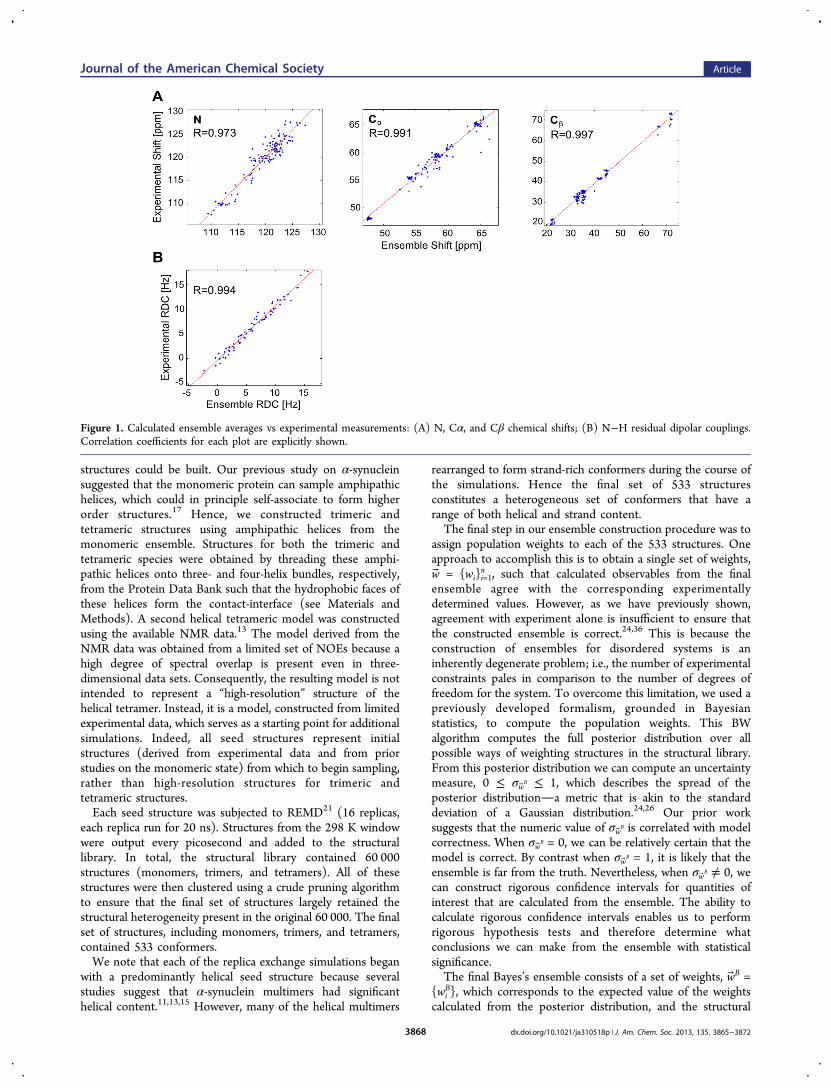
n , such that calculated observables from the finalensemble agree with the corresponding experimentallydetermined values. However, as we have previously shown,agreement with experiment alone is insufficient to ensure thatthe constructed ensemble is correct.24,36 This is because theconstruction of ensembles for disordered systems is aninherently degenerate problem; i.e., the number of experimentalconstraints pales in comparison to the number of degrees offreedom for the system. To overcome this limitation, we used apreviously developed formalism, grounded in Bayesianstatistics, to compute the population weights. This BWalgorithm computes the full posterior distribution over allpossible ways of weighting structures in the structural library.From this posterior distribution we can compute an uncertaintymeasure, 0 ≤ σwB ≤ 1, which describes the spread of theposterior distributiona metric that is akin to the standarddeviation of a Gaussian distribution.24,26 Our prior worksuggests that the numeric value of σwB is correlated with modelcorrectness. When σwB = 0, we can be relatively certain that themodel is correct. By contrast when σwB = 1, it is likely that theensemble is far from the truth. Nevertheless, when σwB ≠ 0, wecan construct rigorous confidence intervals for quantities ofinterest that are calculated from the ensemble. The ability tocalculate rigorous confidence intervals enables us to performrigorous hypothesis tests and therefore determine whatconclusions we can make from the ensemble with statisticalsignificance.The final Bayes’s ensemble consists of a set of weights, wB =
{wiB}, which corresponds to the expected value of the weights
calculated from the posterior distribution, and the structural
Figure 1. Calculated ensemble averages vs experimental measurements: (A) N, Cα, and Cβ chemical shifts; (B) N−H residual dipolar couplings.Correlation coefficients for each plot are explicitly shown.
Journal of the American Chemical Society Article
dx.doi.org/10.1021/ja310518p | J. Am. Chem. Soc. 2013, 135, 3865−38723868
library S = {si}i=1n . The algorithm also ensures that we restrict
our analysis to the most important conformers. More precisely,the ith structure is excluded from the ensemble when we cansay with 95% confidence that wi ≤ c. In the end, a total of 311structures survived this criterion. While the resulting Bayes’sensemble achieves a good fit to the NMR experimental data(Figure 1), the corresponding uncertainty parameter isnonzero: σwB = 0.47. Consequently, we express ensembleaverage values along with their corresponding 95% confidenceintervals.The ensemble is composed mostly of monomeric species
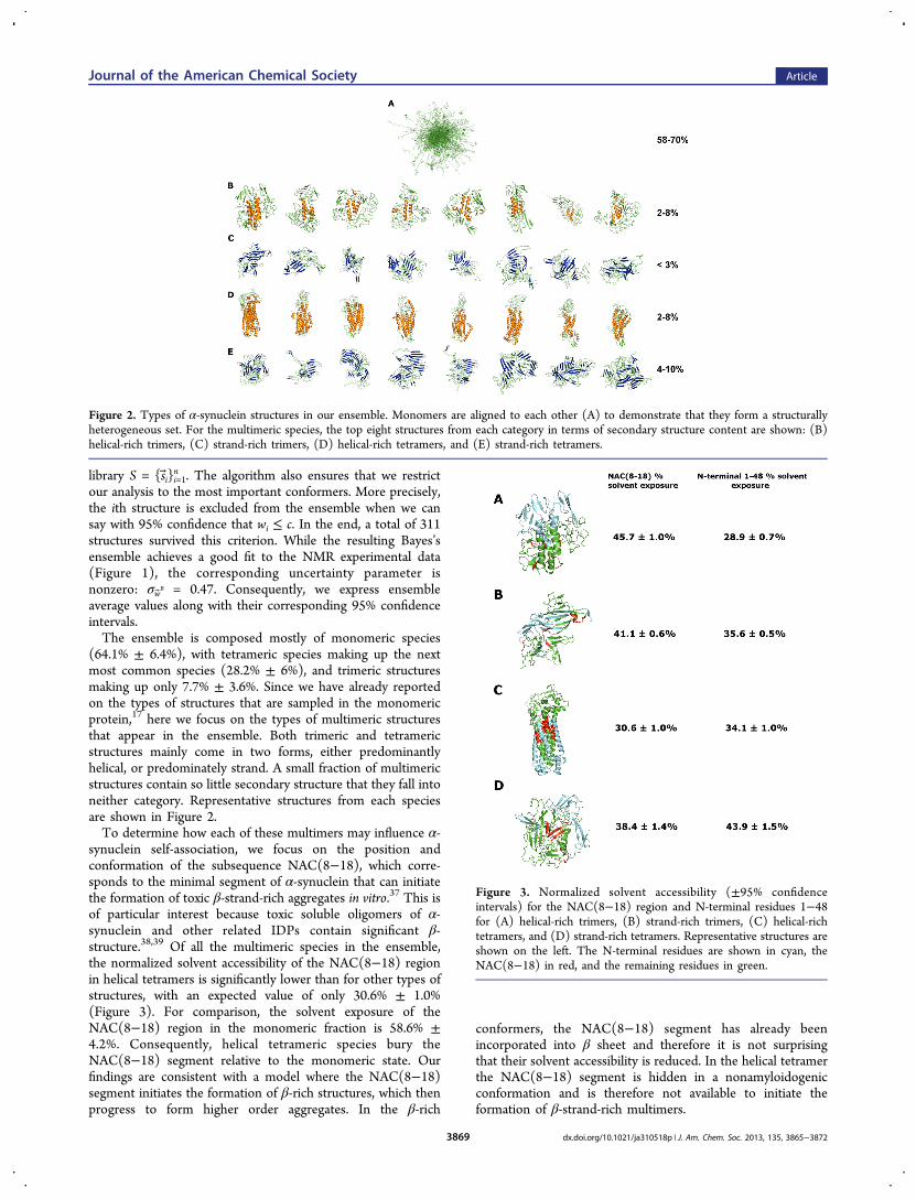
(64.1% ± 6.4%), with tetrameric species making up the nextmost common species (28.2% ± 6%), and trimeric structuresmaking up only 7.7% ± 3.6%. Since we have already reportedon the types of structures that are sampled in the monomericprotein,17 here we focus on the types of multimeric structuresthat appear in the ensemble. Both trimeric and tetramericstructures mainly come in two forms, either predominantlyhelical, or predominately strand. A small fraction of multimericstructures contain so little secondary structure that they fall intoneither category. Representative structures from each speciesare shown in Figure 2.To determine how each of these multimers may influence α-
synuclein self-association, we focus on the position andconformation of the subsequence NAC(8−18), which corre-sponds to the minimal segment of α-synuclein that can initiatethe formation of toxic β-strand-rich aggregates in vitro.37 This isof particular interest because toxic soluble oligomers of α-synuclein and other related IDPs contain significant β-structure.38,39 Of all the multimeric species in the ensemble,the normalized solvent accessibility of the NAC(8−18) regionin helical tetramers is significantly lower than for other types ofstructures, with an expected value of only 30.6% ± 1.0%(Figure 3). For comparison, the solvent exposure of theNAC(8−18) region in the monomeric fraction is 58.6% ±4.2%. Consequently, helical tetrameric species bury theNAC(8−18) segment relative to the monomeric state. Ourfindings are consistent with a model where the NAC(8−18)segment initiates the formation of β-rich structures, which thenprogress to form higher order aggregates. In the β-rich
conformers, the NAC(8−18) segment has already beenincorporated into β sheet and therefore it is not surprisingthat their solvent accessibility is reduced. In the helical tetramerthe NAC(8−18) segment is hidden in a nonamyloidogenicconformation and is therefore not available to initiate theformation of β-strand-rich multimers.
Figure 2. Types of α-synuclein structures in our ensemble. Monomers are aligned to each other (A) to demonstrate that they form a structurallyheterogeneous set. For the multimeric species, the top eight structures from each category in terms of secondary structure content are shown: (B)helical-rich trimers, (C) strand-rich trimers, (D) helical-rich tetramers, and (E) strand-rich tetramers.
Figure 3. Normalized solvent accessibility (±95% confidenceintervals) for the NAC(8−18) region and N-terminal residues 1−48for (A) helical-rich trimers, (B) strand-rich trimers, (C) helical-richtetramers, and (D) strand-rich tetramers. Representative structures areshown on the left. The N-terminal residues are shown in cyan, theNAC(8−18) in red, and the remaining residues in green.
Journal of the American Chemical Society Article
dx.doi.org/10.1021/ja310518p | J. Am. Chem. Soc. 2013, 135, 3865−38723869
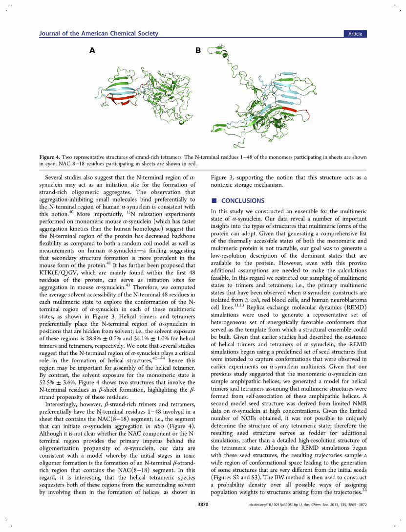
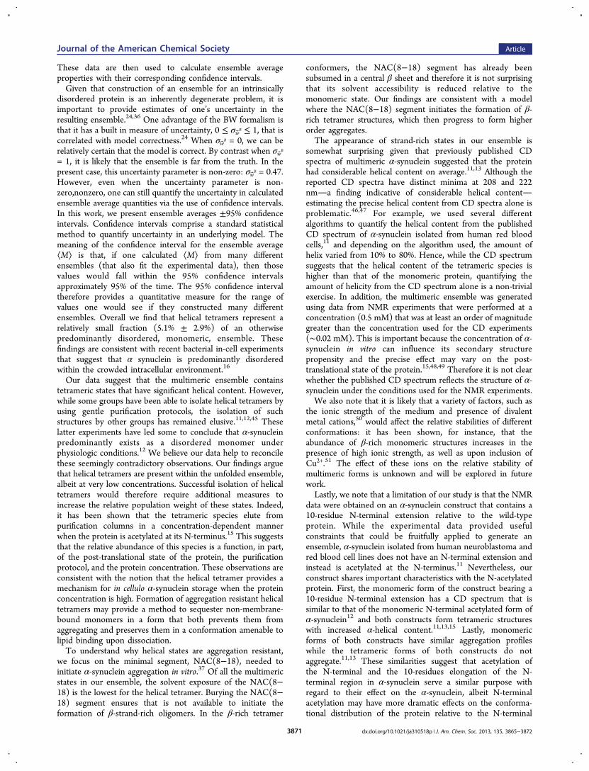
Several studies also suggest that the N-terminal region of α-synuclein may act as an initiation site for the formation ofstrand-rich oligomeric aggregates. The observation thataggregation-inhibiting small molecules bind preferentially tothe N-terminal region of human α-synuclein is consistent withthis notion.40 More importantly, 15N relaxation experimentsperformed on monomeric mouse α-synuclein (which has fasteraggregation kinetics than the human homologue) suggest thatthe N-terminal region of the protein has decreased backboneflexibility as compared to both a random coil model as well asmeasurements on human α-synucleina finding suggestingthat secondary structure formation is more prevalent in themouse form of the protein.41 It has further been proposed thatKTK(E/Q)GV, which are mainly found within the first 48residues of the protein, can serve as initiation sites foraggregation in mouse α-synuclein.41 Therefore, we computedthe average solvent accessibility of the N-terminal 48 residues ineach multimeric state to explore the conformation of the N-terminal region of α-synuclein in each of these multimericstates, as shown in Figure 3. Helical trimers and tetramerspreferentially place the N-terminal region of α-synuclein inpositions that are hidden from solvent; i.e., the solvent exposureof these regions is 28.9% ± 0.7% and 34.1% ± 1.0% for helicaltrimers and tetramers, respectively. We note that several studiessuggest that the N-terminal region of α-synuclein plays a criticalrole in the formation of helical structures,42−44 hence thisregion may be important for assembly of the helical tetramer.By contrast, the solvent exposure for the monomeric state is52.5% ± 3.6%. Figure 4 shows two structures that involve theN-terminal residues in β-sheet formation, highlighting the β-strand propensity of these residues.Interestingly, however, β-strand-rich trimers and tetramers,
preferentially have the N-terminal residues 1−48 involved in asheet that contains the NAC(8−18) segment; i.e., the segmentthat can initiate α-synuclein aggregation in vitro (Figure 4).Although it is not clear whether the NAC component or the N-terminal region provides the primary impetus behind theoligomerization propensity of α-synuclein, our data areconsistent with a model whereby the initial stages in toxicoligomer formation is the formation of an N-terminal β-strand-rich region that contains the NAC(8−18) segment. In thisregard, it is interesting that the helical tetrameric speciessequesters both of these regions from the surrounding solventby involving them in the formation of helices, as shown in
Figure 3, supporting the notion that this structure acts as anontoxic storage mechanism.
■ CONCLUSIONS
In this study we constructed an ensemble for the multimericstate of α-synuclein. Our data reveal a number of importantinsights into the types of structures that multimeric forms of theprotein can adopt. Given that generating a comprehensive listof the thermally accessible states of both the monomeric andmultimeric protein is not tractable, our goal was to generate alow-resolution description of the dominant states that areavailable to the protein. However, even with this provisoadditional assumptions are needed to make the calculationsfeasible. In this regard we restricted our sampling of multimericstates to trimers and tetramers; i.e., the primary multimericstates that have been observed when α-synuclein constructs areisolated from E. coli, red blood cells, and human neuroblastomacell lines.11,13 Replica exchange molecular dynamics (REMD)simulations were used to generate a representative set ofheterogeneous set of energetically favorable conformers thatserved as the template from which a structural ensemble couldbe built. Given that earlier studies had described the existenceof helical trimers and tetramers of α synuclein, the REMDsimulations began using a predefined set of seed structures thatwere intended to capture conformations that were observed inearlier experiments on α-synuclein multimers. Given that ourprevious study suggested that the monomeric α-synuclein cansample amphipathic helices, we generated a model for helicaltrimers and tetramers assuming that multimeric structures wereformed from self-association of these amphipathic helices. Asecond model seed structure was derived from limited NMRdata on α-synuclein at high concentrations. Given the limitednumber of NOEs obtained, it was not possible to uniquelydetermine the structure of any tetrameric state; therefore theresulting seed structure serves as fodder for additionalsimulations, rather than a detailed high-resolution structure ofthe tetrameric state. Although the REMD simulations beganwith these seed structures, the resulting trajectories sample awide region of conformational space leading to the generationof some structures that are very different from the initial seeds(Figures S2 and S3). The BW method is then used to constructa probability density over all possible ways of assigningpopulation weights to structures arising from the trajectories.24
Figure 4. Two representative structures of strand-rich tetramers. The N-terminal residues 1−48 of the monomers participating in sheets are shownin cyan. NAC 8−18 residues participating in sheets are shown in red.
Journal of the American Chemical Society Article
dx.doi.org/10.1021/ja310518p | J. Am. Chem. Soc. 2013, 135, 3865−38723870
These data are then used to calculate ensemble averageproperties with their corresponding confidence intervals.Given that construction of an ensemble for an intrinsically
disordered protein is an inherently degenerate problem, it isimportant to provide estimates of one’s uncertainty in theresulting ensemble.24,36 One advantage of the BW formalism isthat it has a built in measure of uncertainty, 0 ≤ σwB ≤ 1, that iscorrelated with model correctness.24 When σwB = 0, we can berelatively certain that the model is correct. By contrast when σwB
= 1, it is likely that the ensemble is far from the truth. In thepresent case, this uncertainty parameter is non-zero: σwB = 0.47.However, even when the uncertainty parameter is non-zero,nonzero, one can still quantify the uncertainty in calculatedensemble average quantities via the use of confidence intervals.In this work, we present ensemble averages ±95% confidenceintervals. Confidence intervals comprise a standard statisticalmethod to quantify uncertainty in an underlying model. Themeaning of the confidence interval for the ensemble average⟨M⟩ is that, if one calculated ⟨M⟩ from many differentensembles (that also fit the experimental data), then thosevalues would fall within the 95% confidence intervalsapproximately 95% of the time. The 95% confidence intervaltherefore provides a quantitative measure for the range ofvalues one would see if they constructed many differentensembles. Overall we find that helical tetramers represent arelatively small fraction (5.1% ± 2.9%) of an otherwisepredominantly disordered, monomeric, ensemble. Thesefindings are consistent with recent bacterial in-cell experimentsthat suggest that α synuclein is predominantly disorderedwithin the crowded intracellular environment.16
Our data suggest that the multimeric ensemble containstetrameric states that have significant helical content. However,while some groups have been able to isolate helical tetramers byusing gentle purification protocols, the isolation of suchstructures by other groups has remained elusive.11,12,45 Theselatter experiments have led some to conclude that α-synucleinpredominantly exists as a disordered monomer underphysiologic conditions.12 We believe our data help to reconcilethese seemingly contradictory observations. Our findings arguethat helical tetramers are present within the unfolded ensemble,albeit at very low concentrations. Successful isolation of helicaltetramers would therefore require additional measures toincrease the relative population weight of these states. Indeed,it has been shown that the tetrameric species elute frompurification columns in a concentration-dependent mannerwhen the protein is acetylated at its N-terminus.15 This suggeststhat the relative abundance of this species is a function, in part,of the post-translational state of the protein, the purificationprotocol, and the protein concentration. These observations areconsistent with the notion that the helical tetramer provides amechanism for in cellulo α-synuclein storage when the proteinconcentration is high. Formation of aggregation resistant helicaltetramers may provide a method to sequester non-membrane-bound monomers in a form that both prevents them fromaggregating and preserves them in a conformation amenable tolipid binding upon dissociation.To understand why helical states are aggregation resistant,
we focus on the minimal segment, NAC(8−18), needed toinitiate α-synuclein aggregation in vitro.37 Of all the multimericstates in our ensemble, the solvent exposure of the NAC(8−18) is the lowest for the helical tetramer. Burying the NAC(8−18) segment ensures that is not available to initiate theformation of β-strand-rich oligomers. In the β-rich tetramer
conformers, the NAC(8−18) segment has already beensubsumed in a central β sheet and therefore it is not surprisingthat its solvent accessibility is reduced relative to themonomeric state. Our findings are consistent with a modelwhere the NAC(8−18) segment initiates the formation of β-rich tetramer structures, which then progress to form higherorder aggregates.The appearance of strand-rich states in our ensemble is
somewhat surprising given that previously published CDspectra of multimeric α-synuclein suggested that the proteinhad considerable helical content on average.11,13 Although thereported CD spectra have distinct minima at 208 and 222nma finding indicative of considerable helical contentestimating the precise helical content from CD spectra alone isproblematic.46,47 For example, we used several differentalgorithms to quantify the helical content from the publishedCD spectrum of α-synuclein isolated from human red bloodcells,11 and depending on the algorithm used, the amount ofhelix varied from 10% to 80%. Hence, while the CD spectrumsuggests that the helical content of the tetrameric species ishigher than that of the monomeric protein, quantifying theamount of helicity from the CD spectrum alone is a non-trivialexercise. In addition, the multimeric ensemble was generatedusing data from NMR experiments that were performed at aconcentration (0.5 mM) that was at least an order of magnitudegreater than the concentration used for the CD experiments(∼0.02 mM). This is important because the concentration of α-synuclein in vitro can influence its secondary structurepropensity and the precise effect may vary on the post-translational state of the protein.15,48,49 Therefore it is not clearwhether the published CD spectrum reflects the structure of α-synuclein under the conditions used for the NMR experiments.We also note that it is likely that a variety of factors, such as
the ionic strength of the medium and presence of divalentmetal cations,50 would affect the relative stabilities of differentconformations: it has been shown, for instance, that theabundance of β-rich monomeric structures increases in thepresence of high ionic strength, as well as upon inclusion ofCu2+.51 The effect of these ions on the relative stability ofmultimeric forms is unknown and will be explored in futurework.Lastly, we note that a limitation of our study is that the NMR
data were obtained on an α-synuclein construct that contains a10-residue N-terminal extension relative to the wild-typeprotein. While the experimental data provided usefulconstraints that could be fruitfully applied to generate anensemble, α-synuclein isolated from human neuroblastoma andred blood cell lines does not have an N-terminal extension andinstead is acetylated at the N-terminus.11 Nevertheless, ourconstruct shares important characteristics with the N-acetylatedprotein. First, the monomeric form of the construct bearing a10-residue N-terminal extension has a CD spectrum that issimilar to that of the monomeric N-terminal acetylated form ofα-synuclein12 and both constructs form tetrameric structureswith increased α-helical content.11,13,15 Lastly, monomericforms of both constructs have similar aggregation profileswhile the tetrameric forms of both constructs do notaggregate.11,13 These similarities suggest that acetylation ofthe N-terminal and the 10-residues elongation of the N-terminal region in α-synuclein serve a similar purpose withregard to their effect on the α-synuclein, albeit N-terminalacetylation may have more dramatic effects on the conforma-tional distribution of the protein relative to the N-terminal
Journal of the American Chemical Society Article
dx.doi.org/10.1021/ja310518p | J. Am. Chem. Soc. 2013, 135, 3865−38723871
extension. Nonetheless, since the sequence of this constructdiffers from the wild-type protein, we cannot exclude thepossibility that wild-type α-synuclein isolated from other celltypes, such as neurons or red blood cells, may not be welldescribed by the ensemble presented here.
■ ASSOCIATED CONTENT*S Supporting InformationExperimental data, representative multimeric structures fromthe ensemble, and additional information on the structuressampled by the REMD simulations. This material is availablefree of charge via the Internet at http://pubs.acs.org.
■ AUTHOR INFORMATIONCorresponding [email protected] Contributions⊥T.G. and O.U. contributed equally to this work.NotesThe authors declare no competing financial interest.
■ REFERENCES(1) Bellucci, A.; Zaltieri, M.; Navarria, L.; Grigoletto, J.; Missale, C.;Spano, P. Brain Res. 2012, 1476, 183.(2) Spillantini, M. G.; S., M. L.; Lee, V.M.-Y.; Trojanowski, J. Q.;Jakes, R.; Goedert, M. Nature 1997, 388, 839.(3) Uversky, V. N.; Li, J.; Fink, A. L. J. Biol. Chem. 2001, 276, 10737.(4) Conway, K. A.; Lee, S.-J.; Rochet, J.-C.; Ding, T. T.; Williamson,R. E.; Lansbury, P. T. Proc. Natl. Acad. Sci. 2000, 97, 571.(5) Bucciantini, M.; Giannoni, E.; Chiti, F.; Baroni, F.; Formigli, L.;Zurdo, J.; Taddei, N.; Ramponi, G.; Dobson, C. M.; Stefani, M. Nature2002, 416, 507.(6) Kayed, R.; Head, E.; Thompson, J. L.; McIntire, T. M.; Milton, S.C.; Cotman, C. W.; Glabe, C. G. Science 2003, 300, 486.(7) Danzer, K. M.; Haasen, D.; Karow, A. R.; Moussaud, S.; Habeck,M.; Giese, A.; Kretzschmar, H.; Hengerer, B.; Kostka, M. J. Neurosci.2007, 27, 9220.(8) Winner, B.; Jappelli, R.; Maji, S. K.; Desplats, P. A.; Boyer, L.;Aigner, S.; Hetzer, C.; Loher, T.; Vilar, M. a.; Campioni, S.; Tzitzilonis,C.; Soragni, A.; Jessberger, S.; Mira, H.; Consiglio, A.; Pham, E.;Masliah, E.; Gage, F. H.; Riek, R. Proc. Natl. Acad. Sci. 2011, 108, 4194.(9) Uversky, V. N. J. Biomol. Struct. Dyn. 2003, 21, 159.(10) Drescher, M.; Huber, M.; Subramaniam, V. ChemBioChem2012, 13, 761.(11) Bartels, T.; C., J. G.; Selkoe, D. J. Nature 2011, 477, 107.(12) Fauvet, B.; Mbefo, M. K.; Fares, M. B.; Desobry, C.; Michael, S.;Ardah, M. T.; Tsika, E.; Coune, P.; Prudent, M.; Lion, N.; Eliezer, D.;Moore, D. J.; Schneider, B.; Aebischer, P.; El-Agnaf, O. M.; Masliah,E.; Lashuel, H. A. J. Biol. Chem. 2012, 287, 15345.(13) Wang, W.; Perovic, I.; Chittuluru, J.; Kaganovich, A.; Nguyen, L.T. T.; Liao, J.; Auclair, J. R.; Johnson, D.; Landeru, A.; Simorellis, A.K.; Ju, S.; Cookson, M. R.; Asturias, F. J.; Agar, J. N.; Webb, B. N.;Kang, C.; Ringe, D.; Petsko, G. A.; Pochapsky, T. C.; Hoang, Q. Q.Proc. Natl. Acad. Sci. 2011, 108, 17797.(14) Weinreb, P. H.; Zhen, W.; Poon, A. W.; Conway, K. A.;Lansbury, P. T. Biochemistry 1996, 35, 13709.(15) Trexler, A. J.; Rhoades, E. Protein Sci. 2012, 21, 601.(16) Binolfi, A.; Theillet, F. X.; Selenko, P. Biochem. Soc. Trans. 2012,40, 950.(17) Ullman, O.; Fisher, C. K.; Stultz, C. M. J. Am. Chem. Soc. 2011,133, 19536.(18) Mukherjea, M.; Llinas, P.; Kim, H.; Travaglia, M.; Safer, D.;Menetrey, J.; Franzini-Armstrong, C.; Selvin, P. R.; Houdusse, A.;Sweeney, H. L. Mol. Cell 2009, 35, 305.(19) Lawson, D. M.; Artymiuk, P. J.; Yewdall, S. J.; Smith, J. M. A.;Livingstone, J. C.; Treffry, A.; Luzzago, A.; Levi, S.; Arosio, P.;
Cesareni, G.; Thomas, C. D.; Shaw, W. V.; Harrison, P. M. Nature1991, 349, 541.(20) Berman, H. M.; Westbrook, J.; Feng, Z.; Gilliland, G.; Bhat, T.N.; Weissig, H.; Shindyalov, I. N.; Bourne, P. E. Nucleic Acids Res.2000, 28, 235.(21) Sugita, Y.; Okamoto, Y. Chem. Phys. Lett. 1999, 314, 141.(22) Lazaridis, T.; Karplus, M. Proteins: Struct., Funct. Bioinf. 1999, 35,133.(23) Brooks, B. R.; Bruccoleri, R. E.; Olafson, B. D.; States, D. J.;Swaminathan, S.; Karplus, M. J. Comput. Chem. 1983, 4, 187.(24) Fisher, C. K.; Huang, A.; Stultz, C. M. J. Am. Chem. Soc. 2010,132, 14919.(25) Huang, A.; Stultz, C. M. PLoS Comput. Biol. 2008, 4, e1000155.(26) Fisher, C. K.; Ullman, O.; Stultz, C. M. Pac. Symp. Biocomputing2012, 17, 82.(27) Neal, S.; Nip, A. M.; Zhang, H.; Wishart, D. S. J. Biomol. NMR2003, 26, 215.(28) Zweckstetter, M. Nat. Protoc. 2008, 3, 679.(29) Kabsch, W.; Sander, C. Biopolymers 1983, 22, 2577.(30) Kay, L.; Keifer, P.; Saarinen, T. J. Am. Chem. Soc. 1992, 114,10663.(31) Goddard, T. D.; Kneller, D. G. Sparky 3, Mac Intel version;University of California: San Francisco, CA, 2008; http://www.cgl.ucsf.edu/home/sparky/.(32) Bax, A. Protein Sci. 2003, 12, 1.(33) Schwieters, C. D.; Kuszewski, J. J.; Tjandra, N.; Marius Clore, G.J. Magn. Reson. 2003, 160, 65.(34) PyMOL; Schrodinger, LLC: 2010.(35) Wishart, D.; Bigam, C.; Holm, A.; Hodges, R.; Sykes, B. J.Biomol. NMR 1995, 5, 67.(36) Fisher, C. K.; Stultz, C. M. Curr. Opin. Struct. Biol. 2011, 21, 426.(37) el-Agnaf, O. M.; Irvine, G. B. Biochem. Soc. Trans. 2002, 30, 559.(38) Volles, M. J.; Lee, S. J.; Rochet, J. C.; Shtilerman, M. D.; Ding,T. T.; Kessler, J. C.; Lansbury, P. T., Jr. Biochemistry 2001, 40, 7812.(39) Laganowsky, A.; Liu, C.; Sawaya, M. R.; Whitelegge, J. P.; Park,J.; Zhao, M. L.; Pensalfini, A.; Soriaga, A. B.; Landau, M.; Teng, P. K.;Cascio, D.; Glabe, C.; Eisenberg, D. Science 2012, 335, 1228.(40) Cho, M.-K.; Nodet, G.; Kim, H.-Y.; Jensen, M. R.; Bernado, P.;Fernandez, C. O.; Becker, S.; Blackledge, M.; Zweckstetter, M. ProteinSci. 2009, 18, 1840.(41) Wu, K.-P.; Kim, S.; Fela, D. A.; Baum, J. J. Mol. Biol. 2008, 378,1104.(42) Bartels, T.; Ahlstrom, L. S.; Leftin, A.; Kamp, F.; Haass, C.;Brown, M. F.; Beyer, K. Biophys. J. 2010, 99, 2116.(43) Bodner, C. R.; Dobson, C. M.; Bax, A. J. Mol. Biol. 2009, 390,775.(44) Vamvaca, K.; Volles, M. J.; Lansbury, P. T., Jr. J. Mol. Biol. 2009,389, 413.(45) Kang, L.; Moriarty, G. M.; Woods, L. A.; Ashcroft, A. E.;Radford, S. E.; Baum, J. Protein Sci. 2012, 21, 911.(46) Manavalan, P.; Johnson, W. C. J. Biosci. 1985, 8, 141.(47) Greenfield, N. J. Nat. Protoc. 2006, 1, 2876.(48) Iwai, A.; Yoshimoto, M.; Masliah, E.; Saitoh, T. Biochemistry1995, 34, 10139.(49) Jarrett, J. T.; Lansbury, P. T. Cell 1993, 73, 1055.(50) Dudzik, C. G.; Walter, E. D.; Millhauser, G. L. Biochemistry2011, 50, 1771.(51) Sandal, M.; Valle, F.; Tessari, I.; Mammi, S.; Bergantino, E.;Musiani, F.; Brucale, M.; Bubacco, L.; Samorì, B. PLoS Biol. 2008, 6,e6.
Journal of the American Chemical Society Article
dx.doi.org/10.1021/ja310518p | J. Am. Chem. Soc. 2013, 135, 3865−38723872