the fifth cranial nerve in headaches
TRANSCRIPT

REVIEW ARTICLE Open Access
The fifth cranial nerve in headachesJ. C. A. Edvinsson1,2* , A. Viganò3, A. Alekseeva4, E. Alieva5, R. Arruda6, C. De Luca7,8, N. D’Ettore9, I. Frattale10,M. Kurnukhina11,12, N. Macerola13, E. Malenkova14, M. Maiorova15, A. Novikova16, P. Řehulka17, V. Rapaccini18,19,20,O. Roshchina4, G. Vanderschueren21, L. Zvaune22,23,24, A. P. Andreou25,26, K. A. Haanes1 and On behalf of theEuropean Headache Federation School of Advanced Studies (EHF-SAS)
Abstract
The fifth cranial nerve is the common denominator for many headaches and facial pain pathologies currentlyknown. Projecting from the trigeminal ganglion, in a bipolar manner, it connects to the brainstem and suppliesvarious parts of the head and face with sensory innervation. In this review, we describe the neuroanatomicalstructures and pathways implicated in the sensation of the trigeminal system. Furthermore, we present the currentunderstanding of several primary headaches, painful neuropathies and their pharmacological treatments. We hopethat this overview can elucidate the complex field of headache pathologies, and their link to the trigeminal nerve,to a broader field of young scientists.
Keywords: Fifth cranial nerve, Trigeminal ganglion, Headache, CGRP, Treatments, Migraine pathophysiology
IntroductionConsidering that the classification of headache disorders(ICHD-3) contains almost 300 different types of head-aches and facial pains [1], it is quite surprising that alarge part of pathophysiological mechanisms rely on thesame anatomical basis. Since the early work by HaroldWolff and his contemporaries [2] it has been shown that,among intracranial structures, only the dura mater, itsvessels and the cerebral blood vessels are pain sensitiveand can show referred pain on various extracranial posi-tions [3]. This classical view was recently expanded byan observational study [4] to include pia mater and itscortical arterioles as potential pain sensitive structures.Subsequent neuroanatomical and neurochemical studiesrevealed that most sensory fibres from the intracranialand the extracranial tissues originate in the fifth cranialnerve (CN V) ganglion, also called trigeminal ganglion
(TG). However, not all intracranial sensory fibers are tri-geminal. For example, the posterior cranial fossa, ismainly innervated by the occipital nerves.Depending on which part of the head is innervated the
fibres can be traced back to different parts of the TG [5].In general, headache pain is referred to a cutaneous ter-ritory area on the scalp, sharing supply with a nerve in-nervating the intracranial area, which might be theactual source of pain. Similarly, pain can be referred to adifferent territory than the actual nerve receiving thepainful stimulation. This can happen if the two nervesshare a high-order neuron (a process called“convergence”).Primary headaches comprise the most prevalent group
of neurological disorders. Among these, migraine is esti-mated to be present in 14.4% of the global population[6]. The WHO ranks migraine as the most prevalent,disabling, long-term neurological condition when takinginto account years lost due to disability in young individ-uals [7, 8]. The burden on individuals and society isenormous [9], especially if other headaches such astension-type (TTH), the second more common disorderworldwide [7, 8], and medication-overuse headache
© The Author(s). 2020 Open Access This article is licensed under a Creative Commons Attribution 4.0 International License,which permits use, sharing, adaptation, distribution and reproduction in any medium or format, as long as you giveappropriate credit to the original author(s) and the source, provide a link to the Creative Commons licence, and indicate ifchanges were made. The images or other third party material in this article are included in the article's Creative Commonslicence, unless indicated otherwise in a credit line to the material. If material is not included in the article's Creative Commonslicence and your intended use is not permitted by statutory regulation or exceeds the permitted use, you will need to obtainpermission directly from the copyright holder. To view a copy of this licence, visit http://creativecommons.org/licenses/by/4.0/.The Creative Commons Public Domain Dedication waiver (http://creativecommons.org/publicdomain/zero/1.0/) applies to thedata made available in this article, unless otherwise stated in a credit line to the data.
* Correspondence: [email protected] of Clinical Experimental Research, Glostrup Research Institute,Rigshospitalet Glostrup, 2600 Glostrup, Denmark2Department of Drug Design and Pharmacology, Faculty of Health andMedical Sciences, University of Copenhagen, Copenhagen, DenmarkFull list of author information is available at the end of the article
The Journal of Headache and Pain
Edvinsson et al. The Journal of Headache and Pain (2020) 21:65 https://doi.org/10.1186/s10194-020-01134-1

(MOH) are taken into account. Though TTH is moreprevalent (26.1%) [6], migraine is the more debilitating,as migraine has been reported to contribute 16.3% ofdisability-adjusted life-years on the global burden ofneurological disorders [10]. The present work is a com-prehensive description of various aspects of the CN V,the largest of the cranial nerves. Its more common name“trigeminal” (triplet) derives from its clearly visible div-ision into three main branches (Fig. 1). In this review weexplore the trigeminal nerve, its related pain conditionsand current treatments to emphasize its importance toheadache pathophysiology.
The Trigeminovascular systemThe vascular system of the head, face, meninges and thebrain have a variable innervation of autonomic and sen-sory nerves [12]. In general, the arterial system is richlysupplied with sensory nerves whereas the veins areweakly innervated. Capillaries are not innervated. Forthe cerebral vasculature, it is different; while the pial orextracerebral arterial system is richly supplied, once thevessels penetrate into the brain parenchyma their auto-nomic and sensory fibres disappear (at the level of thespace of Virchow), as these are regulated by metabolicdemand [13].The trigeminovascular system has long been a focus of
elucidating primary headache pathophysiology [14]. It
consists of the trigeminal neurons innervating the cere-bral arteries, the pial and dural blood vessels, and si-nuses [15]. Nociceptive activation of C- and Aδ-fibresinnervating these structures is thought to be involved inthe headache phase of migraine. The cranial dura maternerve fibres are mainly supplied by the ophthalmicbranch (V1), though collaterals from the maxillarybranch (V2), the mandibular branch (V3) and cervicalroot ganglion provide dural innervation to smaller cau-dal regions. Afferents from the TG carry this nociceptiveinformation into the brainstem where they mainly ter-minate at second order neurons inhabiting the trigemi-nocervical complex (TCC) [15].Studies have shown that parts of the trigeminovascular
system (notably TG) lack blood-brain barrier (BBB) andhas been hypothesized being the target tissue wheresome anti-migraine drugs (e.g. monoclonal antibodies,gepants, triptans) elicit their effects [16]. Because of thisevidence it is likely that CN V is an integral part in un-derstanding headache pathophysiology [17].
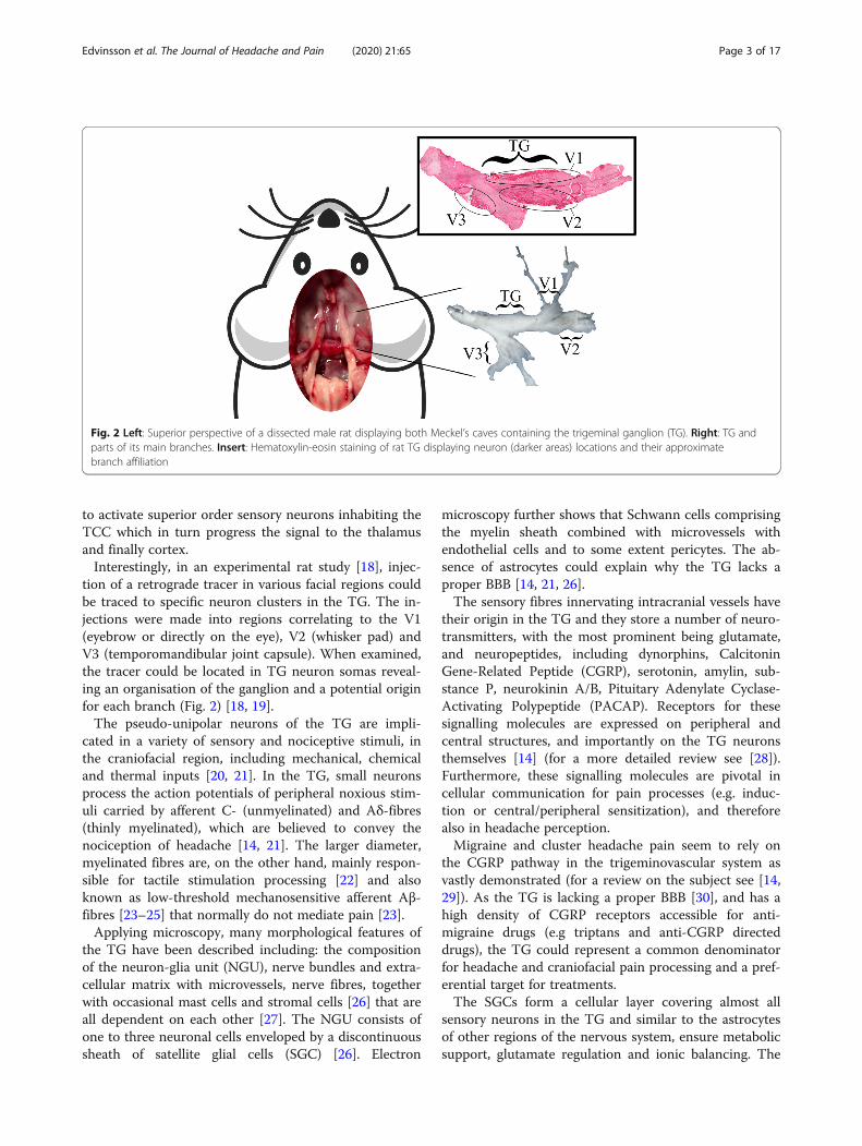
The trigeminal ganglionShortly after CN V protrudes from each side of the su-perior lateral pons the TG can be found residing in eachof Meckel’s caves (Fig. 2). The TG has been termed a“central hub” in the trigeminovascular transmitting path-way as it contains the soma of the peripheral nerves able
Fig. 1 Schematic of the Trigeminal System. a: The somatotopic distribution of trigeminal nociceptive afferents terminating in the trigeminalnucleus caudalis [11]. b: Innervation of facial skin areas and its related three branches (V1, V2 and V3). PSN (Principal sensory nucleus CN V), MN(Mesencephalic nucleus CN V), PA (Spinal nucleus of CN V Pars Oralis), PI (Spinal nucleus of CN V Pars Interpolaris), PC (Spinal nucleus of CN VPars Caudalis). N. = Nerve. G. = Ganglion
Edvinsson et al. The Journal of Headache and Pain (2020) 21:65 Page 2 of 17
to activate superior order sensory neurons inhabiting theTCC which in turn progress the signal to the thalamusand finally cortex.Interestingly, in an experimental rat study [18], injec-
tion of a retrograde tracer in various facial regions couldbe traced to specific neuron clusters in the TG. The in-jections were made into regions correlating to the V1(eyebrow or directly on the eye), V2 (whisker pad) andV3 (temporomandibular joint capsule). When examined,the tracer could be located in TG neuron somas reveal-ing an organisation of the ganglion and a potential originfor each branch (Fig. 2) [18, 19].The pseudo-unipolar neurons of the TG are impli-
cated in a variety of sensory and nociceptive stimuli, inthe craniofacial region, including mechanical, chemicaland thermal inputs [20, 21]. In the TG, small neuronsprocess the action potentials of peripheral noxious stim-uli carried by afferent C- (unmyelinated) and Aδ-fibres(thinly myelinated), which are believed to convey thenociception of headache [14, 21]. The larger diameter,myelinated fibres are, on the other hand, mainly respon-sible for tactile stimulation processing [22] and alsoknown as low-threshold mechanosensitive afferent Aβ-fibres [23–25] that normally do not mediate pain [23].Applying microscopy, many morphological features of
the TG have been described including: the compositionof the neuron-glia unit (NGU), nerve bundles and extra-cellular matrix with microvessels, nerve fibres, togetherwith occasional mast cells and stromal cells [26] that areall dependent on each other [27]. The NGU consists ofone to three neuronal cells enveloped by a discontinuoussheath of satellite glial cells (SGC) [26]. Electron
microscopy further shows that Schwann cells comprisingthe myelin sheath combined with microvessels withendothelial cells and to some extent pericytes. The ab-sence of astrocytes could explain why the TG lacks aproper BBB [14, 21, 26].The sensory fibres innervating intracranial vessels have
their origin in the TG and they store a number of neuro-transmitters, with the most prominent being glutamate,and neuropeptides, including dynorphins, CalcitoninGene-Related Peptide (CGRP), serotonin, amylin, sub-stance P, neurokinin A/B, Pituitary Adenylate Cyclase-Activating Polypeptide (PACAP). Receptors for thesesignalling molecules are expressed on peripheral andcentral structures, and importantly on the TG neuronsthemselves [14] (for a more detailed review see [28]).Furthermore, these signalling molecules are pivotal incellular communication for pain processes (e.g. induc-tion or central/peripheral sensitization), and thereforealso in headache perception.Migraine and cluster headache pain seem to rely on
the CGRP pathway in the trigeminovascular system asvastly demonstrated (for a review on the subject see [14,29]). As the TG is lacking a proper BBB [30], and has ahigh density of CGRP receptors accessible for anti-migraine drugs (e.g triptans and anti-CGRP directeddrugs), the TG could represent a common denominatorfor headache and craniofacial pain processing and a pref-erential target for treatments.The SGCs form a cellular layer covering almost all
sensory neurons in the TG and similar to the astrocytesof other regions of the nervous system, ensure metabolicsupport, glutamate regulation and ionic balancing. The
Fig. 2 Left: Superior perspective of a dissected male rat displaying both Meckel’s caves containing the trigeminal ganglion (TG). Right: TG andparts of its main branches. Insert: Hematoxylin-eosin staining of rat TG displaying neuron (darker areas) locations and their approximatebranch affiliation
Edvinsson et al. The Journal of Headache and Pain (2020) 21:65 Page 3 of 17
role of SGCs in neuropathic pain has been shown to beinvolved in the sensorial malfunctioning leading to “mal-adaptive” plasticity [31–35] that are responsible forchronification process widely described in commonforms of headache (e.g. migraine) [36].In particular, the regulation of extracellular potassium
concentrations and consequently the utilization of ATPby specific ATP-ase pumps seems to be one of the regu-lator mechanisms of pain attributed to SGCs [32, 37].Also, SGCs could modulate the purinergic system in theTG through vesicular nucleotide transporters (VNUT)[38]. In fact, all the purinergic receptors are expressed inthe TG [39]. Purinergic signalling has two-sided effectsin the TG. ATP release and the following purinergic ac-tivation, after peripheral noxious perineural stimulation,activate both SGCs and neurons in TG [34, 38]. Thiscontrasts to the breakdown product ADP, which insteadleads to trigeminovascular deactivation [40].
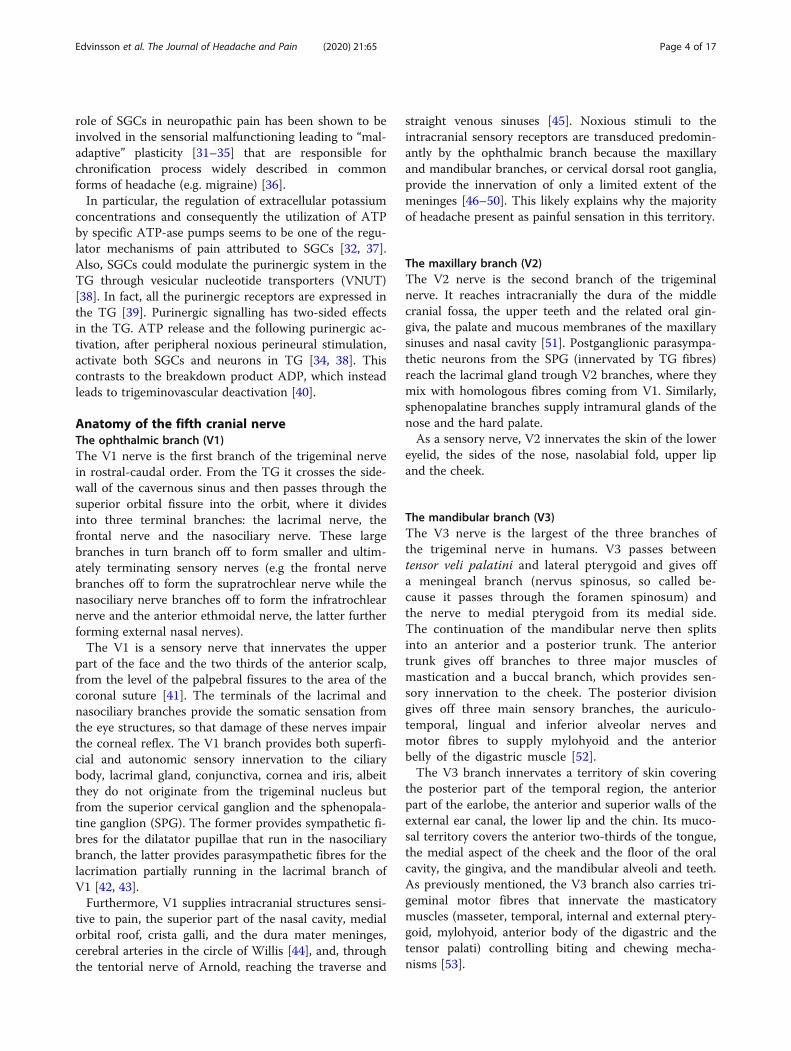
Anatomy of the fifth cranial nerveThe ophthalmic branch (V1)The V1 nerve is the first branch of the trigeminal nervein rostral-caudal order. From the TG it crosses the side-wall of the cavernous sinus and then passes through thesuperior orbital fissure into the orbit, where it dividesinto three terminal branches: the lacrimal nerve, thefrontal nerve and the nasociliary nerve. These largebranches in turn branch off to form smaller and ultim-ately terminating sensory nerves (e.g the frontal nervebranches off to form the supratrochlear nerve while thenasociliary nerve branches off to form the infratrochlearnerve and the anterior ethmoidal nerve, the latter furtherforming external nasal nerves).The V1 is a sensory nerve that innervates the upper
part of the face and the two thirds of the anterior scalp,from the level of the palpebral fissures to the area of thecoronal suture [41]. The terminals of the lacrimal andnasociliary branches provide the somatic sensation fromthe eye structures, so that damage of these nerves impairthe corneal reflex. The V1 branch provides both superfi-cial and autonomic sensory innervation to the ciliarybody, lacrimal gland, conjunctiva, cornea and iris, albeitthey do not originate from the trigeminal nucleus butfrom the superior cervical ganglion and the sphenopala-tine ganglion (SPG). The former provides sympathetic fi-bres for the dilatator pupillae that run in the nasociliarybranch, the latter provides parasympathetic fibres for thelacrimation partially running in the lacrimal branch ofV1 [42, 43].Furthermore, V1 supplies intracranial structures sensi-
tive to pain, the superior part of the nasal cavity, medialorbital roof, crista galli, and the dura mater meninges,cerebral arteries in the circle of Willis [44], and, throughthe tentorial nerve of Arnold, reaching the traverse and
straight venous sinuses [45]. Noxious stimuli to theintracranial sensory receptors are transduced predomin-antly by the ophthalmic branch because the maxillaryand mandibular branches, or cervical dorsal root ganglia,provide the innervation of only a limited extent of themeninges [46–50]. This likely explains why the majorityof headache present as painful sensation in this territory.
The maxillary branch (V2)The V2 nerve is the second branch of the trigeminalnerve. It reaches intracranially the dura of the middlecranial fossa, the upper teeth and the related oral gin-giva, the palate and mucous membranes of the maxillarysinuses and nasal cavity [51]. Postganglionic parasympa-thetic neurons from the SPG (innervated by TG fibres)reach the lacrimal gland trough V2 branches, where theymix with homologous fibres coming from V1. Similarly,sphenopalatine branches supply intramural glands of thenose and the hard palate.As a sensory nerve, V2 innervates the skin of the lower
eyelid, the sides of the nose, nasolabial fold, upper lipand the cheek.
The mandibular branch (V3)The V3 nerve is the largest of the three branches ofthe trigeminal nerve in humans. V3 passes betweentensor veli palatini and lateral pterygoid and gives offa meningeal branch (nervus spinosus, so called be-cause it passes through the foramen spinosum) andthe nerve to medial pterygoid from its medial side.The continuation of the mandibular nerve then splitsinto an anterior and a posterior trunk. The anteriortrunk gives off branches to three major muscles ofmastication and a buccal branch, which provides sen-sory innervation to the cheek. The posterior divisiongives off three main sensory branches, the auriculo-temporal, lingual and inferior alveolar nerves andmotor fibres to supply mylohyoid and the anteriorbelly of the digastric muscle [52].The V3 branch innervates a territory of skin covering
the posterior part of the temporal region, the anteriorpart of the earlobe, the anterior and superior walls of theexternal ear canal, the lower lip and the chin. Its muco-sal territory covers the anterior two-thirds of the tongue,the medial aspect of the cheek and the floor of the oralcavity, the gingiva, and the mandibular alveoli and teeth.As previously mentioned, the V3 branch also carries tri-geminal motor fibres that innervate the masticatorymuscles (masseter, temporal, internal and external ptery-goid, mylohyoid, anterior body of the digastric and thetensor palati) controlling biting and chewing mecha-nisms [53].
Edvinsson et al. The Journal of Headache and Pain (2020) 21:65 Page 4 of 17

The Trigeminocervical complex (TCC)The first order sensory neurons of the TG project cen-trally to the trigeminocervical complex (TCC) in thebrainstem. The TCC includes the second order neuronsof the trigeminal sensory pathway inhabiting the trigemi-nal nucleus caudalis (TNC) and C1 and C2 segments ofthe cervical spinal region [54]. While historically consid-ered as two separate entities, recently the trigeminal sys-tem has been considered both as a morphological [5]and a functional ensemble with first cervical roots [55].The part of the TNC dedicated to pain perception is thelower part, the Pars Caudalis (PC), while rostral partsare mainly deputed to tactile perception. This pain spe-cific part of the TNC extends from C2 or C3 rostrally tothe level of the obex. The TNC has many cytoarchitec-tural similarities with the posterior horn. For this reason,it has been termed “medullary posterior horn” and hasbeen divided into layers that correspond to Rexed spinalcord laminae [56]. The TNC and the posterior horn alsoshow homology in the distribution of neurotransmitters;substance P and CGRP are localized in nociceptive C-fibres that terminate in both of these areas [56]. Themost superior area of the TNC is the inferior medullaand the most inferior area is the upper cervical spinalcord [57]. The spinal trigeminal nucleus is a sensorytract located in the lateral medulla of the brain stem anddescends to the caudal end of the medulla and into thespinal cord (as far as the third or fourth cervical level),where it becomes continuous with Lissauer’s tract [58]and takes sensory information from different cranialnerves, including the trigeminal nerve and its branches[54].The innervation of the face forms a somatotopic map
in the TNC, which is stretched and distorted into theproportions of the PC of the spinal trigeminal nucleus(Fig. 1a). The area of lips and perioral area constitute theoutermost layer of the onion meaning that they liewithin the most superior area of the TNC [57]. The nextinnermost layer lies inferiorly within PC and comprisesthe projections of nose, eyes, and outer oral areas. TheV1 branch travels in the most ventral part of the spinaltract and extends caudally. The V2 branch lies in themost dorsal part of the trigeminal nucleus caudalis andterminates in the most rostral level [54]. In the lowestpart, there are areas reserved to cheeks and forehead;then the vertical area of the ears; and finally the partialsensory innervation of the external ears (from cranialnerves VII, IX, and X) [57]. This pattern of terminationmay account for the onion skin pattern of facial sensoryloss with intramedullary lesions [54]. The TNC that runsmedial to the spinal trigeminal tract, also has an onionskin somatotopy, and divides into three different cyto-architectural regions: Pars Oralis (PO), Pars Interpolaris(PI) and PC. PO is the most superior nucleus, running
from the pons to the mid-medulla. PI is the middle nu-cleus, spanning in the mid-medulla. PC is the most in-ferior nucleus from the lower medulla to the uppercervical spinal cord. Its inferior extent is variably listedfrom C2 to C4 [57].Pradier and McCormick reported in their study, based
on electrophysiological characteristics of neurons of theTNC, that there are five main groups of neurons, includ-ing; tonic, phasic, delayed, H-current and tonic-phasicneurons, groups that exhibit distinct intrinsic propertiesand share some similarity with groups identified in thespinal dorsal horn [59]. The primary function of theTNC is to carry information on temperature, deep orcrude touch (PO and PI), and pain from the portion ofthe face (PC) [54]. Afferents from the TNC terminate atthird-order neurons inhabiting the thalamus (mainlyposterior and ventral posteromedial thalamic nuclei) [58,60].In addition to this major pathway, TCC is also respon-
sible for conveying sensory and nociceptive signallingfrom the meninges and craniovascular structures to sev-eral higher order relays. There are numerous direct as-cending connections within the medulla (e.g. medullarypontine nuclei including the rostral ventromedial me-dulla), brainstem (e.g. nucleus raphe magnus, parabra-chial nucleus and locus coeruleus), midbrain nuclei (e.g.ventrolateral periaqueductal gray and cuneiform nu-cleus), and diencephalon (e.g. hypothalamus and thal-amus) [54, 58].Activation of these structures are believed to contrib-
ute to the perception of pain during migraine, and alsoto autonomic, endocrine, cognitive and affective symp-toms that last throughout the migraine episode [54].Furthermore, the second order neurons receive inputsfrom the occipital nerve. This convergence may havetreatment implications for some primary headache con-ditions as well as referred pain.
Trigeminohypothalamic tract and the parabrachial-limbictractAlthough a detailed description of these relay-functionslies outside the purpose of the present review (for detailssee [61, 62]), we will briefly discuss the trigeminohy-pothalamic tract and the parabrachial-limbic tract.The trigeminohypothalamic tract originates from spe-
cific nociceptive, multimodal intensity-coding wide dy-namic range (fundamental for pain “gating effects”) andnon-nociceptive neurons, albeit about the 80% of its fi-bres are axons from nociceptive neurons [61]. The trige-minohypothalamic tract ascends contralaterally in thebrainstem but about half of the fibres present a decussa-tion in the lateral hypothalamus, reaching both lateraland medial structures of hypothalamus (e.g. prefornical,suprachiamatic, supraoptic nuclei). While non-
Edvinsson et al. The Journal of Headache and Pain (2020) 21:65 Page 5 of 17
nociceptive information are transmitted only by directpathway, nociception is carried both directly and indir-ectly (i.e. trigeminoreticular tract) to hypothalamus, sug-gesting a more resistant mechanism to pathologicalnoxae for nociception [62]. Receiver areas of the hypo-thalamus are those regulating homeostasis and integrat-ing pain with visceral afferent input [63].The trigeminoparabrachial tract is a polysynaptic path-
way connecting CN V to the limbic system, with directtracts ending in the amygdala, lenticular nucleus, nu-cleus accumbens and it is thought to exert, among otherfunctions, the transmission of visceral pain and the emo-tional value of pain sensations [62, 64]. The parabrachialnucleus, in fact, contains a large share of neurons ex-pressing both CGRP and PACAP, especially in its lateralportion, which is the one activated by painful stimula-tion [65–67]. The transmission of CGRP is thought toreach directly the limbic system, where it can mediateaversive behaviour or freezing, as demonstrated in micewith injection of CGRP into the insula region [68].
The trigeminal system in primary headacheconditionsIn the headaches most commonly seen in specializedunits (e.g. migraine, TTH, trigeminal autonomic cepha-lalgias (TACs)) the pain generating mechanism residesin the complex relationship between trigeminal systemand intracranial structures sensible to pain (mostly ves-sels and meninges). For this reason, the functional en-semble of the trigeminovascular system is quite relevantfor the understanding of head and facial pain patho-physiology. We will now continue to describe some ofthe more notorious headache and facial pain diagnoses.Other less common primary headaches, which are not
explored in this article, that also might affect the trigem-inal area are: primary stabbing headache (head pain oc-curs as a single stab or as a series of stabs), nummularheadache (characterized by small circumscribed areas ofcontinuous pain on the head), cold-stimulus headache (adirect result of the rapid cooling and rewarming of thecapillaries in the sinuses leading), and external pressureheadache (external pressure on pain receptors or pain fi-bres) [1].
Tension type headacheAs previously mentioned, TTH is the most prevalentprimary headache [23, 69]. The definition of TTH canbe defined as a mild to moderate bilateral headache witha steady non-pulsating pain which is unaffected bymovement and lasting 30min to 7 days [70]. Further-more, TTH is not associated with nausea or vomitingand can manifest with either photophobia or phonopho-bia. The pathophysiological mechanism of TTH is notyet completely clear and most likely multifactorially
determined. Central sensitization of the trigeminal nerveseems to play an important role, especially in patientswith chronic TTH.Patients with episodic and chronic TTH have a con-
siderably increased tenderness to palpation of pericranialmyofascial tissues [71]. This increased tenderness origi-nates from muscles, fascia and tendons throughout thepericranial region, probably due to sensitization of Aδ-and C-fibres [71]. After a strong peripheral nociceptivestimulus from pericranial myofascial tissues centralsensitization can occur; ineffective synapses can changeto effective contacts of low threshold mechanosensitiveafferent nerves and superficial second order nociceptiveneurons in the trigeminal nucleus, which usually receiveinput from high threshold mechanoreceptors [25]. Thiscentral sensitization may make innoxious stimuli moreaggravating to the pain modulating systems, resulting inallodynia and hyperalgesia [23, 71].
MigraineIn migraine, intracranial vasculature is innervated by tri-geminal fibres (see above for details). Intracranial sen-sory receptors cover the rich plexus of meningealperivascular nerves of pial and dural blood vessels. Cur-rently, this classical vascular theory, naming vasodilationas the migraine pain generator, seems less reliable com-pared to a theory focusing on central sensitization,where activation of neuronal receptors is pivotal as ori-gin of migraine pain. The current paradigm is supportedby evidence of hypothalamic activation [72] and the viewof vasodilation as an epiphenomenon rather than a caus-ation of pain [73–75]. Nevertheless, the current reviewdoes not focus on the origin of the migraine attack, butthe origin of the perceived pain, which most likely re-sides in the TG and the associated sensory fibers.Whichever the trigger, the repeated experimental acti-
vation of the TG leads to release of vasoactive neuropep-tides, such as substance P, CGRP [76] and PACAP [77].CGRP and several other substances have been shown toevoke headaches after intravenous administration bothin healthy subjects and migraineurs, and to trigger a de-layed migraine-like attack in the latter group [78]. Therelease of vasoactive neuropeptides from the peripheralterminals of trigeminal nerve may result in neurogenicvasodilatation, plasma extravasation and trigeminalnerve sensitization, at least in rodents. It remains ques-tionable whether the throbbing quality of migraine painand aggravation by head movements or routine physicalactivity are expression of peripheral [79] or centralsensitization process [46].In addition to the importance of the neuropeptide sig-
nalling, some of the transient receptor potential (TRP)channels, which are identified in trigeminal ganglion,vagal ganglia and in dorsal root ganglia could play an
Edvinsson et al. The Journal of Headache and Pain (2020) 21:65 Page 6 of 17

important role. Those superfamily of receptors expressedin the trigeminal ganglion are mainly TRP vanilloid 1(TRPV1) [80] and TRP ankyrin 1 (TRPA1) channelswhich might be involved not only in pain initiations butalso as future treatment-targets in migraine [81].TRPM8, which mediates the cold sensation has alsobeen linked to migraine pathophysiology by genomewide association studies (GWAS), especially in thenorthern population through a mechanism of evolution-ary selection [82, 83].
Chronification, sensitization and habituationMigraine is a progressive disorder and can transformfrom an episodic state to a chronic state. According toICHD-3 [1], chronic migraine (CM) is defined as thepresence of headache for 15 or more days/month for atleast 3 months with migraine associated symptoms. Awell-accepted mechanism driving the progression fromepisodic to CM is the peripheral sensitization of the pri-mary afferent TG neurons, which leads to centralsensitization of TNC second-order neurons and ultim-ately to central sensitization of third order neurons inthe thalamus [46, 84].In a first phase of activity-dependent central
sensitization, the TNC neurons, under repetitive, persist-ent nociceptive stimuli from the TG, become sensitizedand produce exaggerated and prolonged responses tolower threshold stimuli. Over time, a neuroplastic adap-tation in medullary and cortical pain areas causes a shiftin the pain modulatory system creating a new thresholdand favouring a net pain facilitation rather than pain al-leviation. This shift to activity-independent centralsensitization plays a crucial role in the conversion toCM [85, 86].Based on experiments by Burstein et al, it is hypothe-
sized that cutaneous allodynia serves as a clinical indica-tor of migraine chronification. Particularly, thedevelopment of cutaneous allodynia in the head indi-cates that central sensitization affects mostly neurons inTNC, while the whole-body allodynia is mediated by thecentral sensitization of third-order neurons, suggesting athalamic involvement [87]. On a molecular level, theinterictal levels of trigeminal CGRP are significantly ele-vated in patients with CM when compared to those withepisodic migraine [88]. Interestingly, recent reviews pro-posed an interaction between CGRP and inflammation[89]. This process finally leads to increased productionof pro-inflammatory mediators, which sensitize TG neu-rons [76, 90]. One example is the activation of TG neu-rons by peripheral (dural) inflammation [91], whichmimics some of the features of CM.NMDA-receptors, nitric oxide, and endogenous sub-
stances such as serotonin, bradykinin, substance P andCGRP are involved in the development of this central
sensitization of the trigeminal nucleus and spinal dorsalnucleus [23, 24, 92]. Central sensitization, e.g. of the tri-geminal nucleus will induce an increased pain transmis-sion signal to the thalamus, limbic system and sensorycortex. The descending pathways of the rostral ventro-medial medulla will facilitate the sensitized nociceptivesecond order neurons of the trigeminal nerve [71]. Al-though the majority of studies focused on migraine, thechronification mechanism of TTH seems to be not sodifferent [71].Sensitization has received the most interest in primary
headaches and pathologies of the fifth cranial nerve.However, Groves and Thompson already in 1970’s pro-posed a “dual-process” theory [93]. The basis of this the-ory was based on the balance between depression(habituation) and facilitation (sensitization). Unlikesensitization, the neural mechanisms underlying habitu-ation remain poorly understood [94].Abnormal habituation patterns in migraineurs still
lacks a definitive consensual interpretation. Nevertheless,there are some suggestions in the literature that centralhabituation could play a role in cluster headache (CH)and episodic migraine. For CH a habituation deficit ofbrainstem reflex responses has been observed [95]. Re-garding episodic migraine, it was found that controlshad a habituation response to repetitive sensory stimula-tion in contrast to migraine subjects. Therefore, it seemsthat amplified information processing from spinal tri-geminal relay nuclei is linked to an impaired habituationresponse in migraineurs [96]. The cellular/physiologicalorigin of these responses remains to be determined.
Medication overuse headacheMedication overuse headache (MOH) is considered asecondary headache, with significant implications to pri-mary headache sufferers. MOH is defined as a pre-existing headache (occurring at least 15 days/month)worsening due to regular overuse of medication (used >10–15 days/month depending on the medication) fortreatment of an acute or symptomatic headache formore than 3months [97]. Medication overuse is themajor risk factor for chronification in all primary head-ache forms, although the 80% of MOH patients have mi-graine as original primary headache, a smaller part TTH,and rarely post-traumatic headache [98].Similarly to CM, in an MOH rat-model, persistent
triptan exposure produced cutaneous allodynia and cen-tral upregulation of CGRP and neuronal nitric oxidesynthase (nNOS) [99, 100]. Moreover, reduced seroto-nergic transmission seems to be involved in MOH devel-opment [101], possibly through a facilitation of thesensitization process via a maladaptive plasticity [98]. Inhumans, common neurophysiological investigation ofcentral sensitization shows an abnormal cortical
Edvinsson et al. The Journal of Headache and Pain (2020) 21:65 Page 7 of 17
response to repetitive sensory stimuli, with an increasedresponse amplitude after low numbers of stimuli [102]and a lacking habituation (which is instead normal inchronic migraineurs without MOH) [102], suggesting analtered plasticity.A recent neurophysiological study investigating the se-
rotonergic tone, found a low baseline serotonergic tonein chronic migraineurs with MOH, but it recovers aftera week following anaesthetic block of the greater occipi-tal nerve. Moreover, the size of the recover positivelycorrelated with the clinical benefit after a month [103].
TACs: cluster headache, SUNCT and SUNATrigeminal autonomic cephalalgias (TACs) are rare, buthighly disabling primary headache disorders. The mostcommon of the TACs is CH, known for its severelypainful symptoms. Other subgroups of TACs are hemi-crania continua (HC), paroxysmal hemicrania (PH),short-lasting unilateral neuralgiform headache with con-junctival injection and tearing (SUNCT) and short-lasting unilateral neuralgiform headache with cranialautonomic symptoms (SUNA).The unifying pathophysiological mechanism for TACs
is the role of the trigeminal autonomic reflex with para-sympathetic activation and clinical presentation withstrictly unilateral pain in the distribution of the trigemi-nal nerve and cranial autonomic features ipsilateral tothe pain. Distribution of maximal pain in TACs is at thefirst branch of trigeminal nerve (V1) > upper cervicalroot (C2) > second branch (V2) > third branch (V3)[104].Evidence for the peripheral mechanisms in CH include
increased plasma levels of CGRP, PACAP [105] andvasoactive intestinal peptide (VIP) during acute clusterattack and even interictally [106]. Recently, a clinicalstudy with a monoclonal antibody against CGRP wasfound positive in prevention of episodic CH [107]. Fur-thermore the SPG has connections to the trigeminovas-cular system, superior salivatory nucleus (SSN) andposterior hypothalamus: all areas that have an importantrole in the generation of CH attacks [108].The last decade has brought more insight into patho-
genesis of TACs, but still it is controversial whether thepain in TACs has a peripheral or central origin. Studiesusing animal models have shown that activation of tri-geminal nerve may lead to activation of parasympatheticefferents, producing autonomic symptoms such as lacri-mation, rhinorrhea and nasal congestion via thetrigeminal-autonomic reflex. The origin of the cells forthe parasympathetic autonomic vasodilator pathway is inthe pontine SSN. The efferent projection is predomin-antly through the greater petrosal nerve, a branch of thefacial nerve, and its projection through the SPG.
All primary headaches can be presented with auto-nomic symptoms to some degree, through reflex activa-tion of the cranial autonomic outflow [109, 110]. Aparasympathetic outflow activation probably results fromstimulation of trigeminal afferents. In this trigeminalautonomic reflex, SPG may have a considerable role: inclinical studies, stimulation of SPG reduces intensity andfrequency of CH pain [111]. The cyclical recurrence ofthe disorder (circadian and circannual rhythmicity), be-havioural features, such agitation and restlessness, dur-ing acute cluster attacks, as well recently study onpreictal and postictal symptoms in CH, led to theory ofthe key role of hypothalamus.Genetics and neuroimaging studies has implicated that
the brain and particularly the hypothalamus as a gener-ator of TACs [109, 112]. Animal studies have shown thatthere are direct hypothalamic-trigeminal connections(trigeminohypothalamic tract), and bilateral descendinghypothalamic projections to the spinal trigeminal nu-cleus [61]. Moreover, neuromodulation such as deepbrain stimulation of posterior hypothalamus, occipitalnerve stimulation, SPG stimulation has shown benefit toresistant chronic CH. Furthermore, cutting the trigemi-nal nerve root or ablative methods of TG does not re-solve the pain in TACs [113].In SUNCT and SUNA there are some similarities with
trigeminal neuralgia (TN) that imply the involvement ofneuropathic pain mechanisms, for example, the short-lasting unilateral attacks of pain, the cutaneous trigger-ing and the response to antiepileptic medications [114].TN will be covered below as we move on to conditionsmore plausibly linked to specific trigeminal nervebranches.
Other painful conditionals of the trigeminal nervebranchesThe ophthalmic branchTrigeminal Neuralgia (TN) is defined according toICHD-3 criteria, as “recurrent unilateral brief electricshock-like pains, abrupt in onset and termination, lim-ited to the distribution of one or more divisions of thetrigeminal nerve and triggered by innocuous stimuli” [1].The International Association for the Study of Pain(IASP) defines TN as “sudden, usually unilateral, severe,brief, stabbing, recurrent episodes of pain in the distri-bution of one or more branches of the trigeminal nerve”[115]. TN is a challenging syndrome and a commoncause of head and facial pain, and usually along the dis-tribution of the second or the third branch [116], there-fore TN is covered in more detail below, as only aminority of cases of TN involves the first division of thetrigeminal nerve.Among the few secondary causes of headache in V1
are Tolosa-Hunt syndrome, orbital cellulitis, idiopathic
Edvinsson et al. The Journal of Headache and Pain (2020) 21:65 Page 8 of 17
intracranial hypertension and herpetic neuralgia. Dam-age to V1 can cause complex syndromes, as paratrigem-inal oculosympathetic syndrome (Raeder’s syndrome)and recurrent painful ophthalmoplegic neuropathy(RPON) [117]. Raeder’s syndrome is a constant, unilat-eral pain caused by a disorder in the middle cranial fossaor of the carotid artery. RPON is an uncommon disorderwith repeated attacks of paresis of one or more ocularcranial nerves (commonly the 3rd), with ipsilateral head-aches [1]. The headache features are similar to typicalmigraine with frequent accompanying symptoms, suchas nausea, vomiting photophobia and phonophobia.RPON is a diagnosis of exclusion. The differential diag-nosis comprises all types of inflammatory or space-occupying lesions in the parasellar region and in theorbita [118].One neuralgia that is linked to the V1 branch, is
supraorbital neuralgia, characterized by persistent painover the supraorbital region and medial region forehead[119]. It may be differentiated from supratrochlear neur-algia based on the topography of the pain, which can beconfirmed with anaesthetic blockade [120]. Lacrimalneuralgia, is pain localized to the orbital and periorbitalarea, and was first described in 2013 [121]. All of theseheadaches are linked to V1, but little is known abouttheir molecular pathophysiology. This is also the case fortrochleodynia which is a spectrum of disorders charac-terized by pain arising from the trochlear region [122]and idiopathic ophthalmodynia [123] which is linked topain in the eyeball.Pain due to a cavernous sinus lesion, which is usually
causing total ophthalmoplegia and being accompaniedby a fixed, dilated pupil [124] or compression on thestructures passing through the superior orbital fissure[125] can due to the anatomy also compromise the V1branch. Furthermore, many cranio-cervical structuresmight present with facial pain and it is important alwaysto be sure that the pain is not better accounted for byanother diagnosis [1].
The maxillary branchPain conditions linked to the V2 branch vary frommostly frequent TN to facial presentations of primaryheadaches. The diagnosis of TN is clinical and dependsfundamentally on the description by the patient andcharacterization of pain [126]. TN is typically a unilateralcondition, slightly, but significantly more frequent, onthe right side [127]. Contrary to secondary forms, clas-sical TN includes idiopathic cases as well as thosecaused by neurovascular compression, demonstrated bymagnetic resonance imaging (MRI) or surgery, deter-mining morphological changes to the trigeminal nerveroot that represents about the 50% of cases. The exact
extent of this neurovascular conflict needed to induceTN is still debated [128].Classical TN may be purely paroxysmal, without con-
comitant continuous pain, or it may be with persistentbackground pain.For TN, patients may describe a trigger point that
elicits pain when touched: this could be interpreted as amanifestation of an erratic hyperactive functioning ofthe nerve. Furthermore, central causes have been pro-posed, even if it is difficult to determine which of thethese changes are cause and effect: volume reduction insomatosensory cortex, thalamus and other subcorticalareas has been observed [129], as well as functional con-nectivity alterations were described in sensory trigeminalpathways [130]. Sometimes trigeminal nerve atrophy canbe demonstrated in patients with TN by high-resolutionimaging and it is significantly correlated with the severityof neurovascular compression [131].Another important cause of facial pain in the V2 terri-
tory, which has been considered “the atypical counter-part to trigeminal neuralgia” [132], is the persistentidiopathic facial pain (PIFP), previously termed atypicalfacial pain or atypical odontalgia when occurring in theoral cavity. PIFP is defined as a continuous facial pain,typically localized in a circumscribed area of the face,which is generally not accompanied by any neurologicalor other lesion identified by clinical examination or clin-ical investigations [132]. This facial pain, which occursdaily and persists throughout the day, is generally de-scribed as deep, poorly localized, and is not associatedwith sensory loss or other neurological deficits, whichdifferentiates it from a pure neuropathic process. Thepathophysiology is not fully elucidated and possibly it re-lies on a combination of neuropathic pain, centralsensitization, and local inflammation [132–134].This complex pathophysiology is reflected by the diffi-
culty in treating PIFP successfully, and the concept thatdifferent types of interventions are needed [135]. Whilethe large majority of case are idiopathic with investiga-tions including X-ray of the face and jaws or cranialcomputed tomography (CT) or MRI not demonstratingany relevant abnormality, a part of PIFP-like disorderscan be secondary to dental or oral conditions [136–138].Lastly, neuralgia of the infraorbital nerve (numb cheek
syndrome) is an unusual cause of facial pain, most oftenassociated with the V2 branch [139]. The pain can becharacterized by constant discomfort, often in the formof stabbing pain, often accompanied with hypersensitiv-ity to palpation in the infraorbital notch [140], and canbe linked to an underlying cancer [141].
The mandibular branchA trigeminal nerve injury that mainly affects its V3branch is characterized by acute paroxysmal painful
Edvinsson et al. The Journal of Headache and Pain (2020) 21:65 Page 9 of 17

episodes [142, 143] with a sudden onset that may involveall the aforementioned structures [144]. The typical as-sociated symptoms are PIFP or burning mouth like syn-drome (BMLS) [145]. While PIFP can affects either V2or V3, with a preference for the former, BMLS is definedas a multifactorial chronic pain condition characterizedby a burning or stinging sensation, often accompaniedby xerostomia and preferably located on the tongue or,in a lesser extent, other specific areas of the mouth, in aclinically healthy oral mucosa [146]. The epidemiologyvaries from 0.01% to 40% according the studies, gener-ally observed in middle-aged patients and postmeno-pausal women [147, 148].No definitive aetiology has been established for burn-
ing mouth syndrome, an intramural burning sensationfor which no medical or dental cause can be found: bothcentral and peripheral nervous systems seem to be in-volved and some studies suggested a trigeminal smallfibre sensory neuropathy in innervation territory of max-illary nerve [149]. The diagnosis is generally reachedafter a series of tests, including neurophysiological evalu-ation and peripheral lingual nerve anaesthetic block,allowing the distinction between peripheral and centralforms (for a review, see [150]). Therapeutic options re-main however low, with only topical or systemic low-dose clonazepam as a valuable treatment [151, 152].Topical capsaicin or saliva substitute are second line op-tions in peripheral forms, while amitriptyline or gaba-pentin are considered in central form [150].One of the most “dangerous” neuralgias is the “Numb
chin syndrome” which can occur from a lesion anywherealong the course of the trigeminal nerve. Typically itrepresents loss of the terminal and sensory branch of themandibular branch and is often linked to cancer, such asmetastatic tumours [153].Finally, temporomandibular disorders are also linked
to the V3 branch [154]. TN can be differentiated fromtemporomandibular joint dysfunction by the acute, pier-cing, and stabbing nature of neuralgic pain occurring ata single facial location, spreading along the course of thenerve on one side, leading to differences in the characterand intensity of the pain [155].
Treatments targeting the trigeminal nerveAcute treatmentsAlthough many new therapeutic targets are under inves-tigation [156], the most frequently used acute treatmentfor headaches are non-specific drugs, such as NSAIDs.Cyclooxygenase 1 and 2 (COX-1, COX-2) inhibitors
have peripheral effect on prostaglandin synthesis in-volved in inflammatory processes. Acetylsalicylic acid isfound to have additional inhibitory effect on the centraltrigeminal neurons after sagittal sinus stimulation [157].Ketorolac (a nonselective COX-inhibitor) was found to
prevent sensitization at the trigeminal nucleus neurons[158]. COX-2 inhibitor piroxicam showed good effect inPH and celecoxib in HC [159].It has been shown that some TACs (e.g. PH) respond
well to indomethacin. In a case study, the patient be-came pain-free overnight after the use of indomethacinafter 12 years of failed treatments [160]. Indomethacin isa COX-inhibitor that inhibits evoked firing in the TCCin animal models [110]. Furthermore, indomethacin ex-erts an effect on IL-1β induced prostaglandin E synthesisvia COX-2 in cultured rat trigeminal cells. Consequentlyto the blockage of prostaglandin E release, the release ofCGRP was inhibited [161, 162]. According to studies,indomethacin has higher odds of responders andcomplete responders than any other treatment option inHC and PH [159]. On the other hand, indomethacin canalso inhibit NO-induced vasodilatation. Contrary to NO-induced CH-like and migraine-like headache that startafter a certain delay from NO administration, NO-induced PH symptoms begin immediately after the ad-ministration, and this can be the reason behind the dif-ferent effectiveness of indomethacin [163].Furthermore, there are a subcategory of headaches
that seem to response well to indomethacin, so called“Indomethacin-responsive headaches”. These are sexualheadache, trocheodynia, Valsalva-induced headache, pri-mary stabbing headache, hypnic headache and primaryexertion headache (also called exercise headache) [164].This could be linked to inhibitory effect of indomethacinon trigeminal nociceptive firing and the trigeminoauto-nomic activation, which has been shown in animals byAkerman et al. [165].Ergotamine and dihydroergotamine were the first
specific acute antimigraine drugs in use for several de-cades [166]. Ergot alkaloids are non-specific 5-HT1 re-ceptor agonists that also bind α-adrenoceptors anddopamine receptors. Therapeutic effect of these drugslikely originates from their agonist properties at 5-HT1B
and 5-HT1D receptors that lead to trigeminal inhibitionby, for example, reducing CGRP release [167, 168].Other previously proposed antimigraine mechanisms in-clude constriction of large capacitance arteries, closureof arteriovenous anastomoses, inhibition of neurogenicinflammation, and blockade of transmission in the TNC[169].Triptans have been studied in the context of head-
aches for decades. They are potent 5-HT1B/1D receptoragonists, a majority of them are also 5HT1F receptor ag-onists [170]. There is evidence that triptans exert theirclinical effect peripherally by binding to 5-HT1B recep-tors, resulting in slight vasoconstrictive properties aswell as blocking CGRP release and centrally by blockingtrigeminal transmission through binding at 5-HT1D re-ceptors in the trigeminal nuclei of the brainstem [171].
Edvinsson et al. The Journal of Headache and Pain (2020) 21:65 Page 10 of 17

In addition to being important in treating migraine, trip-tans (especially subcutaneous sumatriptan) are consid-ered the most effective treatment in cluster headaches[172]. Subcutaneous sumatriptan was reported to de-crease pain significantly also in TN [173]. In clinicalpractice, triptans are preferred to ergotamine derivatives,because they are at least as potent, with better tolerabil-ity and fewer side effects [158].Ditans are selective 5-HT1F receptor agonists that
were developed in hope to increase the effectiveness andto lower the risk of cardiovascular side effects of trip-tans. 5-HT1F receptors are located in both peripheraland central sensory trigeminal neurons, and their activa-tion is found to hyperpolarize nerve terminals inhibitingtrigeminal impulses [174], inhibit the CGRP-mediatedvasodilation in vivo, modulate the pain perception path-way and prevents CGRP release [170, 175]. Lasmiditan isthe only compound of this drug class that has been eval-uated in Phase III clinical trials and approved by theFDA [176]. It penetrates the BBB and could thus exerteffects centrally, in addition to the trigeminovascularsystem [174].Gepants antagonize the CGRP receptor in trigeminal
system. These drugs were promising in trials but werediscontinued due to low oral bioavailability (olcegepant)and unexpected hepatotoxicity (telcagepant) [177]. How-ever, ubrogepant and rimegepant tablets have both re-cently received FDA approval (23rd December 2019 and27th February 2020) for acute treatment of migraine inadults [178].Sodium channel blockers, such as lidocaine, blocks
sodium channels in a frequency-dependent and voltage-dependent manner. The nerve block with lidocaine stopsthe nociceptive firing and the neuronal hyperexcitabilityin first order neurons reducing peripheral sensitivity[179]. Intranasal lidocaine administered ipsilaterally tothe pain to anaesthetize the SPG, which is responsiblefor the autonomic symptoms associated to TACs orother headaches via the trigemino-autonomic reflex[180]. Intranasal lidocaine is considered a second linetreatment for CH [181]. The most effective treatment ofSUNCT/SUNA acute attacks is considered to be intra-venous lidocaine, subcutaneous treatment also can beused [159]. Furthermore, TN studies have shown thatlidocaine, rubbed onto the trigger zone of the oral mu-cosa, provided a few hours of pain relief [173].Some voltage-sensitive sodium channel blockers, such
as lamotrigine or amides (carbamazepine, oxcarbazepine,eslicarbazepine), are often the first line treatment forsome painful conditions affecting the trigeminal nerve,and most likely they act by stabilizing neural membranesand inhibit the release of neurotransmitters [182]. In aCochrane review (from 2013) it was concluded thatthere was no reduced headache frequency from
carisbamate, clonazepam, lamotrigine, oxcarbazepine,pregabalin, or vigabatrin [183]. However, carbamazepinehas shown some effect in familial hemiplegic migraine[184]. Further studies are needed to determine the effi-cacy of the newer drugs [185].High-flow oxygen is considered a first line treatment
for CH [172, 181]. The mechanism of action of oxygenin now thought to be related not to its vasoconstrictoreffect, but rather to the inhibition of neuronal activationin the TNC [186]. Oxygen is also thought to normalizethe CGRP levels and thus reduce the activity in the tri-geminovascular system [106].
Prophylactic treatmentsCGRP released from trigeminal terminals results in vaso-dilation via CGRP receptors on the smooth muscle cellsof meningeal and cerebral blood vessels [187] and activa-tion of Aδ-fibres, with the possibility of inducingsensitization [188]. Although antibodies can theoreticallytarget CGRP or its receptors in the brain regions, theBBB permeability is low [189, 190]. Therefore theirtherapeutic action may be entirely peripheral and likelyaffecting targets within the trigeminovascular system[191, 192].Monoclonal antibodies acting on CGRP pathway,
with indications for migraine prevention, have been de-veloped in recent years: one targeting the CGRP recep-tor (erenumab) and three targeting the CGRP peptide(eptinezumab, fremanezumab and galcanezumab) [193].Fremanezumab inhibits activation of central trigemino-vascular neurons with input from the intracranial dura,but not the facial skin or cornea [194] providing evi-dence that antibodies against CGRP can inhibit trigemi-nal neuron activation. However, their site of actionalong the trigeminal pathway remains uncertain, thoughrecently, axon-axon signalling at the node of Ranvier be-tween C- and Aδ-fibres was suggested as a plausible siteof action [167]. A role for the trigeminal nerve in CHand PH is indicated by the increased concentrations ofCGRP in the ipsilateral jugular vein during attacks [106,195]. Galcanezumab was recently reported to reduce thefrequency of episodic CH attacks [107].Onabotulinumtoxin A, beta blockers (e.g propran-
olol), tricyclic antidepressants (e.g amitriptyline), anti-convulsants (e.g topiramate) and calcium channelblockers (e.g flunarizine) continue to be standard therap-ies for migraine prevention [196–198]. Though mainlyknown for its therapeutic effects in CM, onabotulinum-toxin A has been shown positive results in treating TN[199] and refractory, chronic CH [200]. Onabotulinum-toxin A modulates neurotransmitter release, changes insurface expression of receptors and cytokines as well asenhancement of opioidergic transmission [201]. This isdone by cleaving synaptosomal nerve-associated protein
Edvinsson et al. The Journal of Headache and Pain (2020) 21:65 Page 11 of 17
25 (SNAP-25), a vesicle docking protein, within the celland thus disrupting the fusion of neurotransmitter vesi-cles to the synaptic cleft [202]. It is likely that onabotuli-numtoxin A reduces both peripheral and centralsensitization through such mechanisms [203, 204].
Non-pharmacological treatmentsThere are currently several non-invasive and invasivestimulation techniques that may help patients who wishto avoid, are refractory to or intolerant of previous drugtherapies [108]. Non-invasive stimulation options in-clude the supraorbital stimulation, vagus nerve stimula-tion (VNS) and the single-pulse transcranial magneticstimulation [108].The initial use of VNS to treat headaches first came
from the epilepsy field, following several anecdotal re-ports of migraine improvement in patients with comor-bid epilepsy who had been implanted with the device[205]. The vagal nerve is a mixed motor and sensorynerve that is important in controlling autonomic re-sponses; it projects to several higher centres that are im-portant in pain regulation [108]. Indeed, VNS wassufficient to significantly inhibit nocifensive head with-drawal response from mechanical stimulation of V1 tri-geminal nociceptors [206]. The commercial use inmigraine therapy certainly came with the developmentof portable devices, which allow to stimulate the vagalnerve transcutaneously at the neck (GammaCore® de-vice) or in its auricular portion (Nemos® device) in anon-invasive way [205]. Possible uses for VNS is pre-ventative treatment of CH, acute treatment of CH [207]and preventive treatment of CM (controlled studies areneeded to investigate this point) [208].The occipital nerves are a target for stimulation due to
the anatomical overlap between the trigeminal and cer-vical afferents in the TCC [108]. This allows stimulationof the occipital region to modulate pain in the trigeminaldistribution. Occipital nerve stimulation (ONS) is a sur-gical procedure where electrodes are placed subcutane-ously in the occipital region and then wired to a batterypack in the chest or abdomen [108]. Open-label studieshave shown possible efficacy in preventing CM, chronicCH [209]. Possible uses for ONS is preventative treat-ment of refractory CM and chronic CH [108].Trigeminal radiofrequency thermocoagulation
(TRT) is a surgical intervention used for the treatmentof TN. TRT involves puncturing the TG or its brancheswith a CT- or X-ray-guided a radiofrequency thermo-coagulation (RFT) ablation needle [210]. Sensory andmotor stimulation are used to replicate the patient’s painand locate and destroy the responsible nerve. Recurrenceis possible after RFT ablation; some patients need tocontinue medication treatment, while others may requirereoperation, and postoperative facial numbness is a
notable problem combined with developing neuropathicpain [210].Peripheral nerve blocks (PNB) have been used for
the acute and preventive treatment of a variety of pri-mary headache disorders [211]. PNB are generally safeand well-tolerated procedures that may be performed inthe outpatient setting [211]. PNB can be used in primary(migraine, CH, and nummular headache) and secondaryheadaches (cervicogenic headache and headache attrib-uted to craniotomy), as well in cranial neuralgias (tri-geminal neuropathies, glossopharyngeal and occipitalneuralgias) [179]. This procedure can be necessary forboth diagnosis and treatment (e.g a PNB of the inferiordental plexus will halt the pain caused by TN but not atemporomandibular disorder), while in cases it is consid-ered an adjuvant treatment [179]. Interestingly, a retro-spective case-study reported long-lasting (1–8months)and immediate pain-relief for refractory TN patientstreated with PNB [212]. This surprisingly long-lasting re-sult (as the half-life of anaesthetics is usually brief) couldbe due to the dose-dependent neurotoxicity of local an-aesthetics [213].The block of the greater occipital nerve with an anaes-
thetic and corticosteroid compound has proved to be ef-fective in the treatment of CH. Regarding the treatmentof other headaches and cranial neuralgias, controlledstudies are still necessary to clarify the real role of per-ipheral nerve block [179].Although nummular headache is characterized by con-
tinuous pain in a small circumscribed area, it surpris-ingly does not respond well to PNB [214], this contraststo the onabotulinumtoxin A, which seems effective[215]. This suggests that there is difference in the mech-anism of a nerve block, and the use of onabotulinum-toxin A.
ConclusionThe involvement of the fifth cranial nerve in headachehas been thoroughly established, following the originalpostulation by Wolff in the 1940’s. The current reviewsummarizes the anatomical and physiological link be-tween headaches, pain perception and the fifth cranialnerve. The most striking evidence comes from the nu-merous treatments available, where their targets are al-most exclusively found in the nerves of trigeminalganglion; the hub of the fifth cranial nerve. Although webelieve that the headache-trigger most likely have theorigin in the CNS, this review underscores the import-ance of trigeminal neurons in the perception of pain.Only when the activation of the fifth cranial nerve iscombined with knowledge of central pathological mech-anisms, we can start to fully understand the pathology ofheadache.
Edvinsson et al. The Journal of Headache and Pain (2020) 21:65 Page 12 of 17

AbbreviationsCGRP: Calcitonin gene-related peptide; BBB: Blood-brain barrier;TG: Trigeminal ganglion; TN: Trigeminal Neuralgia; WHO: World healthorganization; TTH: Tension-type headache; MOH: Medication-overuseheadache; CN V: Fifth cranial nerve; V1: Ophthalmic branch; V2: Maxillarybranch; V3: Mandibular branch; NGU: Neuronal and glia unit;ECM: Extracellular matrix; SGC: Satellite glial cell; PACAP: Pituitary adenylatecyclase-activating polypeptide; TCC: Trigeminocervical complex;TNC: Trigeminal nucleus caudalis; PC: Pars Caudalis; PO: Pars Oralis; PI: ParsInterpolaris; TACs: Trigeminal autonomic cephalalgias; RPON: Recurrentpainful ophthalmoplegic neuropathy; MRI: Magnetic resonance imaging;PIFP: persistent idiopathic facial pain; CT: computed tomography;BMLS: burning mouth like syndrome; TRP: Transient receptor potential;TRPV1: TRP vanilloid 1; TRPA1: TRP ankyrin 1; TRPM8: TRP melastatin 8;GWAS: Genome wide association studies; ICHD: International Classification ofHeadache Disorders; NMDA: N-methyl-D-aspartate; nNOS: neuronal nitricoxide synthase; CH: Cluster Headache; HC: Hemicrania continua;PH: Paroxysmal hemicranias; SUNCT: Short-lasting unilateral neuralgiformheadache with conjunctival injection and tearing; SUNA: Short-lastingunilateral neuralgiform headache with cranial autonomic symptoms;C2: Upper cervical root; SSN: Pontine superior salivatory nucleus;COX: Cyclooxygenase; IL-β: Interleukin 1 beta; NO: Nitric oxide; 5-HT: 5-hydroxytryptamine; FDA: U.S Food and Drug Administration; SNAP-25: Synaptosomal nerve-associated protein 25; VNS: Vagus nerve stimulation;ONS: Occipital nerve stimulation; TRT: Trigeminal radiofrequencythermocoagulation; RFT: Radiofrequency thermocoagulation; PNB: Peripheralnerve blocks
AcknowledgementsThis manuscript is a product of the program School of Advanced Studiespromoted by the European Headache Federation (EHF-SAS).
Authors’ contributionsAll authors contributed equally to the production of this review. All authorsread and approved the final manuscript. JE, AV, AA, EA, RA, CDL, NDE, IF, MK,NM, EM, MM, AN, PR, VR, OR, GV and LZ are Junior Fellows of EHF-SAS. A. P.Andreou and K. A. Haanes are Senior Fellows of EHF-SAS.
FundingThe authors received no specific funding for this work.
Availability of data and materialsNot applicable.
Ethics approval and consent to participateNot applicable.
Consent for publicationNot applicable.
Competing interestsThe authors declare that they have no competing interests.
Author details1Department of Clinical Experimental Research, Glostrup Research Institute,Rigshospitalet Glostrup, 2600 Glostrup, Denmark. 2Department of DrugDesign and Pharmacology, Faculty of Health and Medical Sciences, Universityof Copenhagen, Copenhagen, Denmark. 3IRCCS Fondazione Don CarloGnocchi, Milan, Italy. 4Department of Neurology, First Pavlov State MedicalUniversity of St.Petersburg, St.Petersburg, Russia. 5GBUZ Regional ClinicalHospital № 2, Krasnodar, Russia. 6Department of Neuroscience, University ofSao Paulo, Ribeirao Preto, Brazil. 7Department of Clinical and ExperimentalMedicine, Neurology Unit, University of Pisa, 56126 Pisa, Italy. 8Department ofPublic Medicine, Laboratory of Morphology of Neuronal Network, Universityof Campania-Luigi Vanvitelli, Naples, Italy. 9Department of Neurology,University of Rome, Tor Vergata, Rome, Italy. 10Department of Applied ClinicalSciences and Biotechnology, University of L’Aquila, 67100 L’Aquila, Italy.11Department of Neurosurgery, First Pavlov State Medical University ofSt.Petersburg, Lev Tolstoy Street 6-8, St.Petersburg, Russia. 12The LeningradRegional State Budgetary Institution of health care “Children’s clinicalhospital”, St.Petersburg, Russia. 13Department of Internal Medicine,
Fondazione Policlinico Universitario Agostino Gemelli IRCCS UniversitàCattolica del Sacro Cuore, Rome, Italy. 14Pain Department, Petrovsky NationalResearch Centre of Surgery, Moscow, Russia. 15Faculty of Medicine, Universityof Tartu, Tartu, Estonia. 16F.F. Erisman Federal Research Center for Hygiene,Mytishchy, Russia. 17Department of Neurology, St. Anne’s University Hospitaland Faculty of Medicine, Masaryk University, Brno, Czech Republic. 18ChildNeurology and Psychiatry Unit, Systems Medicine Department, UniversityHospital Tor Vergata, Viale Oxford 81, 00133 Rome, Italy. 19Unità SanitariaLocale (USL) Umbria 2, Viale VIII Marzo, 05100 Terni, Italy. 20Department ofNeurology, Headache Center, Ospedale Pediatrico Bambino Gesù, IRCCS,Rome, Italy. 21Department of Neurology, ZNA Middelheim, Lindendreef 1,2020 Antwerp, Belgium. 22Department of Anaesthesiology and IntensiveCare, Faculty of Medicine, Riga Stradins University, Riga, Latvia. 23Departmentof Pain Medicine, Hospital Jurmala, Jurmala, Latvia. 24Headache CentreVivendi, Riga, Latvia. 25Headache Research, Wolfson CARD, Institute ofPsychiatry, Psychology and Neuroscience, King’s College London, London,UK. 26The Headache Centre, Guy’s and St Thomas, NHS Foundation Trust,London, UK.
Received: 17 March 2020 Accepted: 25 May 2020
References1. (2018) Headache Classification Committee of the International Headache
Society (IHS) The International Classification of Headache Disorders, 3rdedition. Cephalalgia 38(1):1–211
2. Ray BS, Wolff HG (1940) Experimental studies on headache: pain-sensitivestructures of the head and their significance in headache. Arch Surg 41(4):813–856
3. Silberstein SD, Lipton RB, Dalessio DJ (2001) Wolff's headache and otherhead pain: Oxford University press
4. Fontaine D, Almairac F, Santucci S, Fernandez C, Dallel R, Pallud J et al(2018) Dural and pial pain-sensitive structures in humans: new inputs fromawake craniotomies. Brain 141(4):1040–1048
5. Edvinsson L (2011) Tracing neural connections to pain pathways withrelevance to primary headaches. Cephalalgia 31(6):737–747
6. Stovner LJ, Nichols E, Steiner TJ, Abd-Allah F, Abdelalim A, Al-Raddadi RMet al (2018) Global, regional, and national burden of migraine and tension-type headache, 1990–2016: a systematic analysis for the global burden ofdisease study 2016. Lancet Neurology 17(11):954–976
7. (2018) Global, regional, and national burden of migraine and tension-typeheadache, 1990–2016: a systematic analysis for the Global Burden ofDisease Study 2016. Lancet Neurology 17(11):954–976
8. James SL, Abate D, Abate KH, Abay SM, Abbafati C, Abbasi N et al (2018)Global, regional, and national incidence, prevalence, and years lived withdisability for 354 diseases and injuries for 195 countries and territories,1990–2017: a systematic analysis for the global burden of disease study2017. Lancet 392(10159):1789–1858
9. Linde M, Gustavsson A, Stovner LJ, Steiner TJ, Barré J, Katsarava Z et al(2012) The cost of headache disorders in Europe: the Eurolight project. Eur JNeurol 19(5):703–711
10. Feigin VL, et al. (2020) The global burden of neurological disorders:translating evidence into policy Lancet Neurol 19(3):255-265
11. Finnerup NB, Nikolajsen L, Jensen TS (2012) Are we neglecting spinalreorganization following nerve damage? Pain 153(2):269–272
12. Frederiksen SD, Haanes KA, Warfvinge K, Edvinsson L (2019) Perivascularneurotransmitters: regulation of cerebral blood flow and role in primaryheadaches. J Cereb Blood Flow Metab 39(4):610–632
13. Edvinsson L, Krause D. Cerebral Blood Flow and Metabolism (2002).Philadelphia: Lippincott Williams & Wilkins 17(521):8
14. Edvinsson L (2017) The Trigeminovascular pathway: role of CGRP and CGRPreceptors in migraine. Headache 57(Suppl 2):47–55
15. Noseda R, Burstein R (2013) Migraine pathophysiology: anatomy of thetrigeminovascular pathway and associated neurological symptoms, corticalspreading depression, sensitization, and modulation of pain. Pain 154:S44–S53
16. Eftekhari S, Salvatore CA, Johansson S, Chen TB, Zeng Z, Edvinsson L (2015)Localization of CGRP, CGRP receptor, PACAP and glutamate in trigeminalganglion Relation to the blood-brain barrier. Brain Res 1600:93–109
17. Messlinger K, Russo AF (2019) Current understanding of trigeminal ganglionstructure and function in headache. Cephalalgia 39(13):1661–1674
Edvinsson et al. The Journal of Headache and Pain (2020) 21:65 Page 13 of 17

18. Thalakoti S, Patil VV, Damodaram S, Vause CV, Langford LE, Freeman SE et al(2007) Neuron–glia signaling in trigeminal ganglion: implications formigraine pathology. Headache 47(7):1008–1023
19. Franceschini A, Vilotti S, Ferrari MD, van den Maagdenberg AM, Nistri A,Fabbretti E (2013) TNFα levels and macrophages expression reflect aninflammatory potential of trigeminal ganglia in a mouse model of familialhemiplegic migraine. PLoS One 8(1):e52394 https://doi.org/10.1371/journal.pone.0052394
20. Harriott AM, Gold MS (2009) Contribution of primary afferent channels toneuropathic pain. Curr Pain Headache Rep 13(3):197–207
21. Bista P, Wendy L (2019) Imlach. "Pathological Mechanisms and TherapeuticTargets for Trigeminal Neuropathic Pain." Medicines 6(3):91
22. Pennisi E, Cruccu G, Manfredi M, Palladini G (1991) Histometric study ofmyelinated fibers in the human trigeminal nerve. J Neurol Sci 105(1):22–28
23. Jay GW, Barkin RL (2017) Primary headache disorders- part 2: tension-typeheadache and medication overuse headache. Dis Mon 63(12):342–367
24. Bendtsen L (2003) Central and peripheral sensitization in tension-typeheadache. Curr Pain Headache Rep 7(6):460–465
25. Fumal A, Schoenen J (2008) Tension-type headache: current research andclinical management. Lancet Neurology. 7(1):70–83
26. Rusu MC, Cretoiu D, Vrapciu AD, Hostiuc S, Dermengiu D, Manoiu VS et al(2016) Telocytes of the human adult trigeminal ganglion. Cell Biol Toxicol32(3):199–207
27. De Luca C, Colangelo AM, Alberghina L, Papa M (2018) Neuro-immunehemostasis: homeostasis and diseases in the central nervous system. FrontCell Neurosci 12:459
28. Messlinger K (2018) The big CGRP flood - sources, sinks and signalling sitesin the trigeminovascular system. J Headache Pain 19(1):22
29. Edvinsson L, Haanes KA, Warfvinge K, Krause DN (2018) CGRP as the targetof new migraine therapies — successful translation from bench to clinic.Nat Rev Neurol 14(6):338–350
30. Lundblad C, Haanes KA, Grande G, Edvinsson L (2015) Expeerimentalinflammation following dural application of complete Freund's adjuvant orinflammatory soup does not alter brain and trigeminal microvascularpassage. J Headache Pain 16:91
31. De Luca C, Savarese L, Colangelo AM, Bianco MR, Cirillo G, Alberghina L et al(2016) Astrocytes and microglia-mediated immune response in maladaptiveplasticity is differently modulated by NGF in the ventral horn of the spinal cordfollowing peripheral nerve injury. Cell Mol Neurobiol 36(1):37–46
32. Cirillo G, Colangelo AM, Berbenni M, Ippolito VM, De Luca C,Verdesca F et al (2015) Purinergic modulation of spinal neuroglialmaladaptive plasticity following peripheral nerve injury. Mol Neurobiol52(3):1440–1457
33. Papa M, De Luca C, Petta F, Alberghina L, Cirillo G (2014) Astrocyte-neuroninterplay in maladaptive plasticity. Neurosci Biobehav Rev 42:35–54
34. Virtuoso A, Herrera-Rincon C, Papa M, Panetsos F (2019) Dependence ofNeuroprosthetic stimulation on the sensory modality of the trigeminalneurons following nerve injury. Implications in the Design of Future SensoryNeuroprostheses for correct perception and modulation of neuropathicpain. Front Neurosci 13:389
35. Takeda M, Takahashi M, Nasu M, Matsumoto S (2011) Peripheralinflammation suppresses inward rectifying potassium currents of satelliteglial cells in the trigeminal ganglia. Pain 152(9):2147–2156
36. Lai TH, Protsenko E, Cheng YC, Loggia ML, Coppola G, Chen WT (2015)Neural plasticity in common forms of chronic headaches. Neural Plast 2015:205985
37. Kawaguchi A, Sato M, Kimura M, Ichinohe T, Tazaki M, Shibukawa Y (2015)Expression and function of purinergic P2Y12 receptors in rat trigeminalganglion neurons. Neurosci Res 98:17–27
38. Goto T, Oh SB, Takeda M, Shinoda M, Sato T, Gunjikake KK et al (2016)Recent advances in basic research on the trigeminal ganglion. J Physiol Sci66(5):381–386
39. Haanes KA, Edvinsson L (2014) Expression and characterization of purinergicreceptors in rat middle meningeal artery-potential role in migraine. PLoSOne 9(9):e108782
40. Haanes KA, Labastida-Ramírez A, Blixt FW, Rubio-Beltrán E, Dirven CM,Danser AH et al (2019) Exploration of purinergic receptors as potential anti-migraine targets using established pre-clinical migraine models. Cephalalgia39(11):1421–1434
41. Gates P (2006) Duus’ topical diagnosis in neurology: anatomy, physiology,signs, symptoms, −by M. Baehr and M. Frotscher. Int Med J 36(9):557
42. Toshida H, Suto C (2018) Preganglionic parasympathetic denervation rabbitmodel for innervation studies. Cornea 37(Suppl 1):S106–Ss12
43. Morgan C, DeGroat WC, Jannetta PJ (1987) Sympathetic innervation of thecornea from the superior cervical ganglion. An HRP study in the cat. JAuton Nerv Syst 20(2):179–183
44. Suzuki N, Hardebo JE, Owman C (1989) Trigeminal fibre collaterals storingsubstance P and calcitonin gene-related peptide associate with ganglion cellscontaining choline acetyltransferase and vasoactive intestinal polypeptide inthe sphenopalatine ganglion of the rat. An axon reflex modulatingparasympathetic ganglionic activity? Neuroscience 30(3):595–604
45. Larrier D, Lee A (2003) Anatomy of headache and facial pain. OtolaryngolClin N Am 36(6):1041–1053 v
46. Goadsby PJ, Holland PR, Martins-Oliveira M, Hoffmann J, Schankin C,Akerman S (2017) Pathophysiology of migraine: a disorder of sensoryprocessing. Physiol Rev 97(2):553–622
47. Liu-Chen LY, Han DH, Moskowitz MA (1983) Pia arachnoid containssubstance P originating from trigeminal neurons. Neuroscience 9(4):803–808
48. Liu-Chen LY, Mayberg MR, Moskowitz MA (1983) Immunohistochemicalevidence for a substance P-containing trigeminovascular pathway to pialarteries in cats. Brain Res 268(1):162–166
49. Mayberg MR, Zervas NT, Moskowitz MA (1984) Trigeminal projections tosupratentorial pial and dural blood vessels in cats demonstrated byhorseradish peroxidase histochemistry. J Comp Neurol 223(1):46–56
50. Penfield W, McNAUGHTON F (1940) Dural headache and innervation of thedura mater. Arch Neurol Psychiatr 44(1):43–75
51. Anastasi G, Capitani S, Carnazza M, Cinti S, Cremona O, De Caro R, et al.(2010) Trattato di anatomia umana
52. Burchiel KJ (2003) A new classification for facial pain. Neurosurgery 53(5):1164–1167
53. Donnet A, Simon E, Cuny E, Demarquay G, Ducros A, De Gaalon S et al(2017) French guidelines for diagnosis and treatment of classical trigeminalneuralgia (French headache society and French neurosurgical society). RevNeurol (Paris) 173(3):131–151
54. Brazis PW, Masdeu JC (2011) Localization in Clinical Neurology (sixth ed.),Philadelphia: Lippincott Williams & Wilkins
55. Piovesan EJ, Kowacs PA, Oshinsky ML (2003) Convergence of cervical andtrigeminal sensory afferents. Curr Pain Headache Rep 7(5):377–383
56. Haines DE, Mihailoff GA (2017) Fundamental neuroscience for Basic andclinical applications E-book: Elsevier health sciences
57. Fisch A (2012) Neuroanatomy: draw it to know it: OUP USA58. Rusu MC (2004) The spinal trigeminal nucleus-considerations on the
structure of the nucleus caudalis. Folia Morphol (Warsz) 63(3):325–32859. Pradier B, McCormick SJ, Tsuda AC, Chen RW, Atkinson AL, Westrick MR et al
(2019) Properties of neurons in the superficial laminae of trigeminal nucleuscaudalis. Physiol Rep 7(12):e14112
60. Noseda R, Jakubowski M, Kainz V, Borsook D, Burstein R (2011) Corticalprojections of functionally identified thalamic trigeminovascular neurons:implications for migraine headache and its associated symptoms. J Neurosci31(40):14204–14217
61. Malick A, Strassman RM, Burstein R (2000) Trigeminohypothalamic andreticulohypothalamic tract neurons in the upper cervical spinal cord andcaudal medulla of the rat. J Neurophysiol 84(4):2078–2112
62. Almeida TF, Roizenblatt S, Tufik S (2004) Afferent pain pathways: aneuroanatomical review. Brain Res 1000(1–2):40–56
63. Patel NM, Das JM (2019) Neuroanatomy, Spinal Trigeminal Nucleus.StatPearls Publishing, StatPearls
64. Kocorowski LH, Helmstetter FJ (2001) Calcitonin gene-related peptidereleased within the amygdala is involved in Pavlovian auditory fearconditioning. Neurobiol Learn Mem 75(2):149–163
65. Palmiter RD (2018) The parabrachial nucleus: CGRP neurons function as ageneral alarm. Trends Neurosci 41(5):280–293
66. Han JS, Li W, Neugebauer V (2005) Critical role of calcitonin gene-relatedpeptide 1 receptors in the amygdala in synaptic plasticity and painbehavior. J Neurosci 25(46):10717–10728
67. Shinohara K, Watabe AM, Nagase M, Okutsu Y, Takahashi Y, Kurihara H et al(2017) Essential role of endogenous calcitonin gene-related peptide in pain-associated plasticity in the central amygdala. Eur J Neurosci 46(6):2149–2160
68. Missig G, Mei L, Vizzard MA, Braas KM, Waschek JA, Ressler KJ et al (2017)Parabrachial pituitary adenylate cyclase-activating polypeptide activation ofamygdala endosomal extracellular signal–regulated kinase signalingregulates the emotional component of pain. Biol Psychiatry 81(8):671–682
Edvinsson et al. The Journal of Headache and Pain (2020) 21:65 Page 14 of 17
69. Sohn JH, Choi HC, Kim CH (2013) Differences between episodic and chronictension-type headaches in nociceptive-specific trigeminal pathways.Cephalalgia 33(5):330–339
70. Schwartz BS, Stewart WF, Simon D, Lipton RB (1998) Epidemiology oftension-type headache. Jama 279(5):381–383
71. Bendtsen L (2000) Central sensitization in tension-type headache--possiblepathophysiological mechanisms. Cephalalgia 20(5):486–508
72. May A, Burstein R (2019) Hypothalamic regulation of headache andmigraine. Cephalalgia 39(13):1710–1719
73. Goadsby PJ (2009) The vascular theory of migraine--a great story wreckedby the facts. Brain 132(Pt 1):6–7
74. Amin FM, Asghar MS, Hougaard A, Hansen AE, Larsen VA, de Koning PJ et al(2013) Magnetic resonance angiography of intracranial and extracranialarteries in patients with spontaneous migraine without aura: a cross-sectional study. Lancet Neurology 12(5):454–461
75. Charles A (2013) Vasodilation out of the picture as a cause of migraineheadache. Lancet Neurology 12(5):419–420
76. Goadsby P, Edvinsson L, Ekman R (1988) Release of vasoactive peptides inthe extracerebral circulation of humans and the cat during activation of thetrigeminovascular system. Ann Neurol 23(2):193–196
77. Tuka B, Helyes Z, Markovics A, Bagoly T, Németh J, Márk L et al (2012)Peripheral and central alterations of pituitary adenylate cyclase activatingpolypeptide-like immunoreactivity in the rat in response to activation of thetrigeminovascular system. Peptides 33(2):307–316
78. Lassen L, Haderslev P, Jacobsen V, Iversen HK, Sperling B, Olesen J (2002)CGRP may play a causative role in migraine. Cephalalgia 22(1):54–61
79. Olesen J, Burstein R, Ashina M, Tfelt-Hansen P (2009) Origin of pain inmigraine: evidence for peripheral sensitisation. Lancet Neurology 8(7):679–690
80. Hou M, Uddman R, Tajti J, Kanje M, Edvinsson L (2002) Capsaicin receptorimmunoreactivity in the human trigeminal ganglion. Neurosci Lett 330(3):223–226
81. Benemei S, Fusi C, Trevisan G, Geppetti P (2014) The TRPA1 channel inmigraine mechanism and treatment. Br J Pharmacol 171(10):2552–2567
82. Dussor G, Cao YQ (2016) TRPM8 and migraine. Headache 56(9):1406–141783. Vigano A, Manica A, Di Piero V, Leonardi M (2019) Did going north give us
migraine? An evolutionary approach on understanding latitudinaldifferences in migraine epidemiology. Headache 59(4):632–634
84. Malick A, Burstein R (2000) Peripheral and central sensitization duringmigraine. Funct Neurol 15:28–35
85. Aurora SK, Brin MF (2017) Chronic migraine: an update on physiology,imaging, and the mechanism of action of two available pharmacologictherapies. Headache 57(1):109–125
86. Moskowitz MA (2008) Defining a pathway to discovery from bench tobedside: the trigeminovascular system and sensitization. Headache 48(5):688–690
87. Burstein R, Jakubowski M, Garcia-Nicas E, Kainz V, Bajwa Z, Hargreaves Ret al (2010) Thalamic sensitization transforms localized pain into widespreadallodynia. Ann Neurol 68(1):81–91
88. Cernuda-Morollón E, Larrosa D, Ramón C, Vega J, Martínez-Camblor P,Pascual J (2013) Interictal increase of CGRP levels in peripheral blood as abiomarker for chronic migraine. Neurology 81(14):1191–1196
89. Edvinsson L, Haanes KA, Warfvinge K (2019) Does inflammation have a rolein migraine? Nat Rev Neurol 15(8):483–490
90. Goadsby P, Edvinsson L, Ekman R (1990) Vasoactive peptide release in theextracerebral circulation of humans during migraine headache. AnnNeurology 28(2):183–187
91. Lukács M, Haanes KA, Majláth Z, Tajti J, Vécsei L, Warfvinge K et al (2015)Dural administration of inflammatory soup or complete Freund’s adjuvantinduces activation and inflammatory response in the rat trigeminalganglion. J Headache Pain 16(1):79
92. Fernandez-de-las-Penas C, Cuadrado ML, Arendt-Nielsen L, Simons DG,Pareja JA (2007) Myofascial trigger points and sensitization: an updated painmodel for tension-type headache. Cephalalgia 27(5):383–393
93. Groves PM, Thompson RF (1970) Habituation: a dual-process theory. PsycholRev 77(5):419–450
94. Coppola G, Di Lorenzo C, Schoenen J, Pierelli F (2013) Habituation andsensitization in primary headaches. J Headache Pain 14:65
95. Perrotta A, Serrao M, Sandrini G, Bogdanova D, Tassorelli C, Bartolo M et al(2008) Reduced habituation of trigeminal reflexes in patients with episodiccluster headache during cluster period. Cephalalgia 28(9):950–959
96. Lee J, Lin RL, Garcia RG, Kim J, Kim H, Loggia ML et al (2017) Reduced insulahabituation associated with amplification of trigeminal brainstem input inmigraine. Cephalalgia 37(11):1026–1038
97. Diener HC, Dodick D, Evers S, Holle D, Jensen RH, Lipton RB et al (2019)Pathophysiology, prevention, and treatment of medication overuseheadache. Lancet Neurology 18(9):891–902
98. Diener HC, Holle D, Solbach K, Gaul C (2016) Medication-overuse headache:risk factors, pathophysiology and management. Nat Rev Neurol 12(10):575–583
99. Supornsilpchai W, le Grand SM, Srikiatkhachorn A (2010) Corticalhyperexcitability and mechanism of medication-overuse headache.Cephalalgia 30(9):1101–1109
100. Okada-Ogawa A, Porreca F, Meng ID (2009) Sustained morphine-inducedsensitization and loss of diffuse noxious inhibitory controls in dura-sensitivemedullary dorsal horn neurons. J Neurosci 29(50):15828–15835
101. Srikiatkhachorn A, Anthony M (1996) Platelet serotonin in patients withanalgesic-induced headache. Cephalalgia 16(6):423–426
102. Coppola G, Currà A, Di Lorenzo C, Parisi V, Gorini M, Sava SL et al (2010)Abnormal cortical responses to somatosensory stimulation in medication-overuse headache. BMC Neurol 10(1):126
103. Viganò A, Torrieri MC, Toscano M, Puledda F, Petolicchio B, D’Elia TS et al(2018) Neurophysiological correlates of clinical improvement after greateroccipital nerve (GON) block in chronic migraine: relevance for chronicmigraine pathophysiology. J Headache Pain 19(1):73
104. Wei DY-T, Ong JJY, Goadsby PJ (2018) Overview of trigeminal autonomiccephalalgias: Nosologic evolution, diagnosis, and management. Ann IndianAcad Neurol 21(Suppl 1):S39
105. Tuka B, Szabó N, Tóth E, Kincses ZT, Párdutz Á, Szok D et al (2016) Releaseof PACAP-38 in episodic cluster headache patients–an exploratory study. JHeadache Pain 17(1):69
106. Goadsby PJ, Edvinsson L (1994) Human in vivo evidence fortrigeminovascular activation in cluster headache neuropeptide changes andeffects of acute attacks therapies. Brain 117(3):427–434
107. Goadsby PJ, Dodick DW, Leone M, Bardos JN, Oakes TM, Millen BA et al(2019) Trial of Galcanezumab in prevention of episodic cluster headache. NEngl J Med 381(2):132–141
108. Miller S, Sinclair AJ, Davies B, Matharu M (2016) Neurostimulation in thetreatment of primary headaches. Pract Neurol 16(5):362–375
109. May A, Schwedt TJ, Magis D, Pozo-Rosich P, Evers S, Wang S-J (2018) Clusterheadache. Nat Reviews Dis Prime 4:18006
110. Akerman S, Holland PR, Summ O, Lasalandra MP, Goadsby PJ (2012) Atranslational in vivo model of trigeminal autonomic cephalalgias:therapeutic characterization. Brain 135(12):3664–3675
111. Schoenen J, Jensen RH, Lanteri-Minet M, Láinez MJ, Gaul C, Goodman AMet al (2013) Stimulation of the sphenopalatine ganglion (SPG) for clusterheadache treatment. Pathway CH-1: a randomized, sham-controlled study.Cephalalgia 33(10):816–830
112. Cohen A (2017) SUN: short-lasting unilateral neuralgiform headache attacks.Headache 57(6):1010–1020
113. Pedersen JL, Barloese M, Jensen RH (2013) Neurostimulation in clusterheadache: a review of current progress. Cephalalgia 33(14):1179–1193
114. Snoer A, Lund N, Beske R, Hagedorn A, Jensen RH, Barloese M (2018) Clusterheadache beyond the pain phase: a prospective study of 500 attacks.Neurology 91(9):e822–ee31
115. Merskey HE (1986) Classification of chronic pain: descriptions of chronicpain syndromes and definitions of pain terms. Pain 3:S1-226
116. Maarbjerg S, Di Stefano G, Bendtsen L, Cruccu G (2017) Trigeminalneuralgia–diagnosis and treatment. Cephalalgia 37(7):648–657
117. Evers S (2017) Facial pain: overlapping syndromes. Cephalalgia 37(7):705–713
118. Huang C, Amasanti M, Lovell B, Young T (2017) Recurrent painfulophthalmoplegic neuropathy. Pract Neurol 17(4):318–320
119. Evans RW, Pareja JA (2009) Expert opinion. Supraorbital neuralgia. Headache49(2):278–281
120. Pareja JA, Lopez-Ruiz P, Mayo D, Villar-Quiles RN, Carcamo A, Gutierrez-Viedma A et al (2017) Supratrochlear neuralgia: a prospective case series of15 patients. Headache 57(9):1433–1442
121. Pareja JA, Cuadrado ML (2013) Lacrimal neuralgia: so far, a missing cranialneuralgia. Cephalalgia 33(14):1198–1202
122. Tran TM, McClelland CM, Lee MS (2019) Diagnosis and Management ofTrochleodynia, Trochleitis, and trochlear headache. Front Neurol 10:361
Edvinsson et al. The Journal of Headache and Pain (2020) 21:65 Page 15 of 17
123. Pareja JA, Cuadrado ML, Porta-Etessam J, Fernandez-de-las-Penas C, Gili P,Caminero AB et al (2010) Idiopathic ophthalmodynia and idiopathicrhinalgia: two topographic facial pain syndromes. Headache 50(8):1286–1295
124. Razek AA, Castillo M (2009) Imaging lesions of the cavernous sinus. AJNRAm J Neuroradiol 30(3):444–452
125. Caldarelli C, Benech R, Iaquinta C (2016) Superior orbital fissure syndrome inlateral Orbital Wall fracture: management and classification update.Craniomaxillofac Trauma Reconstr 9(4):277–283
126. Bendtsen L, Zakrzewska JM, Abbott J, Braschinsky M, Di Stefano G, DonnetA et al (2019) European academy of neurology guideline on trigeminalneuralgia. Eur J Neurol 26(6):831–849
127. Maarbjerg S, Gozalov A, Olesen J, Bendtsen L (2014) Trigeminal neuralgia–aprospective systematic study of clinical characteristics in 158 patients.Headache 54(10):1574–1582
128. Maarbjerg S, Wolfram F, Gozalov A, Olesen J, Bendtsen L (2015) Significance ofneurovascular contact in classical trigeminal neuralgia. Brain 138(2):311–319
129. Obermann M, Rodriguez-Raecke R, Naegel S, Holle D, Mueller D, Yoon M-Set al (2013) Gray matter volume reduction reflects chronic pain intrigeminal neuralgia. Neuroimage 74:352–358
130. Tian T, Guo L, Xu J, Zhang S, Shi J, Liu C et al (2016) Brain white matterplasticity and functional reorganization underlying the central pathogenesisof trigeminal neuralgia. Sci Rep 6:36030
131. Leal PRL, Barbier C, Hermier M, Souza MA, Cristino-Filho G, Sindou M (2014)Atrophic changes in the trigeminal nerves of patients with trigeminal neuralgiadue to neurovascular compression and their association with the severity ofcompression and clinical outcomes. J Neurosurg 120(6):1484–1495
132. Benoliel R, Gaul C (2017) Persistent idiopathic facial pain. Cephalalgia 37(7):680–691
133. Forssell H, Tenovuo O, Silvoniemi P, Jaaskelainen SK (2007) Differences andsimilarities between atypical facial pain and trigeminal neuropathic pain.Neurology 69(14):1451–1459
134. Lang E, Kaltenhauser M, Seidler S, Mattenklodt P, Neundorfer B (2005)Persistent idiopathic facial pain exists independent of somatosensory inputfrom the painful region: findings from quantitative sensory functions andsomatotopy of the primary somatosensory cortex. Pain 118(1–2):80–91
135. Du B, Xu J, Hu J, Zhong X, Liang J, Lei P et al (2019) A clinical study of theintra-Neuroendoscopic technique for the treatment of subacute-chronicand chronic septal subdural hematoma. Front Neurol 10:1408
136. Moazzam AA, Habibian M (2012) Patients appearing to dental professionalswith orofacial pain arising from intracranial tumors: a literature review. OralSurg Oral Med Oral Pathol Oral Radiol 114(6):749–755
137. List T, Leijon G, Svensson P (2008) Somatosensory abnormalities in atypicalodontalgia: a case-control study. Pain 139(2):333–341
138. Klasser GD, Grushka M, Su N (2016) Burning mouth syndrome. OralMaxillofac Surg Clin North Am 28(3):381–396
139. Campbell WW Jr (1986) The numb cheek syndrome: a sign of infraorbitalneuropathy. Neurology. 36(3):421–423
140. Lopez Mesonero L, Pedraza Hueso MI, Herrero Velazquez S, Guerrero PeralAL (2014) Infraorbital neuralgia: a diagnostic possibility in patients withzygomatic arch pain. Neurologia 29(6):381–382
141. Okholm C, Frendo M, Kiss K, von Buchwald C (2018) Cheek numbnesscaused by Perineural tumor invasion of the infraorbital nerve: a review of 3diagnostically challenging cases. Am J Case Rep 19:296–300
142. Cesarik M, Zavoreo I, Zadro-Matovina L, Papic M, Basic KV (2016) Effects oftrigeminal nerve dysfunction in various types of headaches. Coll Antropol40(3):183–188
143. Goadsby PJ (2019) Primary headache disorders: five new things. Neurol ClinPract 9(3):233–240
144. Antony AB, Mazzola AJ, Dhaliwal GS, Hunter CW (2019) Neurostimulation forthe treatment of chronic head and facial pain: a literature review. PainPhysician 22(5):447–477
145. Hascalovici JR, Robbins MS (2017) Peripheral nerve blocks for the treatmentof headache in older adults: a retrospective study. Headache 57(1):80–86
146. Coculescu EC, Radu A, Coculescu BI (2014) Burning mouth syndrome: areview on diagnosis and treatment. J Med Life 7(4):512–515
147. Aryeh HB, Gottlieb I, Ish-Shalom S, David A, Szargel H, Laufer D (1996) Oralcomplaints related to menopause. Maturitas 24(3):185–189
148. Klasser GD, Fischer DJ, Epstein JB (2008) Burning mouth syndrome:recognition, understanding, and management. Oral Maxillofac Surg ClinNorth Am 20(2):255–271 vii
149. Lauria G, Majorana A, Borgna M, Lombardi R, Penza P, Padovani A et al(2005) Trigeminal small-fiber sensory neuropathy causes burning mouthsyndrome. Pain. 115(3):332–337
150. Jaaskelainen SK, Woda A (2017) Burning mouth syndrome. Cephalalgia37(7):627–647
151. Heckmann SM, Kirchner E, Grushka M, Wichmann MG, Hummel T (2012) Adouble-blind study on clonazepam in patients with burning mouthsyndrome. Laryngoscope 122(4):813–816
152. Cui Y, Xu H, Chen F, Liu J, Jiang L, Zhou Y et al (2016) Efficacy evaluation ofclonazepam for symptom remission in burning mouth syndrome: a meta-analysis. Oral Dis 22(6):503–511
153. Baskaran RK, Krishnamoorthy SM (2006) Numb chin syndrome--a reflectionof systemic malignancy. World J Surg Oncol 4:52
154. Murphy MK, MacBarb RF, Wong ME, Athanasiou KA (2013)Temporomandibular disorders: a review of etiology, clinical management, andtissue engineering strategies. Int J Oral Maxillofac Implants 28(6):e393–e414
155. Pihut M, Szuta M, Ferendiuk E, Zenczak-Wieckiewicz D (2014) Differentialdiagnostics of pain in the course of trigeminal neuralgia andtemporomandibular joint dysfunction. Biomed Res Int 2014:563786
156. Haanes KA, Edvinsson L (2019) Pathophysiological mechanisms in migraineand the identification of new therapeutic targets. CNS Drugs 33(6):525–537
157. Kröger IL, May A (2014) Central effects of acetylsalicylic acid on trigeminal-nociceptive stimuli. J Headache Pain. 15(1):59
158. Khoury CK, Couch JR (2010) Sumatriptan–naproxen fixed combination foracute treatment of migraine: a critical appraisal. Drug Des Dev Ther 4:9
159. Baraldi C, Pellesi L, Guerzoni S, Cainazzo MM, Pini LA (2017) Therapeuticalapproaches to paroxysmal hemicrania, hemicrania continua and shortlasting unilateral neuralgiform headache attacks: a critical appraisal. JHeadache Pain 18(1):71
160. Sjaastad O (2006) Chronic paroxysmal hemicrania: from the index patient tothe disease. Curr Pain Headache Rep 10(4):295–301
161. Neeb L, Hellen P, Boehnke C, Hoffmann J, Schuh-Hofer S, Dirnagl U et al(2011) IL-1β stimulates COX-2 dependent PGE2 synthesis and CGRP releasein rat trigeminal ganglia cells. PLoS One 6(3):e17360
162. Summ O, Evers S (2013) Mechanism of action of indomethacin inindomethacin-responsive headaches. Curr Pain Headache Rep 17(4):327
163. Summ O, Andreou AP, Akerman S, Goadsby PJ (2010) A potential nitrergicmechanism of action for indomethacin, but not of other COX inhibitors:relevance to indomethacin-sensitive headaches. J Headache Pain 11(6):477–483
164. VanderPluym J (2015) Indomethacin-responsive headaches. Curr NeurolNeurosci Rep 15(2):516
165. Akerman S, Holland PR, Summ O, Lasalandra MP, Goadsby PJ (2012) Atranslational in vivo model of trigeminal autonomic cephalalgias:therapeutic characterization. Brain 135(Pt 12):3664–3675
166. Edvinsson L, Villalón CM, MaassenVanDenBrink A (2012) Basic mechanismsof migraine and its acute treatment. Pharmacol Ther 136(3):319–333
167. Edvinsson JCA, Warfvinge K, Krause DN, Blixt FW, Sheykhzade M, EdvinssonL et al (2019) C-fibers may modulate adjacent Adelta-fibers through axon-axon CGRP signaling at nodes of Ranvier in the trigeminal system. JHeadache Pain 20(1):105
168. Arvieu L, Mauborgne A, Bourgoin S, Oliver C, Feltz P, Hamon M et al (1996)Sumatriptan inhibits the release of CGRP and substance P from the ratspinal cord. Neuroreport 7(12):1973–1976
169. Silberstein SD, McCrory DC (2003) Ergotamine and dihydroergotamine:history, pharmacology, and efficacy. Headache 43(2):144–166
170. Rubio-Beltrán E, Labastida-Ramírez A, Villalón CM, MaassenVanDenBrink A(2018) Is selective 5-HT1F receptor agonism an entity apart from that of thetriptans in antimigraine therapy? Pharmacol Ther 186:88–97
171. Kröger IL, May A (2015) Triptan-induced disruption of trigemino-corticalconnectivity. Neurology 84(21):2124–2131
172. Burish MJ, Rozen TD (2019) Trigeminal Autonomic Cephalalgias. Neurol Clin37(4):847–869
173. Moore D, et al (2019) "A systematic review of rescue analgesic strategies inacute exacerbations of primary trigeminal neuralgia." Br J Anaesth 123.2:e385-e396
174. Vila-Pueyo M (2018) Targeted 5-HT 1F therapies for migraine.Neurotherapeutics 15(2):291–303
175. Labastida-Ramírez A, Rubio-Beltrán E, Haanes KA, et al(2020) Lasmiditaninhibits calcitonin gene-related peptide release in the rodenttrigeminovascular system. Pain. 161(5):1092–1099 https://doi.org/10.1097/j.pain.0000000000001801
Edvinsson et al. The Journal of Headache and Pain (2020) 21:65 Page 16 of 17
176. Curto M, Cipolla F, Cisale GY, Capi M, Spuntarelli V, Guglielmetti M et al(2020) Profiling lasmiditan as a treatment option for migraine. Expert OpinPharmacother 21(2):147-153.
177. Walker CS, Raddant AC, Woolley MJ, Russo AF, Hay DL (2018) CGRP receptorantagonist activity of olcegepant depends on the signalling pathwaymeasured. Cephalalgia 38(3):437–451
178. Scott LJ (2020) Ubrogepant: First Approval. Drugs 80(3):323-328.)179. Dach F, Éckeli ÁL, Ferreira KS, Speciali JG (2015) Nerve block for the
treatment of headaches and cranial neuralgias–a practical approach.Headache 55:59–71
180. Bussone G, Rapoport A (2010) Acute and preventive treatment of clusterheadache and other trigeminal autonomic cephalgias. Handbook ClinNeurol 97:431–442
181. Jürgens TP (2014) Therapie des trigeminoautonomen Kopfschmerzes.Bundesgesundheitsblatt-Gesundheitsforschung-Gesundheitsschutz. 57(8):983–995
182. Obermann M (2010) Treatment options in trigeminal neuralgia. Ther AdvNeurol Disord 3(2):107–115
183. Linde M, Mulleners WM, Chronicle EP, McCrory DC (2013) Antiepilepticsother than gabapentin, pregabalin, topiramate, and valproate for theprophylaxis of episodic migraine in adults. Cochrane Database Syst Rev 6:CD010608.
184. Suzuki-Muromoto S, Kosaki R, Kosaki K, Kubota M (2020) Familial hemiplegicmigraine with a PRRT2 mutation: phenotypic variations and carbamazepineefficacy. Brain and Development 42(3):293–297
185. Parikh SK, Silberstein SD (2019) Current status of antiepileptic drugs aspreventive migraine therapy. Curr Treat Options Neurol 21(4):16
186. Barbanti P, Egeo G, Mitsikostas DD (2019) Trigeminal-targeted treatments inmigraine: is 60% the magic number? Headache 59(9):1659–1661
187. Deen M, Correnti E, Kamm K, Kelderman T, Papetti L, Rubio-Beltrán E et al(2017) Blocking CGRP in migraine patients–a review of pros and cons. JHeadache 18(1):96
188. Melo-Carrillo A, Strassman AM, Nir R-R, Schain AJ, Noseda R, Stratton J et al(2017) Fremanezumab—a humanized monoclonal anti-CGRPantibody—inhibits thinly myelinated (Aδ) but not unmyelinated (C)meningeal nociceptors. J Neurosci 37(44):10587–10596
189. Yuan H, Lauritsen CG, Kaiser EA, Silberstein SD (2017) CGRP monoclonalantibodies for migraine: rationale and progress. BioDrugs 31(6):487–501
190. Noseda R, Schain AJ, Melo-Carrillo A, Tien J, Stratton J, Mai F et al (2020)Fluorescently-labeled fremanezumab is distributed to sensory andautonomic ganglia and the dura but not to the brain of rats withuncompromised blood brain barrier. Cephalalgia 40(3):229–240
191. Charles A (2018) The pathophysiology of migraine: implications for clinicalmanagement. Lancet Neurol 17(2):174–182
192. Edvinsson L (2015) CGRP receptor antagonists and antibodies against CGRPand its receptor in migraine treatment. Br J Clin Pharmacol 80(2):193–199
193. Sacco S, Bendtsen L, Ashina M, Reuter U, Terwindt G, Mitsikostas D-D et al(2019) European headache federation guideline on the use of monoclonalantibodies acting on the calcitonin gene related peptide or its receptor formigraine prevention. J Headache Pain. 20(1):6
194. Melo-Carrillo A, Noseda R, Nir R-R, Schain AJ, Stratton J, Strassman AM et al(2017) Selective inhibition of trigeminovascular neurons by fremanezumab:a humanized monoclonal anti-CGRP antibody. J Neurosci 37(30):7149–7163
195. Goadsby P, Edvinsson L (1996) Neuropeptide changes in a case of chronicparoxysmal hemicrania—evidence for trigemino-parasympathetic activation.Cephalalgia 16(6):448–450
196. Lai KL, Niddam D, Fuh JL, Chen SP, Wang YF, Chen WT et al (2017)Flunarizine versus topiramate for chronic migraine prophylaxis: arandomized trial. Acta Neurol Scand 135(4):476–483
197. Silberstein S, Holland S, Freitag F, Dodick DW, Argoff C, Ashman E (2012)Evidence-based guideline update: pharmacologic treatment for episodicmigraine prevention in adults: report of the quality standardsSubcommittee of the American Academy of neurology and the Americanheadache society. Neurology 78(17):1337–1345
198. Pringsheim T, Davenport W, Mackie G, Worthington I, Aubé M, Christie SNet al (2012) Canadian headache society guideline for migraine prophylaxis.Can J Neurol Sci 39(2 Suppl 2):S1–S59
199. Guardiani E, Sadoughi B, Blitzer A, Sirois D (2014) A new treatmentparadigm for trigeminal neuralgia using botulinum toxin type a.Laryngoscope 124(2):413–417
200. Lampl C, Rudolph M, Bräutigam E (2018) OnabotulinumtoxinA in thetreatment of refractory chronic cluster headache. J Headache Pain 19(1):45
201. Do T, Hvedstrup J, Schytz H (2018) Botulinum toxin: a review of the modeof action in migraine. Acta Neurol Scand 137(5):442–451
202. Edvinsson J, Warfvinge K, Edvinsson L (2015) Modulation of inflammatorymediators in the trigeminal ganglion by botulinum neurotoxin type a: anorgan culture study. J Headache Pain 16:555
203. Gooriah R, Ahmed F (2015) OnabotulinumtoxinA for chronic migraine: acritical appraisal. Therap Clin Risk Manage 11:1003
204. Gazerani P, Pedersen NS, Staahl C, Drewes AM, Arendt-Nielsen L (2009)Subcutaneous botulinum toxin type a reduces capsaicin-induced trigeminalpain and vasomotor reactions in human skin. Pain 141(1–2):60–69
205. Viganò A, Toscano M, Puledda F, Di Piero V (2019) Treating ChronicMigraine With Neuromodulation: The Role of NeurophysiologicalAbnormalities and Maladaptive Plasticity. Front Pharmacol 10:32. https://doi.org/10.3389/fphar.2019.00032
206. Hawkins JL, Cornelison LE, Blankenship BA, Durham PL (2017) Vagus nervestimulation inhibits trigeminal nociception in a rodent model of episodicmigraine. Pain Rep 2(6):e628 https://doi.org/10.1097/PR9.0000000000000628
207. Gaul C, Diener H, Solbach K, Silver N, Straube A, Magis D et al (2014) EHMTI-0364. Non-invasive vagus nerve stimulation using gammacore® forprevention and acute treatment of chronic cluster headache: report fromthe randomized phase of the preva study. J Headache Pain 15(1)
208. Silberstein SD, Da Silva AN, Calhoun AH, Grosberg BM, Lipton RB, CadyRK,Goadsby PJ, Simmons K, Mullin C, Saper JR, Liebler EJ (2014) Non-invasive Vagus NerveStimulation for Chronic Migraine Prevention in aProspective, Randomized, Sham-Controlled Pilot Study (the EVENT Study):report from thedouble-blind phase. Headache 54:1426
209. Magis D, Schoenen J (2012) Advances and challenges in neurostimulationfor headaches. Lancet Neurology 11(8):708–719
210. Li Y, Yang L, Ni J, Dou Z (2019) Microvascular decompression andradiofrequency for the treatment of trigeminal neuralgia: a meta-analysis. JPain Res 12:1937
211. Blumenfeld A, Ashkenazi A, Napchan U, Bender SD, Klein BC, Berliner R et al(2013) Expert consensus recommendations for the performance ofperipheral nerve blocks for headaches–a narrative review. Headache 53(3):437–446
212. Perloff MD, Chung JS (2018) Urgent care peripheral nerve blocks forrefractory trigeminal neuralgia. Am J Emerg Med 36(11):2058–2060
213. Verlinde M, Hollmann MW, Stevens MF, Hermanns H, Werdehausen R, Lirk P(2016) Local anesthetic-induced neurotoxicity. Int J Mol Sci 17(3):339
214. Dach F, Eckeli AL, Ferreira Kdos S, Speciali JG (2015) Nerve block for thetreatment of headaches and cranial neuralgias - a practical approach.Headache 55(Suppl 1):59–71
215. Mathew NT, Kailasam J, Meadors L (2008) Botulinum toxin type a for thetreatment of nummular headache: four case studies. Headache 48(3):442–447
Publisher’s NoteSpringer Nature remains neutral with regard to jurisdictional claims inpublished maps and institutional affiliations.
Edvinsson et al. The Journal of Headache and Pain (2020) 21:65 Page 17 of 17