the novel hypoxic cytotoxin, tx-2098 has antitumor effect in pancreatic cancer; possible mechanism...
TRANSCRIPT

E X P E R I M E N T A L C E L L R E S E A R C H 3 1 8 ( 2 0 1 2 ) 1 5 5 4 – 1 5 6 3
Ava i l ab l e on l i ne a t www.sc i enced i r ec t . com
www.e l sev i e r . com/ loca te /yexc r
Research Article
The novel hypoxic cytotoxin, TX-2098 has antitumor effect inpancreatic cancer; possible mechanism through inhibiting VEGFand hypoxia inducible factor-1α targeted gene expression
Kotaro Miyakea,⁎, Masanori Nishiokaa, Satoru Imuraa, Erdenebulgan Batmunkha,Yoshihiro Utob, Hideko Nagasawac, Hitoshi Horib, Mitsuo Shimadaa
aDepartment of Surgery, Institute of Health Biosciences, The University of Tokushima Graduate School, Tokushima 770-8503, JapanbDepartment of Biological Science and Technology, Institute of Socio Technosciences, The University of Tokushima Graduate School,Tokushima 770-8503, JapancLaboratory of Pharmaceutical and Medicinal Chemistry, Gifu Pharmaceutical University, Gifu 501-1196, Japan
A R T I C L E I N F O R M A T I O N
⁎ Corresponding author at: Department of SurgeTokushima City, Tokushima 770-8503, Japan. Fax
E-mail address: [email protected] (K. MiyAbbreviations: TPZ, Tirapazamine; HIF-1, hyp
ELISA, enzyme-linked immunosorbent; EPO, eryHRE, hypoxia response element.
0014-4827/$ – see front matter © 2012 Elseviedoi:10.1016/j.yexcr.2012.03.013
A B S T R A C T
Article Chronology:
Received 31 January 2012Revised version received16 March 2012Accepted 18 March 2012
Available online 24 March 2012
Tumor hypoxia has been considered to be a potential therapeutic target, because hypoxia is a commonfeature of solid tumors and is associated with their malignant phenotype. In the present study, weinvestigated the antitumor effect of a novel hypoxic cytotoxin, 3-[2-hydroxyethyl(methyl)amino]-2-quinoxalinecarbonitrile 1,4-dioxide (TX-2098) in inhibiting the expression of hypoxia induciblefactor-1α (HIF-1α), and consequently vascular endothelial cell growth factor (VEGF) expression
in pancreatic cancer. The antitumor effects of TX-2098 under hypoxia were tested against varioushuman pancreatic cancer cell lines usingWST-8 assay. VEGF protein induced pancreatic cancer wasdetermined on cell-free supernatant by ELISA. Moreover, nude mice bearing subcutaneously (s.c.)or orthotopically implanted human SUIT-2 were treated with TX-2098. Tumor volume, survivaland expression of HIF-1 and associated molecules were evaluated in treatment versus controlgroups. In vitro, TX-2098 inhibited the proliferation of various pancreatic cancer cell lines. In s.cmodel, tumors from nude mice injected with pancreatic cancer cells and treated with TX-2098showed significant reductions in volume (P<0.01 versus control). Quantitative real-time reversetranscription-PCR analysis revealed that TX-2098 significantly inhibited mRNA expression of theHIF-1 associated molecules, VEGF, glucose transporter 1 and Aldolase A (P<0.01 versus control).These treatments also prolong the survival in orthotopic models. These results suggest that the
effect of TX-2098 in pancreatic cancer might be correlated with the expression of VEGF and HIF-1targeted molecules.
© 2012 Elsevier Inc. All rights reserved.
Keywords:
TX-2098Bioreductive agentHypoxic cytotoxinPancreatic cancerHypoxia inducible factor-1
ry, Institute of Health Biosciences, The University of Tokushima Graduate School, 3-18-15 Kuramoto,: +81 88 631 9698.ake).oxia inducible factor-1; VEGF, vascular endothelial growth factor; GLUT1, glucose transporter 1;thropoietin; GPI, glucose phosphate isomerase; GDEPT, gene-directed enzyme prodrug therapy;
r Inc. All rights reserved.
NN
NNOH
O
O
CN
CH3
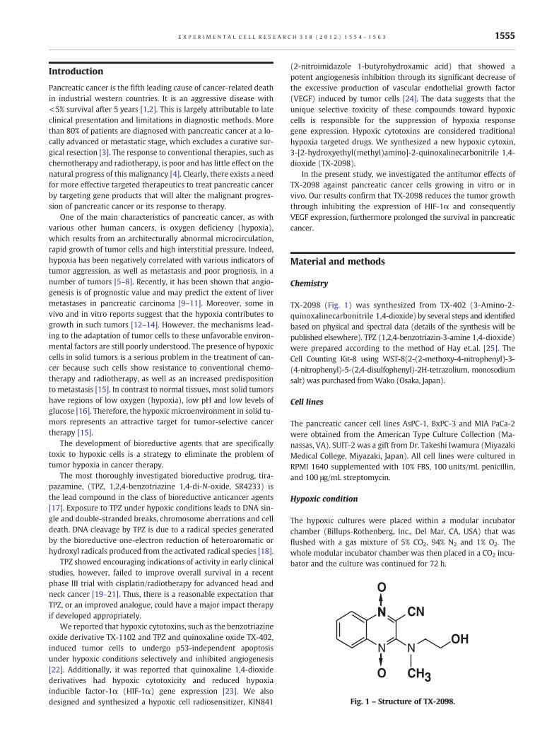
Fig. 1 – Structure of TX-2098.
1555E X P E R I M E N T A L C E L L R E S E A R C H 3 1 8 ( 2 0 1 2 ) 1 5 5 4 – 1 5 6 3
Introduction
Pancreatic cancer is the fifth leading cause of cancer-related deathin industrial western countries. It is an aggressive disease with<5% survival after 5 years [1,2]. This is largely attributable to lateclinical presentation and limitations in diagnostic methods. Morethan 80% of patients are diagnosed with pancreatic cancer at a lo-cally advanced or metastatic stage, which excludes a curative sur-gical resection [3]. The response to conventional therapies, such aschemotherapy and radiotherapy, is poor and has little effect on thenatural progress of this malignancy [4]. Clearly, there exists a needfor more effective targeted therapeutics to treat pancreatic cancerby targeting gene products that will alter the malignant progres-sion of pancreatic cancer or its response to therapy.
One of the main characteristics of pancreatic cancer, as withvarious other human cancers, is oxygen deficiency (hypoxia),which results from an architecturally abnormal microcirculation,rapid growth of tumor cells and high interstitial pressure. Indeed,hypoxia has been negatively correlated with various indicators oftumor aggression, as well as metastasis and poor prognosis, in anumber of tumors [5–8]. Recently, it has been shown that angio-genesis is of prognostic value and may predict the extent of livermetastases in pancreatic carcinoma [9–11]. Moreover, some invivo and in vitro reports suggest that the hypoxia contributes togrowth in such tumors [12–14]. However, the mechanisms lead-ing to the adaptation of tumor cells to these unfavorable environ-mental factors are still poorly understood. The presence of hypoxiccells in solid tumors is a serious problem in the treatment of can-cer because such cells show resistance to conventional chemo-therapy and radiotherapy, as well as an increased predispositionto metastasis [15]. In contrast to normal tissues, most solid tumorshave regions of low oxygen (hypoxia), low pH and low levels ofglucose [16]. Therefore, the hypoxic microenvironment in solid tu-mors represents an attractive target for tumor-selective cancertherapy [15].
The development of bioreductive agents that are specificallytoxic to hypoxic cells is a strategy to eliminate the problem oftumor hypoxia in cancer therapy.
The most thoroughly investigated bioreductive prodrug, tira-pazamine, (TPZ, 1,2,4-benzotriazine 1,4-di-N-oxide, SR4233) isthe lead compound in the class of bioreductive anticancer agents[17]. Exposure to TPZ under hypoxic conditions leads to DNA sin-gle and double-stranded breaks, chromosome aberrations and celldeath. DNA cleavage by TPZ is due to a radical species generatedby the bioreductive one-electron reduction of heteroaromatic orhydroxyl radicals produced from the activated radical species [18].
TPZ showed encouraging indications of activity in early clinicalstudies, however, failed to improve overall survival in a recentphase III trial with cisplatin/radiotherapy for advanced head andneck cancer [19–21]. Thus, there is a reasonable expectation thatTPZ, or an improved analogue, could have a major impact therapyif developed appropriately.
We reported that hypoxic cytotoxins, such as the benzotriazineoxide derivative TX-1102 and TPZ and quinoxaline oxide TX-402,induced tumor cells to undergo p53-independent apoptosisunder hypoxic conditions selectively and inhibited angiogenesis[22]. Additionally, it was reported that quinoxaline 1,4-dioxidederivatives had hypoxic cytotoxicity and reduced hypoxiainducible factor-1α (HIF-1α) gene expression [23]. We alsodesigned and synthesized a hypoxic cell radiosensitizer, KIN841
(2-nitroimidazole 1-butyrohydroxamic acid) that showed apotent angiogenesis inhibition through its significant decrease ofthe excessive production of vascular endothelial growth factor(VEGF) induced by tumor cells [24]. The data suggests that theunique selective toxicity of these compounds toward hypoxiccells is responsible for the suppression of hypoxia responsegene expression. Hypoxic cytotoxins are considered traditionalhypoxia targeted drugs. We synthesized a new hypoxic cytoxin,3-[2-hydroxyethyl(methyl)amino]-2-quinoxalinecarbonitrile 1,4-dioxide (TX-2098).
In the present study, we investigated the antitumor effects ofTX-2098 against pancreatic cancer cells growing in vitro or invivo. Our results confirm that TX-2098 reduces the tumor growththrough inhibiting the expression of HIF-1α and consequentlyVEGF expression, furthermore prolonged the survival in pancreaticcancer.
Material and methods
Chemistry
TX-2098 (Fig. 1) was synthesized from TX-402 (3-Amino-2-quinoxalinecarbonitrile 1,4-dioxide) by several steps and identifiedbased on physical and spectral data (details of the synthesis will bepublished elsewhere). TPZ (1,2,4-benzotriazin-3-amine 1,4-dioxide)were prepared according to the method of Hay et.al. [25]. TheCell Counting Kit-8 using WST-8(2-(2-methoxy-4-nitrophenyl)-3-(4-nitrophenyl)-5-(2,4-disulfophenyl)-2H-tetrazolium, monosodiumsalt) was purchased fromWako (Osaka, Japan).
Cell lines
The pancreatic cancer cell lines AsPC-1, BxPC-3 and MIA PaCa-2were obtained from the American Type Culture Collection (Ma-nassas, VA). SUIT-2 was a gift from Dr. Takeshi Iwamura (MiyazakiMedical College, Miyazaki, Japan). All cell lines were cultured inRPMI 1640 supplemented with 10% FBS, 100 units/mL penicillin,and 100 μg/mL streptomycin.
Hypoxic condition
The hypoxic cultures were placed within a modular incubatorchamber (Billups-Rothenberg, Inc., Del Mar, CA, USA) that wasflushed with a gas mixture of 5% CO2, 94% N2 and 1% O2. Thewhole modular incubator chamber was then placed in a CO2 incu-bator and the culture was continued for 72 h.
1556 E X P E R I M E N T A L C E L L R E S E A R C H 3 1 8 ( 2 0 1 2 ) 1 5 5 4 – 1 5 6 3
Cell proliferation assay
The effect of TX-2098 on cell proliferation was determined byWST-8 assay using Cell Counting Kit-8 as described previously[26]. Briefly, pancreatic cancer cells were washed with PBS andsuspended in a final concentration of 5×10⁴/mL in assay medium,and 100 μL of cell suspension was dispensed into 96-well plates.The plates were incubated at 37 °C for 24 h in a humidified CO2 in-cubator. The medium was aspirated from the wells; 100 μL ofserum-free medium was added into each well. Cells plated in 96-well plates were treated with the indicated concentrations ofTPZ or TX-2098 and cells were incubated for 72 h at 37 °C in a hyp-oxic chamber (1% O2). After the treatment period, WST-8 reagentwas added to cell culture medium (10 μL in 100 μL media), mixedgently, and incubated at 37 °C for 4 h. Plates were shaken vigor-ously on an orbital shaker for 1 min, and absorbance was mea-sured at 450 nm using a 96-well plate reader (Multiskan JX;Labsystems). Experiments were carried out in triplicate in dupli-cate plates.
Determination of VEGF protein levels in cell supernatant
To determine VEGF protein expression levels, equal densities ofcells were analyzed [27]. Pancreatic cancer cells were seeded at2×106 cells per 100 mm tissue culture plate. Cells were grownfor 24 h in RPMI 1640 supplemented with 10% fetal bovineserum (FBS), washed 2 times with phosphate-buffered saline(PBS), and transferred into 10 mL RPMI 1640 supplemented with10% FBS. Identical preparations for normoxic and hypoxic culturethen were incubated for 24 h and harvested at the same timepoint for protein analysis. The amount of VEGF protein found inthe supernatant from the cells was determined using anenzyme-linked immunosorbent (ELISA) assay kit (R&D Systems,Minneapolis, MN) according to the manufacturer's instructions.
Quantitative real-time reverse transcription-PCR
Tissue samples were homogenized with a Multi-Beads Shocker(Yasui-kikai, Osaka, Japan). RNA was extracted using RNeasyMini kit (Qiagen, Valencia, CA). RNA was reverse transcribedwith oligo-dT primers at 42 °C for 50 min using the SuperScriptFirst Strand System (Invitrogen).
Quantitative real-time reverse transcription-polymerase chainreaction (RT-PCR) was done on the ABI Prism 7500 using the com-mercially available gene expression assay for HIF-1α, VEGF,GLUT1, and Aldolase A and 28 S rRNA. A 25 μL final reaction vol-ume containing 1×TaqMan Universal PCR Master Mix (AppliedBiosystems), 1× Multiscribe with RNase inhibitors, and 1× geneexpression assay was used to amplify 25 ng total RNA with the fol-lowing cycling conditions: 30 min at 48 °C, 10 min at 95 °C, then50 cycles of 95 °C for 15 s and 60 °C for 1 min. The 7500 SequenceDetection System 1.3.1 software automatically determined foldchange for each gene in each sample using the ΔΔCT method[28]. Calculations were also done for each gene in tumors relativeto their corresponding matched normal tissue.
Animal model
To produce pancreatic tumors, SUIT-2 cells were harvested fromsubconfluent cultures by a brief exposure to 0.25% trypsin and
0.02% EDTA. Trypsinization was stopped with medium containing10% fetal bovine serum, and the cells were washed once inserum-free medium and resuspended in PBS. Only suspensionsconsisting of single cells with >90% viability were used for the in-jections. Four-five week old female Balb/c nude mice (CLEA Japan,Tokyo, Japan) were inoculated with 2× 10⁶ SUIT-2 cells in 200 μlPBS subcutaneously into the left flank. When the tumor grewapproximately100 mm3 in volume, i.e., the mice were dividedinto 6 groups consisting of 42 animals each with almost equalmean tumor volume, and themicewere given 0.2 ml of physiologicsaline or 13, 26 mg/kg of TPZ or 3, 6, 13 mg/kg of TX-2098 intraper-itoneally consecutively for 8 days. The mice were monitored everyday and the tumor volumewas estimated bymeasuring tumor sizeand using the following formula: tumor volume=0.5×L×W2,where L andW represent the largest diameter and the smallest di-ameter, respectively. After the final treatment, the animals weresacrificed, and tumors were removed; total RNA were isolatedfrom the removed tumors for RT-PCR analysis.
For the orthotopic xenograftmodel, 4–5 week old female Balb/cnude mice were used in accordance with institutional guidelines.2×106 SUIT-2 cells in 50 μl of PBS were injected into the pancreasas described previously [29]. On the 7th day after inoculation, themice were given 0.2 ml of physiologic saline or 13, 26 mg/kg ofTPZ or 3, 6, 13 mg/kg of TX-2098 intraperitoneally on eight consec-utive days. The mice were killed when moribund. Survival and theincidence of liver metastasis were recorded. All mice were main-tained according to the National Institute of Health standardsestablished in the Guidelines for the Care and Use of LaboratoryAnimals (http://oacu.od.nih.gov/regs/guide/guide2.htm), and allexperimental protocols were approved by the Animal Investiga-tion Committee of Tokushima University, Tokushima, Japan.
Statistical analysis
Statistical comparisons of mean values were done using one-wayANOVA. Survival analysis was computed by the Kaplan–Meiermethod and compared by the log-rank test. Statistical analysiswas performed using StatView 5.0J software (SAS Institute, Inc.,Cary, NC, USA). A P value of less than 0.05 was considered to bestatistically significant.
Results
Growth inhibitory effect of TX-2098 in pancreatic cancercells in vitro
To determinewhether TX-2098 inhibits the proliferation of humanpancreatic cancer cells, we first used AsPC-1, BxPC-3, MIA PaCa-2and SUIT-2. These cell lines were tested for viability using WST-8 assay after incubation with 0, 10, 20, 30, 50, 100 or 200 nM ofTPZ or TX-2098 for 72 h under hypoxic condition. As shown inFig. 2, TX-2098 inhibited the proliferation of human pancreaticcancer cell lines in a dose-dependent manner, compared with TPZ.
Expression of VEGF protein expression in the conditionedmedium
Previously, we found that TPZ treatment shows the antiangiogeniceffect through the inhibition of HIF-1α protein expression and

1557E X P E R I M E N T A L C E L L R E S E A R C H 3 1 8 ( 2 0 1 2 ) 1 5 5 4 – 1 5 6 3
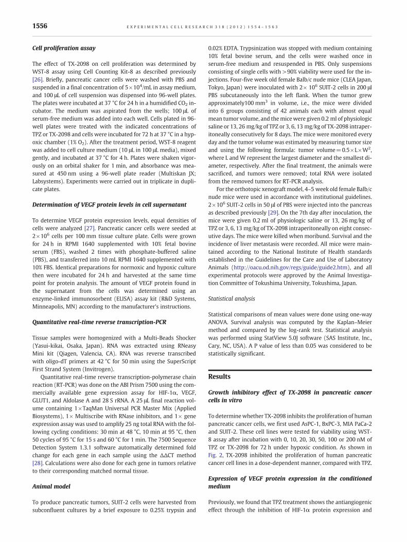
consequent reduction of VEGF protein expression under hypoxicconditions [30]. To assess whether VEGF protein secreted intothe medium decreased or not, we measured VEGF protein levelsin the pancreatic cancer cells conditioned medium. As shown inFig. 3, after 24 h, VEGF protein level in medium from cells culturedunder hypoxic conditions (AsPC-1: 2690 pg/mL, BxPC-3:13532 pg/mL, MIA PaCa-2: 8575 pg/mL, SUIT-2: 13712 pg/mL;
0
100
0 0 10 20 30 50 100 200
0 0 10 20 30 50 100 200 0 0 10 20 30 50 100 2
0 0 10 20 30 50 100 20
50
0
100
50
AsPC-1 BxPC-3
AsPC-1 BxPC-3
TX-2098(nM)
(%)
(%)
Cell
Via
bili
ty (
% o
f co
ntr
ol)
Cell
Via
bili
ty (
% o
f co
ntr
ol)
TX-2098(nM1%O2 1%O220%O2 20%O2
1%O220%O2 1%O220%O2
(a)
(b)
*** *** *** *** *** ***
**** **
* ***
***
******
******
*** *** ***
***
***
****** ***
TPZ(nM) TPZ(nM)
Fig. 2 – Dose-dependent cytotoxicity of TPZ or TX-2098 for humanTPZ or TX-2098, viable cells were counted by WST-8 assay. Experim*P<0.05 versus control under normoxic condition (20% O2), **P<0cultures at each dose.
p<0.05 versus normoxic conditions) increased rather than thatfrom cells cultured under normoxic conditions (AsPC-1: 864 pg/mL, BxPC-3: 8273 pg/mL, MIA PaCa-2: 3296 pg/mL, SUIT-2:7507 pg/mL) on human pancreatic cancer cell lines. Comparedwith VEGF protein level in medium from untreated cells grownunder hypoxic conditions, VEGF protein level in medium fromcells cultured with TX-2098 was reduced, especially in SUIT-2
00 0 0 10 20 30 50 100 2000 0 10 20 30 50 100 200
0 0 0 10 20 30 50 100 200 0 0 10 20 30 50 100 200
MIA PaCa-2 SUIT-2
MIA PaCa-2 SUIT-2
TX-2098(nM) TX-2098(nM))1%O2 1%O220%O2 20%O2
1%O220%O21%O220%O2
****
*** *** ****** ***
***
****** ***
***
***
***
***
***
***
***
****** *** *** ***
TPZ(nM)TPZ(nM)
pancreatic cancer cell lines. At 72 h after the indicated dose ofents were performed in triplicate; data are the mean±SE..05 versus control under hypoxic condition (1% O2) between
1558 E X P E R I M E N T A L C E L L R E S E A R C H 3 1 8 ( 2 0 1 2 ) 1 5 5 4 – 1 5 6 3
cell, VEGF protein level in medium from cells was reduced in adose-dependent manner (p<0.05). Moreover, TX-2098 reducedVEGF protein level in medium from cells cultured rather thanTPZ (p<0.05).
Expression of HIF-1α, VEGF, GLUT1 and Aldolase A in vitro
To elucidate whether TX-2098 inhibit the expression of HIF-1αand consequently VEGF, GLUT1 and Aldolase A in vitro, we per-formed quantitative real-time RT-PCR analysis. SUIT-2 cells weretreated TPZ or TX-2098 on hypoxia or normoxia for 24 h, andtotal cellular RNA was isolated for quantitative real-time RT-PCR.As shown in Fig. 4, quantitative real-time RT-PCR analysis revealeda significant decrease of mRNA expression of VEGF, GLUT1 and
AsPC-1
0
50
100
150
200
250
300
20 50 1000
(pg)
VE
GF
1%O220%O2
1%O220%O2
(a)
* **
*
#
#
MIA PaCa -2
0
100
200
300
400
500
600
700
800
900
1000
20 50 1000
TPZ, TX-2098(nM)
TPZ, TX-2098(nM)
(pg)
VE
GF
(c)
* * **
#
# Control 20%O2
Control 1%O2
TPZ 1%O2TX-2098 1%O2
Control 20%O2
Control 1%O2
TPZ 1%O2TX-2098 1%O2
Fig. 3 – Effect of TPZ and TX-2098 treatment on experimentally induCells were cultured for 24 h under normoxic (20%O2) or hypoxic condof TPZ (0, 20, 50, 100, 200 nM) and TX-2098 (0, 20, 50, 100, 200 nM).using an enzyme-linked immunosorbent assay. Experiments were pecomparison with normoxic cells; * P<0.05 in comparison with untre
Aldolase A in TX-2098 treatment groups compared with the hyp-oxic control group, while there were no significant differences inHIF-1α (p<0.05).
Antitumor efficacy of TX-2098 in xenograft models
We then examined the effects of TX-2098 in vivo model usingSUIT-2 cells. SUIT-2 cells were implanted into the left flank ofnude mice, and animals were vehicle-treated with TPZ or TX-2098. As shown in Fig. 5a, the mean tumor volume was 445±219 mm3 in control, 326±92 mm3 in 13 mg/kg TPZ, 233±39 mm3 in 26 mg/kg TPZ, 276±72 mm3 in 3 mg/kg TX-2098,161±87 mm3 in 6 mg/kg TX-2098, 114±36 mm3 in 13 mg/kgTX-2098. The tumor volume in TPZ group was significantly lower
BxPC-3
0
200
400
600
800
1000
1200
1400
1600
20 50 1000
1%O220%O2
1%O220%O2
(pg)
VE
GF
(b)
* * *
#
##
TPZ, TX-2098(nM)
TPZ, TX-2098(nM)
SUIT-2
0
200
400
600
800
1000
1200
1400
1600
20 50 100 2000
(pg)
VE
GF
(d)
*
*
*
*
#
#
#
#
Control 20%O2
Control 1%O2
TPZ 1%O2TX-2098 1%O2
Control 20%O2
Control 1%O2
TPZ 1%O2TX-2098 1%O2
ced vascular endothelial growth factor (VEGF) protein secretion.itions (1%O2), eitherwithout TPZ and TX-2098 or in the presenceVEGF protein levels were measured in conditioned cell mediumrformed in triplicate; data are the mean±SE. # P<0.05 inated cells; 24 h under hypoxic conditions.

(b)
(c)
0
0.5
1.0
1.5
2.0
2.5
3.0
3.5
4.0
4.5
0 50 100 200
Rel
ativ
e V
EG
F m
RN
A e
xpre
ssio
n(d)
0
0.2
0.4
0.6
0.8
1.0
1.2
1.4
1.6
1.8
2.0
0 50 100 200
Rel
ativ
e A
ldol
ase
Am
RN
A e
xpre
ssio
n
*
#
*
*
**
*
*
*
**
#
#
#
*
**
*
#
(a)
0
1
2
3
4
5
6
0 50 100 200
Rel
ativ
e H
IF-1
amR
NA
exp
ress
ion
0
0.5
1.0
1.5
2.0
2.5
0 50 100 200
Rel
ativ
e G
LUT
1 m
RN
A e
xpre
ssio
n
TPZ, TX-2098(nM) TPZ, TX-2098(nM)1%O2
20%O2 1%O220%O2
TPZ, TX-2098(nM) TPZ, TX-2098(nM)
1%O220%O2 1%O2
20%O2
*
*
* *
**
Control 20%O2
Control 1%O2
TPZ 1%O2TX-2098 1%O2
Control 20%O2
Control 1%O2
TPZ 1%O2TX-2098 1%O2
Control 20%O2
Control 1%O2
TPZ 1%O2TX-2098 1%O2Control 20%O2
Control 1%O2
TPZ 1%O2TX-2098 1%O2
Fig. 4 – In treatment with TPZ or TX-2098, quantitative real-time RT-PCR analysis of HIF-1α, VEGF, GLUT1 and Aldolase A mRNAlevels in the pancreatic cancer cell line SUIT-2. Quantitative real-time RT-PCR revealed down-regulation of HIF-1α, VEGF, GLUT1 andAldolase AmRNA in TPZ or TX-2098 treatment. Columns, mean percentage of mRNA expression; bars, ± SE; # P<0.05 in comparisonwith normoxic cells; * P<0.05 in comparison with untreated cells; 24 h under hypoxic conditions.
1559E X P E R I M E N T A L C E L L R E S E A R C H 3 1 8 ( 2 0 1 2 ) 1 5 5 4 – 1 5 6 3
compared with the control group by day 27 (p<0.05) and, in TX-2098 group, were also statistically significant and much lowercompared with control group (p<0.01). As shown in Fig. 5b,there were no differences in body weight between the four re-spective treatment groups caused by the medication.
Expression of HIF-1α and consequently VEGF, GLUT1, AldolaseA in SUIT-2 xenograft
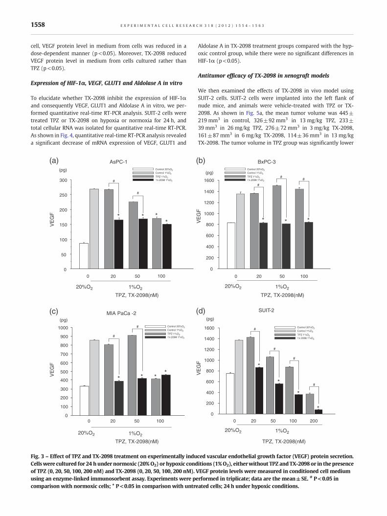
To elucidate whether TX-2098 inhibit the expression of HIF-1αand consequently VEGF, GLUT1 and Aldolase A in SUIT-2 xeno-graft, we preformed quantitative real-time RT-PCR analysis of tu-mors of SUIT-2 xenograft. As shown in Fig. 6, quantitative real-time RT-PCR analysis revealed a significant decrease of mRNA
expression of VEGF, GLUT1 and Aldolase A in the TPZ (26 mg/kg)or TX-2098 (3, 6, 13 mg/kg) treatment groups compared withthe control, while there were no significant differences in HIF-1α(p<0.05).
Survival in orthotopic SUIT-2 xenograft
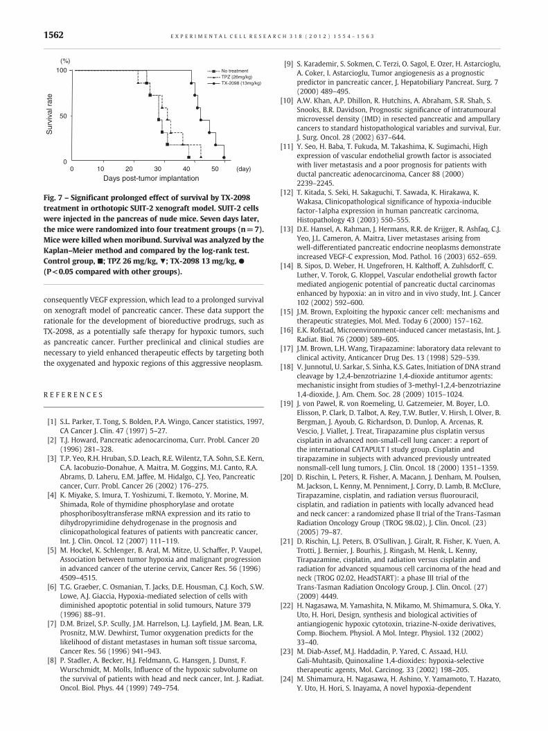
SUIT-2 cells were injected into the pancreas of nude mice to eval-uate whether TX-2098 treatment had an effect on survival. Sevendays later, the mice were randomly placed into four groups of 7mice each. Whenmoribund, mice were killed, and primary tumorsin the pancreas and liver were harvested. As expected, all of themice injected with SUIT-2 cells presented pancreatic tumors. Asshown in Fig. 7, the median survival time for the control group

(a)
(b)
0
5
10
15
20
25
1 2 7 84 653 (day)
(g)
Bod
y w
eigh
t
0
100
200
300
400
500
600
1 2 7 84 653 (day)
(mm3)
Tum
or v
olum
e
No treatmentTPZ 13mg/kgTPZ 26mg/kgTX-2098 3mg/kgTX-2098 6mg/kgTX-2098 13mg/kg
*#
**
#
No treatment
TPZ 13mg/kg
TPZ 26mg/kg
TX-2098 3mg/kg
TX-2098 6mg/kg
TX-2098 13mg/kg
Fig. 5 – TPZ and TX-2098 treatment delays tumor growth inSUIT-2 xenografts. (a) daily injections of TPZ (13 and 26 mg/kg/injection, i.p.) or TX-2098 (3, 6 and 13 mg/kg/injection, i.p.)during the consecutive 8 days in SUIT-2 tumors. After thetumor volume reached 100 mm3, the volume was plottedagainst the time after the initiation of the treatment. Controlgroup, ●; TPZ 13 mg/kg, ▲; TPZ 26 mg/kg, ■; TX-2098 3 mg/kg,▼; TX-2098 6 mg/kg, +; TX-2098 13 mg/kg, ×. Points, means of7 mice; bars, ± SD. * P<0.05 versus control group; #P<0.01versus control group. (b) during the 8 days, there was nodifference in body weights between mice treated with control,TPZ and TX-2098.
1560 E X P E R I M E N T A L C E L L R E S E A R C H 3 1 8 ( 2 0 1 2 ) 1 5 5 4 – 1 5 6 3
was 29 days. After treatment with 26 mg/kg TPZ or 13 mg/kg TX-2098, the median survival time was 31 and 43 days, respectively(control, 26 mg/kg TPZ versus 13 mg/kg TX-2098 group, p<0.001).
Discussion
Regions of acute/chronic hypoxia are present in the majority ofsolid human tumors [29,31,32]. The level of hypoxia in tumorshas a profound influence on the outcome of cancer chemotherapyand radiation therapy and is a strong prognostic factor for diseaseprogression and survival. A hypoxic condition in solid tumorsaccelerates malignant progression and increases metastasis[5,29,33–35]. Pancreatic cancer also shows significant regions ofhypoxia that are often resistant to cell killing by chemotherapeu-tics [36–39]. Therefore, hypoxic cells are important targets for
pancreatic cancer therapy. The major goal for our research is todevelop new hypoxic cytotoxins that possess additional potent cy-tostatic biological effects including antiangiogenic activity.
Tumor hypoxia induces the releases of growth factors such asVEGF that promote vascularization and enhance both tumorgrowth and metastasis [40]. HIF-1 exerts the transcriptional ac-tivity by means of binding to its consensus binding site withinthe hypoxia-responding element in the target genes, such asVEGF, erythropoietin (EPO), glucose phosphate isomerase (GPI)and BNIP3, whose protein products induce the developmentand function of the vascular system, the formation and matura-tion of the blood cells, and the energy metabolism of cells[41–44]. Accordingly, it seems that HIF-1 plays an importantrole in neoangiogenesis, proliferation, infiltration, and metastasisin tumor cells.
Hypoxia-activated prodrugs, such as TX-2098, are developed toselectively target hypoxic tumor tissues while having minimalsystemic toxicity. In the present study, we showed that TX-2098,a new hypoxic cytotoxin had significant anticancer effects includ-ing an antiangiogenic effect and underwent dose-dependent andselective significant inhibition of tumor growth and progressionin pancreatic xenograft models (Figs. 2 and 5). TX-2098 reducedthe increased levels of mRNA and protein expression of VEGFunder hypoxic condition. Similary, TX-2098 inhibited the expres-sion HIF-1α and hypoxia related gene (Figs. 4 and 6). Certainly,while TX-2098 inhibited the HIF-1a mRNA, there was no differ-ence of mRNA expression, even though there were differences ofother hypoxia related gene expression between TPZ and TX-2098. At this point, we need to proceed the experiment for thisdrug and need to clarify the detail of mechanism.
There is no question regarding the importance of VEGF inangiogenesis and expansion of solid tumors, because VEGF partic-ularly affects vascular endothelial cells and, consequently, stimu-lates all steps of angiogenesis. However, several reports revealedthat angiogenic factors other than VEGF play important roles inangiogenesis [45,46]. In melanoma cell lines, a strong correlationbetween VEGF expression and tumorigenesis was not observed,although VEGF production was induced under the hypoxic condi-tion [45]. Angiogenin production was reported to be upregulatedin melanoma cell lines but not in normal melanocytes under hyp-oxic condition. The level of induction of angiogenin correlatedwith the metastatic potential of the cell lines. Furthermore, astrong expression of angiogenin was observed in melanomas andmetastases from patients, but was not observed in benign nevi.These indicated that angiogenin is the most potent angiogenic fac-tor in melanomas. Various approaches to inhibiting angiogenicfactor signaling pathways have been adopted using specific anti-bodies against various angiogenic factors. A monoclonal antibodyagainst VEGF inhibited angiogenesis and prevented tumor growthin solid tumors [47,48]. However, a specific antibody against oneangiogenic factor can not completely inhibit angiogenesis, whichdepends on two or more factors or a different type of angiogenicfactor. This problem may be overcome if hypoxia-induced angio-genic factor-producing cells can be attacked and the productionof angiogenic factors by hypoxic cells can be inhibited. TX-2098may be able to inhibit the production of all types of hypoxia-induced angiogenic factors in tumor cells. In the present study,TX-2098 strongly inhibited the VEGF protein expression (Fig. 3),suggesting that TX-2098 effect is caused by inhibiting VEGF pro-tein expression.
(d)
0
0.2
0.4
0.6
0.8
1.0
1.2
1.4
Rel
ativ
e A
LDLA
SE
A m
RN
A e
xpre
ssio
n
* * **
TPZ13 mg/kg
TPZ26 mg/kg
TX-20983 mg/kg
TX-20986 mg/kg
TX-209813 mg/kg
Control
(b)
TPZ13 mg/kg
TPZ26 mg/kg
TX-20983 mg/kg
TX-20986 mg/kg
TX-209813 mg/kg
Control
0
0.2
0.4
0.6
0.8
1.0
1.2
Rel
ativ
e V
EG
F m
RN
A e
xpre
ssio
n
*
** *
(a)
0
0.2
0.4
0.6
0.8
1.0R
elat
ive
HIF
-1α
mR
NA
exp
ress
ion
TPZ13 mg/kg
TPZ26 mg/kg
TX-20983 mg/kg
TX-20986 mg/kg
TX-209813 mg/kg
Control
(c)
TPZ13 mg/kg
TPZ26 mg/kg
TX-20983 mg/kg
TX-20986 mg/kg
TX-209813 mg/kg
Control
0
0.2
0.4
0.6
0.8
1.0
1.2
1.4
1.6
Rel
ativ
e G
LUT
1 m
RN
A e
xpre
ssio
n
**
*
**
Fig. 6 – Quantitative real-time RT-PCR analysis of HIF-1α, VEGF, GLUT1 and Aldolase AmRNA levels in SUIT-2 xenograft tumors. HIF-1α, VEGF, GLUT1 and Aldolase A mRNA level were decreased in TX-2098 treatment. Columns, mean percentage of mRNA expression;bars, ± SE; * P<0.05 versus control group.
1561E X P E R I M E N T A L C E L L R E S E A R C H 3 1 8 ( 2 0 1 2 ) 1 5 5 4 – 1 5 6 3
In a recent prodrug approach, these hypoxic cytotoxins wereused in combination with suicide genes (gene-directed enzymeprodrug therapy: GDEPT). In the GDEPT strategy, the expressionof a prodrug-activating redox enzyme gene in hypoxic conditionselectively makes the bioreductive anticancer drugs cytotoxicand keeps the concentration of active species high and stablewithin hypoxic tumor cells, even in the heterogeneous tumor mi-croenvironment. To achieve higher gene expression selectively inhypoxic regions, hypoxia response promoters containing the hyp-oxia response element (HRE) were designed and used to controlthe fusion gene expression through HIF-1 pathway [49,50].
At present, the leading hypoxic cytotoxin is the benzotriazine-N-oxide, TPZ, which has been shown to have high hypoxic cytotox-icity and has been promoted to clinical trials [51,52]. TPZ is activat-ed through a one-electron reduction to produce a hydroxyl radicalthat damages DNA. In hypoxic conditions, tirapazamine is reportedto exert 50 to 300 times stronger cytotoxicity than in normoxiccondition. Our studies have shown that TPZ can induce apoptosisat various effective concentrations in either hypoxic or normoxicconditions [24,53]. The apoptosis occurred independently of p53
at low dose in hypoxic condition, while in normoxic conditionsthe apoptosis induced at high dose was partially p53-dependent.
In comparison, TX-2098 showed more potent hypoxic cytotox-icity than TPZ in this study (Fig. 2). Furthermore, we have foundthat these heterocyclic aromatic hypoxic cytotoxins also pre-vented the hypoxia signal transduction pathway mediated byHIF-1α inhibition under hypoxic condition and inhibited angio-genesis significantly at a dose of 50 nM or greater in the VEGFELISA assay. TX-2098 reduced HIF-1α mRNA expression dose-dependently and suppressed the induction of mRNA of VEGF,GLUT1 and Aldolase A. Their potent antiangiogenic effects can beattributed to the suppression of VEGF via their block of the hypox-ic signaling pathway. In addition, as TX-2098 prolonged the syrvi-val in orthotopic model, TX-2098 also has the unlimitedpossibilities for pancreatic cancer therapy (Fig. 7). In this experi-ment, although the detailed mechanism is not clear, except forthe possibility through growth and angiogenesis inhibition, wehave already started next experiment to clarify that.
In conclusion, TX-2098 showed the effects of tumor growthreduction through inhibiting the expression of HIF-1α, and
0
100
0 10 20 30 40 50
(%)
Sur
viva
l rat
e
Days post-tumor implantation
50
(day)
TX-2098 (13mg/kg)TPZ (26mg/kg)No treatment
Fig. 7 – Significant prolonged effect of survival by TX-2098treatment in orthotopic SUIT-2 xenograft model. SUIT-2 cellswere injected in the pancreas of nude mice. Seven days later,the mice were randomized into four treatment groups (n=7).Mice were killed when moribund. Survival was analyzed by theKaplan–Meier method and compared by the log-rank test.Control group, ■; TPZ 26 mg/kg, ▼; TX-2098 13 mg/kg, ●(P<0.05 compared with other groups).
1562 E X P E R I M E N T A L C E L L R E S E A R C H 3 1 8 ( 2 0 1 2 ) 1 5 5 4 – 1 5 6 3
consequently VEGF expression, which lead to a prolonged survivalon xenograft model of pancreatic cancer. These data support therationale for the development of bioreductive prodrugs, such asTX-2098, as a potentially safe therapy for hypoxic tumors, suchas pancreatic cancer. Further preclinical and clinical studies arenecessary to yield enhanced therapeutic effects by targeting boththe oxygenated and hypoxic regions of this aggressive neoplasm.
R E F E R E N C E S
[1] S.L. Parker, T. Tong, S. Bolden, P.A. Wingo, Cancer statistics, 1997,CA Cancer J. Clin. 47 (1997) 5–27.
[2] T.J. Howard, Pancreatic adenocarcinoma, Curr. Probl. Cancer 20(1996) 281–328.
[3] T.P. Yeo, R.H. Hruban, S.D. Leach, R.E. Wilentz, T.A. Sohn, S.E. Kern,C.A. Iacobuzio-Donahue, A. Maitra, M. Goggins, M.I. Canto, R.A.Abrams, D. Laheru, E.M. Jaffee, M. Hidalgo, C.J. Yeo, Pancreaticcancer, Curr. Probl. Cancer 26 (2002) 176–275.
[4] K. Miyake, S. Imura, T. Yoshizumi, T. Ikemoto, Y. Morine, M.Shimada, Role of thymidine phosphorylase and orotatephosphoribosyltransferase mRNA expression and its ratio todihydropyrimidine dehydrogenase in the prognosis andclinicopathological features of patients with pancreatic cancer,Int. J. Clin. Oncol. 12 (2007) 111–119.
[5] M. Hockel, K. Schlenger, B. Aral, M. Mitze, U. Schaffer, P. Vaupel,Association between tumor hypoxia and malignant progressionin advanced cancer of the uterine cervix, Cancer Res. 56 (1996)4509–4515.
[6] T.G. Graeber, C. Osmanian, T. Jacks, D.E. Housman, C.J. Koch, S.W.Lowe, A.J. Giaccia, Hypoxia-mediated selection of cells withdiminished apoptotic potential in solid tumours, Nature 379(1996) 88–91.
[7] D.M. Brizel, S.P. Scully, J.M. Harrelson, L.J. Layfield, J.M. Bean, L.R.Prosnitz, M.W. Dewhirst, Tumor oxygenation predicts for thelikelihood of distant metastases in human soft tissue sarcoma,Cancer Res. 56 (1996) 941–943.
[8] P. Stadler, A. Becker, H.J. Feldmann, G. Hansgen, J. Dunst, F.Wurschmidt, M. Molls, Influence of the hypoxic subvolume onthe survival of patients with head and neck cancer, Int. J. Radiat.Oncol. Biol. Phys. 44 (1999) 749–754.
[9] S. Karademir, S. Sokmen, C. Terzi, O. Sagol, E. Ozer, H. Astarcioglu,A. Coker, I. Astarcioglu, Tumor angiogenesis as a prognosticpredictor in pancreatic cancer, J. Hepatobiliary Pancreat. Surg. 7(2000) 489–495.
[10] A.W. Khan, A.P. Dhillon, R. Hutchins, A. Abraham, S.R. Shah, S.Snooks, B.R. Davidson, Prognostic significance of intratumouralmicrovessel density (IMD) in resected pancreatic and ampullarycancers to standard histopathological variables and survival, Eur.J. Surg. Oncol. 28 (2002) 637–644.
[11] Y. Seo, H. Baba, T. Fukuda, M. Takashima, K. Sugimachi, Highexpression of vascular endothelial growth factor is associatedwith liver metastasis and a poor prognosis for patients withductal pancreatic adenocarcinoma, Cancer 88 (2000)2239–2245.
[12] T. Kitada, S. Seki, H. Sakaguchi, T. Sawada, K. Hirakawa, K.Wakasa, Clinicopathological significance of hypoxia-induciblefactor-1alpha expression in human pancreatic carcinoma,Histopathology 43 (2003) 550–555.
[13] D.E. Hansel, A. Rahman, J. Hermans, R.R. de Krijger, R. Ashfaq, C.J.Yeo, J.L. Cameron, A. Maitra, Liver metastases arising fromwell-differentiated pancreatic endocrine neoplasms demonstrateincreased VEGF-C expression, Mod. Pathol. 16 (2003) 652–659.
[14] B. Sipos, D. Weber, H. Ungefroren, H. Kalthoff, A. Zuhlsdorff, C.Luther, V. Torok, G. Kloppel, Vascular endothelial growth factormediated angiogenic potential of pancreatic ductal carcinomasenhanced by hypoxia: an in vitro and in vivo study, Int. J. Cancer102 (2002) 592–600.
[15] J.M. Brown, Exploiting the hypoxic cancer cell: mechanisms andtherapeutic strategies, Mol. Med. Today 6 (2000) 157–162.
[16] E.K. Rofstad, Microenvironment-induced cancer metastasis, Int. J.Radiat. Biol. 76 (2000) 589–605.
[17] J.M. Brown, L.H. Wang, Tirapazamine: laboratory data relevant toclinical activity, Anticancer Drug Des. 13 (1998) 529–539.
[18] V. Junnotul, U. Sarkar, S. Sinha, K.S. Gates, Initiation of DNA strandcleavage by 1,2,4-benzotriazine 1,4-dioxide antitumor agents:mechanistic insight from studies of 3-methyl-1,2,4-benzotriazine1,4-dioxide, J. Am. Chem. Soc. 28 (2009) 1015–1024.
[19] J. von Pawel, R. von Roemeling, U. Gatzemeier, M. Boyer, L.O.Elisson, P. Clark, D. Talbot, A. Rey, T.W. Butler, V. Hirsh, I. Olver, B.Bergman, J. Ayoub, G. Richardson, D. Dunlop, A. Arcenas, R.Vescio, J. Viallet, J. Treat, Tirapazamine plus cisplatin versuscisplatin in advanced non-small-cell lung cancer: a report ofthe international CATAPULT I study group. Cisplatin andtirapazamine in subjects with advanced previously untreatednonsmall-cell lung tumors, J. Clin. Oncol. 18 (2000) 1351–1359.
[20] D. Rischin, L. Peters, R. Fisher, A. Macann, J. Denham, M. Poulsen,M. Jackson, L. Kenny, M. Penniment, J. Corry, D. Lamb, B. McClure,Tirapazamine, cisplatin, and radiation versus fluorouracil,cisplatin, and radiation in patients with locally advanced headand neck cancer: a randomized phase II trial of the Trans-TasmanRadiation Oncology Group (TROG 98.02), J. Clin. Oncol. (23)(2005) 79–87.
[21] D. Rischin, L.J. Peters, B. O'Sullivan, J. Giralt, R. Fisher, K. Yuen, A.Trotti, J. Bernier, J. Bourhis, J. Ringash, M. Henk, L. Kenny,Tirapazamine, cisplatin, and radiation versus cisplatin andradiation for advanced squamous cell carcinoma of the head andneck (TROG 02.02, HeadSTART): a phase III trial of theTrans-Tasman Radiation Oncology Group, J. Clin. Oncol. (27)(2009) 4449.
[22] H. Nagasawa, M. Yamashita, N. Mikamo, M. Shimamura, S. Oka, Y.Uto, H. Hori, Design, synthesis and biological activities ofantiangiogenic hypoxic cytotoxin, triazine-N-oxide derivatives,Comp. Biochem. Physiol. A Mol. Integr. Physiol. 132 (2002)33–40.
[23] M. Diab-Assef, M.J. Haddadin, P. Yared, C. Assaad, H.U.Gali-Muhtasib, Quinoxaline 1,4-dioxides: hypoxia-selectivetherapeutic agents, Mol. Carcinog. 33 (2002) 198–205.
[24] M. Shimamura, H. Nagasawa, H. Ashino, Y. Yamamoto, T. Hazato,Y. Uto, H. Hori, S. Inayama, A novel hypoxia-dependent
1563E X P E R I M E N T A L C E L L R E S E A R C H 3 1 8 ( 2 0 1 2 ) 1 5 5 4 – 1 5 6 3
2-nitroimidazole KIN-841 inhibits tumour-specific angiogenesisby blocking production of angiogenic factors, Br. J. Cancer 88(2003) 307–313.
[25] M.P. Hay, S.A. Gamage, M.S. Kovacs, F.B. Pruijn, R.F. Anderson, A.V.Patterson, W.R. Wilson, J.M. Brown, W.A. Denny, Structure−activity relationships of 1,2,4-benzotriazine 1,4-dioxides ashypoxia-selective analogues of tirapazamine, J. Med. Chem. 46(2003) 169–182.
[26] K. Miyake, M. Shimada, M. Nishioka, K. Sugimoto, E. Batmunkh, Y.Uto, H. Nagasawa, H. Hori, Downregulation of matrixmetalloprotease-9 and urokinase plasminogen activator byTX-1877 results in decreased tumor growth and metastasis onxenograft model of rectal cancer, Cancer Chemother. Pharmacol.64 (2009) 885–892.
[27] K. Miyake, S. Imura, M. Nishioka, E. Batmunkh, K. Sugimoto, Y.Ohmoto, M. Shimada, Serum evaluation of solubleinterferon-alpha/beta receptor and high-sensitivity C-reactiveprotein for diagnosis of the patients with gastrointestinal andhepatobiliary-pancreatic cancer, Cytokine 49 (2010) 251–255.
[28] M.W. Pfaffl, A new mathematical model for relativequantification in real-time RT-PCR, Nucleic Acids Res. 29 (2001)e45.
[29] K. Miyake, K. Tsuchida, H. Sugino, S. Imura, Y. Morine, M. Fujii, M.Shimada, Combination therapy of human pancreatic cancerimplanted in nude mice by oral fluoropyrimidine anticanceragent (S-1) with interferon-alpha, Cancer Chemother.Pharmacol. 59 (2007) 113–126.
[30] H. Nagasawa, N. Mikamo, Y. Nakajima, H. Matsumoto, Y. Uto, H.Hori, Antiangiogenic hypoxic cytotoxin TX-402 inhibitshypoxia-inducible factor 1 signaling pathway, Anticancer Res. 23(2003) 4427–4434.
[31] K. Miyake, T. Yoshizumi, S. Imura, K. Sugimoto, E. Batmunkh, H.Kanemura, Y. Morine, M. Shimada, Expression ofhypoxia-inducible factor-1alpha, histone deacetylase 1, andmetastasis-associated protein 1 in pancreatic carcinoma:correlation with poor prognosis with possible regulation,Pancreas 36 (2008) e1–e9.
[32] M. Hockel, K. Schlenger, C. Knoop, P. Vaupel, Oxygenation ofcarcinomas of the uterine cervix: evaluation by computerized O2tension measurements, Cancer Res. 51 (1991) 6098–6102.
[33] D.M. Brizel, G.S. Sibley, L.R. Prosnitz, R.L. Scher, M.W. Dewhirst,Tumor hypoxia adversely affects the prognosis of carcinoma of thehead and neck, Int. J. Radiat. Oncol. Biol. Phys. 38 (1997) 285–289.
[34] D.M. Brizel, R.K. Dodge, R.W. Clough, M.W. Dewhirst,Oxygenation of head and neck cancer: changes duringradiotherapy and impact on treatment outcome, Radiother.Oncol. 53 (1999) 113–117.
[35] R. Cairns, I. Papandreou, N. Denko, Overcoming physiologicbarriers to cancer treatment by molecularly targeting the tumormicroenvironment, Mol. Cancer Res. 4 (2006) 61–70.
[36] K. Ranniger, R.M. Saldino, Arteriographic diagnosis of pancreaticlesions, Radiology 86 (1966) 470–474.
[37] N.A. Yassa, J. Yang, S. Stein, M. Johnson, P. Ralls, Gray-scale andcolor flow sonography of pancreatic ductal adenocarcinoma,J. Clin. Ultrasound 25 (1997) 473–480.
[38] A.J. Megibow, Pancreatic adenocarcinoma: designing theexamination to evaluate the clinical questions, Radiology 183(1992) 297–303.
[39] A.C. Koong, V.K. Mehta, Q.T. Le, G.A. Fisher, D.J. Terris, J.M. Brown,A.J. Bastidas, M. Vierra, Pancreatic tumors show high levels ofhypoxia, Int. J. Radiat. Oncol. Biol. Phys. 48 (2000) 919–922.
[40] C. Brahimi-Horn, E. Berra, J. Pouyssegur, Hypoxia: the tumor'sgateway to progression along the angiogenic pathway, TrendsCell Biol. 11 (2001) S32–S36.
[41] G.L. Semenza, P.H. Roth, H.M. Fang, G.L. Wang, Transcriptionalregulation of genes encoding glycolytic enzymes byhypoxia-inducible factor 1, J. Biol. Chem. 269 (1994)23757–23763.
[42] J.A. Forsythe, B.H. Jiang, N.V. Iyer, F. Agani, S.W. Leung, R.D. Koos,G.L. Semenza, Activation of vascular endothelial growth factorgene transcription by hypoxia-inducible factor 1, Mol. Cell. Biol.16 (1996) 4604–4613.
[43] B.H. Jiang, E. Rue, G.L. Wang, R. Roe, G.L. Semenza, Dimerization,DNA binding, and transactivation properties of hypoxia-induciblefactor 1, J. Biol. Chem. 271 (1996) 17771–17778.
[44] H.M. Sowter, P.J. Ratcliffe, P. Watson, A.H. Greenberg, A.L. Harris,HIF-1-dependent regulation of hypoxic induction of the celldeath factors BNIP3 and NIX in human tumors, Cancer Res. 61(2001) 6669–6673.
[45] A. Hartmann,M. Kunz, S. Kostlin, R. Gillitzer, A. Toksoy, E.B. Brocker,C.E. Klein, Hypoxia-induced up-regulation of angiogenin in humanmalignant melanoma, Cancer Res. 59 (1999) 1578–1583.
[46] J. Koga, Y. Kakeji, Y. Sumiyoshi, Y. Kimura, K. Shibahara, Y. Emi, Y.Maehara, K. Sugimachi, Angiogenesis and macrophageinfiltration in Borrmann type IV gastric cancer, Fukuoka IgakuZasshi 92 (2001) 334–339.
[47] P. Borgstrom, K.J. Hillan, P. Sriramarao, N. Ferrara, Completeinhibition of angiogenesis and growth of microtumors byanti-vascular endothelial growth factor neutralizing antibody:novel concepts of angiostatic therapy from intravitalvideomicroscopy, Cancer Res. 56 (1996) 4032–4039.
[48] R.A. Brekken, P.E. Thorpe, Vascular endothelial growth factor andvascular targeting of solid tumors, Anticancer Res. 21 (2001)4221–4229.
[49] A.V. Patterson, K.J. Williams, R.L. Cowen, M. Jaffar, B.A. Telfer, M.Saunders, R. Airley, D. Honess, A.J. van der Kogel, C.R. Wolf, I.J.Stratford, Oxygen-sensitive enzyme-prodrug gene therapy forthe eradication of radiation-resistant solid tumours, Gene Ther. 9(2002) 946–954.
[50] T. Shibata, A.J. Giaccia, J.M. Brown, Hypoxia-inducible regulationof a prodrug-activating enzyme for tumor-specific gene therapy,Neoplasia 4 (2002) 40–48.
[51] A. Rosenberg, S. Knox, Radiation sensitization with redoxmodulators: a promising approach, Int. J. Radiat. Oncol. Biol. Phys.64 (2006) 343–354.
[52] A. Covens, J. Blessing, D. Bender, R. Mannel, M. Morgan, A phase IIevaluation of tirapazamine plus cisplatin in the treatment ofrecurrent platinum-sensitive ovarian or primary peritonealcancer: a Gynecologic Oncology Group study, Gynecol. Oncol. 100(2006) 586–590.
[53] S. Masunaga, H. Nagasawa, Y. Uto, H. Hori, K. Onishi, A. Takahashi,T. Ohnishi, M. Suzuki, K. Nagata, Y. Kinashi, K. Ono, Combinationof the antivascular agent ZD6126 with hypoxic cytotoxintreatment, with reference to the effect on quiescent tumor cellsand the dependency on p53 status of tumor cells, Oncol. Rep. 14(2005) 393–400.