the role of integrin-linked kinase in the molecular architecture of focal adhesions ·...
TRANSCRIPT

Journ
alof
Cell
Scie
nce
The role of integrin-linked kinase in the moleculararchitecture of focal adhesions
Nadav Elad1,*, Tova Volberg2, Israel Patla1, Vera Hirschfeld-Warneken3,4, Carsten Grashoff5,Joachim P. Spatz3,4, Reinhard Fassler5, Benjamin Geiger2,` and Ohad Medalia6,1,`
1Department of Life Sciences and the National Institute for Biotechnology in the Negev, Ben Gurion University of the Negev, Beer-Sheva 84120, Israel2Department of Molecular Cell Biology, Weizmann Institute of Science, Rehovot 76100, Israel3Department of New Materials and Biosystems, Max-Planck Institute for Intelligent Systems, Stuttgart D-70569, Germany4Department of Biophysical Chemistry, University of Heidelberg, Heidelberg D-70569, Germany5Department of Molecular Medicine, Max-Planck Institute of Biochemistry, Martinsried D-82152, Germany6Department of Biochemistry, University of Zurich, CH-8057 Zurich, Switzerland
*Present address: Center for Human Genetics, KULeuven and Department for Molecular and Developmental Genetics, VIB, Leuven 3000, Belgium`Authors for correspondence ([email protected]; [email protected])
Accepted 12 June 2013Journal of Cell Science 126, 4099–4107� 2013. Published by The Company of Biologists Ltddoi: 10.1242/jcs.120295
SummaryIntegrin-mediated focal adhesions (FAs) are large, multi-protein complexes that link the actin cytoskeleton to the extracellular matrixand take part in adhesion-mediated signaling. These adhesions are highly complex and diverse at the molecular level; thus, assigning
particular structural or signaling functions to specific components is highly challenging. Here, we combined functional, structural andbiophysical approaches to assess the role of a major FA component, namely, integrin-linked kinase (ILK), in adhesion formation. Weshow here that ILK plays a key role in the formation of focal complexes, early forms of integrin adhesions, and confirm its involvement
in the assembly of fibronectin-bound fibrillar adhesions. Examination of ILK-null fibroblasts by cryo-electron tomography pointed tomajor structural changes in their FAs, manifested as disarray of the associated actin filaments and an increase in the packing density ofFA-related particles. Interestingly, adhesion of the mutant cells to the substrate required a higher ligand density than in control cells.These data indicate that ILK has a key role in integrin adhesion assembly and sub-structure, and in the regulation of the FA-associated
cytoskeleton.
Key words: Integrin, Cell adhesion, Cryo-electron tomography, Correlated microscopy, Integrin-linked kinase, Focal adhesion
IntroductionCells adhere to the extracellular matrix (ECM) through integrin-
mediated and cytoskeleton-associated structures known as focal
adhesions (FAs) (Burridge et al., 1988; Campbell, 2008; Geiger
et al., 2009). Beyond their scaffolding function, FAs also serve as
sensory structures, which probe the chemical and physical state of
the surrounding microenvironment and consequently activate
signaling processes that affect multiple cellular features,
including survival, proliferation, differentiation and migration
(Legate et al., 2009). The structural integrity of FAs, and their
scaffolding and sensory functions, depend on large multi-protein
complexes linking integrins to F-actin, and regulating both the
assembly of the adhesion structures and their signaling activities.
Over 180 adhesion-associated proteins have thus far been
identified in these adhesion sites (Byron et al., 2011; Schiller
et al., 2011; Zaidel-Bar et al., 2007b); yet their precise
organization, mode of action, and contributions to the adhesion
process are still poorly understood (see: http://www.adhesome.
org).
A prominent component of the adhesome is the protein
integrin-linked kinase (ILK), a multi-functional molecule that
binds directly to the cytoplasmic domains of b1 and b3 integrins,
and may associate indirectly with actin through its binding
partners, including parvin (Hannigan et al., 1996; Legate et al.,
2006; Vakaloglou and Zervas, 2012), paxillin (Nikolopoulos and
Turner, 2001) or PINCH (Stanchi et al., 2009). ILK is also
reported to play a role in regulating actin and microtubule
dynamics and mechanics (Legate et al., 2006; Sayedyahossein
et al., 2012; Wickstrom et al., 2010) by suppressing RhoA-
induced actomyosin contractility (Montanez et al., 2009;
Aspenstrom et al., 2004) and stabilizing microtubule tips at
nascent FAs (Wickstrom and Fassler, 2011; Efimov and
Kaverina, 2009). Consequently, ILK-deficient fibroblasts have
poorly organized actin bundles, associated with attenuated cell
spreading. Loss of ILK expression also impairs fibronectin
fibrillogenesis (Stanchi et al., 2009; Vouret-Craviari et al., 2004).
Knockout of Ilk in mice results in lethality at the preimplantation
stage, characterized by adhesion and polarity defects of the
epiblast, a failure to form the amniotic cavity (Sakai et al., 2003),
and involvement in muscle attachment sites (Zervas et al., 2011).
However, despite this information, the molecular mechanisms
underlying these structural manifestations of ILK depletion have
yet to be resolved.
Recently, the fine architecture of FAs in cultured fibroblasts
was visualized using super-resolution fluorescence microscopy
(Kanchanawong et al., 2010) and cryo-electron tomography
(cryo-ET) (Patla et al., 2010). These findings demonstrated that
FAs are laminated structures, composed of a dense bundle of
actin filaments, well-aligned with their major axis, which is
linked to the ventral cell membrane through a layer of large
Research Article 4099

Journ
alof
Cell
Scie
nce
macro-molecular complexes (termed focal adhesion-related
particles; FARPs) (Patla et al., 2010).
The results presented here show that the attenuated spreading
of ILK-null cells is associated with a loss of focal complexes and
fibrillar adhesions. Furthermore, labeling for multiple adhesome
components indicated that Ilk knockout leads to a reduction in
vinculin levels and an accumulation of the fibrillar adhesions
molecule tensin in FA sites. Cryo-ET of Ilk knockout cells
revealed major disarray of the FA-associated actin filaments, and
a 1.7-fold increase in the density of FARPs at the membrane–
actin interface. Interestingly, FA formation by ILK-null cells
required a similarly higher density of the adhesion-mediating c(-
RGDfK) peptide, compared with control cells. These results shed
light on the key roles of ILK in regulating FA formation and
remodeling, and cytoskeletal organization.
ResultsILK is involved in the formation of integrin-mediated
focal complexes
To determine the function of ILK in the development and
maturation of integrin-mediated adhesions, we compared the
spreading and polarization of cultured mouse kidney fibroblasts
carrying a floxed (loxP-flanked) ILK gene (Ilkf/f; expressing
normal levels of ILK) to their ILK-deficient counterparts
(adenoviral Cre deleted; Ilk2/2) (Azimifar et al., 2012). When
plated on a fibronectin (FN)-coated surface, the Ilkf/f cells
underwent progressive spreading (supplementary material Fig.
S1A) and then polarized (Fig. 1A,B), just like wild-type mouse
fibroblasts. The Ilk2/2 cells, however, showed marked delays in
spreading and remained generally round (Fig. 1C,D, and
supplementary material Fig. S1B). Double-fluorescence labeling
for vinculin (Fig. 1; red) and actin (Fig. 1; green) indicated that the
ILK-deficient cells initially form aberrant FAs, which are almost
exclusively located at the cell periphery, associated with poorly
organized F-actin bundles, and clearly distinct from the robust FAs
and stress fibers found in the Ilkf/f controls.
In cultured fibroblasts, FA assembly is initiated by the
formation of small (typically ,1 mm apparent diameter),
transient nascent adhesions and focal complexes (FXs)
underneath the peripheral lamellipodium (Choi et al., 2008;
Geiger et al., 2009; Geiger and Yamada, 2011; Vicente-
Manzanares and Horwitz, 2011). A subset of these FXs grows
and matures into FAs, which become the primary anchorage sites
of contractile stress fibers (Vicente-Manzanares et al., 2009).
Careful examination of vinculin immunolabeling in Ilkf/f cells
revealed multiple FXs along the leading edges of the cells
(Fig. 1A,B), whereas the Ilk2/2 cells lacked such peripheral
structures (Fig. 1C,D). In these cells, FAs were formed under
Fig. 1. Deletion of ILK prevents the formation of FXs. Ilkf/f cells
(A–B9; ILKf/f in this and subsequent figures) and Ilk2/2 cells
(C–D9; ILK2/2) were cultured on fibronectin-coated coverslips for
30 minutes (A,C) or 120 minutes (B,D), and then permeabilized/fixed
and immunolabeled for vinculin (red) and actin (green). Vinculin
labeling in the boxed areas in the double-labeled images is shown in A9,
B9, C9, D9. Note the multiple FXs along the leading edges of the Ilkf/f
cells at early stages of radial spreading (A, 30 minutes), as well as after
cell polarization (B, 120 minutes), whereas such peripheral FXs were
not formed by the Ilk2/2 cells.
Journal of Cell Science 126 (18)4100

Journ
alof
Cell
Scie
nce
retracted lamellae rather than from FXs, as is commonlyobserved in control cells. This deficiency in FX formation was
apparent both during initial radial spreading (Fig. 1C;30 minutes) and at later stages, when the cells began topolarize (Fig. 1D and supplementary material Fig. S4; Movies1, 2, for Ilk2/2 and Ilkf/f, respectively). The lack of FXs in Ilk2/2
cells probably accounts for the poor alignment of F-actin withinthe mutant cells. Indeed, whereas the Ilkf/f control cells developedrobust stress fibers (supplementary material Fig. S1A, and
Fig. 1A,B), actin bundles in the Ilk2/2 cells were lessprominent, and poorly organized (supplementary material Fig.S1B; Fig. 1C,D).
ILK is essential for FN fibrillogenesis and for the formationof fibrillar adhesions
A prominent subset of integrin-mediated adhesions formed bycultured fibroblasts (including Ilkf/f cells) with extracellular FNfibrils are known as fibrillar adhesions (FBs) (Vicente-Manzanares et al., 2011; Zaidel-Bar et al., 2004; Zamir et al.,
1999). These elongated adhesions extend from the edges of FAs,
point toward the cell center, and are enriched with tensin, a5b1integrin, and dephosphorylated paxillin (Pankov et al., 2000;
Zaidel-Bar et al., 2007b). However, the causal interplay between
FN fibrillogenesis and the formation of distinct FBs remained
unclear.
In line with previous reports (Stanchi et al., 2009; Torgler et al.,
2004), we show here that knockout of the ILK gene leads to theapparent inhibition of FN fibrillogenesis, as well as to the loss of
tensin-rich FBs (Fig. 2A–H). Furthermore, the absence of ILK
(and of FBs) was accompanied by pronounced retention of tensinin FAs, and by a marked reduction in vinculin levels. As shown
in Fig. 2I, tensin intensity in FAs was elevated by ,50%, while
vinculin intensity decreased by ,50%. Accumulation of tensin in
FAs (which are considerably larger than FBs) also resulted in a60% increase in the apparent area of tensin-rich adhesions in the
ILK-null cells, compared with the Ilkf/f controls (Fig. 2J).
Interestingly, the expression of vinculin was not effected uponIlk knockout (supplementary material Fig. S3A), whereas the
Fig. 2. Effect of Ilk knockout on the expression and organization of FA proteins. Upper panel: Ilk2/2 (A–D) and Ilkf/f (E–H) cells were cultured on
FN-coated coverslips for 16 hours, then permeabilized, fixed and immunolabeled with antibodies against tensin (TNS; A,E), fibronectin (FN; B,F) and with
coumarin-tagged phalloidin (AC; C,G). In addition, cells were immunostained with antibodies against tensin, talin, kindlin-2, vinculin, paxillin and zyxin
(not shown). Lower panel: histogram showing the intensities (I) and the apparent areas of adhesion sites (H) in Ilk2/2 cells, relative to Ilkf/f cells (6 standard
deviation) of tensin (TNS), talin (TAL), kindlin-2 (KIND2), vinculin (VIN), paxillin (PAX) and zyxin (ZYX). Note the accumulation of labeled tensin in FAs at
the cell periphery, and the absence of fibronectin fibrils and FBs. P-values are shown at the bottom and point to significant differences in the intensity and area
of the different FA proteins in Ilk2/2 and Ilkf/f cells. Note the significant increase in tensin concentrations and increased adhesion area (due to its retention in FAs),
and the reduced vinculin levels, in the Ilk knockout cells.
Roles of ILK in focal adhesion structure 4101
Journ
alof
Cell
Scie
nce
level of tensin expression increased in the ILK-null cells
(supplementary material Fig. S3B)
To further explore the functional hierarchical interplay
between ILK, tensin and FN in the formation of FBs, we
knocked down FN or tensin in a human fibroblast line, WI-38,
and determined the effect of this perturbation on the formation of
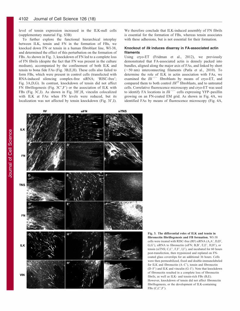
FBs. As shown in Fig. 3, knockdown of FN led to a complete loss
of FN fibrils (despite the fact that FN was present in the culture
medium), accompanied by the confinement of both ILK and
tensin to bona fide FAs (Fig. 3B,E,H). These cells also failed to
form FBs, which were present in control cells (transfected with
RNA-induced silencing complex-free siRNA; ‘RISC-free’;
Fig. 3A,D,G). In contrast, knockdown of tensin did not affect
FN fibrillogenesis (Fig. 3C9,F9) or the association of ILK with
FBs (Fig. 3C,I). As shown in Fig. 3H9,H, vinculin colocalized
with ILK at FAs when FN levels were reduced, but its
localization was not affected by tensin knockdown (Fig. 3I9,I).
We therefore conclude that ILK-induced assembly of FN fibrils
is essential for the formation of FBs, whereas tensin associates
with these adhesions, but is not essential for their formation.
Knockout of Ilk induces disarray in FA-associated actin
filaments
Using cryo-ET (Fridman et al., 2012), we previously
demonstrated that FA-associated actin is densely packed into
bundles, aligned along the major axis of FAs, and linked by short
(,50 nm) interconnecting filaments (Patla et al., 2010). To
determine the role of ILK in actin association with FAs, we
examined the Ilk2/2 fibroblasts by means of cryo-ET, and
compared them to both control Ilkf/f fibroblasts, and to untreated
cells. Correlative fluorescence microscopy and cryo-ET was used
to identify FA locations in Ilk2/2 cells expressing YFP–paxillin
growing on an FN-coated EM grid. As shown in Fig. 4A, we
identified FAs by means of fluorescence microscopy (Fig. 4A,
Fig. 3. The differential roles of ILK and tensin in
fibronectin fibrillogenesis and FB formation. WI-38
cells were treated with RISC-free (RF) siRNA (A,A9, D,D9,
G,G9), siRNA to fibronectin (siFN; B,B9, E,E9, H,H9), or
tensin (siTNS; C,C9, F,F9, I,I9), and incubated for 60 hours
post-transfection, then trypsinized and replated on FN-
coated glass coverslips for an additional 36 hours. Cells
were then permeabilized, fixed and double-immunolabeled
for ILK and fibronectin (A–C9), tensin and fibronectin
(D–F9) and ILK and vinculin (G–I9). Note that knockdown
of fibronectin resulted in a complete loss of fibronectin
fibrils, as well as ILK- and tensin-rich FBs (B,E).
However, knockdown of tensin did not affect fibronectin
fibrillogenesis, or the development of ILK-containing
FBs (C,C9,F9).
Journal of Cell Science 126 (18)4102

Journ
alof
Cell
Scie
nce
inset), froze the samples, inserted them into the cryo-electron
microscope, and identified the same FAs at low magnification.
An angular set of images was then collected, and subjected to
three-dimensional (3D) image reconstruction. One tomographic
slice through the reconstructed volume of the FA, positioned a
few nanometers above the cell–matrix interface at the
cytoplasmic side of the membrane, is shown in Fig. 3A. A
surface-rendered view of the same area, in which all observed
actin filaments were tracked, is shown in Fig. 4C. A
corresponding tomographic slice and a surface-rendered view
of an FA from control Ilkf/f fibroblasts are shown in Fig. 3B,D,
respectively.
The FAs in Ilkf/f fibroblasts were normal: and similar to those
previously reported for REF52 cells (Patla et al., 2010), with an
F-actin bundle, consisting of largely parallel filaments, associated
with the central region of the adhesion site. The actin
cytoskeleton of Ilk2/2 fibroblasts, however, was extremely
disorganized, and the number of actin filaments per unit area
was considerably reduced (by ,70%), compared to wild-type or
Ilkf/f cells (Fig. 4A,C, compared to B and D). The reduction in the
number of filaments was accompanied by a major loss of
filament alignment (Fig. 4E) compared with control cells, where
the actin filaments were largely aligned along the FA major axis.
Quantification of filament alignment indicated a standard
deviation of 15˚ in the Ilkf/f cells, compared with ,25˚ in the
Ilk2/2 cells.
FA-related particles are more densely packed in Ilk2/2
fibroblasts
FARPs, indistinguishable from those previously reported for
REF52 cells (Patla et al., 2010), were observed at the membrane–
cytoskeleton interface of both Ilk2/2 and Ilkf/f fibroblasts
(Fig. 4C,D, green). Quantification of these particles, however,
indicated major differences in their distribution density in control
and mutant cells. In control cells, the inter-particle spacing (mean
distance between the centers of mass of neighboring particles)
was 47629 nm, similar to the inter-particle spacing measured in
REF52 cells (Patla et al., 2010), but in Ilk2/2 cells the mean
inter-particle spacing was 36617 nm (Fig. 4F).
Notably, despite the apparent 1.3-fold decrease in average
FARP spacing in Ilk2/2 cells, the morphological characteristics
of the particles in these mutated cells were highly similar to those
found in the WT and Ilkf/f cells. We analyzed the structure of the
adhesion-related particles in two dimensions by extracting the
relevant volumes, and projecting them onto a common plane
(Fig. 4G). These images were then aligned, and classified
according to principle component analysis. Class averages
revealed ring-shaped densities with radiating filaments, which
presumably connect the FARPs and the actin bundles (Fig. 4G).
In both mutant and control cells, three sources constitute the
apparent structural variability between sub-classes of FARPs: the
particle diameter, the number of ‘whiskers’ extending from the
particles, and the angular positions of the whiskers. Particles of
Fig. 4. The molecular architecture of FAs in Ilk2/2 and Ilkf/f fibroblasts. (A,B) 10 nm-thick x-y sections through representative tomograms of central areas
of FAs in Ilk2/2 cells (A) and Ilkf/f cells (B). FA sites were identified by correlating electron micrographs with fluorescence microscopy images of YFP–paxillin-
expressing cells (A, insert). The red arrowhead in the insert points to the site of the FA tomogram in A. (C,D) Surface rendering view of the FA sites shown
in A and B, respectively, as seen from the direction of the substrate toward the cell center. Actin is depicted in tan, membranes in blue, and focal-adhesion-related
particles (FARPS) in light green. (E) Histograms representing the orientation of actin filaments within the FA-associated bundle in Ilk2/2 cells (left) and Ilkf/f
cells (right). (F) Histograms representing the ‘nearest neighbor’ distance distribution between individual adhesion-related particles in Ilk2/2 (left) and Ilkf/f cells
(right). The average center-to-center distance calculated between particles is 36 nm in Ilk2/2 and 47 nm in Ilkf/f cells. (G) 2D class averages of adhesion-related
particles, which were extracted in silico from FA tomograms, and projected along the z-axis. Both the Ilk2/2 (upper panel) and Ilkf/f cells (lower panel) were clearly
divided into three main classes with distinct ring diameters (supplementary material Fig. S1A); n51037 and n51142 for Ilk2/2 and Ilkf/f cells, respectively.
Roles of ILK in focal adhesion structure 4103

Journ
alof
Cell
Scie
nce
both mutated and control cells similarly vary with respect to these
three structural features (Fig. 4G and supplementary material Fig.
S2). The particles can be classified into three groups of distinct
ring diameter, with ratios of 55%, 24% and 21% in Ilk2/2 cells,
and 30%, 42% and 28% in Ilkf/f cells (Fig. 4G, left to right,
respectively; supplementary material Fig. S3A).
The properties of the whiskers in Ilk2/2 and Ilkf/f cells were
also similar: FARPs of both cell types normally contained 1–5
whiskers per particle, with mean values of 2.8961.04 and
2.6360.98 in Ilk2/2 and Ilkf/f cells, respectively (supplementary
material Fig. S2B). Moreover, the orientations of these whiskers
were largely isotropic, although in the Ilkf/f cells, occasional
alignment of whiskers along the FA major axis was seen
(supplementary material Fig. S2C). Taken together, these data
indicate that the altered actin network in Ilk2/2 cells is
accompanied by higher FARP packing densities; however, the
FARPs in both types of cells were indistinguishable, suggesting
that ILK is not a crucial component of these structures.
Ilk2/2 fibroblasts require a higher density of ECM
molecules for effective surface adhesion
To explore the functional significance of the molecular and
structural changes induced in FAs by ILK depletion, we tested the
adhesion ligand density required for FA formation by Ilk2/2 and
control cells. It has previously been shown that cells cultured on
nano-patterned surfaces, in which the adhesion ligand (cyclic RGD
peptide) is positioned at precise inter-spacings (within the range of
20–200 nm), require a particular threshold density to effectively
adhere to the surfaces (Arnold et al., 2008; Cavalcanti-Adam et al.,
2007). To compare the ligand density requirements of Ilk2/2 cells
and control Ilkf/f cells, we plated both cell types on nanostructured
glass surfaces on which the adhesive peptide ligand [c(-RGDfK);
see Materials and Methods] was linked to nanogold particles
positioned at fixed inter-spacings. As shown in Fig. 5A,D, Ilkf/f
cells plated on surfaces with 32 nm and 52 nm inter-ligand
spacings and incubated for 4 hours, spread well on both surfaces
(Fig. 5B,E), whereas Ilk2/2 cells spread exclusively on surfaces
with a 32 nm inter-ligand spacing (Fig. 5C), and failed to adhere
and spread on the 52 nm pattern (Fig. 5F). These results indicate
that Ilk2/2 cells require closer ligand spacing than control cells in
order to form functional FAs. Interestingly, these ligand densities
are highly similar to the differences in inter-particle spacing found
in the mutant and control cells plated on uniform ECMs, and
visualized by cryo-ET.
DiscussionILK is a canonical component of the integrin cell adhesion
machinery. Based on this and previous studies, it appears that
ILK participates in three distinct stages of integrin adhesion
organization: (1) nucleation of FX under the lamellipodium; (2)
organization of mature FAs and their associated stress fibers; and
(3) induction of FN fibrillogenesis and, consequently, formation
of FBs. Specifically, in the absence of ILK, all forms of integrin
adhesions will assemble inefficiently, leading not only to slow
cell spreading, but also to limited polarization and poor actin
alignment.
FN fibrillogenesis is driven by mechanical forces applied to
extracellular FN through integrin adhesions (Klotzsch et al.,
2009; Ulmer et al., 2008). It was further demonstrated (Zamir
et al., 2000) that this process is accompanied by the formation of
a special type of integrin adhesion, namely FBs, which extend
centripetally from FAs and are transported towards the cell
center. The relationship between FBs and FN fibrils has been
well established; yet the functional hierarchy between them
remained unclear. Here, we show that in the absence of ILK, FN
fibrils do not form. As a consequence, tensin, the hallmark of
FBs, is confined to and enriched in FAs, although its knockdown
has no apparent effect on FN fibrillogenesis, or on the formation
of ILK-rich FBs. These results indicate that ILK is an ‘upstream
regulator’, inducing FN fibrillogenesis that, in turn, drives FB
formation and the recruitment of molecules such as tensin to
these adhesion sites.
The geometry of the ECM has a major effect on the
capabilities of cells to adhere.
Fig. 5. ILK-deficient cells are highly dependent
on the close proximity of surface-bound RGD
molecules. (A,D) Scanning electron micrographs
showing hexagonally ordered gold nanodots on a
glass substrate (dot spacings: 32 and 52 nm,
respectively). Similar surfaces were used to study
the maximum integrin distances necessary for cell
viability. Nano-patterned surfaces were used as a
platform for varying RGD inter-ligand patch
distances of 3260.25 nm (A–C), and 5260.3 nm
(D–F). EGFP–paxillin Ilkf/f and EGFP–paxillin
Ilk2/2 cells were imaged 4 hours after plating on
surfaces with 32 nm (B,C) and 52 nm (E,F) inter-
ligand spacings, respectively. In B and E, the
edges of the functionalized surfaces are marked, to
demonstrate the specificity of the assay.
Journal of Cell Science 126 (18)4104

Journ
alof
Cell
Scie
nce
In previous studies on cell adhesion using nano-patterned
surfaces it was shown that the formation of genuine FAs is highly
sensitive to adhesion ligand density, and occurs when the ligand-
to-ligand spacing is on the order of ,50 nm or less (Cavalcanti-
Adam et al., 2007). Interestingly, the characteristic spacing
between adhesion-related particles within FAs, formed on a
uniform matrix is, essentially, the same (Patla et al., 2010).
Whether this correlation between the apparent densities of the
extracellular molecular tethers to the ECM, and the FARPs
interconnecting the membrane and the actin cytoskeleton, has
some functional significance, was not clear. Nevertheless, our
present findings, showing that in the absence of ILK, both the
adhesive ligand [c(-RGDfK)] density needed to induce cell
adhesion, spreading and FA formation, and the density of
FARPs, increase by ,1.7-fold (from ,450 to 770 FARPs/mm2),
lend further support to the hypothesis that the particles actually
bridge between the ECM-bound integrin and the actin network,
and that their density is functionally related to the ligand density
outside the cell.
The mechanism underlying this ILK-dependent density
sensing is still unclear; yet it may be related to the proposed
role of ILK in regulating the assembly of adhesome components
(Vouret-Craviari et al., 2004; Wang et al., 2008). This is in line
with our current results, showing that ILK regulates FARP
density, probably without being an essential, intrinsic component
of these particles. This conclusion is based on data showing that
the FARPs of Ilk2/2 and control cells are essentially
indistinguishable when examined by cryo-ET. The regulation of
FARP density by ILK may be related to a variety of factors,
including an overall alteration in FA molecular composition, such
as the increase in tensin levels, or the reduction in vinculin levels
shown here (possibly due to the arrest of tensin exit during FB
formation), as well as to the direct and specific effect of ILK on
cytoskeletal organization at the adhesion site.
Taken together, the recent structural mapping of integrin
adhesions at nano-scale resolution (Kanchanawong et al., 2010;
Patla et al., 2010), the characterization of adhesome complexity
and interactivity (Byron et al., 2012; Geiger and Yamada, 2011;
Kuo et al., 2011; Schiller et al., 2011; Zaidel-Bar et al., 2007a),
and the identification of distinct ‘molecular signatures’ of
subdomains within FAs at micro-scale resolution (Zamir et al.,
2008), suggest that integrin adhesions are modular structures,
with various molecular complexes displaying distinct
distributions and functional roles. Our previous siRNA screen
(Winograd-Katz et al., 2009), as well as the findings reported
here, demonstrate that specific adhesome components have
distinct structural and regulatory roles in the assembly of
integrin adhesion structure, and in control of their function. It
is conceivable that systematic perturbation of integrin adhesions
by gene modulation or different environmental cues, combined
with structural analyses such as the one presented here, will
become a mainstream approach for characterizing the functional
molecular architecture of these sites.
Materials and MethodsGeneration of paxillin–EGFP expressing fibroblasts
Ilkf/f and Ilk2/2 fibroblasts, stably expressing similar levels of paxillin–EGFP,were prepared by infecting the mutant cells with a retroviral expression vectorencoding paxillin–YFP (kindly provided by E. Danen, Leiden University). TheVSV-pseudotyped retroviral vectors used for that purpose were produced aspreviously described (Sakai et al., 2003; Azimifar et al., 2012), and theconcentrated viral supernatant, thus produced, was used to infect Ilkf/f
fibroblasts. Single clones of Ilkf/f fibroblasts stably expressing paxillin–EGFPwere isolated, and subsequently treated with an adenovirus expressing Crerecombinase, in order to delete the ILK gene and to obtain Ilk2/2 paxillin–EGFP-expressing fibroblasts.
Cell culture and immunolabeling
Mouse fibroblasts (Ilkf/f) expressing EGFP–paxillin, cells deficient in the ILKprotein (Ilk2/2), and WI-38 cells from human embryonic lung tissue were plated onglass coverslips coated with FN (as indicated). The cells were cultured in Dulbecco’smodified Eagle’s medium (DMEM; Biological Industries) supplemented with 10%(v/v) fetal calf serum (FCS; Biological Industries), L-glutamine (0.2 M; BiologicalIndustries), and penicillin–streptomycin–nystatin (Biological Industries), at 37 Cand in a 5% CO2 humidified atmosphere.
Cultured cells were permeabilized, fixed and double-immunolabeled, aspreviously described (Volberg et al., 2001). Antibodies used included monoclonalantibodies anti-vinculin, rabbit anti-FN and rabbit anti-tensin (produced in theAntibody Production Laboratories, Department of Biological Services, WeizmannInstitute of Science), anti-ILK (Santa Cruz Biotechnology, Inc.) and anti-zyxin(kindly provided by Daniel Louvard, Institut Curie, Paris, France). Secondaryantibodies used included anti-mouse Cy3 (Jackson ImunoResearch; cat. no. 115-166-072), and anti-rabbit Alexa Fluor 488 (Molecular Probes; cat. no. A11034). Tovisualize the F-actin cytoskeleton, cells were treated with phalloidin Alexa Fluor 488(Sigma; cat. no. P1951).
Gene knockdown protocol
WI-38 cells, 36105 cells/well, were cultured in six-well plates coated with 5 mg/mlbovine plasma FN, in DMEM supplemented with 10% FCS (Biological Industries)containing sodium pyruvate, non-essential amino acids, and antibiotics (Sigma) for24 hours at 37 C.
Cells were transfected with siRNA specific for FN (siFN) or tensin (siTNS;SMARTpools; Dharmacon RNAi Technologies).
Correlative fluorescence and electron microscopy
Carbon-coated 200-mesh gold grids (Quantifoil) were rinsed in PBS (BiologicalIndustries) and overlaid on a drop of 50 mg/ml bovine plasma fibronectin (BiologicalIndustries) for 45 minutes. Cells were typically applied to grids in concentrations of100 cells per grid, and cultured for 24 hours. Next, specimens were examined byfluorescence microscopy, washed with PBS, and freeze-plunged instantly intoliquid-nitrogen-cooled ethane (see below). FAs identified by fluorescencemicroscopy were indexed according to their precise positions on the grid.
The coordinates of each FA site were recorded and identified under the electronbeam. Fluorescence images were acquired with a DeltaVision system (AppliedPrecision, USA) equipped with a CoolSnap HQ camera (Photometric, USA)operated by SoftWorx and Resolve3D software (Applied Precision), using anOlympus PlanApoN 606/1.42 NA objective on an Olympus IX70 invertedmicroscope (Olympus, Japan).
Cryo-electron tomography
A 5 ml drop of BSA-coated 15 nm gold colloids in PBS was added to the gridsbefore plunging them into liquid-nitrogen-cooled ethane, as previously described(Dubochet et al., 1988). Specimens were then transferred into a 300 FEG Polaramicroscope (FEI, Eindhoven) equipped with a Gatan post-column GIF 2002energy filter. Tilt series were then collected, typically covering an angular range of260˚ to +66 , sampled in 2˚ tilt increments, and at a 14 mm underfocus. Pixel sizewas 0.82 nm at the specimen level.
Image processing of cryo-ET images
Projection images (204862048 pixels) were aligned to a common origin, using15 nm-sized fiducial gold particles, and reconstructed by means of weighted back-projection, as implemented by a TOM toolbox software package (Nickell et al.,2005). All tomograms were reconstructed with a binning factor, to yield a 1.64 nmvoxel size. For visualization purposes, the reconstructed volumes were processedaccording to an anisotropic de-noising algorithm (Frangakis and Hegerl, 2001).Individual adhesion complexes were identified and extracted in silico (952 and 946from mutant and wild-type cells, respectively), from the five best tomograms fromeach cell line. Two-dimensional images of these elements were calculated byprojecting the volumes, 32632611 voxels, along the z-axis, using the EM softwarepackage. The stack of particles was then masked and normalized prior to PCA, andfollowed by K-means classification (SPIDER). The most detailed class averageswere chosen to be the first references for a multi-reference alignment of the dataset. This strategy was iterated for five rounds, until no major changes occurred inthe classes, and in the alignment of single images. Surface-rendered visualizationswere constructed using AMIRA 5.3 software (Mercury).
Patterned gold surfaces
Gold surfaces were prepared as previously described (Cavalcanti-Adam et al.,2007).
Roles of ILK in focal adhesion structure 4105
Journ
alof
Cell
Scie
nce
Characterization of nano-structured glass interfacesGlass samples were sputter-coated with a carbon layer, in preparation for imagingwith a field-emission scanning electron microscope (LEO-1530, LEO). Tovisualize the gold nanoparticles on the surface, an acceleration voltage of 3 kVwas applied under a pressure of 561026 mbar.
BiofunctionalizationTo passivate the substrate area between the gold nanoparticles, polyethylene glycol(PEG; molecular weight 2000) was used (Blummel et al., 2007). The goldnanoparticles were then functionalized with the cyclic RGD-based peptidec(-RGDfK-), using the spacer aminohexanoic acid, to mercaptopropionic acid.Next, the PEG-functionalized substrates were immersed for 24 hours in a 25-mMc(- RGDfK-)-thiol/water solution. The substrates were then thoroughly rinsed withMilliQH water for 24 hours.
Cell seeding conditions for nanostructured and biofunctionalized substratesCell samples were sterilized in 70% ethanol for 20 minutes, and washed threetimes with phosphate-buffered saline at room temperature. Cells were then platedat a density of 40 cells/mm2 in DMEM containing 1% FBS and 1% L-glutamine.Experiments were performed three times. Images were obtained after 4 hours andafter 24 hours with an Olympus IX inverted microscope.
AcknowledgementsThe authors express gratitude to B. Morgenstern for expert help inpreparing this manuscript. B.G. holds the Erwin Neter ProfessorialChair in Cell and Tumor Biology. J.S. holds a Weston VisitingProfessorship at the Weizmann Institute of Science, Israel.
Author contributionsExperiments were performed by N.E., T.V., I.P., V.H.W. and C.G.The manuscript was written by O.M. and B.G. The study wasdesigned and jointly supervised by J.P.S, R.F., B.G. and O.M.
FundingThis study was supported by the German–Israeli Cooperation Project(DIP H.2.2 to O.M., R.F., J.S. and B.G.); the Israel ScienceFoundation to B.G.; a European Research Council Starting Grant[grant number 243047 INCEL to O.M.]; the Max Planck Society toR.F. and J.S.; and the European Union Seventh FrameworkProgramme (FP7/2007–2013), [grant number NMP4-LA-2009-229289 NanoII to B.G.].
Supplementary material available online at
http://jcs.biologists.org/lookup/suppl/doi:10.1242/jcs.120295/-/DC1
ReferencesArnold, M., Hirschfeld-Warneken, V. C., Lohmuller, T., Heil, P., Blummel, J.,
Cavalcanti-Adam, E. A., Lopez-Garcıa, M., Walther, P., Kessler, H., Geiger,
B. et al. (2008). Induction of cell polarization and migration by a gradient ofnanoscale variations in adhesive ligand spacing. Nano Lett. 8, 2063-2069.
Aspenstrom, P., Fransson, A. and Saras, J. (2004). Rho GTPases have diverse effectson the organization of the actin filament system. Biochem. J. 377, 327-337.
Azimifar, S. B., Bottcher, R. T., Zanivan, S., Grashoff, C., Kruger, M., Legate, K. R.,Mann, M. and Fassler, R. (2012). Induction of membrane circular dorsal ruffles requiresco-signalling of integrin-ILK-complex and EGF receptor. J. Cell Sci. 125, 435-448.
Blummel, J., Perschmann, N., Aydin, D., Drinjakovic, J., Surrey, T., Lopez-Garcia, M.,Kessler, H. and Spatz, J. P. (2007). Protein repellent properties of covalently attachedPEG coatings on nanostructured SiO(2)-based interfaces. Biomaterials 28, 4739-4747.
Burridge, K., Fath, K., Kelly, T., Nuckolls, G. and Turner, C. (1988). Focaladhesions: transmembrane junctions between the extracellular matrix and thecytoskeleton. Annu. Rev. Cell Biol. 4, 487-525.
Byron, A., Humphries, J. D., Bass, M. D., Knight, D. and Humphries, M. J. (2011).Proteomic analysis of integrin adhesion complexes. Sci. Signal. 4, pt2.
Byron, A., Humphries, J. D., Craig, S. E., Knight, D. and Humphries, M. J. (2012).Proteomic analysis of a4b1 integrin adhesion complexes reveals a-subunit-dependentprotein recruitment. Proteomics 12, 2107-2114.
Campbell, I. D. (2008). Studies of focal adhesion assembly. Biochem. Soc. Trans. 36,263-266.
Cavalcanti-Adam, E. A., Volberg, T., Micoulet, A., Kessler, H., Geiger, B. and
Spatz, J. P. (2007). Cell spreading and focal adhesion dynamics are regulated byspacing of integrin ligands. Biophys. J. 92, 2964-2974.
Choi, C. K., Vicente-Manzanares, M., Zareno, J., Whitmore, L. A., Mogilner,
A. and Horwitz, A. R. (2008). Actin and alpha-actinin orchestrate the assembly andmaturation of nascent adhesions in a myosin II motor-independent manner. Nat. Cell
Biol. 10, 1039-1050.
Dubochet, J., Adrian, M., Chang, J. J., Homo, J. C., Lepault, J., McDowall, A. W.
and Schultz, P. (1988). Cryo-electron microscopy of vitrified specimens. Q. Rev.
Biophys. 21, 129-228.
Efimov, A. and Kaverina, I. (2009). Significance of microtubule catastrophes at focaladhesion sites. Cell Adh. Migr. 3, 285-287.
Frangakis, A. S. and Hegerl, R. (2001). Noise reduction in electron tomographicreconstructions using nonlinear anisotropic diffusion. J. Struct. Biol. 135, 239-250.
Fridman, K., Mader, A., Zwerger, M., Elia, N. and Medalia, O. (2012). Advances intomography: probing the molecular architecture of cells. Nat. Rev. Mol. Cell Biol. 13,736-742.
Geiger, B. and Yamada, K. M. (2011). Molecular architecture and function of matrixadhesions. Cold Spring Harb. Perspect. Biol. 3, a005033.
Geiger, B., Spatz, J. P. and Bershadsky, A. D. (2009). Environmental sensing throughfocal adhesions. Nat. Rev. Mol. Cell Biol. 10, 21-33.
Hannigan, G. E., Leung-Hagesteijn, C., Fitz-Gibbon, L., Coppolino, M. G., Radeva, G.,Filmus, J., Bell, J. C. and Dedhar, S. (1996). Regulation of cell adhesion and anchorage-dependent growth by a new beta 1-integrin-linked protein kinase. Nature 379, 91-96.
Kanchanawong, P., Shtengel, G., Pasapera, A. M., Ramko, E. B., Davidson, M. W.,
Hess, H. F. and Waterman, C. M. (2010). Nanoscale architecture of integrin-basedcell adhesions. Nature 468, 580-584.
Klotzsch, E., Smith, M. L., Kubow, K. E., Muntwyler, S., Little, W. C., Beyeler, F.,Gourdon, D., Nelson, B. J. and Vogel, V. (2009). Fibronectin forms the mostextensible biological fibers displaying switchable force-exposed cryptic binding sites.Proc. Natl. Acad. Sci. USA 106, 18267-18272.
Kuo, J. C., Han, X., Hsiao, C. T., Yates, J. R., III and Waterman, C. M. (2011).Analysis of the myosin-II-responsive focal adhesion proteome reveals a role for b-Pixin negative regulation of focal adhesion maturation. Nat. Cell Biol. 13, 383-393.
Legate, K. R., Montanez, E., Kudlacek, O. and Fassler, R. (2006). ILK, PINCH andparvin: the tIPP of integrin signalling. Nat. Rev. Mol. Cell Biol. 7, 20-31.
Legate, K. R., Wickstrom, S. A. and Fassler, R. (2009). Genetic and cell biologicalanalysis of integrin outside-in signaling. Genes Dev. 23, 397-418.
Montanez, E., Wickstrom, S. A., Altstatter, J., Chu, H. and Fassler, R. (2009).Alpha-parvin controls vascular mural cell recruitment to vessel wall by regulatingRhoA/ROCK signalling. EMBO J. 28, 3132-3144.
Nickell, S., Forster, F., Linaroudis, A., Net, W. D., Beck, F., Hegerl, R., Baumeister,W. and Plitzko, J. M. (2005). TOM software toolbox: acquisition and analysis forelectron tomography. J. Struct. Biol. 149, 227-234.
Nikolopoulos, S. N. and Turner, C. E. (2001). Integrin-linked kinase (ILK) binding topaxillin LD1 motif regulates ILK localization to focal adhesions. J. Biol. Chem. 276,23499-23505.
Pankov, R., Cukierman, E., Katz, B. Z., Matsumoto, K., Lin, D. C., Lin, S., Hahn,C. and Yamada, K. M. (2000). Integrin dynamics and matrix assembly: tensin-dependent translocation of alpha(5)beta(1) integrins promotes early fibronectinfibrillogenesis. J. Cell Biol. 148, 1075-1090.
Patla, I., Volberg, T., Elad, N., Hirschfeld-Warneken, V., Grashoff, C., Fassler, R.,
Spatz, J. P., Geiger, B. and Medalia, O. (2010). Dissecting the moleculararchitecture of integrin adhesion sites by cryo-electron tomography. Nat. Cell Biol.
12, 909-915.
Sakai, T., Li, S., Docheva, D., Grashoff, C., Sakai, K., Kostka, G., Braun, A., Pfeifer,
A., Yurchenco, P. D. and Fassler, R. (2003). Integrin-linked kinase (ILK) is requiredfor polarizing the epiblast, cell adhesion, and controlling actin accumulation. Genes
Dev. 17, 926-940.
Sayedyahossein, S., Nini, L., Irvine, T. S. and Dagnino, L. (2012). Essential role of integrin-linked kinase in regulation of phagocytosis in keratinocytes. FASEB J. 26, 4218-4229.
Schiller, H. B., Friedel, C. C., Boulegue, C. and Fassler, R. (2011). Quantitativeproteomics of the integrin adhesome show a myosin II-dependent recruitment of LIMdomain proteins. EMBO Rep. 12, 259-266.
Stanchi, F., Grashoff, C., Nguemeni Yonga, C. F., Grall, D., Fassler, R. and VanObberghen-Schilling, E. (2009). Molecular dissection of the ILK-PINCH-parvintriad reveals a fundamental role for the ILK kinase domain in the late stages of focal-adhesion maturation. J. Cell Sci. 122, 1800-1811.
Torgler, C. N., Narasimha, M., Knox, A. L., Zervas, C. G., Vernon, M. C. andBrown, N. H. (2004). Tensin stabilizes integrin adhesive contacts in Drosophila. Dev.
Cell 6, 357-369.
Ulmer, J., Spatz, J. P. and Geiger, B. (2008). Force-induced fibronectin fibrillogenesisin vivo. Soft Matter 4, 1998-2007.
Vakaloglou, K. and Zervas, C. (2012). Parvin-ILK: An intimate relationship.BioArchitecture 2, 91-94.
Vicente-Manzanares, M. and Horwitz, A. R. (2011). Adhesion dynamics at a glance.J. Cell Sci. 124, 3923-3927.
Vicente-Manzanares, M., Choi, C. K. and Horwitz, A. R. (2009). Integrins in cellmigration – the actin connection. J. Cell Sci. 122, 199-206.
Vicente-Manzanares, M., Newell-Litwa, K., Bachir, A. I., Whitmore, L. A. and
Horwitz, A. R. (2011). Myosin IIA/IIB restrict adhesive and protrusive signaling togenerate front-back polarity in migrating cells. J. Cell Biol. 193, 381-396.
Volberg, T., Romer, L., Zamir, E. and Geiger, B. (2001). pp60(c-src) and relatedtyrosine kinases: a role in the assembly and reorganization of matrix adhesions. J. Cell
Sci. 114, 2279-2289.
Vouret-Craviari, V., Boulter, E., Grall, D., Matthews, C. and Van Obberghen-
Schilling, E. (2004). ILK is required for the assembly of matrix-forming adhesionsand capillary morphogenesis in endothelial cells. J. Cell Sci. 117, 4559-4569.
Wang, H. V., Chang, L. W., Brixius, K., Wickstrom, S. A., Montanez, E.,Thievessen, I., Schwander, M., Muller, U., Bloch, W., Mayer, U. et al. (2008).
Journal of Cell Science 126 (18)4106

Journ
alof
Cell
Scie
nce
Integrin-linked kinase stabilizes myotendinous junctions and protects muscle fromstress-induced damage. J. Cell Biol. 180, 1037-1049.
Wickstrom, S. A. and Fassler, R. (2011). Regulation of membrane traffic by integrinsignaling. Trends Cell Biol. 21, 266-273.
Wickstrom, S. A., Lange, A., Hess, M. W., Polleux, J., Spatz, J. P., Kruger, M.,
Pfaller, K., Lambacher, A., Bloch, W., Mann, M. et al. (2010). Integrin-linkedkinase controls microtubule dynamics required for plasma membrane targeting ofcaveolae. Dev. Cell 19, 574-588.
Winograd-Katz, S. E., Itzkovitz, S., Kam, Z. and Geiger, B. (2009). Multiparametricanalysis of focal adhesion formation by RNAi-mediated gene knockdown. J. Cell
Biol. 186, 423-436.Zaidel-Bar, R., Cohen, M., Addadi, L. and Geiger, B. (2004). Hierarchical assembly
of cell-matrix adhesion complexes. Biochem. Soc. Trans. 32, 416-420.
Zaidel-Bar, R., Itzkovitz, S., Ma’ayan, A., Iyengar, R. and Geiger, B. (2007a).Functional atlas of the integrin adhesome. Nat. Cell Biol. 9, 858-867.
Zaidel-Bar, R., Milo, R., Kam, Z. and Geiger, B. (2007b). A paxillin tyrosinephosphorylation switch regulates the assembly and form of cell-matrix adhesions.J. Cell Sci. 120, 137-148.
Zamir, E., Katz, B. Z., Aota, S., Yamada, K. M., Geiger, B. and Kam, Z. (1999).Molecular diversity of cell-matrix adhesions. J. Cell Sci. 112, 1655-1669.
Zamir, E., Katz, M., Posen, Y., Erez, N., Yamada, K. M., Katz, B. Z., Lin, S., Lin,D. C., Bershadsky, A., Kam, Z. et al. (2000). Dynamics and segregation of cell-matrix adhesions in cultured fibroblasts. Nat. Cell Biol. 2, 191-196.
Zervas, C. G., Psarra, E., Williams, V., Solomon, E., Vakaloglou, K. M. and Brown,N. H. (2011). A central multifunctional role of integrin-linked kinase at muscleattachment sites. J. Cell Sci. 124, 1316-1327.
Roles of ILK in focal adhesion structure 4107