the transplantation of auerbach's …repository.kulib.kyoto-u.ac.jp/dspace/bitstream/2433/...of...
TRANSCRIPT

Title
THE TRANSPLANTATION OF AUERBACH'S PLEXUS INTHE SMALL INTESTINE OF A DOG TO THE BLADDERWALL AND OVARY, AND A NEURO-HISTOLOGICALSTUDY ON THE ATTITUDE OF THE TRANSPLANTEDGANGLION CELLS IN THE PLEXUS
Author(s) NOMURA, GENZO
Citation 日本外科宝函 (1959), 28(9): 3488-3502
Issue Date 1959-11-01
URL http://hdl.handle.net/2433/207031
Right
Type Departmental Bulletin Paper
Textversion publisher
Kyoto University

3488
THE TRANSPLANTATION OF AUERBACH’S PLEXUS IN
THE SMALL INTESTINE OF A DOG TO THE BLADDER
WALL AND OVARY, AND A NEURO-HISTOLOGICAL
STUDY ON THE ATTITUDE OF THE TRANSPLANTED
GANGLION CELLS IN THE PLEXUS
by
GENzo No:1IUI{A
From the 2nd Surgical Division, Kyoto University l¥lcclical School
(Director : Prof. Dr. YASUMASA AOYAGI)
(E巴ceivec¥for publication Aug. 8, 1959)
Contents
I Introcluct10n 3) At the encl of the 6th month II i¥laterials and :'llcthocls III i¥licroscopic Obscrvaton of the Transplanted
λucrbach’s plexus of the Small Intestine on the Urinary blac¥c¥er and o、ary
(八) The Auerbach’s plexus transplanted on the blaclc¥er wall 1) At the encl of the 1st month 2) At the encl of the 3rd month
(B) The 人肌rbach’討 plex山 transplantedon the ovary 1) At the encl of th巴 1stmonth 2) At the encl of the 3rd mo川 h
IV Discussion V Summary and Conclusion
Referencじs,Figures, Explanation of Figures
I INTRODUCTION
The digestive trεct is influenced by the autonomic nerve, namely, on principle
it is promoted by the excitement of the parasympathetic nerve and inhibited by
the sympathetic nerve. The intestine performing the peristaltic motion by
mechanic stimulations without the extrinsic nerves is due to the action of intra-mural plexuses.
Since AuEEBACH (1862) described the network structures of the nervous
tissue in the muscle layer of the intestine, GREYING, DocmL, KUNTZ and others
have investigated this plexus histologically and also, MAG>iUS, E:onLD, YAN人SE
and others have observed the peristaltic motion controled by AuEI{JL¥C山 plexus
physiologically. But among the many reports on the anatomy, physiology and
histology of AUERBACH’s plexus, the author have never seen a single report describing its transplantation.
Accordingly, the author carried out the transplantation experiments of
AUERBACH’s plexus in the small intestine to the bladder wall and ovary, and the
attitude of the transplanted ganglion cells in the plexus were studied histologically.
THE TRANSP ANTATION OF AUERBACH’S & CELLS IN THE PLEXUS 3489
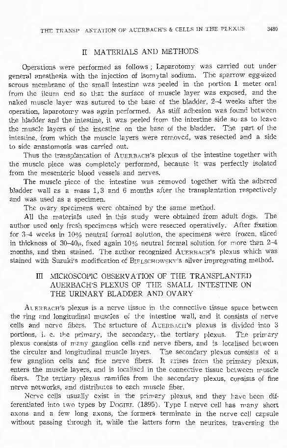
II MATERIALS AND METHODS
Operations were performed as follows ; Laparotomy was carried out under general anesthesia with the injection of isomytal sodium. The sparrow eggもizedserous membrane of the small intestine was peeled in the portion 1 meter oral from the ileum end so that the surface of muscle layer was exposed, and the naked muscle layer was sutured to the base of the bladder, 2-4 weeks after the
operation, laparotomy was again performed. As sti妊 adhesionwas found between the bladder and the intestine, it wεs peeled from the intestine side so as to leave the muscle layers of the intestine on the base of the bladder. The pa抗 ofthe intestine, from which the muscle layers were removed, was resected and a side
to side a紅白tomosiswas carried out. Thus the transplantation of AUERBACH’s plexus of the intestine together with
the muscle piece was completely performed, because it was perfectly isolated
from the mesenteric blood vessels and nerves. The muscle piece of the intestine was removed together with the adhered
bladder wall as a mass 1, 3 and 6 months after the transplantation respectively and was used as a specimen.
The ovary specimens were obtained by the same method. All the materials used in this study were obtained from adult dogs. The
author used only fresh specimens which were resected operatively. After五xationfor 3-4 weeks in 10% neutral formal solution, the specimens were frozen, sliced in thickness of 30 401.i,,五xedagain 10% neutral formal solution for more than 2-4 months, and then stained. The author recognized AUERBACH’s plexus which was stained with Suzuki’s modi五cationof BrELSCHOWSKγs silver impregnating method.
III MICROSCOPIC OBSERVATION OF THE TRANSPLANTED AUERBACH’S PLEXUS OF THE SMALL INTESTINE ON THE URINARY BLADDER AND OVARY
AUERBACH’s plexus is a nerve tissue in the connective tissue space between the ring 2nd longitudinal muscles of the intestine wall, and it consists of nerve cells and nerve fibers. The srtucture of AUERBACH’s plexus is divided into 3
portions, i. e. the primary, the secondary, the tertiary plexus. The priffべaryplexus consists of mεny ganglion cells εnd nerve五bers,and is localised between the circular and longitudinal muscle layers. The secondary plexus consists of a few ganglion cells and fine nerve fibers. It arises from the primary plexus, enters the muscle layers, and is loc21ised in the connective tissue between muscle 五bers. The tertiary plexus rami古田 fromthe secondary plexus, consists of五.nenerve networks, and distributes to e;:;.
Nerve cells usuε.lly exist in the primary plexus, and they have been dif ・ ferentiated into two types by DocrnL (1895). Type I nerve cell has many short axons and a few long axons, the formers termin2te in the nerve cell capsule without passing through it, while the Jatters form the neurites, traversing the

3490 日本外科宝繭第28巻第9号
capsule. Type I nerve cell mainly has plural axons, sometimes as many as 4 5
axons. Type II nerve cell has both long and short axons, however both of them run out of the capsule. Short axons in type II nerve cell are far less in num her than in type I. Most of them ramify and terminate in tapering endings. The greater part of the long axons in type II show a plurality such 2s in type I
nerve cell. As a control of experiments, the author made an adhesion between the small
intestine and the bladder wall, without cutting o百themesenteric blood and nerve
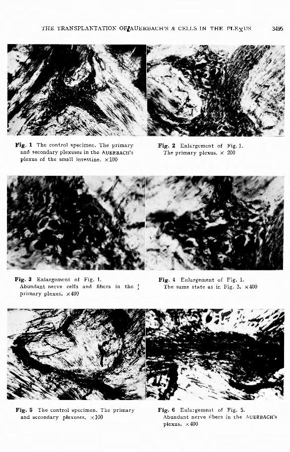
supply, and ci.dhered part of the intestine was studied. In the control specimens, almost normal nerve structures of AuERBACH’s
plexus as mentioned above were observed (Fig. 1~6). Next, observing the specimens in which the transplantation was carried out,
the following findings were obtained :
(A) The AuERB.¥CH’s plexus transplanted on the bladder wall 1) At the end of the 1st month In AuERBACH’s plexus, various degrees of degenerations were found in nerve
cells ; dysharmony of axons, dislocation of nucleus, and fenstrated cell, etc., were observed. In some specimen the number of nerve fibers in AUERBACH’s plexus decreased, in another they proliferated, while in some others decrease and increase
in the number of of nerve fibers were found in different portions of AuERBACH’s plexus (Fig. 7 22).
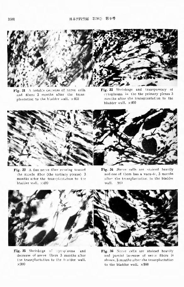
2) At the end of the 3rd month Mild degenerated nerve cells were found. A few specimens showed decreased
number of nerve五bers,but most of specimens showed marked increase of nerve 五berswith a vigorous growth toward the scar (Fig. 23 36).
3) At the end of the 6th month It was hard to discriminate the transplante'.l muscle piece in the scar, Au-
ERB AC H’s plexus remained as a carcase, and nerve cells or白herswere scarcely found there (Fig. 37, 38, 39).
(B) The AuERBACH’s plexus transplanted on the ovary 1) At the end of 1st month
Severe degenero.ted nerve cells were observed,εnd nerve fibers were generally found to have decreased consider2.bly without a tendency to increase (Fig. 40, 41, 42).
2) At the end of the 3rd month
Most of degenerated nerve cells commonly showed up, light stainning, falling of the argent a伍nity,indistinct日guresand invisible axons. Nerve fibers were found to have almost disappeared (Fig. 43 48).
IV DISCUSSION
Considering the re3ults of various experiments performed by many investi-
gators from the anatomical or physiological standpoint, the urinary bladder is mainly innervated by pelvic nerves and by hypogastric nerves. The pelvic nerves
THE TRANSPLANTATION OF AUERBACH’S & CELLS IN THE PLEXUS 3491
belong to the sacral parasympathetic system, and the hypogastric nerves belong to the sympathetic system, while the formers are more dominant in the bladder than the latters, and the pelvic nerves play a leading innervation in the bladder. The ovary is under the innervation of the ovarian plexus, and especially is innervated by sympathetic nerves which are derived from the spinal segments of Th. 10-L. 2 like the kidney and the testis, while the parasympathetic innervation is very
poor.
From these facts and neurohumoralism (DALE), it can be assumed that autonomic nerves of the bladder muscles belong to the parasympathetic system, i. e. the cholinergic nerve system, like AUERBACH’s plexus. Both are homotype. The autonomic nerves of the ovary belong to the sympathetic system, i. e. the adrenergic nerve system, therefore, AuERBACH’s plexus and the ovary are heterotype.
Next, one may thi叫( that intramural nerve cells in the digestive tract wall, i. e. cells of AUERBACH’s and MEISSNER’s plexus, are the starting point of parasy-mpathetic-postganglionic fibers.
Then in the experiments, at the end of the 1st month after the transplantation of AUERBACH’s plexus to the bladder, the author recognized a remarkable cell infiltration, various degenerated nerve cells and 五.bers. It was thought that these degenerations were affected not only by cutting o百 theblood supply but also by the inflammation, as was expected. In the preparations o:t the 3rd month after the transplantation, mild degenerated nerve cells and remarkable proliferation of nerve fibers were found. Though the transplantation seemed to be possible from these results, in the preparations of the 6th month, AuERBACH’s plexus remained as a carcase, and the nerve cells and fibers had completely disappeared.
Next, in the 1st month after transplantation to the ovary, nerve cells already showed severe degenerations and nerve fibers showed a one sided remarkable decreasing without a tendency to increase. In the 3rd month, nerve cells showed a high degree of degeneration and nerve日bersalmost disappeared.
From these results which the author observed, the transplantation of AUE RB人CH’splexus may be impossible and once degenerated, nerve cells are irreversible.
In regard to the postganglionic五bers,they began to decrease in the 1st month. In the bladder wall which belongs to the cholinergic nerve system, they lived for a long time. They showed once proliferation, and finally in 6 months completely disappeared. In the ovary which belongs to the adrenergic nerve system, they decreased from the beginning without showing any increase, and in 3 months they disappeared.
The postganglionic nerve fibers, which were isolated from the preganglionic fibers and transplanted to other organs, show a di旺erentattitude in those organs. In a homotype organ, from the view point of chemical mediator of the innervating nerves, the nerve fibers in the transplanted muscle can be alive for a long time, i. e. for over 3 months, once showing proliferation during this time period. In a

3492 日本外科宝函第28巻第9号
heterotype organ, the nerve fibers in the transplanted muscle decrease the number
from the beginning and completely degenerate in 3 months.
V SUMMARY AND CONCLUSION
The author carried out the transplantation experiments of AuERBACH’s plexus in the small intestine to the bladder wall and ovary, and observed the attitude
of the transplanted ganglion cells in the plexus by neurohistological study. And
then he investigated the change of postganglionic fibers which were isolated from the preganglionic五bers.
Summarizing the results of experiments, the following conclusions are obtained. 1) The transpl訂itationof AUERBACH’s plexus to the urinary bladder and
ovary may be impossible. If it is possible, it will be in the bladder. 2) The postganglionic五bersof the parasympathetic nerve begin to decrease
in about one month after the isolation, and then go through different courses by the type of nerve innervation ;
(a) In the homotype organ (the bladder) which belongs to the cholinergic nerve system, they live a relatively long time.
(b) In the heもerotypeorgan (the ovary) which belongs to the adrenergic nerve
system, they continue to decrease to earlier death.
I wish to express my deepest gratitude toward A",i寸.Prof. Dr. Ch. KIMURA for his helpful
advice and kind guidance throughout this study.
References
1) 八iba,人 OberBewegung d引 Dickdarms(in Japanese)・ Jap.J. i¥led. I'rog., 22. 1684. 1933 2) Bayliss, ¥¥'. i¥l. and Starling, E. 11. The movement and the innervation of the small
intesine. J. Physiol., 26 (1901) 125-138
3) Bozier, E .. Reflex peristalsis of the intestine.人口1. J. Physiol., 157 (1949), 338-342
めぐhoda,T. : Die Studien口berdie Innervation des Dlinnclarms. Ok<Lyama' I. Z., 51. 1484. 1939 5) Dale, H. H. and ¥V. Felclberg. : Chemical transmission at motor nerve endings in voluntary
muscle. J. Physiol., 81 : 39
6) Feyrter, F.: lber die Pathologie cler vegetativcn nervosen Peripherie und ihrer gan・
glioniircn Regulationsst社Hen.:¥Tandrich, ¥¥"ien. 1952
7) Fukuda, K. Human Physiology (Jintai seiri gaku, in Japanese). 212-220. s. 1949 8) Fukui, T .. Histopathological Studies on the Stomach and Intc,tin巴(in Japanese). J.
Kyoto Pref. Med. Univ., 58. 735., 1955
9) Ha,c, T.・The Action of vagus n巴rve on the intestine (Meiso shinkt、ino chosayo). J.
Kyoto Pref. :¥feel. Univ., 24 1191 (1938)
10) Herrmann, H. : Pathologishe I-listologie cler peripheren vじgetati,・cnN crvensystems. Berliner :¥fed, ¥'crlagsanstalt 1956
11) Inouye, R. : Pathohistologische Studien Uber intramuralen J》lexusdes Colons bei chronischer
Obstipation, nebst cinigen Bemerkungen zur chirurgischen Inclikations stellung. Arch. f,.
jap. Chir., 27. 13'11. 1958
12) Jabonero, V. Der anatomische Aufbau des periphcren neurovegetativen勾’stems. 人仁ta
Neuro-veg ; Suppl. IV. Springer. ¥¥'ien 1953
13) Kimura, (、:h. Surgery of the Autonomic Nervous System (Jiritsu Shinkei no Geka, in
Japanese)・ J.Jap. Surg. Soc., 52. '150. 1951 ; J¥ihon Gcka Z巴nsho・9; 341. 1956 14) Kimura, Ch. : The Problem of Abdominal Pain・.\rch. f. jap. Chir., 22. 59. 1953
THE TRANSPLANTATION OF AUERBACH’S & CELLS IN THE PLEXUS 3493
15) Kimura, Ch. : Physiology of Abdominal Pain (Fukutsu no S巴iri,in Japan巴se).Rinsho no
Shimpo. 7. 1953 16) Kimura, Ch. Abdominal P乱in(Shink巴i-Biorito Rinsho, in Japanese)・ J・Jap. Surg. Soc.,
6. 947. 57. 1956 17) Kure, T. and Okinaka, S .. Autonomic Nervous System (in Japan巴se).1949
18) Langley, J. N. : a) The Autonomic Nervous System. Brain, 26, 1. 1903
b) The Autonomic nervous system. Part, I, Cambridge, W. Heffer and
Sons Ltd. 1921
19) Muller, L, R. Lebcnsnerven und Leb巴nstriebe.3. Aufl., 1931
20) Okinaka, S .. l¥Iorphology of autonomic nerve fiber (Jiritsu shinkei s巴n-ino k巴itai).Nihon
Rinsho., 8. 11. 1950. 21) Sato, H .. A Histological Study of the Afferent Innervation of the ovary of the Dog. Arch.
f. jap. Chir., 24 ; 456, 1955
22) Seto, H. : a) Advance of Medicin巴. 5, 223, 1949.
b) Histological Observation of Human S巴nsibility.Rinsho no Nihon. I; 32. 1955
23) Stoehr, Jr. Ph. : a) L巴hrbuchder Histologie und der Mikroskopische Anatomie des l¥Ien-
schen. Springer Verlag. 1951
b) Mikroskopische Anatomie des v巴g巴tativen N巴rvcnsystems. 1951 u.
1957
24) Shishido, S. : Abdominal Pain (Naizochikaku no Dendoro o chusin to shite, in Japan巴se)・
J. Jap. Surg. Soc., 6. 922. 57. 1956 25) Suzuki, K. Note of Techinique to make Tissue-Preparations (Soshiki Hyohon S巴isaku
Gijyutsu Not巴, inJapanese)・(IV).Noshinkei ryoiki, 5. 184. 1952
26) Thomas, J. E. and A. Kuntz. : A study of gastro-intestinal motility in relation to the
巴ntericnervous system. Am. J. Physiol. 76 (1926), 606-626
27) Takayasu, T. Histologische Studien Uber die Innervation der Darmwand b巴iml¥Tenschen.
(1) (2) (3) (in Japanese) J. Tokyo Soc. l¥Ied. Sci., 48. 837; 1955. 1934. 49. 259 ; 901.1935.
28) Uchino, T. : Studien Uber die Degeneration der intramuralen Darmnerven bei exp巴ri-
mentellem. Ileus. (in Japanese) Nagasaki IgKZ., 19, 1959 ; 2423, 1941
29) Yoshida, T. : A Histological Study of Sensory Nerves in the Urinary Organs. Arch. f. jap.
Chir., 26. 55. 1957
Suzuki’s Method
The specimens, aft巴rhaving been sliced with the freezing method and kept in 10% neutral
formal solution, are
1) Washed 3 times with distilled water, each time for about 10 minut巴s,
2) Put into 20% sil、・ernitrate solution for about 1 hour, in the darkness,
3) ¥¥'ashed with distill巴dwater for a few seconds,
4) Put into ammonical silver solution until th巴 sp巴cimensare colored light yellow,
5) Placed in 10% sodium-potassium tartrate solution for a few minutes until the specimens are
color巴dgold yellow,
6) Washed with distilled water for a few minutes,
7) Placed in 0.05-0.1% gold choride solution for 1 2 hours,
8) Washed with distilled wat巴rfor a few minutes,
9) Placed in 20% sodium thiosulfate solution,
10) Washed in distilled water,
11) Dehydrat巴dand mounted.

3494 日本外科宝函第28巻第9号
和女抄録
アウエルバッハ氏神経叢の勝目光及び卵巣への移植
並びに移植神経叢心消長に関する組織学的研究
京都大学医学,';j;外科引教室第2講座(指導:青柳安誠教綬)
野 村 源 蔵
成犬小腸のアウエjレバッハ氏神経殺を勝l此及び卵巣 で,神経細胞及び神経線維は全く消F人していた.
へ移値し.移縞後1カ月, 3カ月, 6カ月の標本に於 4)次に卵巣への移摘は移組後 1カ月に於て,既lζ
て,移植伸経畿の消長並びに節前線維よ り港町『された 神経細胞には粛はの変性が;泌め られ,神経線維は全く
副交感神経節後線維に就て検討し,次のような結論を Wi/i1'itD傾向なく一方的に著しく減少しているのが認め
得た. られた.
1)移他後 1カ月,跨脱IL於ては著明な細胞浸潤を 5) 3カ月の標本に於ては.神経細胞の変性が更に
認め,アウエルバッハ氏神経殺の神経細胞には核備 高度となり,神経線維は全く消失していた.
位,有窓細胞等種々の変性が強く,神経線維はp)Z標本 6)以上の結果から,アウエルバッハ氏神経設の移
に於て減少し.叉他の原本に於ては僧殖しているのが 植は不可能である.若し可能であるとすれば,それは
認められた.更に神経線維の僧yp:i減少が同一際本K於 勝目光に於てであろう.
て異なった場所fr_認められることもあった.乙れらの 7)節前線維より滋断され, 他の臓器に移植された
変化は血流活断によるだけではなく,炎症の影響を強 節後線維は約 1カ月内に減少し始め,その後神経実配
く受けていると考えられ, の型によって態度を異にする.
2) 3ヵ月の標本fr_於て,神経細胞の変性は少な (a) 同型の臓m,即ちlfiJコーリ ン神経系に属する肪
く,神経線維は極く少数の原本に於て減少している 脱に於ては,副交感神経節後線維は比較的長く 3
が.多くの標本1ζ於ては著しくt凶作lし搬痕へ伸びて行 カ月以上も生存し.
くのが必め られた.との結果から移値可能と思われた (b) 異型の臓熔,即ち向アド lノナリン神経系K属す
る卵巣に於ては,副交感神経節後線維は減少の-
3) 6カ月の標本に於て, 移植筋肉片は剤使痕と判 途を辿り早期に消火する.
別し難く,アウエルバッハ氏神経哉は残骸を残す状態
THE TRANSPLANTATION OFlAUERBACH’S & CELLS IN THE PLExUS 3495
Fig. 1 The control specimen. The primary
and secondary plexuses in the AUERBACH’s plexus of the small intestine.×100
Fig. 3 Enlargement of Fig. 1. Abundant nerve cells and fibers in the ~
primary plexus・.×400
Fig. 5 The control specimen. The primary
and secondary plexuses. x 100
Fig. 2 Enlargement of Fig. 1. The primary plexus.× 200
Fig. 4 Enlargement of Fig. 1. The same state as in Fig. 3.×400
Fig. 6 Enlargement of Fig. 5. Abundant nerve fibers in the 九UERBACH’splexus. x 400

3496 日本外科宝函第28巻第9号・
Fig. 7 A notable cell infiltration 1 month
after the transplantation of AUERBACH’s
plexus to the bladder wall. ×100
Fig. 9 Enlargement of Fig. 7.
Decreased nervヒ自bersin the AUERBACH’s
plexus. x 400
Fig. 11 A notable cell infiltration 1 month
after the transplantation of AUERBACH’s
plexus to the bladrln "ι111. X llJIJ
Fig. 8 Enlargement of Fig. 7. ×200
Fig. 10 Degenerativeどhangesin the Au
ERBACtt's plexus 1 mom th after the trans・
plantation If> the bladder wall. ×200
Fig. 12 Enlargement of Fig. 11.
Partial proliferation of nerve fibers.×200

THE TRANSPLANTATION OF AUERBACH’S & CELLS IN THE PLEXUS 3497
Fig. 13 Enlargem巴nt of Fig. 11.
Shrinkage and the hyperchromasia of
cytoplasma of nerve cells.×400
Fig. 15 Deer巴ased nerv巴 fibers 1 month
aft巴rthe transplantation to the bladder
wall.×400
Fig. 17 Slight increase of ncr、c fibers 1
month after the transplantation to the
bladder wall.×400
Fig. 14 Dysharmonious axons and decreased
nerv巴五bers of degen巴rat巴d nerve cells,
1 month after the transplantation to
the bladder wall. ×400
Fig. 16 Decrease of ner、ecells and ner、'C
fibers, 1 month after the transplantation
to the bladder wall.×400
Fig. 18 .¥ fenstrated cell in人UERBACH’spl巴xus1 month after the transplantation
to the bladder 日all.×400

3498 日本外科宝函第28巻第9号
Fig. 19 Dislocation of nucleus in type I
nerve cell (DOGIEL ) 1 month after the
transplantation to the bladder wall.×900
Fig. 21 Normal type JI nerve cells (DOG!EL)
and shrinkage of cytoplasma in type I
nerve cells, 1 momth after the trans-
plantation to the bladder wall. x 900
Fig. 23 Mild cell infiltration 3 months
after the transplantation to th巴 bladder
wall.×100
--一守J".'-
Fig. 20 Decrease of the satellite cells of
ganglionic c巴lls(DOGIEL type I neuron) an
dislocation of nucleus 1 month after the
transplantation to the bladder wall. x900
Fig. 22 A type JI nerv巴 cell(DOGIEL ) s
remained without a marked change, 1
month after the transplantation to the
bladder wall.×900
Fig. 24 Enlargement of Fig. 23.
Proliferation of nerve抗bers. ×400

THE TRANSPLANTATION OF AUERBACH’S & CELLS IN THE PLEXUS 3499
Fig. 25 Enlargement of Fig. 23.
Strongly increased nerve fibers.×400
Fig. 27 Slight decrease of nerv巴負bers
with mild cell infiltration 3 months after
the transplantation to the bladder wall.
×100
Fig. 29 Enlargement of Fig. 27.
Nerve fibers increase in one portion and
decrease in another. ×400
Fig. 20 Nerve cells remaining intact wi-
thout marked changes, and slight decrease
of nerve fibers 3 months after the trans-
plantation to the bladd巴rwall.×400
:Fig. 23 Enlargement of Fig. 27.
Almost normal AUERBACH’s plexus (The
primary plexus)・×400
Fig. 30 Enlargement of Fig. 27.
Partial disappearance of nerv巴 cellsrema-
ining spaces in the AUERBACH’s pl巴xus.×400
3500 日本外科宝函第28G 第9号
Fig. 31 人 notabledecrease of nerve cells
and 五bers 3 months after the trans-
plantation to the bladder wall. x 400
13e
o
一hgh
+ι
u
VJX
コ
cek
mが[
M
Y明
、J
V
A
ー
「
ベ
句
凶
M
n町
t
aIn
rr孔
t
P
L
p
auQM
・d
・hn
n
t
a
3
L
4
T
A
e+Lnu
h
o
e&L
nuaA宮
g
h
く
a
1
t〉
’k
u
f
n
F』日
口
a弘」
a
h
m叶
ω
rい
円、ム~ミ
asr
胃
Ai1e
2pA七
d
qυ0
日唱d
toa
-塚町
m
M
F
Fig. 33 A fine ner、e五berrunning toward
the muscle 品ber(the tertiary plexus) 3 months after the tra口、plantationto the
bladder u’all. ×400
Fig. 34 Ner、e cells are st" in eel heavily
and one of them has a vacuけ le.3 months after the transplantation to the bladder
wall. 900
Fig. 35 Shrinkage of cyt11ドlasma and
decrease of ner、e fibers 3 months after
the transplantation to the hlarl<ler wall.
x900
Fig. 36 :N er、e cells are stained hea、ilyand partial increase of nen・e 五bers is
shown, 3 months after the transplantation
to the bladder wall. x900

THE TRANSPLANTATION OF AUERBACH’S & CELLS I >J" THE PLEXUS 3501
Fig. 37 A carcase of AUERBACH’s plexus in
th巴 scar, 6 months after the trans-
plantation to the bladder wall. x 30
Fig. 39 Enlargement of Fig. 37.
Destroyed nerve cells and disappearance
of nerve fibers.× 900
Fig. 41 Enlargement of Fig. 40.
A considerable decrease of nerve fibers.×400
Fig. 38 Enlargement of Fig. 37. ×200
Fig. 40 Atrophied AUERBACH’s plexus 1
month after the transplantation to the
ovary. ×100
Fig. 42 Enlargement of Fig. 40.
The indistinct figur巴 ofnerve cells due
to degenerations and broken nuclei.×900

3502 日本外科宝函第28巻第9号
Fig. 43 A notable cell infiltration 3 mon-ths after the transplantation to the ovary.
×100
Fig. 45 Enlargement of Fig. 43.
Breaking of nucleus in nerve cells.×900
Fig. 47 Enlargement of Fig. 4.6. DisappearanLc of nerve fibers and axuno.べ400
Fig. 44 Enlargement of Fig. 43.
Disappearance of axons and nerve fibers. x400
Fig. 46 Atrophied AuEKBACH’s plexus 3 months after the transplantation to the ovary.×100
ν Fig. 48 Enlargement of Fig. 46.
A indistinct figure of nerve cells and
shrinkage of cytoplasma. x 900