title a case of organized scrotal hematocele with high...
TRANSCRIPT

Title A case of organized scrotal hematocele with high serum CA19-9 level
Author(s) Mitsuhashi, Makoto; Nakatani, Tatsuya; Wada, Seiji;Kanagawa, Kenji; Kyo, Munenori; Tsurusaki, Kiyoshi
Citation 泌尿器科紀要 (2003), 49(4): 231-234
Issue Date 2003-04
URL http://hdl.handle.net/2433/114947
Right
Type Departmental Bulletin Paper
Textversion publisher
Kyoto University

Acta Urol. Jpn. 49: 231-234, 2003 231
A CASE OF ORGANIZED SCROTAL HEMATOCELE WITH HIGH SERUM CA19-9 LEVEL
Makoto MITSUHASHI, Tatsuya NAKATANI, Seiji WADA and Kenji KANAGAWA From the Department of Urology, Osaka City University Medical School
Munenori Kyo and Kiyoshi TSURUSAKI From the Department of Urology, Teramoto Memorial Hospital
A 67 year-old male consulted om department for examination a painless left scrotal mass accompanied by a continuously high serum carbohydrate antigen 19-9 level (CA19-9, 563.2 IU/ml). The patient underwent radical orchiectomy of the mass. The histopathological diagnosis was an organized hematocele, and left testicular tissue was found in the cyst wall. There was no evidence of malignancy in the cyst wall or cyst contents, and immunohistochemical analysis showed no CA19-9-positive cells. However, the CA19-9 level lowered to the normal range immediately after surgery. The patient's CA19-91evel has remained normal, with no recurrence of tumor to date. Considering the clinical course, we suspected the resected mass to have been the cause of the high serum CA19-9 level, and to our knowledge, this is the first case report of organized hematocele in the scrotum with a high CA19-9 level.
(Acta Urol. Jpn. 49: 231-234, 2003)
Key words: Carbohydrate antigen 19-9, Scrotal mass, Organized hematocele
INTRODUCTION
CA19-9 (carbohydrate antigen 19-9), a Lewis-(a) blood group-related antigen, was described by Koprowski et al. in 1979 as a tumor-associated antigen in a colorectal can~er cellline1
) In 1983, Del Villano et al. first established an assay against the serum soluble antigen2) The clinical correlation circulating in human serum is a mucin with a molecular weight of 10,000 daltons3
) CA19-9 occurs in many human tissues: it has been detected in salivary glands, thyroid, lung tissue, mammary glands, liver tissue, exocrine pancreas, ovaries, endometrial tissue, gastrointestinal tract, the prostate and the seminal vesicles as well as in fetal tissue3
).
Therefore its organ specificity is unlimited3)
In recent years, CA19-9 has been widely applied in clinical practice as a tumor marker for numerous types of malignancies4
) It is particularly useful in
diagnosis, monitoring and follow-up of patients with carcinoma of the exocrine pancreas4
) In other gastrointestinal tumors, especially in colorectal
carcinomas, and in gastric cancer, CA19-9 has also shown to be an efficient tumor marker4
)
We report a patient with a large scrotal mass
diagnosed as an organized hematocele and having a
significantly elevated CA19-9 level.
CASE REPORT
A 67-year-old man was diagnosed with a high
serum CA19-9 level (563.2 IV/ml) during a health examination at our hospital and was admitted for further examination. His left scrotal mass was
enlarged and he consulted our department for
treatment and for elucidation of the relationship between his high serum CA 19-9 level and the mass. Approximately 15 years before this examination, he had undergone surgery for left scrotal hydrocele and experienced re-swelling of the left scrotum 6 months post operation. It continued to enlarge to approximately 20 cm in diameter. However he felt no pain and did not seek medical attention. On investigation, we could not distingush the left testis or epididymis from the mass by palpation and the surface was regular. There were no other abnormal findings such as pigmentations on scrotal skin. No pulsation could be palpable from outside and the
surface was regular. After admission, a high serum CAl9-9level was observed persistently, but the serum
levels of other tumor markers (a-fetoprotein, carcinoembryonic antigen, f1-human chorionic gonadtropin) were within their normal range. Left scrotal ultrasound showed a large hyperechogenic tumor with nearly homogenous structure and smqpth
margins. Pelvic computed tomography revealed a large unilocular cystic lesion measuring 8X6X6 em in the left scrotum, and there was no apparent nodule
in the cyst wall. There was no abnormal uptake in Garium scintigraphy. Because we could not exclude the possibility of a malignant tumor, especially testicular carcinoma, the tumor was resected by radical orchiectomy. The tumor was a unilocular
cyst filled with approximately 200 ml of dark red
purulent fluid (Fig. 1). Pathohistological diagnosis revealed an organized hematocele. The lumen of the cyst was covered with fibrin, and infiltrations of many
macrophages containing iron and giant cells. There
was an insignificant number of inflammatory cells.

232 Acta Urol. Jpn. Vol. 49, No.4, 2003
Fig. I. Plain pelvic CT scan showing a cystic lesion In left scrotum (arrow).
A
B Fig. 2. Histopathological examination of the
tumor revealing organized hematocele. The cyst wall is almost constituted of tunica vaginal is testis, and its intralumen is covered with fibrin precipitates and epithelial cells can not be found (A, H-E X 100). Several chorecterol clefts can be seen within the fibrins. In a part of the cyst wall, left testicular tissue can be seen (B, H-E X 100). Some seminiferous tubules are extant while others are atrophic.
A distorted left testis with some atrophic changes was seen in a part of the cyst wall (Fig. 2). We could see
no evidence of malignant cells in the cyst wall or fluid, and immunohistochemical analysis revealed no CAI9-9-positive cells . The postoperative course was uneventful and soon after surgery, the serum CA19-9 level decreased immediately. Within 2 months, it
lowered to within the normal range and to date, we
have observed no re-elevation of serum CA19-9 level
nor recurrence of the mass.
DISCUSSION
Unspecific elevations of CA19-9 have been described in various benign conditions4
) Very high
levels have been detected, particularly in acute and chronic inflammations of exocrine pancreas4
).
Recent studies have shown that CA19-9 antigen is a specific ligand ofE-secretine molecule that appears in
the membrane of the endothelial cells of blood vessels by inflammatory stimulations of cytokines. The adhesion of cancer cells to the endothelium of blood vessels is an essential part of the mechanism of metastasis through blood circulation, and CA19-9 antigen plays an important role in this mechanism5
)
It is suspected that a CAI9-9-antigen-producing malignant tumor can spread more easily to other organs compared to non-antigen producing tumors5
).
In the field of ~rology, several cases of transitional cell carcinoma of the urinary tract with a high serum CA19-9 level have been reported6
), but Ohshio et al. reported that CA19-9 antigen is present in the normal mucosa of renal pelvis and normal tubules 7) . Ito et
al. also reported a case of hydronephrosis caused by a renal stone with a high serum CA19-9 levelS) Some
cases of CAI9-9-producing mixed gonadal germ cell tumors in testis have been reported9
) Increased levels of CA19-9 have been reported in many benign cystic lesions; for example, benign teratoma in various organs IO
), bronchogenic cystll), benign
epithelial cyst of spleen 12), and liver cyst I3 ). In these
cases, CA 19-9 antigen expressing cells were identified immunohistochemically and high CAl9-9levels of the cystic fluid were often detected in them. However, we could not find CAI9-9-antigen-expressing cells in our sample and strongly suspect that the scrotal mass was the cause of a high serum CA19-9 level considering the clinical course.
Muraki et al. reported a case of primary adenocarcinoma of tunica vaginalis testis expressing CA19-9 antigen 14)
To our knowledge, this is the first report on organized scrotal hematocele associated with a high serum CA19-9 level. Unexpectedly, we could not
find any CAI9-9-positive cells in the present case. There might have been some CAI9-9-producing cells in our samples, but they could not be detected immunohistochemically by our method. The fact that the CA19-9 level returned to the normal range after resection of the tumor suggests that the

MITSUHASHI, et al.: high serum CA19-9 level' organized scrotal hematocele 233
continuous ~igh serum level of CA19-9 derived from
the organized scrotal hematocele. The present case
supports the view that a high serum CA19-91evel may
present not only in a malignant lesion, but also in a
benign lesion. Attention should be paid when
interpreting the significance of an abnormal level of
CAI9-9.
ACKNOWLEDGEMENT
We thank Shoji Fukushima and Wei Min,
Department of Pathology, Osaka City University
School of Medicine for their valuable and kind advice.
REFERENCES
I) Koprowski H, Steplewski Z, Mitchell K, et al.:
Colorectal carcinoma antigens detected by
hybridoma antibodies. Somat Cell Mol Genet 5 :
957-972, 1972
2) Del Villano BC, Brennan S, Brock P, et al.:
Radioimmunometric assay for a monoclonal
antibody-defined tumor marker, CAI9-9. Clin
Chern 29: 549-552, 1983
3) Mokovitzky J : The occurrence of the carbohydrate
antigen 19-9 in the organism of man. In : Current
tumor diagnosis: Applications, clinical relevance,
research, trends Edited by Klapdor R. 1st ed.,
Munchen Bern Wien New York, W.Zuckschwerdt
Verlag, pp 808-8lO, 1994
4) Von Kleist S, Hess Y and Kananeeh H:
Comparative evaluation of four tumor markers,
CA242, CAI9-9, TPA and CEA in carcinomas of the
colon. Anticancer Res 12: 2325-2332, 1996
5) Shimano R, Mori M, Adachi Y, et al.:
Zmmunohistochemical expression of carbohydrate
antigen 19-9 in Colorectal carcinoma. Am J
Gastroenterol 89: 101-lO5, 1994
6) Kodama K, Sadakata H, Mitomo 0, et al. : CA19-9
producing transitional cell carcinoma of renal pelvis
and ureter. Jpn J Clin Urol 45/13: lO48-lO50,
1991
7) Ohshio G, Ogawa K and Kudo H: Immunohisto
chemical distribution of CA19-9 in normal tumor
tissue of kidney. Urol lnt 45: 1-3, 1990
8) Ito S, Nishikawa K and Goto T' Hydronephrosis
caused renal stone with high serum CA19-9 and
CA 125 level: a case report. Acta U rol J pn 40:
885-888, 1994
9) Minamide M, Hosoi I and Yanagi S: CAI9-9-
producing testicular tumor: a case report. Acta
Urol Jpn 46: 45-47, 2000
lO) Ito K: CA19-9 in mature cystic teratoma. Tohoku
J Exp Med 172: 133-138, 1994
11) Otto K, Sendler A, Heidecke CD, et al.:
Bronchogenic cyst of the esophagus with high tumor
marker levels a case report and review of the
literature. Disease of esophagus 11 : 130-133, 1998
12) Ishibashi R, Sakai T, Yamashita Y, et al. : Benign
epitherial cyst of the spleen with a high production
of carbohydrate antigen 19-9. Int Surg 84: 151-
154, 1994
13) Horsmans Y, Laka A, Gigot JF, et al. : Serum and
cystic fluid CA19-9 determinations as a diagnostic
help in liver cyst of nucertain nature. Liver 16: 255-257, 1996
14) Muraki J, Hashimoto S, Morita T, et al.: Primary
adenocarcinoma of the tunica vaginalis testis
expressing CA19-9 antigen. Nippon Hinyoukika
Kaishi 86: 1398-1401, 1995
(Received on September 10, 2002)
Accepted on December 17, 2002
234 Acta Urol. Jpn. Vol. 49, No.4, 2003
和文抄録
高 CA19-9血症を伴った陰嚢内器質化血腫の I例
大阪市立大学大学院泌尿器科学教室(主任:仲谷達也助教授)
三橋 誠,仲谷達也,和田誠次,金川賢司
寺元記念病院泌尿器科(部長:萎宗憲)
鶴崎清之,萎 宗憲
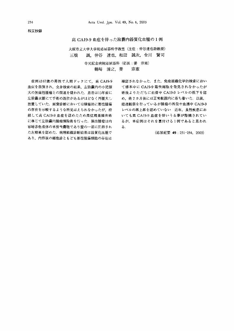
症例は67歳の男性で人間ドックにて,高 CA19-9
血症を指摘され,全身検索の結果,左陰嚢内の小児頭
大の無痛性腫癌との関連を疑われた.患者は15年前に
左陰嚢水腫にて手術の既往があるがほどなく再腫大し
放置していた.画像診断においては積極的に悪性腫蕩
の存在を示唆するような所見はえられなかったが,持
続して高 CA19-9血症を認めたため高位精巣摘出術
に準じて左陰嚢内腫癌摘除術を行った.摘出腫癌は内
容暗赤色液体の単房性嚢胞であり壁の一部に圧排され
た左精巣を認めた.病理組織診断結果は器質化血躍で
あり,内容液の細胞診ともども悪性腫蕩細胞の存在は
確認されなかった.また,免疫組織化学的検索におい
て標本中に CA19-9陽性細胞を発見されなかったが
術後よりただちに血清中 CA19-9レベルの低下を認
め,術 2カ月後には正常範囲内に落ち着いた.以後,
経過観察を行っているが腫癌の再発や血清中 CA19-9
レベルの再上昇を認めていない 近年,良性疾患にお
いても高 CA19-9血症を伴いうる事が指摘されてい
るが,本症例はそれを裏付ける I例であると思われ
る.
(泌尿紀要 49: 231-234, 2003)