title soft tissue recurrence of giant cell tumor of...
TRANSCRIPT
Title Soft Tissue Recurrence of Giant Cell Tumor of Bone : A Pitfallin Treating the Giant Cell Tumors of Bone
Author(s) MIKI, TAKAAKI; YAMAMURO, TAKAO; KOTOURA,YOSHIHIKO; SUGITANI, SHIGEKI; AMANO, TOSHIO
Citation 日本外科宝函 (1988), 57(1): 17-24
Issue Date 1988-01-01
URL http://hdl.handle.net/2433/203932
Right
Type Departmental Bulletin Paper
Textversion publisher
Kyoto University
Arch Jpn Chir 57(1), 17~23, Jan., 1988
Soft Tissue Recurrence of Giant Cell Tumor of Bone:
A Pitfall in Treating the Giant Cell Tumors of Bone
TAKAAKI MrKr1>, TAKAO YAMAMUR02>‘ YosHrHrKo KoTOURA2>,
SHIGEKI SuGITANr2>, and TosHJo AMANoa>
l) Department of Orthopaedics, Tamatsukuri-Kouseinenkin Hospital 2> Department of Orthopaedic Surgery, Faculty of Medicine, Kyoto University町 The
Division of Orthopaedics, Himeji National Hospital
Thourough curr巴ttageand bone grafting is the treatment of choice for most of the cases of
giant cell tumor of bone although high recurrence rate is known (GOLDENBERG”et al. 1970,
HUTTER, et al. 1962, }HNSON, et al. 1982, Marcove, et al. 1978). This tumor has an aggressive
biological characteristics (GOLDENBERG, et al. 1970, Hutter, et al. 1962, }HNSON, et al. 1982,
MARCOVE, et al. 1978, MIRRA, et al. 1982) and one of the pitfalls in treating this tumor is its
recurrence in the soft tissue of the operation五eld. When scattered to the soft tissue, giant cell
tumor of bone attracts new blood vessels and start proliferation there. We recently experienced
three cases of giant cell tumor of bone which recurred in the soft tissue of surgical field. We
believe such cases might not be rare and it is rather surprising that we could find out only a few
documented cases of soft tissue recurrence of giant cell tumor of bone in English literature
(FRAGAKIS, 1981, RILEY et al. 1967, SERRA, et al. 1985). It is a serious problem particularly in
the forearm or hand where many tendons and nerv巴srun and the removal of the recurrent
tumor is technically ve可 di伍cult. ¥Ve believe it could be preventable if we are careful enough
not to contaminate the soft tissue of th巴 op巴ration五eldby tumorous tissue.
Case Reports
Case 1. A 29-year-old woman noticed a dull pain in th巴rightpopliteal region in April,
1980 and visited a hospital on July 5, 1980. Roentgenograms showed an osteolytic lesion in th巴
distal epiphyseo-metaphyseal region of the right femur. The tumor was curretted and an iliac
bone graft was performed on September 12. The histological diagnosis was giant cell tumor of
bone. Follow up roentgenograms showed an recurrent tumor at th巴 siteof operation. The
patient underwent currettage and bone grafting again in April, 1981. Reccurrence of the tumor
was again apparent radiologically in July and she was referred to us. On November 25, when
whe was admitted to our hospital, two surgical scars were seen around her right knee, a medial
parapatellar incision and a lazy S-shaped scar in the popliteal region. Roentgenograms showed
Key words: Giant cell tumor ofしone,Recurrence in soft tissue.
索引語:骨巨細胞腫,軟部での再発.Present address: Department of Orthopaedics, Tamatsukuri-Kouseinenkin Hospital, 1-2, Yumachi唱 Tamayu-cho,Y atsuka-gun, Shimane Prefecture, 699-02 Japan.
18 日外宝第57巻第l号(昭和63年 1月)
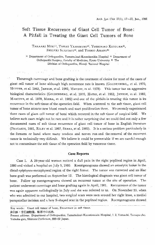
Fi~. 1-1. Lateral ,.i<・"・ of the kn氏、hリ、、in日an山 tcolyticlesion in the di,ta! cpiphyseometaphyseal 川 CIol the femUI . .¥rroWS indicate a的 ftti"m・ tumor m front of the n・けusfcmo11s.
an osteolytir lesion in the distal epiph)、eo-metaphysealregion of the right femur. The right
femoral condyle was mainly involved with little subchondral bone remaining目 Althoughthe
lateral femoral rnrtex w山 destroyed、thetumor was covered hy a thin bony layer, and there was
no u、iden何 ofsoft ti;,,uc extension of the tumor. Just anterior to the rectus femoris, there was
d 哨 fttissue m山 、whichwas 26 mm×13 mm on rocntgenogrnrns on :¥larch 3 (Fig. 1-1). The
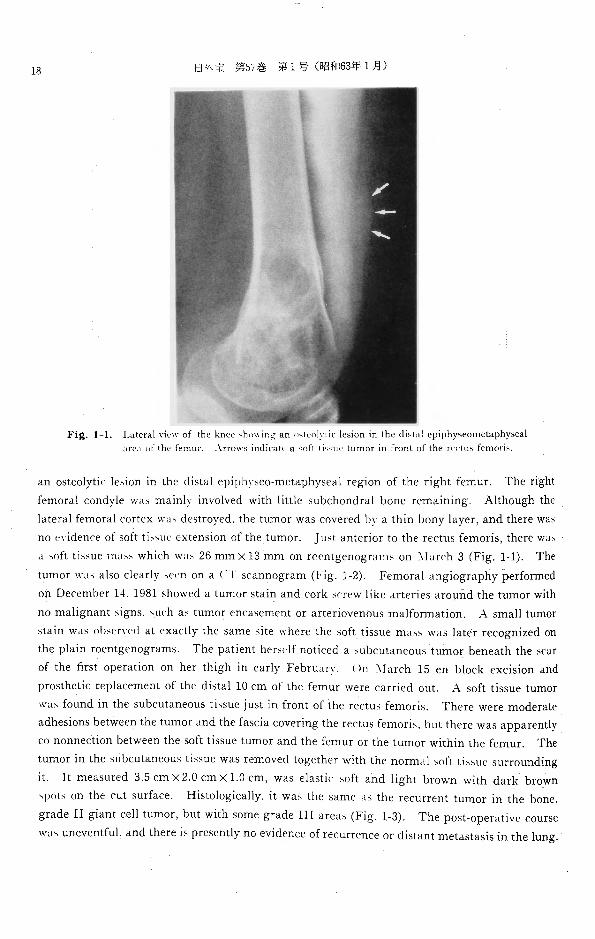
tumor w山 a!so clearly 吋叩 ona ( T scannogram (Fig. 1 2). Femoral angiography performed
on December 14. 1981 showed a tumor stain and cork screw like arteries around the tumor with
no malignant signs.、urhas tumor encasement or arteriovenous malformation. A small tumor
stain was olisl・rycd at exactly the same討itewhere the soft tissue mass was later recognized on
the plはinroentgenograms. The patient her吋 lfnoticed a subcutaneous tumor beneath the ~car
of the 五rstoperation on her thigh in early February. On :¥larch 15 en block excision and
prosthetic replacement of the dist乱l10 cm of the femur were carried out. A soft tissue tumo r
u,山 foundin the subcutaneous ti‘.、
adhesior】sbetween the tumor and the fascia covering the rectus femoris、butthere was apparently
co nonnection between the soft tissue tumor and the femur or the t山 norwithin the femur. The
tumor in the 叫1bcutaneousti州 uewas removed together with the normは1soft ti川 1csurrounding
it. It measured 3.5 cm×2.0 cm×1.0 cm, was elastic‘的ftand light brown with dark brown
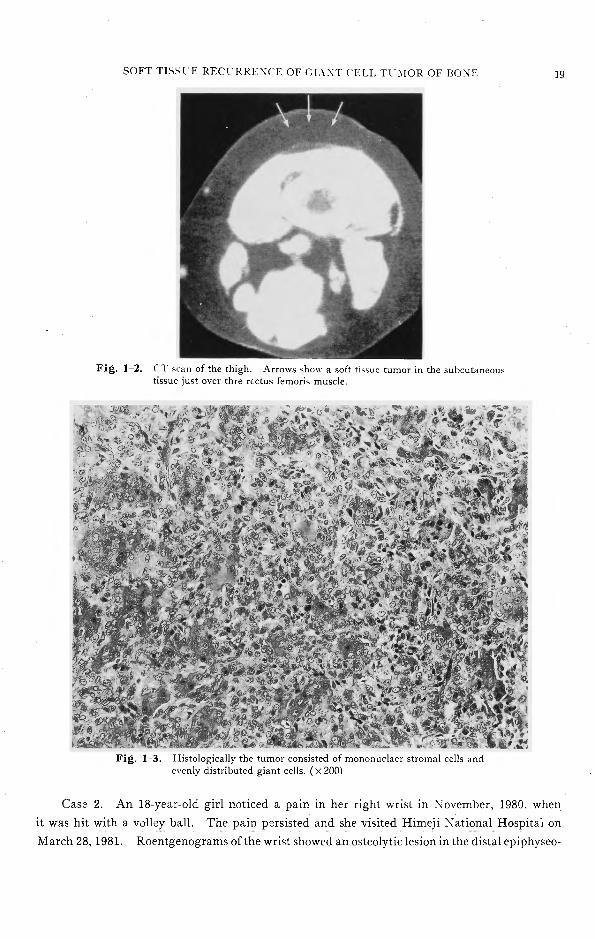
、potson the cut surface. Histologically、itwas the same川 therecurrent tumor in the bone,
grade II giant cell tumor, but with some grade III areas (Fig. 1-3). The post-operative course
W 山 uneventful,and there is presently no evidence of recurrence or distant metastasis in the lung.
SOFT TISSl E RECl-RRE'¥C、EOF CL¥'¥T CELL TC:¥IOR OF BO¥TF
Fig. 1 2. CT scan of the thigh. Arrows show a soft ti同 uctumor in the subcutaneous tissue just over thre rcctus femoris muscle.
司』滋ム 胸 、己主
Fig. 1 3. Histologically the tumor consisted of mononudaer stromal cells and evenly distributed giant cells. (×200)
19
Case 2. An 18-year-old girl noticed a pain in her right wrist in November, 1980, when
it was hit with a v0lley ball. The pain p~rsisted and she vi5ited Himeji :¥ ational Hospital on
March 28, 1981. Roentgenograms of the wrist showed an osteolytic lesion in the distal epiphyseo-
20
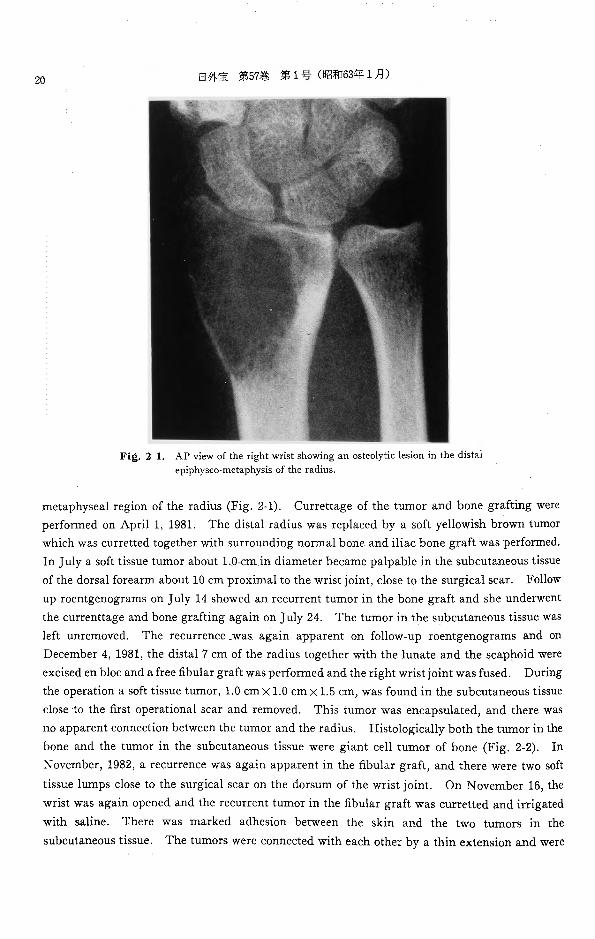
Fi邑.2-1.
日外宝第57巻第1号(昭和63年1月)
AP view of the right wrist showing an osteolytic lesion in the distal epiphyseo-metaphysis of the radius.
metaphyseal region of the radius (Fig. 2-1). Currettage of the tumor and bone grafting were
performed on April 1, 1981. The distal radius was replaced by a soft yellowish brown tumor
which was curretted together with surrounding normal bone and iliac bone graft was perfoロned.
In July a soft tissue tumor about 1.0 cm in diameter became palpable in the subcutaneous tissue
of the dorsal forearm about 10 cm proximal to the wrist joint, close to the surgical scar. Follow
up roentgenograms on July 14 showed an recurrent tumor in the bone graft and she underwent
the currenttage and bon巴graftingagain on July 24. The tumor in the subcutaneous tissue was
left unremoved. The recurrence was again apparent on follow-up roentgenograms and on
December 4, 1981, the distal 7 cm of the radius together with the lunate and the scaphoid were
excised en bloc and a free fibular graft was performed and the right wrist joint was fused. During
the operation a soft tissue tumor, 1.0 cm×l.Ocm×1.5 cm, was found in the subcutaneous tissue
close to the first operational scar and removed. This tumor was encapsulated, and there was
no apparent connection between the tumor and the radius. Histologically both the tumor in the
bone and the tumor in the subcutaneous tissue were giant cell tumor of bone (Fig. 2-2). In
~ovember, 1982, a recurrenc巴wasagain apparent in the fibular graft, and there were two soft
tissue lumps close to the surgical scar on the dorsum of the wrist joint. On November 16, the
wrist was again opened and the recurrent tumor in the fibular graft was curretted and irrigated
with saline. There was marked adhesion between the skin and the two tumors in the
subcutaneous tissue. The tumors were connected with each other by a thin extension and were
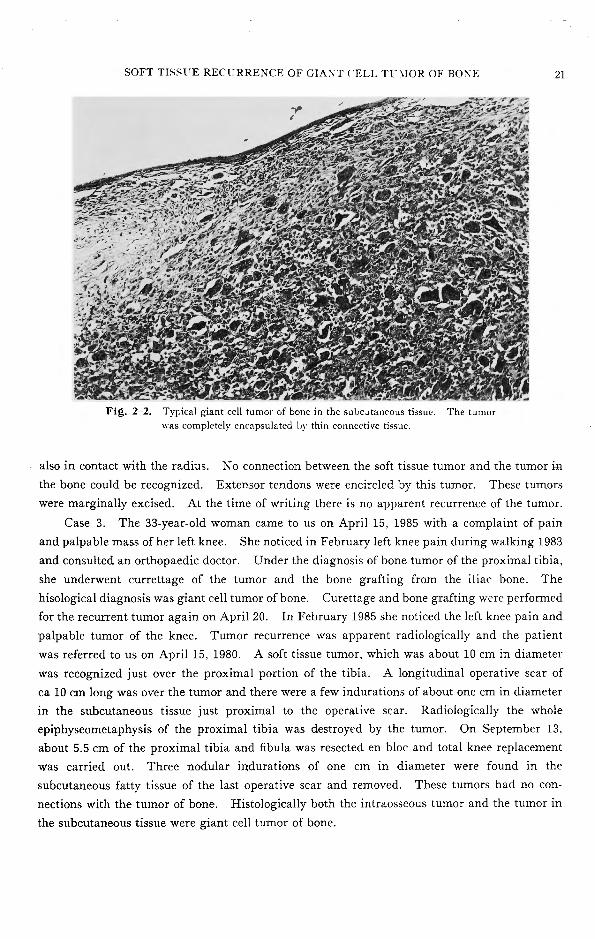
SOFT TI討おl'EREC℃RRENCE OF GIA¥!T CELL TC:¥IOR OF BOI¥E 21
Fig. 2 2. Typical giant cell tumor of bone in the subc~taneous tissue. The tumor ¥¥'aS completely encapsulated ],y thin connective tissue.
also in contact with the radius. No connection between the soft tissue tumor and the tumor in
the bone could be recognized. Extensor tendons were encircled by this tumor. These tumors
were marginally excised. At the time of writing there is no apparent recurrence of the tumor.
Case 3. The 33-year-old woman came to us on April 15, 1985 with a complaint of pain
and palpable mass of her left knee. She noticed in February left knee pain during walking 1983
and consulted an orthopaedic doctor. Under the diagnosis of bone tumor of the proximal tibia,
she underw巴ntcurrettage of the tumor and the bone grafting from the iliac bone. The
hisological diagnosis was giant cell tumor of bone. Curettage and bone grafting were performed
for the recurrent tumor again on April 20. In February 1985 she noticed the left knee pain and
palpable tumor of the knee. Tumor recurrence was apparent radiologically and the patient
was referred to us on April 15, 1980. A soft tissue tumor, which was about 10 cm in diameter
was recognized just over the proximal portion of the tibia. A longitudinal operative scar of
ca 10 cm long was over the tumor and there were a few indurations of about one cm in diameter
in the subcutaneous tissue just proximal to the operative scar. Radiologically the whole
epiphyseometaphysis of the proximal tibia was destroyed by the tumor. On September 13,
about 5.5 cm of the proximal tibia and五bulawas resected en bloc and total knee replacement
was carried out. Three nodular indurations of one cm in diameter were found in the
subcutaneous fatty tissue of the last operative scar and removed. These tumors had no con-
nections with the tumor of bone. Histologically both the intraosseous tumor and the tumor in
the subcutaneous tissue were giant cell tumor of bone.
22 日外宝第57巻第1号 (昭和63年 1月)
Discussion
Tumor contamination does not necessarily lead to local recurrence even in malignant tumors
(SPRT:"GFIELD, 1982, SuGARBAKER & KETO HAM. 1977). ¥fost of the scattered tumor cells die
because of nutritional deficit after the operation, immunological reactions or other reasons. Thus
soft tissue recurrence of benign bone tumors is unusual, even if some tumor cells or lumps of tumor
tissue are implanted in the soft tissue of the surgical field. Tumor cells or small lumps of tumor
tissue scattered in the surgical field may die or remain alive without proliferation in a dormant
state through the diffusion of nutrients for a while. Only when they attract vessels from the
surrounding tissue by Yirtue of tumor angiogenesis factor they start proliferation to form a
macromξtastasis. Tumor angiogenesis factor is considered to be a kind of R:¥ A molecule with
the molecular weight of 70,000 and usually secreted by malignant tumors or embryonal tissue,
and not by benign tumors or normal tissue (CLARK. 1979, FIDLER、etal. 1981, FISHER, et al.
1975, GALASKO, 1981. S!'.tITH, et al. 1958. SPRINGFIELD, 1982) It is well known that giant
cell tumors of bone (MIRRA, 1982) sometimes metastasize to the lung after currettage. The
metastasis does not generally endanger the patient’s life and is called benign metastasis (HUTTER,
1962. ¥lARCOVE, et al 1978, '.VIIRRA, 1980). Soft tissue recurrence of giant cell tumor of bone as
i,; seen in the three cases and also documented by FR.>.GAKIS (1981) and RILEY, et al (1967) and
SERRAS, et al (1985) is fundamentally the same phenomeneon as the benign metastasis in that the
scattered tumor cells obtain their blood supply supposedly by virtue of tumor angiogenesis
factor and proliferate. Considering the clinical facts of the high incidence of tumor contamination
of the wound in cancer patients (SMITH, et al. 1958), frequent subclinical fat embolism in long
bone fractures (ARx1:.1 & GRA1'T、1951.Gosul'¥G & PELLEGRJ:-11, 1982, SALDEEK, 1970), the
demonstration of pulmonary cement embolism after total hip replacement (BREED, 1974), or a
sudden incr何回 inthe number of tumor cells circulating in the blood of patients after surgical or
diagnostic procedured (FISLER, rt al 1981). the tumor contamination is inevitable, either locally
or systemically, during the currettage of bone tumors. The volume of the scattered tumor is
an important factor. SOUTHAM, et al (1961) demonstrated the dose and take relationship in
subcutaneous autotransplantations of human tumors; the larger the tumor dose or the volume
of implanted tumor, the more probable its local growth and innoculation of at least one million
cells are necessary for local growth of the tumor. Leaving a visilde size of the tumorous tissue
in the operation日eldis particularly dangerous. We should try to minimize contamination of
the surgical field by covering the exposed area with towels, frequently changing gloves and
instruments and irrigating with plenty of saline, although complete prevention may be impossible.
ft might be very difficult to wash out the tumor tissue sacattered over the soft tissue even if we
use the jet stream of water. So routinely covering of the soft tissue operation field by thick
towels may be most important. .-¥.lso we should excise giant cell tumor of bone en bloc whenever feasible.
References
1) Arnim J, Grant RT: Observation on gross pulmonary fat embolism in man and the rabbit. Cli Sci 10・441. 1951.
SOFT TISSUE RECじRRENCEOF GIANT CELL TUMOR OF BONE 23
2) Breed AL: Experimental production of vascular hypotension and bone marrow and fat embolism with
methylmethacrylate cement. Clin Orthop 102: 227, 1974.
3) Clark RL: Systemic cancer and the metastatic process. Cancer 43: 790, 1979.
4) Fidler IJ, Gersten DM, Hart IR: The biology of cancer invasion and metastasis. Adv Cancer Re• 28: 149, 1981.
5) Fisher B, Fisher Folkman J, Greenspan HP: Influence of geometry on control of cell growth. Biochim
Biophy Acta 417: 211, 1975.
6) Fragakis E: Soft tissue spread of giant cell tumor. A case report., J Bone Joint Surg 53-A・994,1981.
7) Galasko CBS: Bone metastasis studied in experimental animals. Clin Orthop 155: 269, 1981.
8) Goldengerg RR, Campbell CJ, Bor凸o:VI: Giant cell tumor of bone. An analysis of two hundred and
eighteen cases. J Bone Joint Surg 52-A: 619, 1870.
9) Gossling HR, Pellegrini VD: Fat embolism syndrome. A review of pathophysiology and physiological
basis of treatment. Clin Orthop 165: 68, 1982.
10) Hutter RV, Worchester JN, Francic KC, et al: Benign and malignant giant cell tumor of bone. Cancer
15: 653, 1962.
11) Jhnson EW, Kuo DP, Shu WP, et al: Giant cell tumor of bone. Analysis of two hundred eight cases in
Chinese literature. J Bone Joint Surg 64-A: 755, 1982.
12) Marcove RC, Weis LD, Vaghainwalla MR, et al: Cryosurgery in the treatment of giant cell tumors of bone.
A review of五ftytwo consecutive cases. Cancer 41: 957, 1978.
13) Mirra JM, Ulich T, Magidson J, et al: A case of probable pulmonary metastases or implants arising
from a giant cell tumor of bone. Clin Orthop 162: 245, 1982.
14) l』1irraJl¥I: Bone Tumors, diagnosis and treatment. 230, 1980. Lippincott.
15) Riley LH, Hartman ¥VH, Robinson RA: Soft tissue recurrence of giant cell tumor of bone after irrigation
and excision. J Bone Joint Surg 49-A: 365, 1967.
16) Saldeen T: Fat embolism and signs of intravascular coagulation in post traumatic autopsy material. J
Trauma 10: 272, 1970.
17) Serra JM, Mirragui A, Tadjalli H: Extensive sidta] subcutaneous metastases of a benign giant cell tumor
of the radius. Plast and Reconstructive surgery75 263 267, 1985.
18) Smith RR, Thomas LB, Hilbers AW: Cancer cell contamination of operative wounds. Cancer 11: 53,
1958.
19) Southam CM, Brunschwig A: Quantitative studies of autotransp!antation of human cancer. Cancer 14:
971' 1961. 20) Spring五eldDS: Mechanisms of metastasis. Cl in Orthop 169: 15, 1982.
21) Sugarbaker EV, Ketcham AS: :Vlechanisms and prevention of cancer dissemination. An overview
Seminars in Oncology 4: 19, 1977.
24 日外宝第57巻第l号 (昭和63年1月)
和文抄録
骨巨細胞腫の軟部組織での再発一一骨巨細胞腫治療上の落し穴一一
京都大学医学部整形外科学教室
三木 尭明,山室隆夫,琴浦良彦, 杉 谷 茂 樹 , 天 野 俊 男
術野軟部組織に再発した骨巨細胞腫を三症例を供覧 された小腫場塊の一部が増殖再発するのも“benign
する . 骨巨細胞腫は掻~, 骨移植術を施行した場合の metastasis,, と同様の機序と思われる.いずれにして
再発率40-5096と,良性腫蕩の中では治療に難渋する も治療上問題は大きく,殊に本論文の症例2のように
乙との多い骨腫蕩である.また掻問術後に 1-296の骨 多くの鍵,神経の走行する前腕での軟部組織での再発
巨細胞援は肺へ遠隔転移することも知られている.乙 は根治が困難となるので,術中術野の汚染を防ぐべき
の転移は,悪性腫療の場合と異なり生命的予後が良好 十分な努力をすべきである.即ち,可能な限り enbloc
であるため “benignmetastasis,, とも云われる.術中 lζ切除する,術野をタオルで、覆う, jetstream等で術
操作で血行中に入った腫場塊の一部が肺 まで遠隔転 野を十分洗浄する,手袋を頒固に交換する等の注意を
移しそこで一般的lζ良性腫場は生産しな いとされる 払うべきである. 殊に大きい腫場塊ほど生着しやすい
lumor Angigenesis Factorをおそらくは自ら生産し, から少なくとも肉眼に見える位以上の大きさの腫場塊
乙れを介して周囲組織より血行を得て増殖を開始した を術野IC残さぬことが大切で、あろう.
結果と思われる.掻J恨の際l乙術里子の軟部組織に撤種