utilitat de la membrana periprotètica en el diagnòstic de...
TRANSCRIPT

Utilitat de la membrana periprotètica en el diagnòstic de la infecció en el recanvi
d’una artroplàstia de maluc
Ernesto Muñoz Mahamud
ADVERTIMENT. La consulta d’aquesta tesi queda condicionada a l’acceptació de les següents condicions d'ús: La difusió d’aquesta tesi per mitjà del servei TDX (www.tdx.cat) i a través del Dipòsit Digital de la UB (diposit.ub.edu) ha estat autoritzada pels titulars dels drets de propietat intel·lectual únicament per a usos privats emmarcats en activitats d’investigació i docència. No s’autoritza la seva reproducció amb finalitats de lucre ni la seva difusió i posada a disposició des d’un lloc aliè al servei TDX ni al Dipòsit Digital de la UB. No s’autoritza la presentació del seu contingut en una finestra o marc aliè a TDX o al Dipòsit Digital de la UB (framing). Aquesta reserva de drets afecta tant al resum de presentació de la tesi com als seus continguts. En la utilització o cita de parts de la tesi és obligat indicar el nom de la persona autora. ADVERTENCIA. La consulta de esta tesis queda condicionada a la aceptación de las siguientes condiciones de uso: La difusión de esta tesis por medio del servicio TDR (www.tdx.cat) y a través del Repositorio Digital de la UB (diposit.ub.edu) ha sido autorizada por los titulares de los derechos de propiedad intelectual únicamente para usos privados enmarcados en actividades de investigación y docencia. No se autoriza su reproducción con finalidades de lucro ni su difusión y puesta a disposición desde un sitio ajeno al servicio TDR o al Repositorio Digital de la UB. No se autoriza la presentación de su contenido en una ventana o marco ajeno a TDR o al Repositorio Digital de la UB (framing). Esta reserva de derechos afecta tanto al resumen de presentación de la tesis como a sus contenidos. En la utilización o cita de partes de la tesis es obligado indicar el nombre de la persona autora. WARNING. On having consulted this thesis you’re accepting the following use conditions: Spreading this thesis by the TDX (www.tdx.cat) service and by the UB Digital Repository (diposit.ub.edu) has been authorized by the titular of the intellectual property rights only for private uses placed in investigation and teaching activities. Reproduction with lucrative aims is not authorized nor its spreading and availability from a site foreign to the TDX service or to the UB Digital Repository. Introducing its content in a window or frame foreign to the TDX service or to the UB Digital Repository is not authorized (framing). Those rights affect to the presentation summary of the thesis as well as to its contents. In the using or citation of parts of the thesis it’s obliged to indicate the name of the author.

TESI DOCTORAL
Utilitat de la membrana periprotètica en el
diagnòstic de la infecció en el recanvi d’una
artroplàstia de maluc
Tesi doctoral presentada per obtenir el títol de Doctor per la
Universitat de Barcelona
Ernesto Muñoz Mahamud
DIRECTORS: Dr. Guillem Bori Tuneu
Dr. Àlex Soriano Viladomiu
TUTOR: Dr. Sebastián García Ramiro
Programa doctorat medicina: Agressió biològica i mecanismes de resposta
DEPARTAMENT DE CIRURGIA I ESPECIALITATS QUIRÚRGIQUES
FACULTAT DE MEDICINA, UNIVERSITAT DE BARCELONA
BARCELONA, 2015


«Res del que succeeix s’oblida mai,
encara que un mateix no pugui recordar-ho»
Sen to Chihiro No Kamikakushi


V
El Dr. Guillem Bori Tuneu del Servei de Cirurgia Ortopèdica i
Traumatologia de l’Hospital Clínic de Barcelona, i el Dr. Àlex
Soriano Viladomiu del Servei de Malalties Infeccioses de l’Hospital
Clínic de Barcelona,
CERTIFIQUEN:
Que Ernesto Muñoz Mahamud ha elaborat el treball titulat Utilitat
de la membrana periprotètica en el diagnòstic de la infecció en
el recanvi d’una artroplàstia de maluc, sota la seva direcció, i
que compleix les exigències metodològiques i científiques per
poder optar al grau de Doctor en Medicina.
Els estudis presentats han estat desenvolupats al Grup de Patologia
Sèptica de l’Aparell Locomotor del Servei de Cirurgia Ortopèdica i
Traumatologia de l’Hospital Clínic de Barcelona.
Aquest treball s’ha realitzat amb l’ajuda d’una beca de la Societat
Catalana de Cirurgia Ortopèdica i Traumatologia (SCCOT)
concedida el mes de maig de 2014.
Dr. Guillem Bori Tuneu Dr. Àlex Soriano Viladomiu
Barcelona, 2015


VII
Agraïments
En primer lloc vull donar les gràcies al Guillem Bori per la seva
amistat i per la inestimable ajuda que sempre m’ha ofert des del
moment en què jo començava la meva etapa com a resident i ell tot
just l’acabava.
Vull també donar el meu sincer agraïment al Dr. García i al
Dr. Soriano, estimadíssims companys de feina que m’han fet
créixer ensenyant-me no tan sols coneixements del món de la
infecció osteoarticular sinó també valors i actituds com a metge i
com a persona. L’elaboració d’aquesta tesi no hauria estat possible
sense la seva admirable empenta.
També vull donar les gràcies a la resta de companys de la Unitat de
Maluc (Dr. Combalia, Dr. Fernández-Valencia, Dr. Gallart,
Dr. Riba i els residents que hi han passat), que van ajudar a la
recollida de tot el material necessari, i sense l’aportació dels quals
aquesta tesi no hauria estat possible.
Vull agrair a la Societat Catalana de Cirurgia Ortopèdica i
Traumatologia la beca que em va concedir per compensar les
despeses que ha ocasionat l’elaboració d’aquesta tesi.
Vull donar les gràcies a tots aquells amics i companys que,
d’alguna manera, m’han recolzat, orientat i acompanyat durant la
realització d’aquest treball. Gràcies especialment a Ruben Córdoba
pel disseny de la portada.

VIII
No tinc prou paraules d’agraïment per a aquelles persones que més
estimo. En primer lloc, al meu pare i a la meva mare, que tant
m’han estimat i que m’han educat i fet ser qui sóc. També a la meva
germana Blanca, a qui tantíssima estima tinc. Finalment, vull
dedicar aquesta tesi a la meva dona i al meu fill, Emma i Nico, que
amb el seu amor i afecte m’aporten cada dia allò necessari per ser
feliç i viure amb il·lusió. Sin vosotros, nada de esto tendría sentido.
Ernesto
Barcelona, 2015

IX
Resum
La infecció és una de les complicacions més greus que poden
aparèixer després de la implantació d’una artroplàstia total de
maluc. Fer un recanvi en un terreny infectat comporta un major risc
d’infecció de la nova pròtesi. No existeix cap prova que tingui
suficient sensibilitat i especificitat com per determinar si
l’afluixament protètic és degut a una causa sèptica o mecànica.
Aquesta tesi està dedicada a l’estudi de la utilitat de la membrana
periprotètica per al diagnòstic d’infecció durant el recanvi d’una
pròtesi de maluc.
El primer objectiu d’aquesta tesi ha estat avaluar si el rendiment de
la membrana periprotètica com a mostra per a l’estudi histològic
peroperatori d’infecció en un recanvi d’una artroplàstia de maluc és
superior al de la mostra de pseudocàpsula. Els resultats han
demostrat que el percentatge de la histologia positiva de la
membrana en pacients amb infecció de pròtesi és significativament
més gran que el percentatge de la histologia positiva de
pseudocàpsula. D’aquesta manera se suggereix que la membrana és
millor com a mostra histològica.
El segon objectiu ha estat determinar si el rendiment de la
membrana periprotètica com a mostra sòlida per al cultiu
convencional és superior al de la mostra de pseudocàpsula per al
diagnòstic d’infecció en un recanvi d’una artroplàstia de maluc. Els
X
resultats han demostrat que no existeixen diferències significatives
entre tots dos tipus de mostra.
El tercer objectiu ha estat avaluar la utilitat de la histologia fent
servir la membrana periprotètica per al diagnòstic de la infecció
durant un recanvi d’una artroplàstia de maluc per una fractura
periprotètica. Els resultats han demostrat que la utilització de la
histologia en casos de fractura està associada a una elevada taxa de
falsos positius (baixa especificitat).

XI
Abstract
Prosthetic infection is one of the most severe complications that can
appear after a total hip arthroplasty. To perform the replacement in
a septic environment entails a significantly higher risk of prosthesis
infection. Currently, there are no preoperative or perioperative
reliable tests able to determine whether the prosthesis loosening is
caused by septic or mechanical reasons. This thesis is devoted to
study the usefulness of the periprosthetic membrane for predicting
infection during a total hip replacement.
The first objective of this thesis has been to evaluate if the
usefulness of the periprosthetic membrane as a histologic sample is
more accurate than the pseudocapsule. The results have confirmed
that the percentage of positive histology of the membrane is
significantly higher than the pseudocapsule, suggesting that
periprosthetic membrane is the best specimen for the histological
diagnosis of prosthetic joint infection.
The second objective has been to assess whether conventional
cultures from the periprosthetic membrane are superior to
pseudocapsule samples in the diagnosis of infection in hip revision
arthroplasty. The results allow to concluding that the membrane
sample for a conventional culture is not superior to the
pseudocapsule sample in detecting microorganisms.
The third objective has been to analyze the usefulness of the
histology for the diagnosis of infection during a hip prosthesis

XII
replacement for the treatment of a periprosthetic fracture. The
results have demonstrated that periprosthetic fractures are a cause
of false-positive histology results for the diagnosis of infection
during revision of a hip prosthesis for the treatment of a
periprosthetic fracture when conventional cultures are used for
diagnosis of infection.

XIII
Índex
!Pàg.
1 Introducció ................................................................................ 1 1.1 Investigacions preoperatòries ....................................................... 3
1.1.1 Anamnesi i exploració física ................................................. 3
1.1.2 Radiologia ............................................................................. 4
1.1.3 Analítica ................................................................................ 5
1.1.4 Medicina nuclear ................................................................... 6
1.1.5 Anàlisi cel·lular del líquid articular ...................................... 8
1.1.6 Anàlisi microbiològica del líquid articular ........................... 8
1.2 Investigacions peroperatòries ....................................................... 9
1.2.1 Tinció de Gram del líquid articular ....................................... 9
1.2.2 Cultius intraoperatoris ........................................................... 9
1.2.3 Histologia ............................................................................ 10
2 Hipòtesi i Objectius ................................................................ 13
2.1 Hipòtesi ....................................................................................... 13
2.2 Objectius ..................................................................................... 14
3 Material i Mètodes .................................................................. 15 3.1 Disseny de l’estudi ...................................................................... 15
3.2 Pacients inclosos a l’estudi ......................................................... 15
3.3 Protocol d’obtenció de les mostres histològiques ....................... 16
3.4 Microbiologia ............................................................................. 17
3.5 Diagnòstic preoperatori .............................................................. 18
3.6 Diagnòstic definitiu .................................................................... 19
3.7 Anàlisi estadística ....................................................................... 20

XIV
4 Resultats .................................................................................. 23
4.1 Estudi 1 ....................................................................................... 23
4.2 Estudi 2 ....................................................................................... 28
4.3 Estudi 3 ....................................................................................... 32
5 Discussió .................................................................................. 37 5.1 Estudi 1 ....................................................................................... 37
5.2 Estudi 2 ....................................................................................... 41
5.3 Estudi 3 ....................................................................................... 45
6 Conclusions ............................................................................. 49
7 Bibliografia .............................................................................. 51
8 Publicacions ............................................................................. 63 !!!!!!!!!!!!!

XV
Llista de figures
Pàg.
Figura 1. Presència de fístula en una cicatriu.......................... 3
Figura 2. Afluixament del component acetabular................... 4
Figura 3. Afluixament de la tija femoral................................. 5
Figura 4. Imatge gammagràfica d’una pròtesi de maluc
infectada..................................................................................
7
Figura 5. Leucòcits polimorfonuclears en un tall histològic... 11
Figura 6. Histologia de membrana amb presència de
leucòcits polimorfonuclears....................................................
24
Figura 7. Histologia de pseudocàpsula sense leucòcits
polimorfonuclears....................................................................
24
Figura 8. Microorganismes identificats als recanvis sèptics
amb cultius positius.................................................................
31
Figura 9. Fractura periprotètica de maluc............................... 34
Figura 10. Fibrosi a la pseudocàpsula..................................... 40

XVII
Llista de taules
Pàg.
Taula 1. Relació entre els cultius i la histologia de la
membrana periprotètica.........................................................
26
Taula 2. Relació entre els cultius i la histologia en el
recanvi protètic......................................................................
27
Taula 3. Recanvis sèptics amb cultius positius...................... 29
Taula 4. Característiques i evolució dels pacients amb
fractura periprotètica..............................................................
33
Taula 5. Relació entre els cultius i la histologia de les
fractures periprotètiques........................................................
34
Taula 6. Mostres histològiques utilitzades per al diagnòstic
d’infecció protètica................................................................
38


XIX
Abreviatures
ECN Estafilococ coagulasa-negativa
HMPAO Hexametil propilen amino oxima
IS Infecció superficial
MIS Musculoskeletal Infection Society
PMN Leucòcits polimorfonuclears
PCR Proteïna C reactiva
SPSS Statistical Product and Service Solutions
VSG Velocitat de sedimentació globular


1
1 1 Introducció
La infecció és una de les complicacions més greus que poden
aparèixer després de la implantació d’una artroplàstia total de
maluc. Avui en dia, es considera com a taxa acceptable d’infecció
un valor per sota del 2 %, en funció de la durada de l’acte quirúrgic
i de la condició de base del pacient. En canvi, aquesta incidència
pot ascendir fins a un 15 % si es tracta d’un recanvi protètic (1).
Una infecció d’aquest tipus acostuma a associar-se a una elevada
morbiditat. En el millor dels casos, una neteja quirúrgica i
l’administració perllongada d’antibiòtics podria ser suficient per
curar la infecció (2). Malauradament, sovint és necessari retirar la
pròtesi infectada i substituir-la per una altra. Freqüentment cal
mantenir el pacient durant diverses setmanes sense articulació
funcional i amb tractament antibiòtic, i fer posteriorment una
segona intervenció per col·locar la nova pròtesi definitiva, la qual
ara tindrà un risc aproximadament cinc vegades més alt de patir una
nova infecció (3). Les repercussions funcionals, psíquiques,
familiars i econòmiques són per tant de gran magnitud (4, 5). Amb
l’envelliment de la població occidental i l’increment del nombre de

1. Introducció
2
pròtesis de maluc que es col·loquen anualment (6), és previsible
que la necessitat de recanviar pròtesis afluixades sigui cada vegada
més gran. Així mateix, el nombre de fractures que es produeixen al
voltant d’una pròtesi de maluc va en augment. La fractura
periprotètica es pot produir per un traumatisme important sobre una
pròtesi no afluixada o per un traumatisme mínim sobre una pròtesi
que ja presenti un afluixament (asèptic o sèptic) previ. Si el pacient
presenta una fractura periprotètica sobre un implant afluixat
[fractura tipus B2 segons la classificació internacional de
Vancouver (7)], llavors el tractament consisteix en el recanvi de la
pròtesi. Si aquest recanvi es duu a terme en el context d’un
afluixament sèptic la probabilitat que s’infecti el recanvi és alta. Per
tot això, la necessitat de millorar la prevenció, el diagnòstic i el
tractament d’aquesta complicació és essencial.
El diagnòstic diferencial entre una infecció crònica (afluixament
sèptic) i un afluixament mecànic (afluixament asèptic) és molt
difícil i alhora essencial, atès que el tractament quirúrgic en el
primer cas requereix un recanvi del material en dos temps i un
tractament antibiòtic prolongat (8, 9). En el segon cas es fa el
recanvi en un sol temps i no és necessari prolongar el tractament
antibiòtic més enllà de la profilaxi establerta. No reconèixer una
infecció, i per tant dur a terme un recanvi en un terreny infectat,
comporta un major risc d’infecció de la nova pròtesi.
Malauradament, no existeix cap prova que tingui una sensibilitat i
especificitat del 100 % per determinar si l’afluixament protètic és
degut a una causa sèptica o mecànica (1). Si l’afluixament de la
pròtesi s’ha produït en el context d’una fractura, el diagnòstic de la

1. Introducció
3
infecció pot ser un problema encara més difícil de resoldre. Les
proves per diagnosticar infecció al voltant de la pròtesi es poden
dividir en preoperatòries i peroperatòries.
1.1 Investigacions preoperatòries
1.1.1 Anamnesi i exploració física
Una història clínica i física detallada és important per al diagnòstic,
però no deixa de ser subjectiva. Únicament la troballa d’una fístula
és patognomònica d’infecció, però la seva presència és poc habitual
(Figura 1). Actualment la fístula està considerada un criteri major
d’infecció segons els nous criteris descrits per la Musculoskeletal
Infection Society (MIS) (10, 11). El que és més freqüent és que la
manifestació principal sigui dolor de característiques poc
específiques. Si el context és una fractura periprotètica, el dolor
produït per la fractura fa que la simptomatologia aguda perdi
utilitat.
Figura 1. Presència de fístula en una cicatriu. A la imatge es pot apreciar la presència d’una fístula a la cara externa de la cuixa després d’una pròtesi total de maluc, signe patognomònic d’infecció de l’implant.

1. Introducció
4
1.1.2 Radiologia
L’estudi radiològic aporta pocs canvis específics suggestius
d’infecció. En alguns casos es poden observar signes com osteòlisi,
reabsorció periòstica i imatges radiolúcides al voltant de l’implant,
però habitualment apareixen en fases avançades de l’afluixament i
cap d’ells són útils per diagnosticar entre afluixament mecànic o
sèptic (Figures 2 i 3).
Figura 2. Afluixament del component acetabular. Radiografia anteroposterior de pelvis on es mostra una pròtesi total de maluc dret. A la imatge es poden observar signes d’osteòlisi i imatges radiolúcides al voltant d’una còtila cimentada, que es tradueix en afluixament d’aquest component protètic, però sense poder diferenciar si és d’etiologia mecànica o sèptica.

1. Introducció
5
Figura 3. Afluixament de la tija femoral. Radiografia anteroposterior d’ambdós malucs on es mostra una pròtesi total de maluc dret. A la imatge poden observar-se signes d’osteòlisi i imatges radiolúcides al voltant d’una tija femoral no cimentada. La presència d’una osteòlisi tan important podria suggerir la presència d’una infecció.
1.1.3 Analítica
El nombre de leucòcits en la sang és un paràmetre molt poc sensible
(1, 12). En canvi la velocitat de sedimentació globular (VSG) i la
proteïna C reactiva (PCR) no només tenen una sensibilitat,
especificitat, valor predictiu positiu i valor predictiu negatiu al
voltant de 85 %, 92 %, 95 % i 66 % respectivament, segons les
sèries (1), sinó que també tenen un cost econòmic baix, per tant són
de gran utilitat en l’estudi preoperatori (13). No obstant això, s’ha

1. Introducció
6
de recordar que tant la VSG com la PCR són criteris menors
d’infecció segons la MIS (10, 11). A més de no tenir una fiabilitat
del 100 %, la seva principal limitació es troba en els pacients amb
malalties inflamatòries i en els que pateixen una fractura al voltant
de la pròtesi, ja que els valors dels paràmetres poden estar elevats
per la mateixa malaltia i per la mateixa fractura. La interleucina-6
també s’ha descrit com un bon marcador correlacionat amb el
diagnòstic d’infecció, però amb les mateixes limitacions que la
VSG i la PCR. Per tant, és necessari disposar de més informació per
determinar quin és el veritable paper diagnòstic que pot
desenvolupar aquest paràmetre (14).
1.1.4 Medicina nuclear
La gammagrafia òssia amb 99mTc-HMPAO (hexametil propilen
amino oxima) i utilitzant leucòcits marcats amb 99mTc-HMPAO,
així com la gammagrafia amb Ga-67, disposen en general d’elevada
sensibilitat i especificitat (Figura 4). No és així en infeccions
cròniques amb poca resposta inflamatòria. La gammagrafia de
medul·la macrofàgica pot incrementar significativament els
resultats si es compara amb la gammagrafia amb leucòcits marcats
(15). La seva utilització fent servir 99mTc-ciprofloxacina sembla
d’utilitat per al diagnòstic d’infecció de pròtesi de maluc, i la
sensibilitat, especificitat, valor predictiu positiu i valor predictiu
negatiu és de 74 %, 90 %, 86 % i 86 % respectivament (16). El
rendiment de la gammagrafia és, en general, molt baix en el context
d’una fractura periprotètica (17). Recentment s’ha descrit la
1. Introducció
7
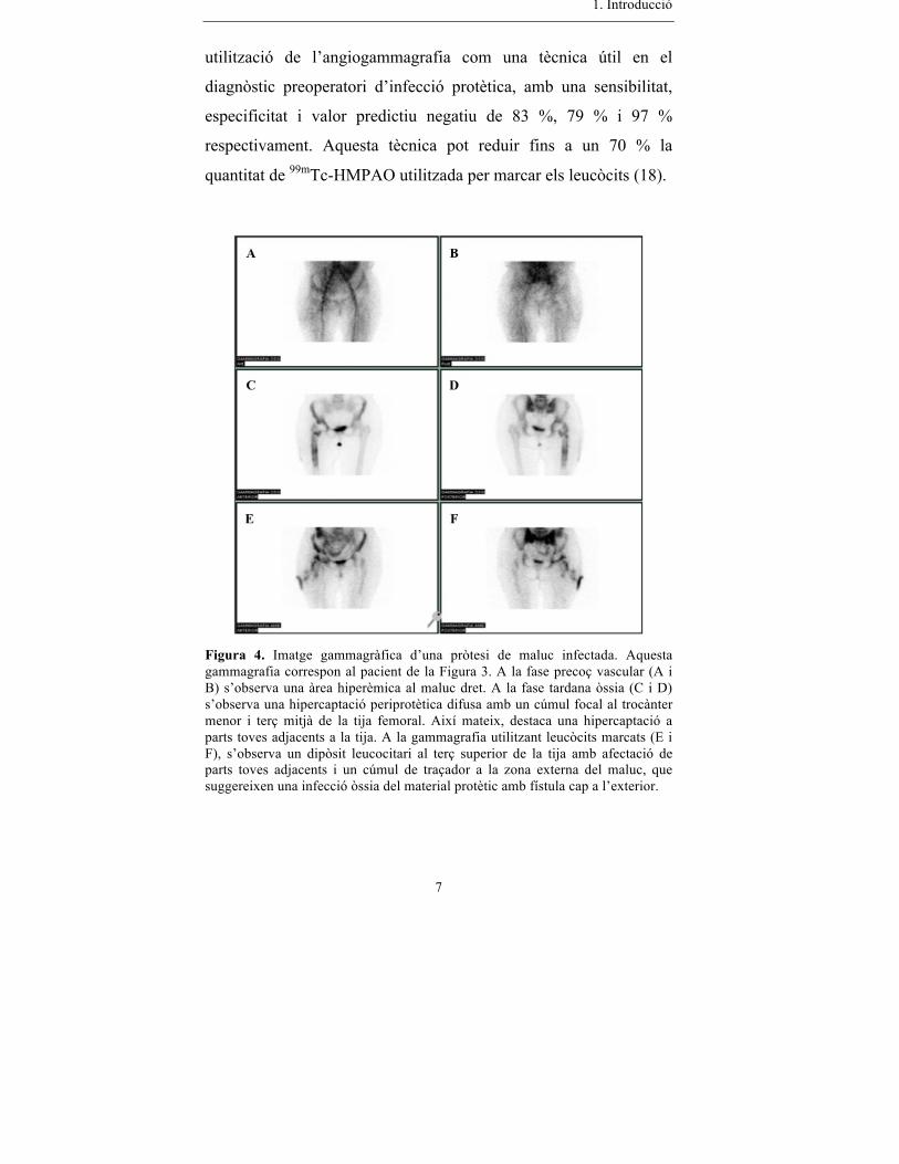
utilització de l’angiogammagrafia com una tècnica útil en el
diagnòstic preoperatori d’infecció protètica, amb una sensibilitat,
especificitat i valor predictiu negatiu de 83 %, 79 % i 97 %
respectivament. Aquesta tècnica pot reduir fins a un 70 % la
quantitat de 99mTc-HMPAO utilitzada per marcar els leucòcits (18).
Figura 4. Imatge gammagràfica d’una pròtesi de maluc infectada. Aquesta gammagrafia correspon al pacient de la Figura 3. A la fase precoç vascular (A i B) s’observa una àrea hiperèmica al maluc dret. A la fase tardana òssia (C i D) s’observa una hipercaptació periprotètica difusa amb un cúmul focal al trocànter menor i terç mitjà de la tija femoral. Així mateix, destaca una hipercaptació a parts toves adjacents a la tija. A la gammagrafia utilitzant leucòcits marcats (E i F), s’observa un dipòsit leucocitari al terç superior de la tija amb afectació de parts toves adjacents i un cúmul de traçador a la zona externa del maluc, que suggereixen una infecció òssia del material protètic amb fístula cap a l’exterior.

1. Introducció
8
1.1.5 Anàlisi cel·lular del líquid articular
El recompte del nombre de leucòcits i el percentatge de neutròfils
en el líquid articular ha despertat gran interès en els últims anys per
diagnosticar infecció al voltant de la pròtesi ja que aquests dos
paràmetres es consideren criteris menors d’infecció segons la MIS
(10, 11). Segons les sèries, tenen una sensibilitat, especificitat, valor
predictiu positiu i valor predictiu negatiu al voltant de 83 %, 93 %,
63 % i 93 %, respectivament (19-21), i per tant poden ser d’utilitat
en l’estudi preoperatori. Tot i així, aquests paràmetres no tenen una
fiabilitat del 100 %, sinó només del 83 %. A més, la taxa d’aspirat
blanc és aproximadament del 23 % (21), la qual cosa impedeix
sovint que es pugui fer aquesta anàlisi. S’ha descrit que un aspirat
blanc no significa que no hi hagi infecció articular (22). Per últim,
no està ben estudiat quina és la fiabilitat d’aquests tests en pacients
amb malalties inflamatòries i/o fractures periprotètiques.
1.1.6 Anàlisi microbiològica del líquid articular
El cultiu del líquid articular obtingut per teleradioscòpia, ecografia
o tomografia computada (23) no ha mostrat ni una elevada
sensibilitat ni especificitat. Aquesta anàlisi microbiològica del
líquid articular únicament té un rendiment elevat quan es duu a
terme en pacients amb una elevada sospita d’infecció i hi ha altres
indicadors suggestius d’infecció (24). En el context d’una fractura
periprotètica aquesta anàlisi no és viable ja que retardaria la cirurgia
diversos dies.

1. Introducció
9
1.2 Investigacions peroperatòries
1.2.1 Tinció de Gram del líquid articular
La tinció de Gram del líquid articular té una molt baixa sensibilitat
(varia entre el 0 % i el 19 % segons les sèries) i un pobre valor
predictiu negatiu, ja que la majoria de microorganismes es troben
adherits a la superfície protètica formant un biofilm (25). La seva
utilització en el diagnòstic d’infecció protètica està desaconsellada a
causa de l’elevada taxa de falsos negatius (26).
1.2.2 Cultius intraoperatoris
Els cultius intraoperatoris són actualment el patró or per establir el
diagnòstic de la infecció i són considerats un criteri major
d’infecció segons la MIS (10, 11). Per tal de disminuir al màxim la
taxa de falsos negatius, és imprescindible que el malalt no hagi
rebut antibiòtics durant les setmanes prèvies a la cirurgia. A causa
de la poca quantitat de microorganismes no adherits a la superfície
protètica, és recomanable obtenir de tres a cinc mostres de diferents
localitzacions (10).
S’ha descrit que el cultiu del líquid sinovial inoculat en flascons
d’hemocultiu és el tipus de cultiu convencional més sensible
comparat amb el cultiu de material sòlid periprotètic o els frotis
(27-30). En la nostra pràctica habitual les tres mostres
convencionals periprotètiques s’obtenen just en el moment d’obrir

1. Introducció
10
la càpsula i visualitzar la pròtesi, és a dir, s’obtenen del líquid
sinovial, del teixit sòlid de la pseudocàpsula que envolta el coll de
la pròtesi i fent un frotis del coll o el cap de la pròtesi.
Se sap que la major part dels microorganismes que infecten un
implant es troben creixent sobre el mateix implant formant biofilms
(sessile bacteria) i només una petita part es troben lliures
(planktonic bacteria) en els teixits periprotètics (29). Per tant, el fet
d’utilitzar material sòlid provinent de la pseudocàpsula i no de la
membrana periprotètica, que és el teixit sòlid més pròxim i amb
més contacte a la superfície de l’implant, podria explicar aquesta
menor sensibilitat del material sòlid respecte al líquid inoculat amb
flascons d’hemocultiu. Avui en dia, les guies aconsellen obtenir
com a mínim tres mostres per a cultiu, però no deixen clar de quin
tipus i d’on s’han d’obtenir aquestes mostres (10, 31-33).
1.2.3 Histologia
L’estudi histològic consisteix en la determinació del nombre de
leucòcits polimorfonuclears (PMN) existents en una mostra de
teixit periprotètic. En un teixit no infectat, la seva presència és
mínima o nul·la, mentre que és abundant quan el teixit està infectat
(34) (Figura 5). Actualment s’ha documentat que l’anatomia
patològica per al diagnòstic d’infecció protètica té un elevat
rendiment (17, 35-37), però en canvi en altres publicacions ha estat
descrita una baixa sensibilitat (32, 38-42). La baixa especificitat
(elevada taxa de falsos positius) pot ser deguda al fet que les
mostres per a l’estudi histològic i les mostres per a l’estudi

1. Introducció
11
microbiològic són obtingudes de diferents localitzacions (8, 27, 40,
41). La baixa sensibilitat (elevada taxa de falsos negatius) s’ha
atribuït a: 1) presència de microorganismes de baixa virulència com
Staphylococcus epidermidis o Propionibacterium spp., que no
arriben a estimular la infiltració de neutròfils en el teixit, 2)
contaminació bacteriològica de la mostra obtinguda per a l’estudi
microbiològic, i/o 3) punt de tall (nombre de neutròfils per camp de
gran augment) que s’utilitza per establir el diagnòstic d’infecció (8,
27, 40, 43-45). S’ha de recordar que la histologia també és un criteri
menor per al diagnòstic d’infecció segons la MIS (10, 11).
Figura 5. Leucòcits polimorfonuclears en un tall histològic. Fotomicrografia a camp de gran augment (x400) de tall histològic en parafina amb tinció d’hematoxilina-eosina, on es pot observar la presència d’abundants leucòcits polimorfonuclears (fletxes).

1. Introducció
12
Així mateix, un fet que pot provocar un possible biaix dels resultats
de la histologia són els pacients que es recanvien la pròtesi perquè
presenten una fractura periprotètica. Una revisió de la literatura
evidencia que molts autors (36, 38, 41, 46) afirmen que la fractura
periprotètica és una causa de falsos positius per al diagnòstic de la
infecció mitjançant la histologia però cap d’ells mostra dades
objectives sobre aquesta qüestió.

13
2
2 Hipòtesi i Objectius
2.1 Hipòtesi
Atès que, com s’ha comentat, la major part dels microorganismes
que infecten un implant es troben creixent sobre aquest formant
biofilms i només una petita part es troben lliures en els teixits del
voltant de la pròtesi, la nostra hipòtesi és que la utilització de la
membrana periprotètica com a mostra sòlida per al cultiu
convencional en el diagnòstic d’infecció en un recanvi d’una
artroplàstia de maluc pot tenir una major rendibilitat que no pas la
utilització de la pseudocàpsula. Així mateix, com que la important
formació de fibrosi a la pseudocàpsula pot dificultar la infiltració de
neutròfils en el teixit, també establim la hipòtesi que la utilització
de la membrana periprotètica com a mostra per a l’estudi histològic
peroperatori d’infecció en un recanvi d’una artroplàstia de maluc
pot tenir una major rendibilitat que no pas la utilització de la
pseudocàpsula.

2. Hipòtesi i Objectius
14
D’altra banda, una fractura periprotètica pot produir el pas de
neutròfils des del torrent intravascular cap als teixits periprotètics
degut tant a fenòmens de quimiotaxi com a la lesió mecànica dels
vasos sanguinis. Per tant, hipotetitzem que la utilització de la
histologia com a mètode diagnòstic peroperatori d’infecció durant
un recanvi d’una artroplàstia de maluc per una fractura periprotètica
pot estar associada a una elevada taxa de falsos positius (és a dir, a
una baixa especificitat).
2.2 Objectius
1. Analitzar si el rendiment de la membrana periprotètica com a
mostra per a l’estudi histològic peroperatori d’infecció en un
recanvi d’una artroplàstia de maluc és superior a la mostra de
pseudocàpsula.
2. Avaluar si el rendiment de la membrana periprotètica com a
mostra sòlida per al cultiu convencional per al diagnòstic d’infecció
en un recanvi d’una artroplàstia de maluc és superior a la mostra de
pseudocàpsula.
3. Determinar la utilitat de la histologia fent servir la membrana
periprotètica per al diagnòstic d’infecció en un recanvi d’una
artroplàstia de maluc per una fractura periprotètica.

15
3 3 Material i Mètodes
3.1 Disseny de l’estudi
Estudi prospectiu.
3.2 Pacients inclosos a l’estudi
Els pacients s’han inclòs a l’estudi segons l’objectiu.
Primer objectiu
Pacients sotmesos a un recanvi d’una pròtesi de maluc, entre gener
de 2007 i juny de 2009. Els pacients van ser classificats en dos
grups: grup A) pacients sotmesos a cirurgia de revisió protètica de
maluc amb diagnòstic preoperatori d’afluixament asèptic i
diagnòstic definitiu d’afluixament asèptic, i grup B) pacients
sotmesos a cirurgia de revisió protètica de maluc amb diagnòstic
preoperatori d’afluixament sèptic i diagnòstic definitiu
d’afluixament sèptic. No es van incloure pacients sotmesos a

3. Material i Mètodes
16
cirurgia de revisió de maluc per una fractura periprotètica ni tampoc
pacients amb diagnòstic preoperatori d’afluixament asèptic i
diagnòstic definitiu d’afluixament sèptic.
Segon objectiu
Pacients sotmesos a un recanvi d’una pròtesi de maluc, entre
octubre de 2009 i octubre de 2011. Es van crear dos grups en funció
de la mostra sòlida utilitzada: grup A) membrana periprotètica, i
grup B) pseudocàpsula. En el grup A, les mostres utilitzades per
identificar la infecció eren dues mostres líquides, dos frotis i dues
mostres de la membrana periprotètica. En el grup B, les mostres
utilitzades per identificar la infecció eren dues mostres líquides, dos
frotis i dues mostres de la pseudocàpsula. Les dues mostres líquides
i els dos frotis eren les mateixes mostres per als dos grups.
Tercer objectiu
Pacients sotmesos a un recanvi d’una pròtesi de maluc degut a una
fractura periprotètica tipus B2 segons la classificació de Vancouver
(7) entre gener de 2007 i juliol de 2011.
3.3 Protocol d’obtenció de les mostres histològiques
Es van obtenir mostres de pseudocàpsula i de membrana
periprotètica de cada pacient intervingut. Les mostres de
pseudocàpsula es van obtenir de la part en contacte amb el coll de la
pròtesi fent seccions perpendiculars. Les mostres de membranes es

3. Material i Mètodes
17
van obtenir de la interfície de la tija femoral i de la copa acetabular.
En fractures periprotètiques, la mostra es va obtenir de la tija
femoral on s’havia produït la fractura. Les mostres histològiques
van ser fixades amb formalina i parafina abans de ser tenyides amb
hematoxilina-eosina. El Servei d’Anatomia Patològica de l’Hospital
Clínic de Barcelona va utilitzar el criteri de Mirra amb l’adaptació
feta per Feldman (37, 47), de manera que la histologia va ser
considerada positiva per infecció quan hi havia cinc o més
neutròfils per camp de gran augment (x400) en almenys cinc camps
microscòpics diferents.
3.4 Microbiologia
En el moment de la retirada dels implants i prèviament a
l’administració de la profilaxi antibiòtica, sis mostres
periprotètiques de diferents localitzacions van ser enviades al
laboratori per al cultiu: dues mostres de líquid periprotètic, dues
mostres sòlides (pseudocàpsula) i dos frotis. Un cop obtingudes
aquestes sis mostres, es va iniciar la profilaxi antibiòtica. Quan el
recanvi no era degut a una fractura, una vegada retirat l’implant, es
van obtenir dues mostres sòlides més de la membrana periprotètica:
una provinent de la còtila i una altra provinent de la tija femoral. Si
el recanvi només era de la còtila o de la tija, es van obtenir dues
mostres de membrana del mateix lloc.
Les mostres líquides van ser obtingudes mitjançant aspiració amb
una xeringa estèril, immediatament inoculades en flascons

3. Material i Mètodes
18
d’hemocultiu Bactec 9000® (Becton Dickinson Diagnostic
Instruments®, Sparks, Maryland) i posteriorment incubades durant
cinc dies. Dels flascons amb creixement de microorganismes es van
fer subcultius en medis d’agar tant aeròbic com anaeròbic. També
es van obtenir cultius de frotis, fregant un isòtop estèril (Deltalab®,
hisop estèril Eurotube® en medi Stuart, Rubí, Catalunya, Espanya) a
l’àrea del teixit periprotètic, os o líquid sospitós d’infecció. Les
mostres sòlides de teixit periprotètic van ser posades
immediatament en un recipient estèril. Les mostres sòlides i els
frotis van ser cultivats en medis d’agar tant aerobi com anaerobi
així com en solució en medi de tioglicolat, tots ells enriquits amb
vitamina K i hemina, i posteriorment cultivats durant deu dies. Els
cultius positius van ser enviats per a la identificació i antibiograma.
3.5 Diagnòstic preoperatori
El diagnòstic preoperatori d’afluixament asèptic es va considerar
quan el pacient presentava aquests símptomes: dolor inespecífic,
VSG < 30 mm/h, concentració sèrica de la PCR < 1,3 mg/dL,
signes radiològics d’afluixament i/o gammagrafia òssia amb 99mTc-
HMPAO i leucòcits marcats amb 99mTc-HMPAO negatives per
infecció. En aquests pacients, es va fer un recanvi en un temps.
En canvi el diagnòstic preoperatori d’afluixament sèptic es va
considerar quan el pacient presentava els símptomes següents: dolor
al maluc i/o fístula, VSG > 30 mm/h, concentració sèrica de la
PCR > 1,3 mg/dL, signes radiològics d’afluixament, gammagrafia

3. Material i Mètodes
19
òssia amb 99mTc-HMPAO i leucòcits marcats amb 99mTc-HMPAO
positives per infecció i/o cultiu de líquid sinovial obtingut per
aspiració positiu. En aquests pacients, es va fer un recanvi en dos
temps.
3.6 Diagnòstic definitiu
El diagnòstic definitiu d’infecció del recanvi es va considerar: 1)
quan després d’analitzar el conjunt de sis mostres obtingudes (dues
mostres líquides, dues mostres sòlides i dos frotis), dues o més
mostres van ser positives per al mateix microorganisme, o bé 2)
quan hi havia presència de pus franc al voltant de la pròtesi (48).
Per tant, els pacients amb un o menys cultius intraoperatoris
positius van ser classificats com a no infectats.
Es van crear dos grups de sis mostres en funció de la mostra sòlida
utilitzada: membrana periprotètica (grup A) o pseudocàpsula (grup
B). En el grup A, les mostres utilitzades per identificar la infecció
van ser dues mostres líquides, dos frotis i dues mostres de la
membrana periprotètica. En el grup B, les mostres utilitzades per
identificar la infecció van ser dues mostres líquides, dos frotis i
dues mostres de la pseudocàpsula. Les dues mostres líquides i els
dos frotis van ser les mateixes mostres per als dos grups. Un cop
creats aquests dos grups de sis mostres, es va analitzar si hi havia
diferències en els resultats obtinguts en funció de si s’havia utilitzat
la mostra sòlida de membrana (grup A) o la mostra sòlida de
pseudocàpsula (grup B).

3. Material i Mètodes
20
Alhora, es van crear dos grups per avaluar quina era la mostra
histològica més útil per al diagnòstic d’infecció. Grup 1: pacients
sotmesos a un recanvi de pròtesi de maluc amb diagnòstic
preoperatori d’afluixament asèptic i diagnòstic definitiu
d’afluixament asèptic (pròtesi no infectada). Grup 2: pacients
sotmesos a un recanvi de pròtesi de maluc amb diagnòstic
preoperatori d’afluixament sèptic i diagnòstic definitiu
d’afluixament sèptic (pròtesi infectada). Aquells pacients amb un
diagnòstic preoperatori d’afluixament asèptic i un diagnòstic
definitiu d’afluixament sèptic van ser exclosos de l’estudi. Les
fractures periprotètiques també van ser excloses d’aquests dos
grups.
3.7 Anàlisi estadística
Es va dur a terme una anàlisi estadística amb la finalitat d’avaluar si
el rendiment de la membrana periprotètica com a mostra sòlida per
al cultiu convencional era superior a la mostra de pseudocàpsula per
al diagnòstic d’infecció en un recanvi d’una pròtesi de maluc.
Acceptant un risc alfa de 0,05 i un risc beta de 0,20 en un contrast
bilateral, es va calcular el nombre mínim de subjectes necessaris per
formar part de l’estudi. Es va utilitzar la prova Chi-quadrat (o la
prova exacta de Fisher si era necessari) assumint una significació
estadística amb un nivell de confiança del 95 % (p < 0,05).
D’una banda, es volia avaluar si el rendiment de la membrana
periprotètica com a mostra per a l’estudi histològic peroperatori

3. Material i Mètodes
21
d’infecció en un recanvi d’una pròtesi de maluc és superior al de la
mostra de pseudocàpsula. D’altra banda, es pretenia avaluar la
utilitat de la histologia fent servir la membrana periprotètica per al
diagnòstic d’infecció durant un recanvi d’una pròtesi de maluc per
una fractura periprotètica. És per això que es va calcular
l’especificitat (veritables negatius / falsos positius + veritables
negatius), la sensibilitat (veritables positius / falsos negatius +
veritables positius), el valor predictiu positiu (veritables positius /
veritables positius + falsos positius) i el valor predictiu negatiu
(veritables negatius / veritables negatius + falsos negatius) del
criteri de Mirra. Per a l’anàlisi comparativa de proporcions es va
utilitzar la prova exacta de Fisher assumint significació estadística
amb un nivell de confiança del 95 % (p < 0,05). L’anàlisi
estadística es va dur a terme amb el programa SPSS® (versió 20.0;
SPSS, Inc., Chicago, IL, EUA).


23
4 4 Resultats
4.1 Estudi 1
«Interface membrane is the best sample for histological study to
diagnose prosthetic joint infection»
Autors: Guillem Bori, Ernesto Muñoz-Mahamud, Sebastián García,
Carme Mallofré, Xavier Gallart, Jordi Bosch, Ester García, Josep
Riba, Josep Mensa, Àlex Soriano
Mod Pathol. 2011;24:579-584
Síntesi dels resultats
Es van incloure un total de 69 casos de recanvi de pròtesi de maluc,
57 classificats en el grup A (diagnòstic preoperatori d’afluixament
asèptic i diagnòstic definitiu d’afluixament asèptic) i 12 en el grup
B (diagnòstic preoperatori d’afluixament sèptic i diagnòstic
definitiu d’afluixament sèptic). En el grup B, el percentatge de
positivitat de la histologia de la membrana va ser significativament

4. Resultats
24
superior al de la pseudocàpsula (83 % i 42 % respectivament,
p = 0,04 a la prova exacta de Fisher) (Figures 6 i 7).
Figura 6. Histologia de membrana amb presència de leucòcits polimorfonuclears. Fotomicrografia a camp de gran augment (x400) de tall histològic en parafina amb tinció d’hematoxilina-eosina, on es pot observar la presència de més de 5 leucòcits polimorfonuclears.
Figura 7. Histologia de pseudocàpsula sense leucòcits polimorfonuclears. Fotomicrografia a camp de gran augment (x400) de tall histològic en parafina amb tinció d’hematoxilina-eosina, on no s’observen leucòcits polimorfonuclears. Aquest tall histològic correspon al mateix pacient que el de la Figura 6.

4. Resultats
25
Els detalls dels resultats obtinguts dels pacients amb diagnòstic
preoperatori d’afluixament asèptic i diagnòstic definitiu
d’afluixament asèptic (grup A) i dels pacients amb diagnòstic
preoperatori d’afluixament sèptic i diagnòstic definitiu
d’afluixament sèptic (grup B) es presenten a la Taula 1.

4. Resultats
26
Taula 1. Relació entre els cultius i la histologia de la membrana periprotètica Histologia Cultiu nc MOd Mostra
membrana pseudocàpsula
líquida sòlida frotis
GRUP Aa
- - - 55 - - - -
- + - 1 - - - -
+ - - 1 - - - -
GRUP Bb
+ + + 1 SAURe 1/2 1/2 1/2
+ + + 1 PAERf 2/2 0/2 0/2
+ + + 1 KPg 2/2 2/2 2/2
+ + + 1 PAER / ECNh 2/2 2/2 2/2
+ + + 1 ECN 2/2 2/2 2/2
+ - + 1 ECN 2/2 1/2 0/2
+ - + 1 ECN 1/2 3/3 0/2
+ - + 1 E. coli 1/1 1/2 2/2
+ - + 1 ECN 1/2 2/4 1/3
+ - + 1 ECN 2/2 0/2 0/2
- - + 1 ECN 1/2 1/2 0/2
- - + 1 SAUR 2/2 2/2 0/2
a Pacients sotmesos a cirurgia de revisió protètica de maluc amb diagnòstic preoperatori d’afluixament asèptic i diagnòstic definitiu d’afluixament asèptic
b Pacients sotmesos a cirurgia de revisió protètica de maluc amb diagnòstic preoperatori d’afluixament sèptic i diagnòstic definitiu d’afluixament sèptic
c Nombre de pacients d Microorganismes aïllats als cultius e Staphylococcus aureus f Pseudomonas aeruginosa g Klebsiella pneumoniae h Estafilococ coagulasa-negativa

4. Resultats
27
La sensibilitat, especificitat, valor predictiu positiu i valor predictiu
negatiu van ser respectivament de 83 %, 98 %, 91 % i 96% per a la
membrana i de 42 %, 98 %, 83 % i 83 % per a la pseudocàpsula. La
relació entre els resultats dels cultius i de la histologia es detallen a
la Taula 2.
Taula 2. Relació entre els cultius i la histologia en el recanvi protètic
Histologiaa Diagnòstic definitiu (cultius)b
Total Positiu Negatiu
Membrana periprotètica
Positiu 10 1 11
Negatiu 2 56 58
Total 12 57
Pseudocàpsula
Positiu 5 1 6
Negatiu 7 56 63
Total 12 57
a El resultat va ser considerat com a positiu per infecció quan es van identificar cinc o més PMN per camp de gran augment (x400) en almenys cinc camps microscòpics en seccions de parafina b El diagnòstic definitiu d’infecció va ser considerat positiu quan dos o més cultius intraoperatoris van ser positius per al mateix microorganisme, i/o quan es va detectar la presència de pus al voltant de la pròtesi

4. Resultats
28
4.2 Estudi 2
«Comparison of bacterial results from conventional cultures of
the periprosthetic membrane and the synovial or pseudocapsule
during hip revision arthroplasty»
Autors: Ernesto Muñoz-Mahamud, Àlex Soriano, Andrés
Combalia, Cristina Medrano, Jordi Bosch, Sebastián García,
Guillem Bori
Arch Orthop Trauma Surg. 2014;134:577-583
Síntesi dels resultats
Es van incloure un total de 86 casos de recanvi de pròtesi de maluc
amb el diagnòstic preoperatori següent: 50 recanvis asèptics, 1
recanvi sèptic en un temps, 16 primers temps de recanvi sèptic en
dos temps, 18 segons temps de recanvi sèptic en dos temps i 1
recanvi en un temps degut a fractura periprotètica. La sèrie incloïa
32 homes i 54 dones, amb una mitjana d’edat de 54 anys.
Del total de 86 recanvis, 22 van ser definitivament considerats com
a sèptics de forma postoperatòria i, d’aquests, 16 van tenir cultius
positius. Dels 16 recanvis amb cultius positius, 14 es van considerar
positius utilitzant tant la membrana com la pseudocàpsula com a
mostra sòlida per a cultiu (p = 0,484). En altres paraules, no es van
trobar diferències significatives entre els grups A i B. Els detalls
dels 16 casos de recanvi amb cultius positius es presenten a la
Taula 3.

4. Resultats
29
Taula 3. Recanvis sèptics amb cultius positius. Pacients sotmesos a recanvi de
pròtesi de maluc, diferenciant en cada cas si el pacient hagués estat considerat
infectat o no depenent de si la mostra sòlida utilitzada per a cultiu era la
membrana (grup A) o bé la pseudocàpsula (grup B)
n Diagnòstic
preoperatori LAa Frotis
Mb (grup A) Infecciód
(grup A)
MOe
(grup A)
Pc (grup B) Infecciód
(grup B)
MOe
(grup B)
1 Recanvi
asèptic 1/2 2/2
2/2 Sí ECNf
2/2 Sí ECN
2 Recanvi
asèptic 2/2 1/2
2/2 Sí ECN
2/2 Sí ECN
3 2n temps de
recanvi sèptic 0/2 0/2
0/2 No -
2/2 Sí S. capitis
4 1r temps de
recanvi sèptic 2/2 0/2
1/2 Sí STCg
2/2 Sí ECN
5 Recanvi
asèptic 2/2 0/2
0/2 Sí CBh
2/2 Sí CB
6 1r temps de
recanvi sèptic 1/2 2/2
2/2 Sí SAURi
0/2 Sí SAUR
7 1r temps de
recanvi sèptic 2/2 2/2
1/2 Sí SAUR
2/2 Sí SAUR
8 Recanvi
asèptic 0/2 1/2
0/2 No -
1/2 Sí ECN
9 1r temps de
recanvi sèptic 2/2 0/2
0/2 Sí ECN
2/2 Sí ECN
10 1r temps de
recanvi sèptic 2/2 1/2
1/2 Sí E. coli
KLj
1/2 Sí
E. coli
KL
ECN

4. Resultats
30
11 1r temps de
recanvi sèptic 2/2 2/2
1/2 Sí SAUR
1/2 Sí SAUR
12 1r temps de
recanvi sèptic 2/2 2/2
0/2 Sí SAUR
2/2 Sí SAUR
13 Recanvi
asèptic 2/2 1/2
1/2 Sí ECN
1/2 Sí ECN
14 1r temps de
recanvi sèptic 2/2 1/2
2/2 Sí SAUR
2/2 Sí SAUR
15 1r temps de
recanvi sèptic 2/2 2/2
2/2 Sí SAUR
2/2 Sí SAUR
16 1r temps de
recanvi sèptic 2/2 2/2
0/2 Sí SAUR
2/2 Sí SAUR
a Líquid articular b Membrana periprotètica c Pseudocàpsula d El diagnòstic definitiu d’infecció va ser considerat positiu quan dos o més cultius intraoperatoris van ser positius per al mateix microorganisme i/o quan es va detectar la presència pus al voltant de la pròtesi e Microorganismes aïllats als cultius f Estafilococ coagulasa-negativa g Streptococcus spp. h Corynebacterium spp. i Staphylococcus aureus j Klebsiella spp.
Hi va haver dos casos de discrepància diagnòstica quant al tipus de
mostra de material sòlid utilitzat (casos 3 i 8). En tots dos casos, si
la mostra sòlida utilitzada hagués estat la membrana, el recanvi
hauria estat considerat com a asèptic, mentre que utilitzant la
mostra de pseudocàpsula el recanvi hauria estat considerat sèptic.
Hi va haver cinc casos (casos 5, 6, 9, 12 i 16) on també hi va haver
discrepància quant al resultat obtingut utilitzant la membrana o bé
la pseudocàpsula, però el diagnòstic definitiu hauria estat igualment

4. Resultats
31
de recanvi sèptic ja que la resta de cultius (líquid articular i/o frotis)
ja eren positius (p = 0,083).
Hi va haver dos casos de discrepància quant al microorganisme
identificat a la mostra sòlida: en un cas (cas 4), en les dues mostres
de pseudocàpsula va créixer un estafilococ coagulasa-negativa
(ECN) (igual que en una de les mostres de líquid articular), mentre
que en una de les mostres de membrana va créixer un
Streptococcus spp. (igual que en una de les mostres de líquid
articular). En l’altre cas (cas 10), en una mostra de pseudocàpsula
va créixer un ECN (igual que en una de les mostres de frotis),
mentre que en una de les mostres de membrana va créixer un
Escherichia coli i una Klebsiella spp. (igual que en les dues mostres
de líquid articular).
El microorganisme més freqüentment identificat va ser l’ECN en
set casos (vuit casos, considerant que Staphylococcus capitis és un
ECN), seguit de Staphylococcus aureus en sis casos. Hi va haver
dos casos (4 i 10) d’infecció polimicrobiana (Figura 8).
Figura 8. Microorganismes identificats als recanvis sèptics amb cultius positius.

4. Resultats
32
4.3 Estudi 3
«Usefulness of Histology for Predicting Infection at the Time of
Hip Revision for the Treatment of Vancouver B2 Periprosthetic
Fractures»
Autors: Ernesto Muñoz-Mahamud, Guillem Bori, Sebastián García,
José Ramírez, Josep Riba, Àlex Soriano
J Arthroplasty. 2013;28:1247-1250
Síntesi dels resultats
Es van incloure un total d’onze casos de recanvi de pròtesi total de
maluc degut a fractura periprotètica tipus B2 de la classificació de
Vancouver (Figura 9). La sèrie incloïa vuit dones i tres homes, amb
una mitjana d’edat de 78,1 anys. Dels onze casos, sis presentaven
una histologia positiva, dels quals quatre presentaven cultius
negatius (falsos positius de la histologia). Els resultats es detallen a
la Taula 4.
La sensibilitat, especificitat, valor predictiu positiu i valor predictiu
negatiu de la histologia va ser de 100 %, 55,5 %, 33,3 % i 100 %
respectivament (Taula 5). D’aquells sis pacients que van presentar
una histologia positiva, només dos van tenir cultius positius: 66,6 %
de falsos positius.

4. Resultats
33
a El resultat va ser considerat com a positiu per infecció quan s’identificaven cinc o més PMN per camp de gran augment (x400) en almenys cinc camps microscòpics en seccions de parafina b El diagnòstic definitiu d’infecció va ser considerat positiu quan dos o més cultius intraoperatoris van ser positius per al mateix microorganisme, i/o quan es va detectar la presència de fístula o pus al voltant de la pròtesi c Estafilococ coagulasa-negativa d IS: Infecció superficial e SGV: Estreptococ del grup viridans
Taula 4. Característiques i evolució dels pacients amb fractura periprotètica
Cas Histologiaa Cultiusb Microorganisme Evolució
1 + + ECNc bona
2 + - - bona
3 - - - bona
4 - - - ISd 5 + - - bona
6 + + ECN + SGVe bona
7 - - - èxitus
8 + - - luxació
9 - - - bona
10 + - - bona
11 - - - bona

4. Resultats
34
Figura 9. Fractura periprotètica de maluc. Radiografia anteroposterior de maluc dret amb la presència d’una fractura al voltant d’una tija femoral afluixada.
Taula 5. Relació entre els cultius i la histologia de les fractures periprotètiques
Histologiaa
Histologiaa
Diagnòstic definitiu (cultius)b
Total
Total
Positiu Negatiu
Positiu 2 4 6
Negatiu 0 5 5
Total 2 9 11
a El resultat va ser considerat com a positiu per infecció quan s’identificaven cinc o més PMN per camp de gran augment (x400) en almenys cinc camps microscòpics en seccions de parafina b El diagnòstic definitiu d’infecció va ser considerat positiu quan dos o més cultius intraoperatoris van ser positius per al mateix microorganisme, i/o quan es va detectar la presència de pus al voltant de la pròtesi

4. Resultats
35
Dels onze pacients inclosos a la sèrie, un total de tres casos van
presentar algun tipus de complicació: en un cas (cas 4), el pacient
va presentar una infecció superficial de la ferida quirúrgica, que es
va tractar amb antibiòtic via oral i va presentar una posterior bona
evolució. En un altre cas (cas 7), el pacient va morir al cap de 20
dies de la intervenció a causa de problemes respiratoris no
relacionats directament amb la intervenció quirúrgica. Un altre cas
(cas 8) va presentar una luxació una setmana després de la
intervenció, que es va solucionar amb el recanvi del component
acetabular de la pròtesi. Els cultius de les mostres obtingudes durant
aquest últim recanvi van ser tots negatius.


37
5 5 Discussió
5.1 Estudi 1
Utilitat de la membrana periprotètica com a mostra histològica
en el diagnòstic de la infecció en el recanvi d’una artroplàstia de
maluc.
Establir el correcte diagnòstic d’infecció protètica és de gran
importància per tal de dur a terme un tractament correcte. La utilitat
de la histologia intraoperatòria ha estat analitzada en diferents
treballs i, avui en dia, forma part dels criteris diagnòstics d’infecció
protètica (10, 11). No obstant això, s’ha observat una notable
variabilitat quant a la seva sensibilitat depenent dels autors (32, 38-
42, 49, 50). Aquestes inconsistències poden atribuir-se al tipus de
pacient inclòs a l’estudi (32, 39, 40), als criteris microbiològics (1,
35, 51) i/o histològics (35-37, 52-54) aplicats per establir el
diagnòstic d’infecció o bé al tipus de mostra utilitzada per a
l’anàlisi (36). Revisant la literatura, existeix una enorme variabilitat
quant al tipus de mostra histològica utilitzada. Així, alguns autors

5. Discussió
38
han utilitzat teixits diferents de la membrana periprotètica (17, 52,
55, 56), o bé simplement no especifiquen quina és la mostra
utilitzada (47, 50, 57). A la Taula 6 es detallen els articles més
importants on es va utilitzar mostra histològica per al diagnòstic
d’infecció protètica.
Taula 6. Mostres histològiques utilitzades per al diagnòstic d’infecció protètica
Autor Mostra utilitzada Mirra i col. (58) Sinovial i teixit capsular Fehring i col. (49) Pseudocàpsula, membrana periprotètica i qualsevol àrea
amb aspecte sospitós d’infecció Feldman i col. (37) Pseudocàpsula i membrana periprotètica Athanasou i col. (36)
Pseudocàpsula i membrana periprotètica
Lonner i col. (46) Pseudocàpsula, membrana periprotètica i qualsevol àrea amb aspecte sospitós d’infecció
Pace i col. (59) Pseudocàpsula i membrana periprotètica Abdul-Karim i col. (41)
Sospita asèptica: membrana periprotètica. Sospita sèptica: membarana periprotètica, teixit sinovial i teixit amb algun inusual canvi de coloració
Spangehl i col. (1) Superfície sinovial Pandey i col. (53, 60)
Pseudocàpsula i membrana periprotètica
Pons i col. (17) Superfície sinovial Della Valle i col. (39)
Pseudocàpsula, teixit de granulació i qualsevol àrea amb aspecte sospitós d’infecció
Banit i col. (52) Pseudocàpsula i qualsevol àrea amb aspecte sospitós d’infecció
Musso i col. (42) Pseudocàpsula, membrana periprotètica i qualsevol àrea amb aspecte sospitós d’infecció
Ko i col. (38) Pseudocàpsula, membrana periprotètica i qualsevol àrea amb aspecte sospitós d’infecció
Wong i col. (61) Superfície sinovial, pseudocàpsula i membrana periprotètica
Frances i col. (57) Teixits tous periprotètics Bori i col. (32, 40, 62)
Pseudocàpsula, membrana periprotètica i qualsevol àrea amb aspecte sospitós d’infecció

5. Discussió
39
Morawietz i col. (35, 54)
Membrana periprotètica
Nuñez i col. (63) Pseudocàpsula, membrana periprotètica i qualsevol àrea amb aspecte sospitós d’infecció
Nilsdotter i col. (64) Superfície sinovial i membrana periprotètica Della Valle i col. (56)
Superfície sinovial
Kanner i col. (50) Teixits tous periprotètics Müller i col. (65, 66)
Membrana periprotètica
Schinsky i col. (55) Superfície sinovial Woo-Shin Cho i col. (67)
Qualsevol àrea amb aspecte sospitós d’infecció, canal medul·lar, teixit intraarticular
Tohtz i col. (51) Membrana periprotètica
En l’estudi comparatiu s’evidencia que la membrana té una major
sensibilitat i valors predictius que no pas la pseudocàpsula. De fet,
la proporció de pacients amb infecció protètica i amb membrana
positiva va ser significativament superior a aquells amb
pseudocàpsula positiva (83 % i 42 % respectivament, p = 0,04).
Així doncs, utilitzant només la pseudocàpsula, set dels dotze
pacients amb infecció no haurien estat correctament diagnosticats.
Athanasou i col. (36) ja van suggerir que, en general, hi ha una
major presència de signes inflamatoris a la membrana que a la
pseudocàpsula; no obstant això, aquesta informació mai no va ser
quantificada. Una possible explicació dels nostres resultats pot ser
la presència de fibrosi (Figura 10) a la pseudocàpsula, que dificulta
la infiltració dels neutròfils, així com el fet que la major part del
biofilm es troba entre l’implant i l’os (32).

5. Discussió
40
Figura 10. Fibrosi a la pseudocàpsula. Fotomicrografia de secció en parafina de pseudocàpsula tenyida amb hematoxilina-eosina obtinguda en un primer temps d’un recanvi sèptic d’una pròtesi de maluc. A la imatge no s’observa infiltrat de cèl·lules polimorfonuclears. Just sota la superfície de la pseudocàpsula (fletxa), s’aprecia la presència d’un dens teixit fibrós compost per fibres de col·lagen madur i fibroblasts ordenats.
Hi ha una sèrie d’autors (35, 51, 54, 65, 66) que han fet servir
només la membrana periprotètica i han proposat una classificació
histològica per avaluar de forma estandarditzada els teixits
periprotètics. Els nostres resultats recomanen la utilització de la
membrana periprotètica com a teixit de referència.
Tot i que l’anàlisi de talls congelats de la mostra histològica és la
tècnica habitualment utilitzada per obtenir un ràpid diagnòstic
durant la intervenció quirúrgica, en aquest estudi s’han fet servir
talls de parafina per tal de prevenir biaixos de la tècnica, ja que s’ha
descrit que els talls congelats tenen una pitjor qualitat que no pas

5. Discussió
41
els talls de parafina (1, 57). Tohtz i col. (51) van descriure un 19 %
de discrepàncies (en 14 de 64 casos) entre els resultats obtinguts
fent servir talls congelats i talls de parafina.
5.2 Estudi 2
Utilitat de la membrana periprotètica com a mostra per a
l’estudi microbiològic en el diagnòstic de la infecció en el
recanvi d’una artroplàstia de maluc.
El tractament antibiòtic d’un afluixament d’una pròtesi de maluc
degut a una infecció crònica és molt perllongat. Determinar el
microorganisme causant de la infecció és essencial per tal de poder
escollir l’antibiòtic més adient en cada cas. En infeccions
protètiques de tipus crònic, el nombre de bacteris lliures en els
teixits periprotètics és baix, ja que la majoria es troben formant part
d’un biofilm al voltant de l’implant (4, 68-72). Per tant, sembla
coherent obtenir diverses mostres durant la intervenció per tal de
poder identificar el microorganisme causant de la infecció. Existeix
un consens general que durant un recanvi d’una pròtesi afluixada és
necessari agafar més d’una mostra i, de fet, diversos autors obtenen
fins a sis mostres (31, 32, 73, 74). En canvi, existeix una gran
discrepància entre els autors sobre quin és el tipus de mostra que cal
obtenir. Aquest fet és de gran importància ja que existeixen
diferències significatives entre la sensibilitat dels diferents tipus de
mostra periprotètica (líquid articular, mostra sòlida i frotis) (27, 28).
A la literatura, els diferents autors utilitzen varietat de mostres i

5. Discussió
42
combinacions d’aquestes: mostra sòlida (19, 60), frotis (28, 75-77),
mostra sòlida i mostra líquida (78), mostra sòlida i frotis (1) o
combinació dels tres tipus (32).
Recentment s’han publicat alguns estudis que emfatitzen la baixa
sensibilitat dels frotis (28, 30, 79) i, de fet, les guies actuals (10,
73) no inclouen aquest tipus de mostra en els criteris per definir
infecció periprotètica. Els nostres resultats tenen concordança amb
els treballs publicats, ja que quatre dels setze casos infectats van
tenir frotis falsament negatius (25 % de taxa de falsos negatius).
Addicionalment, en altres quatre casos, només un dels dos cultius
de frotis va ser positiu, mentre que de dos a quatre dels cultius de la
resta de teixits van ser positius. D’altra banda, hi ha alguns autors
que defensen que els frotis poden tenir el seu rol ajudant en el
diagnòstic, ja que alguns pacients efectivament no haurien estat
diagnosticats correctament com a infectats si els frotis no
s’haguessin fet (28, 75). Així, un dels pacients de la sèrie (pacient
8) no hauria estat diagnosticat sense la mostra de frotis.
La literatura publicada afirma que la mostra amb una major
sensibilitat és el líquid sinovial o líquid periprotètic inoculat en
flascons d’hemocultiu, en comparació amb el material sòlid o els
frotis. Levine i Evans (27), en un estudi incloent 24 casos, van
descriure un 92 % de sensibilitat del líquid inoculat en flascons
d’hemocultiu, 46 % de sensibilitat de la mostra sòlida i 64 % de
sensibilitat dels frotis. Font-Vizcarra i col. (28) van corroborar
aquests resultats, publicant un 86 % de sensibilitat per a la mostra
líquida en flascó d’hemocultiu, 69 % per a la mostra sòlida i 61 %

5. Discussió
43
per als frotis. No obstant això, en aquest treball l’objectiu era
determinar si la mostra de membrana periprotètica com a mostra
sòlida per a cultiu té un major rendiment que la mostra de
pseudocàpsula per al diagnòstic d’infecció protètica.
En les infeccions protètiques, els microorganismes causants poden
trobar-se en dues formes diferents: de forma lliure en els teixits al
voltant de la pròtesi (s’anomenen planktonic bacteria) o bé creixent
sobre el mateix implant formant els biofilms (s’anomenen sessile
bacteria). Totes dues formes coexisteixen de manera simultània, no
sent excloents, però la majoria dels microorganismes es troben
sobre l’implant formant biofilms (80). Aquest fet desencadena un
primer problema des del punt de vista clínic, amb referència a la
identificació del microorganisme causant de la infecció, ja que els
mètodes de cultiu de rutina van ser desenvolupats per identificar
planktonic bacteria en infeccions agudes. En canvi, en infeccions
protètiques de tipus crònic, la majoria dels gèrmens són molt
difícils de fer créixer en cultius ja que romanen en un estat sèssil.
Aquests gèrmens poden ser clarament identificats utilitzant
tècniques avançades, mentre que sovint no són detectats pels
mètodes tradicionals de cultiu (27, 28, 68-70, 80, 81). Per aquest
motiu, es podria pensar que la membrana d’interfase, és a dir, aquell
teixit amb major íntim contacte amb l’implant, hauria de tenir un
major rendiment que no pas la pseudocàpsula, tenint en compte que
en infeccions cròniques la majoria de microorganismes es troben a
la superfície de la pròtesi formant biofilms.

5. Discussió
44
D’altra banda, en termes anatomopatològics, s’ha descrit que
existeix una major reacció inflamatòria amb presència de neutròfils
a la membrana que no pas a la pseudocàpsula (10, 72, 73, 80, 81).
Per tant, la possibilitat de trobar microorganismes podria ser també
major a la membrana. En canvi, els resultats de l’estudi mostren que
els resultats de les mostres sòlides de membrana no van
diagnosticar més casos d’infecció que els de la pseudocàpsula. De
fet, si només s’haguessin utilitzat mostres sòlides per al diagnòstic,
hi hauria hagut dos casos (casos 3 i 8) en què la infecció hauria
passat desapercebuda si la mostra hagués estat la membrana, però
no amb la pseudocàpsula. Una possible explicació per a aquest fet
podria ser que el volum de pseudocàpsula enviat per a l’estudi
microbiològic és major que el de la de membrana, ja que la
quantitat de la primera és molt més abundant i de més fàcil accés
per al cirurgià, mentre que la quantitat de membrana a vegades és
escassa i sovint de difícil accés. A més a més, com que la mostra
líquida es considera la més sensible de totes (10, 27, 28, 73), cal
tenir en compte que aquell teixit en més íntim contacte amb el
líquid és la pseudocàpsula, i no la membrana.
La pseudocàsula és un teixit de fàcil accés que pot obtenir-se de
forma ràpida al cap de pocs minuts d’haver començat la cirurgia. En
canvi, la membrana d’interfase només pot obtenir-se un cop s’ha
retirat l’implant, fet que pot tardar molta estona des de l’inici de la
intervenció. Això comporta un factor important per comentar, i és el
fet que, durant la intervenció quirúrgica, les mostres de
pseudocàpsula es van obtenir prèviament a l’administració de la
profilaxi antibiòtica, mentre que les mostres de membrana es van

5. Discussió
45
obtenir una vegada ja s’havia administrat la profilaxi. La profilaxi
antibiòtica és un factor crític per disminuir la taxa d’infecció en
cirurgia protètica. L’administració d’aquesta profilaxi s’intenta
demorar fins a l’obtenció de les mostres, ja que es creu que una sola
dosi d’antibiòtic pot afectar el resultat dels cultius intraoperatoris.
Així, és congruent demorar la profilaxi uns minuts fins a haver
obtingut les mostres de pseudocàpsula, el líquid articular i els frotis,
però sembla inacceptable demorar l’antibiòtic molta estona fins a
poder obtenir la membrana. No obstant això, tot i que no existeix
una gran evidència sobre quin és el millor moment per administrar
l’antibiòtic amb millor rendiment (82, 83), estudis recentment
publicats suggereixen que la profilaxi antibiòtica no té una
influència significativa sobre el resultat dels cultius (76, 77), i per
tant el fet d’administrar o no l’antibiòtic abans de l’obtenció de
mostres no hauria de suposar cap biaix.
5.3 Estudi 3
Utilitat de la membrana periprotètica com a mostra histològica
en el diagnòstic de la infecció en el recanvi d’una artroplàstia de
maluc per un fractura periprotètica.
La utilitat de la histologia per al diagnòstic d’infecció periprotètica
ha estat àmpliament descrita per diversos autors (17, 35-37, 84).
Tanmateix, en diferents treballs s’ha descrit una baixa sensibilitat
associada a aquest mètode diagnòstic (32, 38-42, 50). Les
discrepàncies entre la informació publicada pels diversos autors

5. Discussió
46
poden ser degudes als diferents criteris per diagnosticar infecció
[criteris microbiològics (1, 35, 51) i criteris histològics (35-37, 52-
54)], o bé als distints tipus de pacients estudiats [pacients sotmesos
a un recanvi protètic degut a un afluixament asèptic (40), a un
afluixament sèptic (62) o pacients sotmesos a un segon temps d’un
recanvi en dos temps per un afluixament sèptic (32, 39)].
A la literatura hi ha diversos autors que consideren les fractures
periprotètiques una potencial causa de fals positiu de la mostra
histològica per al diagnòstic d’infecció (42, 46, 49, 52, 53, 59), però
no hi ha cap estudi que hagi demostrat aquesta teoria. En aquest
treball s’ha tractat d’avaluar de forma específica la utilitat de la
histologia de la membrana periprotètica per al diagnòstic de
pacients sotmesos a un recanvi per una fractura sobre una pròtesi
afluixada. En sis dels onze casos amb histologia positiva, dos van
tenir cultius positius i quatre cultius negatius (66,6 % de falsos
positius). Els quatre casos amb histologia positiva i cultius negatius
no van rebre tractament antibiòtic addicional i l’evolució de la
pròtesi de revisió va ser bona, la qual cosa suggereix que
veritablement eren falsos positius.
Els neutròfils són cèl·lules intravasculars, que poden ser trobades
als teixits tous quan són atretes per quimiotaxi durant un procés
inflamatori i/o quan existeix lesió dels vasos sanguinis i es produeix
una extravasació d’aquests als teixits circumdants (34). Així, una
possible explicació dels resultats obtinguts pot ser la infiltració dels
neutròfils a la membrana periprotètica, procedents de la inflamació

5. Discussió
47
secundària així com dels vasos sanguinis lesionats a causa de la
fractura.
En l’actualitat no existeix cap prova preoperatòria ni peroperatòria
que sigui capaç de dilucidar amb una fiabilitat del 100 % si una
fractura periprotètica s’ha produït en un context d’infecció o no. Els
símptomes previs a la fractura són inespecífics i massa subjectius
(37), els reactants de fase aguda (PCR i VSG) i la gammagrafia es
veuen alterats per la mateixa fractura, i els resultats dels cultius de
líquid articular (obtingut mitjançant aspiració guiada per ecografia o
tomografia computada) retarden diversos dies la intervenció, i la
seva no suficientment elevada sensibilitat i especificitat no
justifiquen la demora d’una intervenció urgent (85). Hi ha autors
que han publicat sèries de fractures periprotètiques tractades
mitjançant recanvi de la pròtesi amb taxes d’infecció que
ascendeixen fins al 33 % (86-88). Holley i col. (89) van publicar
una taxa d’infecció del 10 % (2 de 20 casos) en fractures
periprotètiques de maluc tipus B2 de la classificació de Vancouver,
i del 33 % (3 de 9 casos) en fractures tipus B3. Mukundan i col.
(86) van tenir una taxa d’infecció del 17 % (3 de 17 casos) en
fractures tipus B3, mentre que Ko i col. (38) la van tenir del 14 %
(2 de 14 casos) en fractures tipus B2. Totes aquestes taxes
d’infecció són més elevades que les taxes publicades després d’un
recanvi per afluixament asèptic. Aquestes diferències podrien
explicar-se per la presència d’un focus sèptic al voltant de la pròtesi
sobre la qual s’ha produït la fractura. Per tant, identificar si una
fractura al voltant d’una pròtesi s’ha produït en un context sèptic o
asèptic pot ser de gran importància de cara a escollir el millor

5. Discussió
48
tractament i de cara al pronòstic. Així doncs, per exemple, si una
fractura periprotètica al voltant de la tija femoral s’identifica com a
no sèptica, serà suficient recanviar el component femoral de la
pròtesi. En canvi, si es detecta infecció al voltant de la pròtesi, s’ha
de recanviar també el component acetabular de la pròtesi. Un fals
positiu en la histologia podria fer pensar erròniament que la fractura
s’ha produït sobre una pròtesi infectada, i per tant es faria el recanvi
del component acetabular de forma innecessària, amb la
comorbiditat que això implica.
S’ha descrit que les fractures periprotètiques tipus B1 de la
classificació de Vancouver que són tractades mitjançant reducció
oberta i osteosíntesi presenten un pitjor pronòstic que les tipus B2
tractades mitjançant recanvi de la pròtesi, i la infecció és un dels
motius d’aquesta diferència en el pronòstic (90). Per tant, davant
una fractura periprotètica de maluc tipus B2 de la classificació de
Vancouver, sembla recomanable tractar-la mitjançant un recanvi de
la pròtesi, i obtenir almenys sis mostres intraoperatòries diferents
per a cultiu microbiològic, iniciar antibioteràpia endovenosa
d’ampli espectre de forma empírica i mantenir-la fins a l’obtenció
dels resultats dels cultius. En cas que els cultius siguin positius, i
d’acord amb l’antibiograma, es podrà escollir l’antibiòtic més
adient i fer el pas a antibiòtic via oral. Si els cultius són tots
negatius, llavors el tractament antibiòtic es podrà suspendre.

49
6 6 Conclusions
Objectiu 1
Analitzar si el rendiment de la membrana periprotètica com a
mostra per a l’estudi histològic peroperatori d’infecció en un
recanvi d’una artroplàstia de maluc és superior a la mostra de
pseudocàpsula.
Conclusió 1
El percentatge de positivitat de la histologia de la membrana
periprotètica en pacients amb infecció protètica és superior al de la
pseudocàpsula, i per tant es pot concloure que la millor mostra
histològica per al diagnòstic de la infecció durant un recanvi d’una
pròtesi de maluc és la membrana periprotètica.
Objectiu 2
Avaluar si el rendiment de la membrana periprotètica com a mostra
sòlida per al cultiu convencional per al diagnòstic d’infecció en un
recanvi d’una artroplàstia de maluc és superior a la mostra de
pseudocàpsula.

6. Conclusions
50
Conclusió 2
Quan per al diagnòstic d’una infecció s’utilitza el criteri d’un mínim
de dos cultius positius per al mateix microorganisme per
diagnosticar infecció periprotètica i es prenen un mínim de sis
mostres, incloent dos líquids inoculats en flascons d’hemocultiu,
dos frotis i dos sòlids, el lloc d’on s’obté la mostra sòlida
(pseudocàpsula o membrana periprotètica) és indiferent i no
modifica els resultats. Per tant, la mostra de membrana per al cultiu
convencional no és superior per a la detecció de microorganismes a
la mostra de pseudocàpsula, quan la profilaxi antibiòtica és
administrada després de l’obtenció de la mostra sòlida de
pseudocàpsula i abans de la mostra sòlida de membrana.
Objectiu 3
Determinar la utilitat de la histologia fent servir la membrana
periprotètica per al diagnòstic de la infecció durant un recanvi d’una
artroplàstia de maluc per una fractura periprotètica.
Conclusió 3
La fractura periprotètica és una causa de fals positiu de la histologia
en el diagnòstic d’infecció en un recanvi com a tractament d’una
fractura periprotètica de maluc tipus B2 de la classificació de
Vancouver. Per tant, no es recomana la utilització de la histologia
per al diagnòstic d’infecció durant el tractament d’una fractura
periprotètica a causa de la seva baixa especificitat.

51
7 7 Bibliografia
1. Spangehl MJ, Masri BA, O'Connell JX, Duncan CP.
Prospective analysis of preoperative and intraoperative investigations for the diagnosis of infection at the sites of two hundred and two revision total hip arthroplasties. J Bone Joint Surg Am 1999;81:672–83.
2. Soriano A, García S, Bori G, Almela M, Gallart X, Maculé F, Sierra J, Martínez JA, Suso S, Mensa J. Treatment of acute post-surgical infection of joint arthroplasty. Clin Microbiol Infect 2006;12:930–3.
3. Masri BA, Panagiotopoulos KP, Greidanus NV, Garbuz DS, Duncan CP. Cementless two-stage exchange arthroplasty for infection after total hip arthroplasty. J Arthroplasty 2007;22:72–8.
4. Garrido-Gomez J, Arrabal-Polo MA, Giron-Prieto MS, Cabello-Salas J, Torres-Barroso J, Parra-Ruiz J. Descriptive analysis of the economic costs of periprosthetic joint infection of the knee for the public health system of Andalusia. J Arthroplasty 2013;28:1057–60.
5. Klouche S, Sariali E, Mamoudy P. Total hip arthroplasty revision due to infection: a cost analysis approach. Orthop Traumatol Surg Res 2010;96:124–32.

7. Bibliografia
52
6. Nemes S, Gordon M, Rogmark C, Rolfson O. Projections of total hip replacement in Sweden from 2013 to 2030. Acta Orthop 2014;85:238–43.
7. Masri BA, Meek RMD, Duncan CP. Periprosthetic fractures evaluation and treatment. Clin Orthop Relat Res 2004;420:80–95.
8. Kraay MJ, Goldberg VM, Fitzgerald SJ, Salata MJ. Cementless two-staged total hip arthroplasty for deep periprosthetic infection. Clin Orthop Relat Res 2005;441:243–9.
9. Hart WJ, Jones RS. Two-stage revision of infected total knee replacements using articulating cement spacers and short-term antibiotic therapy. J Bone Joint Surg Br 2006;88:1011–5.
10. Parvizi J, Zmistowski B, Berbari EF, Bauer TW, Springer BD, Della Valle CJ, Garvin KL, Mont MA, Wongworawat MD, Charalampos G. New definition for periprosthetic joint infection: from the Workgroup of the Musculoskeletal Infection Society. Clin Orthop Relat Res 2011;469:2992–4.
11. Parvizi J, Gehrke T. Definition of periprosthetic joint infection. J Arthroplasty 2014;29:1331.
12. Canner GC, Steinberg ME, Heppenstall RB, Balderston R. The infected hip after total hip arthroplasty. J Bone Joint Surg Am 1984;66:1393–9.
13. Austin MS, Ghanem E, Joshi A, Lindsay A, Parvizi J. A simple, cost-effective screening protocol to rule out periprosthetic infection. J Arthroplasty 2008;23:65–8.
14. Bottner F, Wegner A, Winkelmann W, Becker K, Erren M, Gotze C. Interleukin-6, procalcitonin and TNF-alpha: markers of peri-prosthetic infection following total joint replacement. J Bone Joint Surg Br 2007;89:94–9.
7. Bibliografia
53
15. Fuster D, Duch J, Soriano A, García S, Setoain X, Bori G, Rubí S, Rodríguez D, Doménech D, Piera C, Mensa J, Pons F. Potential use of bone marrow scintigraphy in suspected prosthetic hip infection evaluated with 99mTc-HMPAO-leukocytes. Rev esp med nucl (English Edition) 2008;27:430–5.
16. Fuster D, Soriano A, Garcia S, Piera C, Suades J, Rodriguez D, Martínez JC, Mensa J, Campos F, Pons F. Usefulness of 99mTc-ciprofloxacin scintigraphy in the diagnosis of prosthetic joint infections. Nucl Med Commun 2011;32:44–51.
17. Pons M, Angles F, Sanchez C, Matamala A, Cuchi E, Salavert M, Forcada P, Ferrer H. Infected total hip arthroplasty. The value of intraoperative histology. Int Orthop (SICOT) 1999;23:34–6.
18. Granados U, Fuster D, Soriano A, García S, Bori G, Martinez JC, Mayoral M, Perlaza P, Tomas X, Pons F. Screening joint arthroplastic infection using blood flow scintigraphy prior to Tc-HMPAO labelled leukocyte. Rev Esp Med Nucl Imagen Mol 2015; Jan 3. pii: S2253-654X(14)00187-5. doi: 10.1016/j.remn.2014.10.003 [Epub ahead of print].
19. Mason JB, Fehring TK, Odum SM, Griffin WL, Nussman DS. The value of white blood cell counts before revision total knee arthroplasty. J Arthroplasty 2003;18:1038–43.
20. Trampuz A, Hanssen AD, Osmon DR, Mandrekar J, Steckelberg JM, Patel R. Synovial fluid leukocyte count and differential for the diagnosis of prosthetic knee infection. Am J Med 2004;117:556–62.
21. Squire MW, Valle CJD, Parvizi J. Preoperative diagnosis of periprosthetic joint infection: role of aspiration. Am J Roentgenol 2011;196:875–9.

7. Bibliografia
54
22. Ali F, Wilkinson JM, Cooper JR, Kerry RM, Hamer AJ, Norman P, Stockley I. Accuracy of joint aspiration for the preoperative diagnosis of infection in total hip arthroplasty. J Arthroplasty 2006;21:221–6.
23. Tomas X, Bori G, Garcia S, Garcia-Diez AI, Pomes J, Soriano A, Rios J, Almela M, Mensa J, Gallart X, Martinez JC, Riba J. Accuracy of CT-guided joint aspiration in patients with suspected infection status post-total hip arthroplasty. Skeletal Radiol 2011;40:57–64.
24. Tigges S, Stiles RG, Meli RJ, Roberson JR. Hip aspiration: a cost-effective and accurate method of evaluating the potentially infected hip prosthesis. Radiology 1993;189:485–8.
25. Updegrove GF, Armstrong AD, Kim HMM. Preoperative and intraoperative infection workup in apparently aseptic revision shoulder arthroplasty. J Shoulder Elbow Surg 2015;24:491–500.
26. Zywiel MG, Stroh DA, Johnson AJ, Marker DR, Mont MA. Gram stains have limited application in the diagnosis of infected total knee arthroplasty. Int J Infect Dis 2011;15:702–5.
27. Levine BR, Evans BG. Use of blood culture vial specimens in intraoperative detection of infection. Clin Orthop Relat Res 2001;382:222–31.
28. Font-Vizcarra L, Garcia S, Martínez-Pastor JC, Sierra JM, Soriano A. Blood culture flasks for culturing synovial fluid in prosthetic joint infections. Clin Orthop Relat Res 2010;468:2238–43.
29. Costerton JW. Biofilm theory can guide the treatment of device-related orthopaedic infections. Clin Orthop Relat Res 2005;437:7–11.

7. Bibliografia
55
30. Aggarwal VK, Higuera C, Deirmengian G, Parvizi J, Austin MS. Swab cultures are not as effective as tissue cultures for diagnosis of periprosthetic joint infection. Clin Orthop Relat Res 2013;471:3196–203.
31. Atkins BL, Athanasou N, Deeks JJ, Crook DW, Simpson H, Peto TE, McLardy-Smith P, Berendt AR. Prospective evaluation of criteria for microbiological diagnosis of prosthetic-joint infection at revision arthroplasty. The OSIRIS Collaborative Study Group. J Clin Microbiol 1998;36:2932–9.
32. Bori G, Soriano A, Garcia S, Mallofre C, Riba J, Mensa J. Usefulness of histological analysis for predicting the presence of microorganisms at the time of reimplantation after hip resection arthroplasty for the treatment of infection. J Bone Joint Surg Am 2007;89:1232–7.
33. Leone JM, Hanssen AD. Management of infection at the site of a total knee arthroplasty. J Bone Joint Surg Am 2005;87:2335–48.
34. Lindbom L, Werr J. Integrin-dependent neutrophil migration in extravascular tissue. Semin Immunol 2002;14:115–21.
35. Morawietz L, Classen R-A, Schroder JH, Dynybil C, Perka C, Skwara A, Neidel J, Gehrke T, Frommelt L, Hansen T, Otto M, Barden B, Aigner T, Stiehl P, Schubert T, Meyer-Scholten C, Konig A. Proposal for a histopathological consensus classification of the periprosthetic interface membrane. J Clin Pathol 2006;59:591–7.
36. Athanasou NA, Pandey R, de Steiger R, Crook D, Smith PM. Diagnosis of infection by frozen section during revision arthroplasty. J Bone Joint Surg Br 1995;77:28–33.
37. Feldman DS, Lonner JH, Desai P, Zuckerman JD. The role of intraoperative frozen sections in revision total joint arthroplasty. J Bone Joint Surg Am 1995;77:1807–13.

7. Bibliografia
56
38. Ko PS, Ip D, Chow KP, Cheung F, Lee OB, Lam JJ. The role of intraoperative frozen section in decision making in revision hip and knee arthroplasties in a local community hospital. J Arthroplasty 2005;20:189–95.
39. Valle Della CJ, Bogner E, Desai P, Lonner JH, Adler E, Zuckerman JD, Di Cesare PE. Analysis of frozen sections of intraoperative specimens obtained at the time of reoperation after hip or knee resection arthroplasty for the treatment of infection. J Bone Joint Surg Am 1999;81:684–9.
40. Bori G, Soriano A, Garcia S, Gallart X, Casanova L, Mallofre C, Almela M, Martínez JA, Riba J, Mensa J. Low sensitivity of histology to predict the presence of microorganisms in suspected aseptic loosening of a joint prosthesis. Mod Pathol 2006;19:874–7.
41. Abdul-Karim FW, McGinnis MG, Kraay M, Emancipator SN, Goldberg V. Frozen section biopsy assessment for the presence of polymorphonuclear leukocytes in patients undergoing revision of arthroplasties. Mod Pathol 1998;11:427–31.
42. Musso AD, Mohanty K, Spencer-Jones R. Role of frozen section histology in diagnosis of infection during revision arthroplasty. Postgrad Med J 2003;79:590–3.
43. Tsaras G, Maduka-Ezeh A, Inwards CY, Mabry T, Erwin PJ, Murad MH, Montori VM, West CP, Osmon DR, Berbari EF. Utility of intraoperative frozen section histopathology in the diagnosis of periprosthetic joint infection: a systematic review and meta-analysis. J Bone Joint Surg 2012;94:1700–11.
44. Grosso MJ, Frangiamore SJ, Ricchetti ET, Bauer TW, Iannotti JP. Sensitivity of frozen section histology for identifying Propionibacterium acnes infections in revision shoulder arthroplasty. J Bone Joint Surg Am 2014;96:442–7.

7. Bibliografia
57
45. Zhao X, Guo C, Zhao G-S, Lin T, Shi ZL, Yan SG. Ten versus five polymorphonuclear leukocytes as threshold in frozen section tests for periprosthetic infection: a meta-analysis. J Arthroplasty 2013;28:913–7.
46. Lonner JH, Desai P, Dicesare PE, Steiner G, Zuckerman JD. The reliability of analysis of intraoperative frozen sections for identifying active infection during revision hip or knee arthroplasty. J Bone Joint Surg Am 1996;78:1553–8.
47. Mirra JM, Amstutz HC, Matos M, Gold R. The pathology of the joint tissues and its clinical relevance in prosthesis failure. Clin Orthop Relat Res 1976;117:221–40.
48. Leone JM, Hanssen AD. Management of infection at the site of a total knee arthroplasty. J Bone Joint Surg Am 2005;87:2335–48.
49. Fehring TK, McAlister JAJ. Frozen histologic section as a guide to sepsis in revision joint arthroplasty. Clin Orthop Relat Res 1994;304:229–37.
50. Kanner WA, Saleh KJ, Frierson HFJ. Reassessment of the usefulness of frozen section analysis for hip and knee joint revisions. Am J Clin Pathol 2008;130:363–8.
51. Tohtz SW, Muller M, Morawietz L, Winkler T, Perka C. Validity of frozen sections for analysis of periprosthetic loosening membranes. Clin Orthop Relat Res 2010;468:762–8.
52. Banit DM, Kaufer H, Hartford JM. Intraoperative frozen section analysis in revision total joint arthroplasty. Clin Orthop Relat Res 2002;401:230–8.
53. Pandey R, Drakoulakis E, Athanasou NA. An assessment of the histological criteria used to diagnose infection in hip revision arthroplasty tissues. J Clin Pathol 1999;52:118–23.

7. Bibliografia
58
54. Morawietz L, Tiddens O, Mueller M, Tohtz S, Gansukh T, Schroeder JH, Perka C, Krenn V. Twenty-three neutrophil granulocytes in 10 high-power fields is the best histopathological threshold to differentiate between aseptic and septic endoprosthesis loosening. Histopathology 2009;54:847–53.
55. Schinsky MF, Valle Della CJ, Sporer SM, Paprosky WG. Perioperative testing for joint infection in patients undergoing revision total hip arthroplasty. J Bone Joint Surg 2008;90:1869–75.
56. Valle Della CJ, Sporer SM, Jacobs JJ, Berger RA, Rosenberg AG, Paprosky WG. Preoperative testing for sepsis before revision total knee arthroplasty. J Arthroplasty 2007;22:90–3.
57. Frances Borrego A, Martinez FM, Cebrian Parra JL, Graneda DS, Crespo RG, Lopez-Duran Stern L. Diagnosis of infection in hip and knee revision surgery: intraoperative frozen section analysis. Int Orthop (SICOT) 2007;31:33–7.
58. Mirra JM, Marder RA, Amstutz HC. The pathology of failed total joint arthroplasty. Clin Orthop Relat Res 1982;170:175–83.
59. Pace TB, Jeray KJ, Latham JTJ. Synovial tissue examination by frozen section as an indicator of infection in hip and knee arthroplasty in community hospitals. J Arthroplasty 1997;12:64–9.
60. Pandey R, Berendt AR, Athanasou NA. Histological and microbiological findings in non-infected and infected revision arthroplasty tissues. The OSIRIS Collaborative Study Group. Oxford Skeletal Infection Research and Intervention Service. Arch Orthop Trauma Surg 2000;120:570–4.
61. Wong YC, Lee QJ, Wai YL, Ng WF. Intraoperative frozen section for detecting active infection in failed hip and knee arthroplasties. J Arthroplasty 2005;20:1015–20.

7. Bibliografia
59
62. Bori G, Soriano A, Garcia S, Gallart X, Mallofre C, Mensa J. Neutrophils in frozen section and type of microorganism isolated at the time of resection arthroplasty for the treatment of infection. Arch Orthop Trauma Surg 2009;129:591–5.
63. Nunez LV, Buttaro MA, Morandi A, Pusso R, Piccaluga F. Frozen sections of samples taken intraoperatively for diagnosis of infection in revision hip surgery. Acta Orthop 2007;78:226–30.
64. Nilsdotter-Augustinsson A, Briheim G, Herder A, Ljunghusen O, Wahlstrom O, Ohman L. Inflammatory response in 85 patients with loosened hip prostheses: a prospective study comparing inflammatory markers in patients with aseptic and septic prosthetic loosening. Acta Orthop 2007;78:629–39.
65. Muller M, Morawietz L, Hasart O, Strube P, Perka C, Tohtz S. Diagnosis of periprosthetic infection following total hip arthroplasty. Evaluation of the diagnostic values of pre- and intraoperative parameters and the associated strategy to preoperatively select patients with a high probability of joint infection. J Orthop Surg Res 2008;3:31.
66. Muller M, Morawietz L, Hasart O, Strube P, Perka C, Tohtz S. Histopathological diagnosis of periprosthetic joint infection following total hip arthroplasty: use of a standardized classification system of the periprosthetic interface membrane. Orthopade 2009;38:1087–96.
67. Cho WS, Byun SE, Cho WJ, Yoon YS, Dhurve K. Polymorphonuclear cell count on frozen section is not an absolute index of reimplantation in infected total knee arthroplasty. J Arthroplasty 2013;28:1874–7.
68. Arciola CR, Montanaro L, Costerton JW. New trends in diagnosis and control strategies for implant infections. Int J Artif Organs 2011;34:727–36.

7. Bibliografia
60
69. Costerton JW, Stewart PS, Greenberg EP. Bacterial biofilms: a common cause of persistent infections. Science 1999;284:1318–22.
70. Hill KE, Malic S, McKee R, Rennison T, Harding KG, Williams DW, Thomas DW. An in vitro model of chronic wound biofilms to test wound dressings and assess antimicrobial susceptibilities. J Antimicrob Chemother 2010;65:1195–206.
71. Drago L, Romano CL, Mattina R, Signori V, De Vecchi E. Does dithiothreitol improve bacterial detection from infected prostheses? A pilot study. Clin Orthop Relat Res 2012;470:2915–25.
72. Zameer F, Gopal S. Evaluation of antibiotic susceptibility in mixed culture biofilms. Intern J Biotech Biochem 2010;6:93–9.
73. Osmon DR, Berbari EF, Berendt AR, Lew D, Zimmerli W, Steckelberg JM, Rao N, Hanssen A, Wilson WR. Executive summary: diagnosis and management of prosthetic joint infection: clinical practice guidelines by the Infectious Diseases Society of America. Clin Infect Dis 2013;56:1–10.
74. Trampuz A, Piper KE, Jacobson MJ, Hanssen AD, Unni KK, Osmon DR, Mandrekar JN, Cockerill FR, Steckelberg JM, Greenleaf JF, Patel R. Sonication of removed hip and knee prostheses for diagnosis of infection. N Engl J Med 2007;357:654–63.
75. Mehra A, Hemmady MV, Nelson R, Hodgkinson JP. Bacteriology swab in primary total hip arthroplasty: does it have a role? Int J Clin Pract 2006;60:665-6.
76. Burnett RSJ, Aggarwal A, Givens SA, McClure JT, Morgan PM, Barrack RL. Prophylactic Antibiotics Do Not Affect Cultures in the Treatment of an Infected TKA: A Prospective Trial. Clin Orthop Relat Res 2009;468:127–34.

7. Bibliografia
61
77. Tetreault MW, Wetters NG, Aggarwal V, Mont M, Parvizi J, Valle Della CJ. The Chitranjan Ranawat Award: should prophylactic antibiotics be withheld before revision surgery to obtain appropriate cultures? Clin Orthop Relat Res 2014;472:52-6.
78. Panousis K, Grigoris P, Butcher I, Rana B, Reilly JH, Hamblen DL. Poor predictive value of broad-range PCR for the detection of arthroplasty infection in 92 cases. Acta Orthop 2005;76:341–6.
79. Trampuz A, Zimmerli W. Prosthetic joint infections: update in diagnosis and treatment. Swiss Med Wkly 2005;135:243–51.
80. Romanò C. Antibiofilm agents and implant-related infections in orthopaedics: where are we? J Chemother 2013;25:67-80.
81. Gómez-Barrena E, Esteban J, Medel F, Molina-Manso D, Ortiz-Pérez A, Cordero-Ampuero J, Puértolas JA. Bacterial adherence to separated modular components in joint prosthesis: a clinical study. J Orthop Res 2012;30:1634–9.
82. Steinberg JP, Braun BI, Hellinger WC, Kusek L, Bozikis MR, Bush AJ, Dellinger EP, Burke JP, Simmons B, Kritchevsky SB. Timing of antimicrobial prophylaxis and the risk of surgical site infections: results from the Trial to Reduce Antimicrobial Prophylaxis Errors. Ann Surg 2009;250:10–6.
83. Stefansdottir A, Robertsson O, W-Dahl A, Kiernan S, Gustafson P, Lidgren L. Inadequate timing of prophylactic antibiotics in orthopedic surgery. We can do better. Acta Orthop 2009;80:633–8.
84. Stroh DA, Johnson AJ, Naziri Q, Mont MA. How do frozen and permanent histopathologic diagnoses compare for staged revision after periprosthetic hip infections? J Arthroplasty 2012;27:1663-8.
7. Bibliografia
62
85. Somme D, Ziza JM, Desplaces N, Chicheportiche V, Chazerain P, Leonard P, Lhotellier L, Jaquenod P, Mamoudy P. Contribution of routine joint aspiration to the diagnosis of infection before hip revision surgery. Joint Bone Spine 2003;70:489–95.
86. Mukundan C, Rayan F, Kheir E, Macdonald D. Management of late periprosthetic femur fractures: a retrospective cohort of 72 patients. Int Orthop (SICOT) 2009;34:485–9.
87. Zuurmond RG, Van Wijhe W, Van Raay JJAM, Bulstra SK. High incidence of complications and poor clinical outcome in the operative treatment of periprosthetic femoral fractures: an analysis of 71 cases. Injury 2010;41:629–33.
88. Ko PS, Lam JJ, Tio MK, Lee OB, Ip FK. Distal fixation with Wagner revision stem in treating Vancouver type B2 periprosthetic femur fractures in geriatric patients. J Arthroplasty 2003;18:446–52.
89. Holley K, Zelken J, Padgett D, Chimento G, Yun A, Buly R. Periprosthetic fractures of the femur after hip arthroplasty: an analysis of 99 patients. HSS J 2007;3:190–7.
90. Lindahl H, Garellick G, Regner H, Herberts P, Malchau H. Three hundred and twenty-one periprosthetic femoral fractures. J Bone Joint Surg Am 2006;88:1215–22.

63
8 8 Publicacions


Interface membrane is the best sample forhistological study to diagnose prostheticjoint infection
Guillem Bori1, Ernesto Munoz-Mahamud1, Sebastian Garcia1, Carme Mallofre2, Xavier Gallart1,Jordi Bosch3, Ester Garcia1, Josep Riba1, Josep Mensa4 and Alex Soriano4
1Department of Orthopaedic and Traumatology, Hospital Clınic, University of Barcelona, Barcelona, Spain;2Department of Pathology, Hospital Clınic, University of Barcelona, Barcelona, Spain; 3Department of
Microbiology, Hospital Clınic, University of Barcelona, Barcelona, Spain and 4Department of Infectious
Diseases, Hospital Clınic, University of Barcelona, Barcelona, Spain
The objective of our study was to study which is the most accurate specimen for histological diagnosis ofprosthetic joint infections (pseudocapsule or interface membrane). This is a prospective study including hiprevision arthroplasties performed from January 2007 to June 2009. Specimens from pseudocapsule and frominterface membrane were obtained from each patient. The histology was considered positive for infection whenZ5 neutrophils per high-power field (! 40) were found. Definitive diagnosis of infection was considered whenZ2 cultures were positive for the same microorganism. According to the definition of infection, patients wereclassified in two groups: (A) patients with aseptic loosening in whom cultures obtained during surgery werenegative and (B) patients with prosthetic joint infection. A total of 69 revisions were included in the study; 57were classified in group A and 12 in group B. In group B, the percentage of positive interface membranehistology was significantly higher than the percentage of positive pseudocapsule histology (83 vs 42%,P¼ 0.04, Fisher’s exact test). The results suggest that periprosthetic interface membrane is the best specimenfor the histological diagnosis of prosthetic joint infection.Modern Pathology (2011) 24, 579–584; doi:10.1038/modpathol.2010.219; published online 3 December 2010
Keywords: aseptic loosening; hip replacement; histology; infection; prosthesis; septic loosening
Infection after total joint arthroplasty is a challen-ging problem.1 Microorganisms colonizing the im-plant surface are associated with prosthesisloosening. Cultures and polymorphonuclear leuko-cytes count from periprosthetic tissue are the goldstandard for the diagnosis of prosthetic joint infec-tions.2 The specificity and sensitivity of histologyhas never been 100%3–21 and this may be fordifferent reasons.5,12,22,23 False-positive results ofhistology could be because culture and histologicalspecimens are taken from different areas.5,12,22 False-negative results could be attributed to: (1) lowvirulent microorganisms like Staphylococcusepidermidis6,7,22,24,25 or Propionibacterium spp26,27
that do not stimulate neutrophile infiltration, (2)bacteriological contamination of the specimenobtained for culture12 or (3) the cut-off point(number of neutrophils per field) to establish thediagnosis of infection.2,4,5,10,13,28–30 Another possibi-lity for inconsistencies in the histological resultscould be the type of specimen submitted to thelaboratory. The majority of investigators obtainspecimens from pseudocapsule, synovial surface,interface membrane and any tissue area suspectedof infection.6–8,12,15,16,22 The objective of ourstudy was to study which is the most accuratespecimen for histological diagnosis of prostheticjoint infection.
Materials and methods
Prospective study including hip revision arthroplas-ties performed in our hospital from January 2007 toJune 2009.
Received 28 April 2010; revised 25 October 2010; accepted 25October 2010; published online 3 December 2010
Correspondence: Dr G Bori, PhD, Department of Orthopedic andTraumatology, Hospital Clınic. University of Barcelona, C/Villar-roel 170 Escalera 12 Piso 4, 08036 Barcelona (Catalunya), Spain.E-mail: [email protected]
Modern Pathology (2011) 24, 579–584
& 2011 USCAP, Inc. All rights reserved 0893-3952/11 $32.00 579
www.modernpathology.org

Histological Protocol
Specimens from pseudocapsule and interface mem-brane were obtained for each patient. The pseudo-capsule specimens studied were obtained from thepart in contact with the neck of the prosthesis. Thesurface of the pseudocapsule that faces the jointcavity was identified and the sections were takenperpendicular to it. These surfaces were histologi-cally identified in each specimen examined. Inter-face membranes from the femoral stem and from theacetabular cup were taken. The specimens forparaffin histology sections were formalin-fixed andparaffin-embedded before hematoxylin-eosin stain-ing. The pathologists of our hospital used theMirras’ criteria (adapted by Feldman).2,13 The histol-ogy was considered positive for infection when Z5neutrophils per high-power field (! 40) in at leastfive separate microscopic fields were found. Thestudy was performed in paraffin sections and not infrozen sections avoiding histological technical bias.
Microbiology
The specimens for the microbiological study werealways taken before the administration of antibioticprophylaxis. At the time of implant removal, at leastfive periprosthetic specimens from different siteswere submitted to the laboratory for culture. Liquidsamples were aspirated from the operative site usinga sterile syringe and immediately inoculated intoblood culture flasks (Bactec 9400, Becton DickinsonDiagnostic Instruments, Sparks, MD, USA) andincubated for 5 days. Positive flasks were sub-cultured in aerobic and anaerobic agar media. Swabcultures were obtained by rubbing a sterile swab(Deltalab, invasive sterile eurotube collection swabwith Stuart transport medium, Rubı, Catalonia,Spain) over the tissue area, bone or fluid suspectedof infection. Solid tissue samples from pseudocap-sule, periprosthetic membranes or tissue suspectedto be infected were immediately placed into aseparate sterile universal bottle. Solid tissue andswabs were cultured in aerobic and anaerobic agarmedia and in thioglicolate broth enriched withvitamin K and hemin and incubated for 10 days.Positive cultures were sent for identification andsensitivity testing.
Patient Classification
Preoperative diagnosis of aseptic loosening wasmade when the patient had pain, erythrocytesedimentation rate o30 mm/h and a serum concen-tration of C-reactive protein o1.3 mg per 100 ml,radiological signs of loosening, Technetium99m
methylene diphosphonate scintigraphy and theTecnetium99m hexamethylpropylene-amineoxine-labelled leukocytes scintigraphy were negative for
infection. In these patients revision was performedusing one stage exchange.
Preoperative diagnosis of septic loosening wasmade when the patient had pain in the hip and/orfistula, erythrocyte sedimentation rate 430 mm/hand serum concentration of C-reactive protein41.3 mg per 100 ml, radiological signs of loosening,Technetium99m methylene diphosphonate scintigra-phy and the Tecnetium99m hexamethylpropylene-amineoxine-labelled leukocytes scintigraphy werepositive for infection and when a culture of synovialfluid obtained by joint aspiration was positive. Inthese patients, revision was performed using a twostage exchange.
Definitive diagnosis of infection was consideredwhen Z2 intraoperative cultures were positive forthe same microorganism or when there was pussurrounding the prosthesis.31 Patients with r1intraoperative positive culture were classified asnon-infected.
Patients included in the study were classified intwo groups:
Group A: patients submitted to hip revisionarthroplasty because of a preoperative diagnosis ofan aseptic loosening in whom the definitive diag-nosis was as non-infected.
Group B: patients submitted to hip revisionarthroplasty because of a preoperative diagnosis ofa septic loosening in whom the definitive diagnosiswas confirmed as infection.
Patients who underwent hip revision arthroplastybecause of periprosthetic fracture4,12,15 and patientswith a preoperative diagnosis of aseptic looseningand a definitive diagnosis of infection were ex-cluded for the study.6
Statistical Analysis
The specificity (true negatives/false positives þtrue negatives), sensitivity (true positives/false ne-gatives þ true positives), positive predictive value(true positives/true positives þ false positives) andnegative predictive value (true negatives/true nega-tives þ false negatives) of Mirras’ criteria wereevaluated. For comparison of proportions, a Fisher’sexact test was applied and the differences wereconsidered significant when Po0.05.
Results
During the study period, a total of 69 revisions hiparthroplasties were included; 57 were classified ingroup A and 12 in group B. The mean age was 65years (range 45–85 years), 34 were women and 35were men. The results of interface membrane,pseudocapsule histology and microbiology fromgroup A and B are shown in Table 1.
The sensitivity, specificity, positive and negativepredictive value of interface membrane histologywere 83, 98, 91 and 96%, respectively (Table 2) and
Interface membrane in prosthetic loosening
580 G Bori et al
Modern Pathology (2011) 24, 579–584

of pseudocapsule histology were 42, 98, 83 and83%, respectively (Table 3). In group B, thepercentage of positive interface membrane histology(Figure 1) was significantly higher than the percen-tage of positive pseudocapsule histology (83 vs 42%,P¼ 0.04, Fisher’s exact test).
The types of specimens used for the histologicalstudy in previous articles that evaluate the useful-ness of histology in prosthetic loosening are sum-marized in Table 4.
Table 1 Patients with preoperative diagnosis of aseptic loosening and definitive diagnosis of non-infected prosthesis (group A) andpatients with preoperative diagnosis of septic loosening and definitive diagnosis of infected prosthesis (group B)
Interface membranehistologya
Pseudocapsulehistologya
Culture NP Microorganismb Li So Sw
Group ANegative Negative " 55 — — — —Negative Positive " 1 — — — —Positive Negative " 1 — — — —
Group BPositive Positive + 1 Staphylococcus aureus 1/2 1/2 1/2Positive Positive + 1 Pseudomonas aeruginosa 2/2 0/2 0/2Positive Positive + 1 Klebsiella pneumoniae 2/2 2/2 2/2Positive Positive + 1 Pseudomonas aeruginosa/CNS 2/2 2/2 2/2Positive Positive + 1 CNS 2/2 2/2 2/2Positive Negative + 1 CNS 2/2 1/2 0/2Positive Negative + 1 CNS 1/2 3/3 0/2Positive Negative + 1 Escherichia coli 1/1 1/2 2/2Positive Negative + 1 CNS 1/2 2/4 1/3Positive Negative + 1 CNS 2/2 0/2 0/2Negative Negative + 1 CNS 1/2 1/2 0/2Negative Negative + 1 Staphylococcus aureus 2/2 2/2 0/2
Abbreviations: CNS, coagulase-negative staphylococci; NP, number of patients; Li, liquid samples; So, solid samples; Sw, swab samples.aThe result was considered positive for infection if there were more than five PMN per high-power field (#40) in at least five separatemicroscopic fields.bNumber of positive samples/number of samples taken for culture.
Table 2 Interface membrane histology and culture results in the69 patients
Interface membranehistologya
Definitive diagnosis (culture)b Total
Positive Negative
Positive 10 1 11Negative 2 56 58Total 12 57
aThe result was considered positive for infection if there were morethan 5 PMN per high-power field (# 40) in at least five separatemicroscopic fields in paraffin histology sections.bDefinitive diagnosis was considered positive for infection when Z2intraoperative cultures were positive for the same microorganism orthe presence of pus around the prosthesis.
Table 3 Pseudocapsule histology and culture results in the 69patients
Pseudocapsulehistologya
Definitive diagnosis (culture)b Total
Positive Negative
Positive 5 1 6Negative 7 56 63Total 12 57
aThe result was considered positive for infection if there were morethan 5 PMN per high-power field (# 40) in at least five separatemicroscopic fields in paraffin histology sections.bDefinitive diagnosis was considered positive for infection when Z2intraoperative cultures were positive for the same microorganism orthe presence of pus around the prosthesis.
Figure 1 This photomicrograph is a paraffin section fromperiprosthetic interface membrane using hematoxylin-eosin inthe first-stage arthroplasty. There are more than five neutrophilsout of the vessels per high-power field (#40). It is a positivecriteria for infection, using Feldman’s criteria.
Interface membrane in prosthetic loosening
G Bori et al 581
Modern Pathology (2011) 24, 579–584
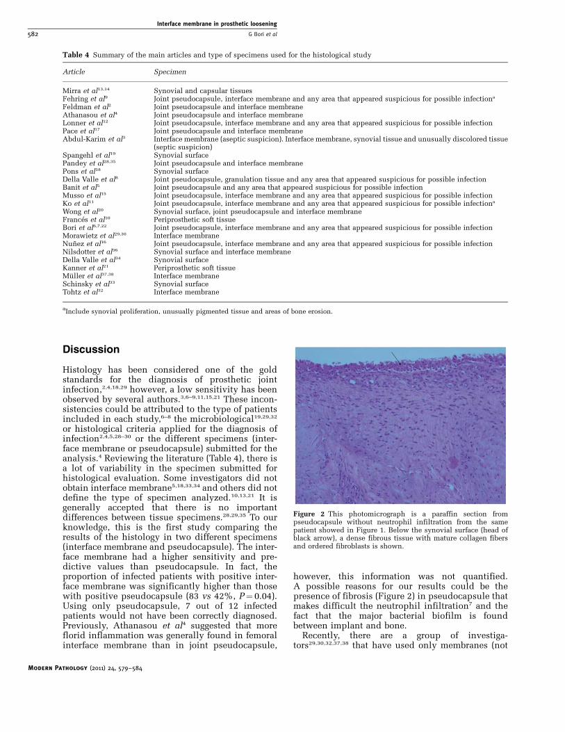
Discussion
Histology has been considered one of the goldstandards for the diagnosis of prosthetic jointinfection,2,4,18,29 however, a low sensitivity has beenobserved by several authors.3,6–9,11,15,21 These incon-sistencies could be attributed to the type of patientsincluded in each study,6–8 the microbiological19,29,32
or histological criteria applied for the diagnosis ofinfection2,4,5,28–30 or the different specimens (inter-face membrane or pseudocapsule) submitted for theanalysis.4 Reviewing the literature (Table 4), there isa lot of variability in the specimen submitted forhistological evaluation. Some investigators did notobtain interface membrane5,18,33,34 and others did notdefine the type of specimen analyzed.10,13,21 It isgenerally accepted that there is no importantdifferences between tissue specimens.28,29,35 To ourknowledge, this is the first study comparing theresults of the histology in two different specimens(interface membrane and pseudocapsule). The inter-face membrane had a higher sensitivity and pre-dictive values than pseudocapsule. In fact, theproportion of infected patients with positive inter-face membrane was significantly higher than thosewith positive pseudocapsule (83 vs 42%, P¼ 0.04).Using only pseudocapsule, 7 out of 12 infectedpatients would not have been correctly diagnosed.Previously, Athanasou et al4 suggested that moreflorid inflammation was generally found in femoralinterface membrane than in joint pseudocapsule,
however, this information was not quantified.A possible reasons for our results could be thepresence of fibrosis (Figure 2) in pseudocapsule thatmakes difficult the neutrophil infiltration7 and thefact that the major bacterial biofilm is foundbetween implant and bone.
Recently, there are a group of investiga-tors29,30,32,37,38 that have used only membranes (not
Table 4 Summary of the main articles and type of specimens used for the histological study
Article Specimen
Mirra et al13,14 Synovial and capsular tissuesFehring et al9 Joint pseudocapsule, interface membrane and any area that appeared suspicious for possible infectiona
Feldman et al2 Joint pseudocapsule and interface membraneAthanasou et al4 Joint pseudocapsule and interface membraneLonner et al12 Joint pseudocapsule, interface membrane and any area that appeared suspicious for possible infectionPace et al17 Joint pseudocapsule and interface membraneAbdul-Karim et al3 Interface membrane (aseptic suspicion). Interface membrane, synovial tissue and unusually discolored tissue
(septic suspicion)Spangehl et al19 Synovial surfacePandey et al28,35 Joint pseudocapsule and interface membranePons et al18 Synovial surfaceDella Valle et al8 Joint pseudocapsule, granulation tissue and any area that appeared suspicious for possible infectionBanit et al5 Joint pseudocapsule and any area that appeared suspicious for possible infectionMusso et al15 Joint pseudocapsule, interface membrane and any area that appeared suspicious for possible infectionKo et al11 Joint pseudocapsule, interface membrane and any area that appeared suspicious for possible infectiona
Wong et al20 Synovial surface, joint pseudocapsule and interface membraneFrances et al10 Periprosthetic soft tissueBori et al6,7,22 Joint pseudocapsule, interface membrane and any area that appeared suspicious for possible infectionMorawietz et al29,30 Interface membraneNunez et al16 Joint pseudocapsule, interface membrane and any area that appeared suspicious for possible infectionNilsdotter et al36 Synovial surface and interface membraneDella Valle et al34 Synovial surfaceKanner et al21 Periprosthetic soft tissueMuller et al37,38 Interface membraneSchinsky et al33 Synovial surfaceTohtz et al32 Interface membrane
aInclude synovial proliferation, unusually pigmented tissue and areas of bone erosion.
Figure 2 This photomicrograph is a paraffin section frompseudocapsule without neutrophil infiltration from the samepatient showed in Figure 1. Below the synovial surface (head ofblack arrow), a dense fibrous tissue with mature collagen fibersand ordered fibroblasts is shown.
Interface membrane in prosthetic loosening
582 G Bori et al
Modern Pathology (2011) 24, 579–584

pseudocapsule) and have proposed a histopatholo-gical consensus classification for a standardizedevaluation of the periprosthetic tissues. Our resultssupport the selection of the interface membrane as areference tissue for histological classification.
Although frozen section gives surgeons intrao-perative information about the diagnosis, our studywas performed on paraffin sections to avoid histo-logical technical bias. It has been described thatfrozen sections have inferior quality than paraffinones.10,19,32 For instance, Tohtz et al32 described a19% of discrepancies (in 14 out of 64 cases) betweenfrozen section and paraffin sections.32
The main drawback of our study was the lownumber of infected patients included. However, thisstudy had enough statistical power to reveal differ-ences between interface membrane and pseudocap-sule specimens. Patients who underwent hiprevision arthroplasty because of periprosthetic frac-ture were not included since it is a cause of false-positive histology results.4,12,15 Patients with apreoperative diagnosis of aseptic loosening and adefinitive diagnosis of infection were also notincluded since in our experience,6 the sensitivityof the histology (using Mirras’ criteria) in this groupof patients is low.
In conclusion, our results suggest that the bestspecimen of periprosthetic soft tissue for histologi-cal diagnosis of the infection in a total hip revisionarthroplasty is the periprosthetic interface mem-brane.
Disclosure/conflict of interest
The authors declare no conflict of interest.
References
1 Bauer TW, Parvizi J, Kobayashi N, et al. Diagnosis ofperiprosthetic infection. J Bone Joint Surg Am2006;88:869–882.
2 Feldman DS, Lonner JH, Desai P, et al. The role ofintraoperative frozen sections in revision total jointarthroplasty. J Bone Joint Surg Am 1995;77:1807–1813.
3 Abdul-Karim FW, McGinnis MG, Kraay M, et al.Frozen section biopsy assessment for the presence ofpolymorphonuclear leukocytes in patients undergoingrevision of arthroplasties. Mod Pathol 1998;11:427–431.
4 Athanasou NA, Pandey R, de SR, et al. Diagnosis ofinfection by frozen section during revision arthro-plasty. J Bone Joint Surg Br 1995;77:28–33.
5 Banit DM, Kaufer H, Hartford JM. Intraoperative frozensection analysis in revision total joint arthroplasty.Clin Orthop Relat Res 2002;401:230–238.
6 Bori G, Soriano A, Garcia S, et al. Low sensitivity ofhistology to predict the presence of microorganisms insuspected aseptic loosening of a joint prosthesis. ModPathol 2006;19:874–877.
7 Bori G, Soriano A, Garcia S, et al. Usefulness ofhistological analysis for predicting the presence ofmicroorganisms at the time of reimplantation after hip
resection arthroplasty for the treatment of infection.J Bone Joint Surg Am 2007;89:1232–1237.
8 la Valle CJ, Bogner E, Desai P, et al. Analysis of frozensections of intraoperative specimens obtained at thetime of reoperation after hip or knee resectionarthroplasty for the treatment of infection. J Bone JointSurg Am 1999;81:684–689.
9 Fehring TK, McAlister Jr JA. Frozen histologic sectionas a guide to sepsis in revision joint arthroplasty. ClinOrthop Relat Res 1994;304:229–237.
10 Frances BA, Martinez FM, Cebrian Parra JL, et al.Diagnosis of infection in hip and knee revisionsurgery: intraoperative frozen section analysis. IntOrthop 2007;31:33–37.
11 Ko PS, Ip D, Chow KP, et al. The role of intraoperativefrozen section in decision making in revision hip andknee arthroplasties in a local community hospital.J Arthroplasty 2005;20:189–195.
12 Lonner JH, Desai P, Dicesare PE, et al. The reliability ofanalysis of intraoperative frozen sections for identify-ing active infection during revision hip or kneearthroplasty. J Bone Joint Surg Am 1996;78:1553–1558.
13 Mirra JM, Amstutz HC, Matos M, et al. The pathologyof the joint tissues and its clinical relevance inprosthesis failure. Clin Orthop Relat Res 1976;117:221–240.
14 Mirra JM, Marder RA, Amstutz HC. The pathology offailed total joint arthroplasty. Clin Orthop Relat Res1982;170:175–183.
15 Musso AD, Mohanty K, Spencer-Jones R. Role of frozensection histology in diagnosis of infection duringrevision arthroplasty. Postgrad Med J 2003;79:590–593.
16 Nunez LV, Buttaro MA, Morandi A, et al. Frozensections of samples taken intraoperatively for diag-nosis of infection in revision hip surgery. Acta Orthop2007;78:226–230.
17 Pace TB, Jeray KJ, Latham Jr JT. Synovial tissueexamination by frozen section as an indicator ofinfection in hip and knee arthroplasty in communityhospitals. J Arthroplasty 1997;12:64–69.
18 Pons M, Angles F, Sanchez C, et al. Infected total hiparthroplasty–the value of intraoperative histology. IntOrthop 1999;23:34–36.
19 Spangehl MJ, Masri BA, O’Connell JX, et al. Prospec-tive analysis of preoperative and intraoperative inves-tigations for the diagnosis of infection at the sites oftwo hundred and two revision total hip arthroplasties.J Bone Joint Surg Am 1999;81:672–683.
20 Wong YC, Lee QJ, Wai YL, et al. Intraoperative frozensection for detecting active infection in failed hip andknee arthroplasties. J Arthroplasty 2005;20:1015–1020.
21 Kanner WA, Saleh KJ, Frierson Jr HF. Reassessment ofthe usefulness of frozen section analysis for hip andknee joint revisions. Am J Clin Pathol 2008;130:363–368.
22 Bori G, Soriano A, Garcia S, et al. Neutrophils in frozensection and type of microorganism isolated at the timeof resection arthroplasty for the treatment of infection.Arch Orthop Trauma Surg 2009;129:591–595.
23 Athanasou NA, Pandey R, de SR, et al. The role ofintraoperative frozen sections in revision total jointarthroplasty. J Bone Joint Surg Am 1997;79:1433–1434.
24 Tunney MM, Patrick S, Curran MD, et al. Detection ofprosthetic hip infection at revision arthroplasty byimmunofluorescence microscopy and PCR amplifica-tion of the bacterial 16S rRNA gene. J Clin Microbiol1999;37:3281–3290.
Interface membrane in prosthetic loosening
G Bori et al 583
Modern Pathology (2011) 24, 579–584

25 Savarino L, Baldini N, Tarabusi C, et al. Diagnosis ofinfection after total hip replacement. J Biomed MaterRes B Appl Biomater 2004;70:139–145.
26 Zeller V, Ghorbani A, Strady C, et al. Propionibacter-ium acnes: an agent of prosthetic joint infection andcolonization. J Infect 2007;55:119–124.
27 Zappe B, Graf S, Ochsner PE, et al. Propionibacteriumspp. in prosthetic joint infections: a diagnosticchallenge. Arch Orthop Trauma Surg 2008;128:1039–1046.
28 Pandey R, Drakoulakis E, Athanasou NA. An assess-ment of the histological criteria used to diagnoseinfection in hip revision arthroplasty tissues. J ClinPathol 1999;52:118–123.
29 Morawietz L, Classen RA, Schroder JH, et al. Proposalfor a histopathological consensus classification of theperiprosthetic interface membrane. J Clin Pathol2006;59:591–597.
30 Morawietz L, Tiddens O, Mueller M, et al. Twenty-three neutrophil granulocytes in 10 high-power fieldsis the best histopathological threshold to differentiatebetween aseptic and septic endoprosthesis loosening.Histopathology 2009;54:847–853.
31 Leone JM, Hanssen AD. Management of infection atthe site of a total knee arthroplasty. J Bone Joint SurgAm 2005;87:2335–2348.
32 Tohtz SW, Muller M, Morawietz L, et al. Validity offrozen sections for analysis of periprosthetic looseningmembranes. Clin Orthop Relat Res 2009;468:762–768.
33 Schinsky MF, la Valle CJ, Sporer SM, et al. Periopera-tive testing for joint infection in patients undergoingrevision total hip arthroplasty. J Bone Joint Surg Am2008;90:1869–1875.
34 la Valle CJ, Sporer SM, Jacobs JJ, et al. Preoperativetesting for sepsis before revision total knee arthro-plasty. J Arthroplasty 2007;22:90–93.
35 Pandey R, Berendt AR, Athanasou NA. Histologicaland microbiological findings in non-infected andinfected revision arthroplasty tissues. The OSIRISCollaborative Study Group. Oxford Skeletal InfectionResearch and Intervention Service. Arch OrthopTrauma Surg 2000;120:570–574.
36 Nilsdotter-Augustinsson A, Briheim G, Herder A, et al.Inflammatory response in 85 patients with loosenedhip prostheses: a prospective study comparing inflam-matory markers in patients with aseptic and septicprosthetic loosening. Acta Orthop 2007;78:629–639.
37 Muller M, Morawietz L, Hasart O, et al. Diagnosis ofperiprosthetic infection following total hip arthro-plasty—evaluation of the diagnostic values of pre-and intraoperative parameters and the associatedstrategy to preoperatively select patients with a highprobability of joint infection. J Orthop Surg 2008;3:31.
38 Muller M, Morawietz L, Hasart O, et al. Histopatholo-gical diagnosis of periprosthetic joint infection follow-ing total hip arthroplasty: use of a standardizedclassification system of the periprosthetic interfacemembrane. Orthopade 2009;38:1087–1096.
Interface membrane in prosthetic loosening
584 G Bori et al
Modern Pathology (2011) 24, 579–584

1 3
Arch Orthop Trauma Surg (2014) 134:577–583DOI 10.1007/s00402-014-1921-z
HIP ARTHROPLASTY
Comparison of bacterial results from conventional cultures of the periprosthetic membrane and the synovial or pseudocapsule during hip revision arthroplasty
E. Muñoz-Mahamud · A. Soriano · A. Combalia · C. Medrano · J. Bosch · S. García · G. Bori
Received: 27 July 2013 / Published online: 14 January 2014 © Springer-Verlag Berlin Heidelberg 2014
Results Of a total of 86 hip revision surgeries, 22 were considered septic revisions postoperatively, of which 16 resulted in positive cultures. Of these 16 revisions, 14 obtained the same microbiologic diagnostic when consider-ing either the synovial/pseudocapsule culture results or the solid membrane sample.Conclusions We conclude that the membrane sam-ple for a conventional culture is not superior to the synovial/pseudocapsule sample in detecting microorganisms.
Keywords Membrane · Hip arthroplasty · Revision · Cultures · Infection
Introduction
Bacterial identification is essential to diagnose and treat a revision for prosthetic loosening of an infected hip. In chronic prosthetic joint infection, the majority of microor-ganisms are attached to the implant surface forming bio-films (sessile bacteria), whereas the amount of free floating (planktonic bacteria) in the periprosthetic tissue is small [1–9]. Current guidelines for diagnosis of periprosthetic joint infection consider that it is imperative that tissue for culture be obtained from representative periprosthetic tis-sue or fluid [10, 11]. Among these samples, it has been described that synovial fluid inoculated into blood culture flasks is the one with the highest sensitivity and specificity [1, 3]. However, these guidelines do not provide objective data regarding which one is the best solid tissue to identify microorganisms. It is accepted that at least three and opti-mally five or six periprosthetic intraoperative samples from the most suspicious areas of tissue as deemed by the ortho-pedic surgeon should be obtained for aerobic and anaero-bic culture for the optimal diagnosis of PJI. Nonetheless,
Abstract Introduction Bacterial identification is essential to diag-nose and treat a revision for prosthetic loosening of an infected hip. The purpose of this study was to determine whether conventional cultures from the periprosthetic membrane are superior to synovial/pseudocapsule samples in the diagnosis of infection in hip revision arthroplasty.Materials and methods We performed a prospective study including all hip revisions from October 2009 to October of 2011. Once the implants were removed and prior to the administration of the antibiotic prophylaxis, six periprosthetic samples from different sites were sent to the laboratory for culturing: two periprosthetic fluid sam-ples, two solid material (synovial/pseudocapsule) samples and two swabs. Once the six samples were taken, antibi-otic prophylaxis was administered and just as the implant was removed, two more solid samples of the periprosthetic membrane were obtained.
E. Muñoz-Mahamud (*) · A. Combalia · C. Medrano · S. García · G. Bori Bone and Joint Infectious Diseases Unit, Department of Orthopaedics and Trauma Surgery, Hospital Clinic of Barcelona, University of Barcelona, C/Villarroel 170, 08036 Barcelona, Spaine-mail: [email protected]
E. Muñoz-Mahamud · A. Soriano · J. Bosch · S. García · G. Bori Bone and Joint Infectious Diseases Unit, Hospital Clinic of Barcelona, University of Barcelona, Barcelona, Spain
A. Soriano Department of Infectious Diseases, Hospital Clinic of Barcelona, University of Barcelona, Barcelona, Spain
J. Bosch Department of Microbiology, Hospital Clinic of Barcelona, University of Barcelona, Barcelona, Spain
Author's personal copy

578 Arch Orthop Trauma Surg (2014) 134:577–583
1 3
to dilucidate which one is that most suspicious tissue still remains unspecific.
On a physiopathological level, it can be assumed that the membrane sample should be superior to the synovial/pseudocapsule sample, since the membrane is the closest tissue to the implant and hence to the biofilm. Moreover, in pathological terms, it has been described that there is more acute inflammatory reaction in the presence of neutrophils in the membrane than in the synovial/pseudocapsule [12]. Therefore, the possibility of finding more microorganisms in the membrane should also be greater.
Our hypothesis is that the membrane might be the best solid tissue so as to identify the presence of microorgan-isms. The objective of this study was to determine whether conventional cultures from the periprosthetic membrane are superior to synovial/pseudocapsule samples in the diagno-sis of infection in hip revision arthroplasty.
Materials and methods
This was a prospective study that included all the hip revi-sions that were performed in our hospital from October 2009 to October of 2011.
Microbiology
When the implants were removed and before antibiotic prophylaxis was administered, six periprosthetic samples from different sites were sent to the laboratory for cultur-ing: two periprosthetic fluid samples, two solid material (synovial/pseudocapsule) samples and two swabs. Once the six samples were taken antibiotic prophylaxis was adminis-tered and just as the implant was removed, two more solid samples of the periprosthetic membrane were obtained, one from the cup and one from the stem. If the revision was either of the cup or the stem independently of each other then two membrane samples were taken from the same site.
Synovial fluid was obtained through aspiration with a sterile syringe, immediately inoculated into Bactec9000® (Becton–Dickinson Diagnostic Instruments®, Sparks, Maryland) blood culture flasks (one 8–10 mL aerobic flask and one 8–10 mL anaerobic flask per synovial fluid sam-ple) and then incubated for 5 days. If the system automati-cally detected microorganism growth, subcultures were done in aerobic and anaerobic agar media. Swab cultures were obtained by swabbing the periprosthetic tissue area, bone and fluids where infection was suspected with a ster-ile swap (Deltalab®, sterile Eurotube® collection swab with Stuart transport medium, Rubí, Catalonia, Spain). Solid periprosthetic tissue samples were immediately placed into a separate sterile recipient. Solid samples and swabs
were cultivated in both aerobic and anaerobic agar media (Columbia Blood Agar and Schaedler Blood Agar, respec-tively) as well as in fluid thioglycollate medium, which were all enriched with vitamin K and hemin; all the sam-ples were then incubated for 10 days. Microorganism iden-tification and proper antibiogram were performed in those cultures in which the Bactec9000® automatically detected the presence of microorganisms.
Definitive diagnosis of infection was determined when (1) there was a sinus tract communicating with the pros-thesis, and/or (2) a pathogen was isolated by culture from at least two separate samples obtained intraoperatively [10, 11].
Two groups were created out of the six samples accord-ing to the solid sample that was taken: periprosthetic mem-brane group (Group A) and synovial/pseudocapsule group (Group B). In Group A, two fluid samples, two swabs and two periprosthetic membrane samples were taken to iden-tify infection. In Group B two fluid samples, two swabs and two synovial/pseudocapsule samples were taken to identify infection. Therefore, the two fluid samples and the two swabs were the same for both groups. Once these two groups of six samples each were created, they were sta-tistically analyzed so as to dilucidate if there were differ-ences in the results that were obtained according to whether a solid membrane sample was considered (Group A) or a solid synovial/pseudocapsule sample was considered (Group B).
Quantitative results are reported as median and inter-quartilic range (IQR). Qualitative, nominal and ordinal, results are reported as absolute frequencies and percentage (%). Qualitative variables were analyzed by means of Fish-er’s exact test. Analyses were performed using the SPSS® v. 20.0 statistical package (SPSS, Inc. Chicago, IL, USA). Two-sided p ≤ 0.05 was considered statistically significant.
Results
A total of 86 hip revision surgeries were performed during the period of the study with a preoperative diagnostic of 50 aseptic revisions, 1 one-stage septic revision, 16 primary two-stage septic revisions, 18 secondary two-stage septic revisions and 1 one-stage revision due to a Vancouver type B2 periprosthetic fracture. The series included 32 men and 54 women with a median age of 74.0 (IQR 66–80) years.
Out of the 86 performed revisions, a total of 22 were considered as septic revisions postoperatively, and 16 had positive cultures (Table 1). Out of these 16 revi-sions, 14 were considered as positive considering both the synovial/pseudocapsule sample or the membrane sam-ple as solid tissue (p = 0.484); hence there were no sta-tistically significant differences between group A and
Author's personal copy

579Arch Orthop Trauma Surg (2014) 134:577–583
1 3
group B. There were only two cases of discrepancies between the definitive diagnosis of an aseptic or sep-tic revision according to whether the sample was either from the synovial/pseudocapsule or the membrane: in both cases (cases three and eight), if the solid mem-brane sample had been considered the revision would not have been diagnosed as infected, whereas if the solid synovial/pseudocapsule sample had been considered the revision would have been diagnosed as septic.
There were five cases (cases 5, 6, 9, 12 and 16) where a discrepancy was found with respect to the solid cul-tures depending on whether the sample was from the synovial/pseudocapsule or the membrane, but the final diagnosis was the same for both of the solid samples since the other cultures (fluids and swabs) were all positive as well (p = 0.083).
There were two cases where discrepancies were found in relation to the microorganism that was identified in the solid
Table 1 Cultures of the patients diagnosed of septic revision due to positive culture
CNS coagulase-negative staphylococcia Infection was determined when: (1) there was a sinus tract communicating with the prosthesis; and/or (2) a pathogen was isolated by culture from at least two separate samples obtained intraoperatively [8, 9]
n Preoperative diagnosis Fluid Swab Membrane (group A) Infectiona (group A) Microorganism (group A)
Capsule (group B) Infectiona (group B) Microorganism (group B)
1 Aseptic revision 1/2 2/2 2/2 Yes CNS
2/2 Yes CNS
2 Aseptic revision 2/2 1/2 2/2 Yes CNS
2/2 Yes CNS
3 2nd stage of septic revision 0/2 0/2 0/2 No –
2/2 Yes S. capitis
4 1st stage of septic revision 2/2 0/2 1/2 Yes Streptococcus spp
2/2 Yes CNS
5 Aseptic revision 2/2 0/2 0/2 Yes Corynebacterium spp
2/2 Yes Corynebacterium spp
6 1st stage of septic revision 1/2 2/2 2/2 Yes S. aureus
0/2 Yes S. aureus
7 1st stage of septic revision 2/2 2/2 1/2 Yes S. aureus
2/2 Yes S. aureus
8 Aseptic revision 0/2 1/2 0/2 No –
1/2 Yes CNS
9 1st stage of septic revision 2/2 0/2 0/2 Yes CNS
2/2 Yes CNS
10 1st stage of septic revision 2/2 1/2 1/2 Yes E. coli
Klebsiella spp
1/2 Yes E. coli
Klebsiella spp
CNS
11 1st stage of septic revision 2/2 2/2 1/2 Yes S. aureus
1/2 Yes S. aureus
12 1st stage of septic revision 2/2 2/2 0/2 Yes S. aureus
2/2 Yes S. aureus
13 Aseptic revision 2/2 1/2 1/2 Yes CNS
1/2 Yes CNS
14 1st stage of septic revision 2/2 1/2 2/2 Yes S. aureus
2/2 Yes S. aureus
15 1st stage of septic revision 2/2 2/2 2/2 Yes S. aureus
2/2 Yes S. aureus
16 1st stage of septic revision 2/2 2/2 0/2 Yes S. gordonii
2/2 Yes S. gordonii
Author's personal copy

580 Arch Orthop Trauma Surg (2014) 134:577–583
1 3
samples: in one case (case four), on the synovial/pseudocapsule grew one coagulase-negative staphylococci (CNS) in two of the samples (the same as in one of the fluid samples), but on the membrane grew a Streptococcus spp in one sample (the same as in one of the fluid samples); in another case (case ten), whereas on the synovial/pseudocapsule grew one CNS in one sample (the same as in one of the swab samples) and on the membrane grew one E. coli and one Klebsiella spp in one sample (the same as in two of the fluid samples). This latter case was the only case of polymicrobial infection. The most frequent microorganism was CNS in seven cases, followed by S. aureus in six of the cases.
Discussion
The antibiotic treatment of an infection of a prosthetic loosening of a chronic infected hip is prolonged and determining the implicated etiological microorgan-ism is essential to select the most suitable antimicrobial therapy. In chronic periprosthetic infections the number of free-floating bacteria in the periprosthetic tissues is low since the majority is part of a biofilm [2, 5–9]. As a result, it is a logical procedure for the surgeon to take more than one sample during the revision arthroplasty to identify the microorganism that is causing the infec-tion. There is a general consensus that during a revision arthroplasty for chronic loosening more than one sample must be taken and many authors take up to six differ-ent samples [10, 13–15]. However, there is a difference of opinion among the authors as to what type of sample should be taken for conventional cultures. This fact is of paramount importance since the sensitivity of the differ-ent periprosthetic samples (fluid, solid or swab) is dif-ferent [1, 3]. According to the literature, authors use all kinds of different combinations of the type of samples that are used for conventional cultures: solid samples [16, 17], swabs [3, 18–20], solid and fluid [21], solid and swabs [22] or a combination of the three types of sam-ples [13]. Some studies have recently highlighted swabs’ low sensitivity [3, 23] and current guidelines [10, 11] do not include them in the criteria to define periprosthetic joint infection. In fact, our results remain in concordance with the published data, as 4 of the 16 cases had false-negative swabs (25 % false-negative rate). Furthermore, in four additional patients, only one out of two swab cultures were positive, whereas 2–4 of the rest of tissue cultures were positive. There are still some authors who emphasize that swabs may have a role and may help in diagnosis, since some patients would not have been diag-nosed as infected if swabs had not been performed [3, 18]. Accordingly, one of our patients (case eight) would have been misdiagnosed if swab samples had not been
cultured. Anyway, as current guidelines dictate [10, 11], swabs should not be considered for the diagnosis of pros-thetic infection anymore and, therefore, they should be avoided.
According to the literature, the samples with the highest sensitivity seem to be synovial or periprosthetic fluid inoc-ulated into blood culture flasks, when compared with the cultures of solid periprosthetic material or swabs. Levine and Evans [1], in a study involving 24 patients, described a 92 % sensitivity for fluids inoculated into blood culture flasks, 46 % for solid periprosthetic material and 64 % for swabs. Recently, our research group corroborated these results and found an 86 % sensitivity for fluids inoculated into blood culture flasks, 69 % for solid periprosthetic material and 61 % for swabs [3]. However, in the current study the aim was to determine if the periprosthetic mem-brane as a solid sample for conventional cultures increases the results in conventional cultures in comparison with the solid samples obtained from the synovial or pseudocapsule when diagnosing infection during revision.
In prosthetic joint infections, microorganisms may be present in one of two forms: either suspended floating (planktonic bacteria) or attached to the biomaterial surface (sessile bacteria). Both forms exist simultaneously being not mutually exclusive. Most of microorganisms are found attached rather than suspended and often reside in biofilms [4]. This phenomenon raises a first clinical problem, con-cerning pathogen identification. Routine culture methods were in fact developed for identifying planktonic bacte-ria in acute infections. However, in prosthetic joint infec-tions, a majority of bacteria are viable yet not culturable since they are in their sessile state. These microorganisms can be clearly identified only using advanced techniques, whereas traditional sampling and cultural examination do often fail [1–7]. For this reason, one would assume that the membrane sample (the closest tissue to the implant) should be superior to the synovial or pseudocapsule sample, since in a chronic joint prosthesis infection the majority of the microorganisms are found on the surface of the implant forming biofilms.
Moreover, in pathological terms, it has been described that there is more acute inflammatory reaction with presence of neutrophils in the membrane than in the synovial/pseudocapsule [2, 4–9, 12]. Therefore, the pos-sibility of finding more microorganisms in the membrane should also be greater. Despite these facts, our results show that the solid membrane samples did not increase the num-ber of patients diagnosed as infected. Actually, when exclu-sively considering the solid membrane sample (ignoring the synovial/pseudocapsule samples), there were two patients (cases three and eight) that were not diagnosed as infected.
A possible explanation for the obtained results might be that the quantity of solid synovial/pseudocapsule sample
Author's personal copy

581Arch Orthop Trauma Surg (2014) 134:577–583
1 3
that was sent to the microbiology laboratory was greater than the solid membrane sample since it is more easily accessible to the surgeon during the intervention. Moreo-ver, as the fluid sample is considered to be the most sen-sitive [1, 3, 10, 11], it should be kept in mind that the sample that is in contact and immersed in this fluid is the synovial/pseudocapsule, sample and not the membrane sample. At the moment in which we started the study, we were concerned about the fact that when the solid synovial/pseudocapsule samples were being taken the patient had not yet received antibiotic prophylaxis but when the periprosthetic membrane sample was taken the patient had already received antibiotic prophylaxis. Preoperative antibiotics are known to be critical for decreasing the risk of periprosthetic joint infection (PJI) in primary THA and TKA. However, antibiotics often are withheld before revi-sion surgery, as there is concern that even a single dose of prophylactic antibiotics may affect intraoperative cultures. Despite lacking clear evidence regarding the best moment to infuse an antibiotic during arthroplasty surgery yet [24, 25], recent randomized trials suggested that prophylactic antibiotics do not affect cultures [19, 20], and, therefore, the administration of the antibiotics should not pose any bias for the results of the cultures. Thus, it can be con-cluded that the solid sample taken from the periprosthetic membrane is not superior to the solid sample taken from the synovial/pseudocapsule, when diagnosing a peripros-thetic infection.
Last, it is worth mentioning that even with improved sampling procedures the detection of infection by culture may still underestimate the real incidence of infection. As a matter of fact, there are currently multiple approaches to solve this problem including direct diagnostics of the bio-film and biomarkers. The detection of bacteria in sonicates from patients with culture-negative implants indicates that these implants were colonized by bacteria which were not isolated by the microbiological techniques used. This may be because the implants were infected with viable but non-culturable bacteria. The process of 16S ribosomal deoxy-ribonucleic acid (rDNA) polymerase chain reaction (PCR) and sequencing of biofilm removed from the explanted prosthesis is an important addition to conventional culture methods in prosthetic joint infection, detecting additional pathogens and improving diagnostic sensitivity [26]. It has been described that molecular detection can increase (by one-tenth) the number of patients diagnosed as having an infection [27]. So as to improve both the sensitivity and the specificity of the PCR, innovations such as the PCR-Elec-trospray ionization mass spectrometry have been described [28]. The detection of bacterial rRNA genes as an indicator of the presence of bacteria is an established technique that has been used for the detection of both environmental and medically important bacteria. In fact, PCR amplification
of bacterial 16S rRNA genes has been used successfully to detect bacteria that cause a variety of acute or suba-cute medical infections [29]. In infections such as those of prosthetic joints, however, in which the infectious agents are opportunistic, the potential contamination of samples must be addressed. The direct immunological detection of bacteria in clinical samples can be achieved by the use of monoclonal antibodies (MAbs) and polyclonal antise-rum prepared against the bacteria implicated in the clinical infection [29]. This technique allows the direct visualiza-tion of the bacterial morphology and visual comparison with potential contamination. The role of molecular tech-niques in the diagnosis of prosthetic joint infection remains unclear and still reveals several limitations, such as poorly formulated gold standard criteria or the difficult interpreta-tion of false-positive results when post-PCR sequencing is not used [30].
Nevertheless, both tissue culture and PCR can identify bacteria but are susceptible to contamination in the sur-gical field, during sample handling, and in the laboratory setting. Therefore, confidence in treating a patient based solely on these tests is mitigated by the concern for a false-positive result. For this reason, protein profiling as biomarkers for the purpose of prosthetic joint infection seems to hold great promise. Several proteins that have been previously associated with inflammatory condi-tions and that appear to be useful as clinical indicators for the presence of infection have been identified [31]. If a reliable, effective marker or set of markers for diagnos-ing prosthetic joint infection can be identified, a quick test for its diagnosis may be on the horizon. It has been demonstrated that inflammatory proteins can be detected in synovial fluid and its concentrations ascertained and these varying concentrations align well with the presence or absence of prosthetic joint infection [31]. Leukocyte esterase reagent strips have been proposed as a useful point-of-care screening test with a 100 % negative predic-tive value in the setting of revision surgery. It has been reported that even with its subjectivity, these rapid, inex-pensive and sensitive strips can quickly rule out infection in cases that show a negative result [32]. Because the syn-ovial fluid white blood cells express specific genes when the etiology for inflammation is bacterial, a more specific measure of the host response to infection could potentially be developed. In fact, proteins such as IL-1B, IL1-α, IL-6, IL-17, G-CSF, IL-8, VEGF or α2 M have shown excellent diagnostic performance [31, 33]. Other new biomarkers such as α-defensin have shown encouraging results which provide an additional valuable resource in the diagnsosis of periprosthetic infection [34].
Conflict of interest At the time of publication none of the authors disclosed any potential conflicts of interest. All authors declare that
Author's personal copy

582 Arch Orthop Trauma Surg (2014) 134:577–583
1 3
they have no affiliations that might affect the objective and scientific presentation of this research.
References
1. Levine BR, Evans BG (2001) Use of blood culture vial speci-mens in intraoperative detection of infection. Clin Orthop Relat Res 382:222–231
2. Gómez-Barrena E, Esteban J, Medel F, Molina-Manso D, Ortiz-Pérez A, Cordero-Ampuero J, Puértolas JA (2012) Bacterial adherence to separated modular components in joint prosthesis: a clinical study. J Orthop Res 30(10):1634–1639
3. Font-Vizcarra L, Garcia S, Martínez-Pastor JC, Sierra JM, Soriano A (2010) Blood culture flasks for culturing syno-vial fluid in prosthetic joint infections. Clin Orthop Relat Res 468(8):2238–2243
4. Romanò C (2013) Antibiofilm agents and implant-related infec-tions in orthopaedics: where are we? J Chemother 25(2):67–80
5. Arciola CR, Montanaro L, Costerton JW (2011) New trends in diagnosis and control strategies for implant infections. Int J Artif Organs 34:727–736
6. Costerton JW, Stewart PS, Greenberg EP (1999) Bacterial biofilms: a common cause of persistent infections. Science 284:1318–1322
7. Hill KE, Malic S, McKee R, Rennison T, Harding KG, Williams DW (2010) An in vitro model of chronic wound biofilms to test wound dressings and assess antimicrobial susceptibilities. J Anti-microb Chemother 65:1195–1206
8. Drago L, Romanò CL, Mattina R, Signori V, De Vecchi E (2012) Does dithiothreitol improve bacterial detection from infected prosthesis? A pilot study. Clin Orthop Relat Res 470(10):2915–2925
9. Zameer F, Gopal S (2010) Evaluation of antibiotic susceptibility in mixed culture biofilms. Intern J Biotech Biochem 6:93–99
10. Osmon DR, Berbari EF, Berendt AR, Lew D, Zimmerli W, Steck-elberg JM, Rao N, Hanssen A, Wilson WR (2013) Diagnosis and management of prosthetic joint infection: clinical practice guide-lines by the infectious diseases society of America. Clin Infect Dis 1(56):1–25
11. Parvizi J, Zmistowski B, Berbari EF, Bauer TW, Springer BD, Valle Della CJ, Garvin KL, Mont MA, Wongworawat MD, Zala-vras CG (2011) New definition for periprosthetic joint infection: from the workgroup of the musculoskeletal infection society. Clin Orthop Relat Res 469(11):2992–2994
12. Bori G, Muñoz Mahamud E, Garcia S, Mallofre C, Gallart X, Bosch J, García E, Riba J, Mensa J, Soriano A (2011) Interface membrane is the best sample for histological study to diagnose prosthetic joint infection. Mod Pathol 24(4):579–584
13. Bori G, Soriano A, Garcia S, Mallofre C, Riba J, Mensa J (2007) Usefulness of histological analysis for predicting the presence of microorganisms at the time of reimplantation after hip resection arthroplasty for the treatment of infection. J Bone Jt Surg Am 89(6):1232–1237
14. Atkins BL, Athanasou N, Deeks JJ, Crook DW, Simpson H, Peto TE, McLardy-Smith P, Berendt AR (1998) Prospective evalua-tion of criteria for microbiological diagnosis of prosthetic-joint infection at revision arthroplasty. The OSIRIS collaborative study group. J Clin Microbiol 36(10):2932–2939
15. Trampuz A, Piper KE, Jacobson MJ, Hanssen AD, Unni KK, Osmon DR, Mandrekar JN, Cockerill FR, Steckelber JM, Green-leaf JF, Patel R (2007) Sonication of removed hip and knee pros-theses for diagnosis of infection. N Engl J Med 357(7):654–663
16. Pandey R, Berendt AR, Athanasou NA (2000) Histological and microbiological findings in non-infected and infected revision
arthroplasty tissues. The OSIRIS collaborative study group. Oxford skeletal infection research and intervention service. Arch Orthop Trauma Surg 120(10):570–574
17. Mason JB, Fehring TK, Odum SM, Griffin WL, Nussman DS (2003) The value of white blood cell counts before revision total knee arthroplasty. J Arthroplast 18(8):1038–1043
18. Mehra A, Hemmady MV, Nelson R, Hodgkinson JP (2006) Bac-teriology swab in primary total hip arthroplasty: does it have a role? Int J Clin Pract 60(6):665–666
19. Burnett RSJ, Aggarwal A, Givens SA, McClure JT, Morgan PM, Barrack RL (2009) Prophylactic antibiotics do not affect cultures in the treatment of an infected TKA: a prospective trial. Clin Orthop Relat Res 468(1):127–134
20. Tetreault MW, Wetters NG, Aggarwal V, Mont M, Parvizi J, Valle Della CJ (2013) The Chitranjan Ranawat Award: should prophy-lactic antibiotics be withheld before revision surgery to obtain appropriate cultures? Clin Orthop Relat Res 30. doi:10.1007/s11999-013-3016-5 (Epub ahead of print)
21. Panousis K, Grigoris P, Butcher I, Rana B, Reilly JH, Ham-blen DL (2005) Poor predictive value of broad-range PCR for the detection of arthroplasty infection in 92 cases. Acta Orthop 76(3):341–346
22. Spangehl MJ, Masri BA, O’Connell JX, Duncan CP (1999) Prospective analysis of preoperative and intraoperative inves-tigations for the diagnosis of infection at the sites of two hun-dred and two revision total hip arthroplasties. J Bone Jt Surg Am 81(5):672–683
23. Trampuz A, Zimmerli W (2005) Prosthetic joint infections: update in diagnosis and treatment. Swiss Med Wkly 24: 1–10
24. Steinberg JP, Braun BI, Hellinger WC, Kusek L, Bozikis MR, Bush AJ, Dellinger EP, Burke JP, Simmons B, Kritchevsky SB (2009) Timing of antimicrobial prophylaxis and the risk of surgi-cal site infections: results from the trial to reduce antimicrobial prophylaxis errors. Ann Surg 250(1):10–16
25. Stefánsdóttir A, Robertsson O, W-Dahl A, Kiernan S, Gustaf-son P, Lidgren L (2009) Inadequate timing of prophylactic anti-biotics in orthopedic surgery. We can do better. Acta Orthop 80(6):633–638
26. Suda A, Kommerell M, Geiss HK, Burckhardt I, Zimmermann S, Zeifang F, Lehner B (2013) Prosthetic infection: diagnostic pro-cedures using 16S ribosomal deoxyribonucleic acid polymerase chain reaction. Int Orthop 37(12):2515–2521
27. Esteban J, Alonso-Rodriguez N, del-Prado G, Ortiz-Pérez A, Molina-Manso D, Cordero-Ampuero J, Sandoval E, Fernández-Roblas R, Gómez-Barrena E (2012) PCR-hybridization after sonication improves diagnosis of implant-related infection. Acta Orthop 83:299–304
28. Greenwood-Quaintance KE, Uhl JR, Hanssen AD, Sampath R, Mandrekar JN, Patel R (2013) Diagnosis of prosthetic joint infec-tion (PJI) using polymerase chain reaction-electrospray ioniza-tion mass spectrometry (PCR-ESI/MS). J Clin Microbiol. doi:10.1128/JCM.03217-13
29. Tunney MM, Patrick S, Curran MD, Ramage G, Hanna D, Nixon JR, Gorman SP, Davis RI, Anderson N (1999) Detection of pros-thetic hip infection at revision arthroplasty by immunofluores-cence microscopy and PCR amplification of the bacterial 16S rRNA gene. J Clin Microbiol 37(10):3281–3290
30. Marin M, Garcia-Lechuz JM, Alonso P, Villanueva M, Alcala L, Gimeno M, Cercenado E, Sánchez-Somolinos M, Radice C, Bouza E (2012) Role of universal 16S rRNA gene PCR and sequencing in diagnosis of prosthetic joint infection. J Clin Microbiol 50(3):583–589
31. Jacovides CL, Parvizi J, Adeli B, Jung KA (2011) Molecular markers for diagnosis of periprosthetic joint infection. J Arthro-plast 26(6):99–103
Author's personal copy

583Arch Orthop Trauma Surg (2014) 134:577–583
1 3
32. Wetters NG, Berend KR, Lombardi AV, Morris MJ, Tucker TL, Valle Della CJ (2012) Leukocyte esterase reagent strips for the rapid diagnosis of periprosthetic joint infection. J Arthroplast 27(8):8–11
33. Deirmengian C, Hallab N, Tarabishy A, Valle Della C, Jacobs JJ, Lonner J, Booth RE Jr (2010) Synovial fluid biomarkers for periprosthetic infection. Clin Orthop Relat Res 468(8):2017–2023
34. Ahn JK, Huang B, Bae EK, Park EJ, Hwang JW, Lee J, Koh EM, Cha HS (2013) The role of α-defensin-1 and related signal trans-duction mechanisms in the production of IL-6, IL-8 and MMPs in rheumatoid fibroblast-like synoviocytes. Rheumatol (Oxford) 52(8):1368–1376
Author's personal copy


Usefulness of Histology for Predicting Infection at the Time of Hip Revision for theTreatment of Vancouver B2 Periprosthetic Fractures
Ernesto Muñoz-Mahamud, MD a,b, Guillem Bori, MD, PhD a,b, Sebastián García, MD, PhD a,b,José Ramírez, MD, PhD c, Josep Riba, MD, PhD a, Alejandro Soriano, MD, PhD b,d
a Department of Orthopaedic Surgery and Traumatology, Hospital Clínic of Barcelona, University of Barcelona, Barcelona, Spainb Bone and Joint Infectious Diseases Unit, Hospital Clínic of Barcelona, University of Barcelona, Barcelona, Spainc Department of Pathology, Hospital Clínic of Barcelona, University of Barcelona, Barcelona, Spaind Department of Infectious Diseases, Hospital Clínic of Barcelona, University of Barcelona, Barcelona, Spain
a b s t r a c ta r t i c l e i n f o
Article history:Received 27 July 2012Accepted 18 December 2012
Keywords:periprosthetic fractureshiparthroplastyhistologyculturesinfection
When facing a Vancouver B2 periprosthetic fracture, the most recommended treatment is the prosthesisreplacement. Current tests do not provide enough reliability to identify whether the fracture has beenproduced on a septic or an aseptic loosened prosthesis. Our aimwas to evaluate the usefulness of the histologyfor the diagnosis of infection in these cases. A total of 11 hip revision procedures were performed (mean age:78.1 years, 8 women and 3 men). Sensitivity, specificity, positive predictive value and negative predictivevalue of the histology were 100%, 55.5%, 33.3% and 100% respectively. Of the six patients presenting with apositive histology, four of them had negative cultures (66.6% of false positives). Our results suggest thatperiprosthetic fractures are a cause of false-positive histology results for the diagnosis of infection duringrevision of a hip prosthesis for the treatment of Vancouver B2 periprosthetic fractures.
© 2013 Elsevier Inc. All rights reserved.
To establish the correct diagnosis of prosthetic joint infection(PJI) is challenging, especially when a periprosthetic fracture isassociated [1]. A periprosthetic fracture can result as a consequenceof a high-energy traumatism on a non-loosened prosthesis or eithera low-energy traumatism on a prosthesis that is already loosened.When the patient presents with a Vancouver B2 periprostheticfracture, the most recommended treatment is the prosthesisreplacement [2]. If the replacement is performed on a prosthesisseptically loosened, the risk of prosthetic infection is high. Whenfacing a loosened prosthesis, the most used tests in order todifferentiate between aseptic and septic etiology are the clinicaldata [3], laboratory tests [C-reactive protein (CRP), erythrocytesedimentation rate (ESR), interleukin-6] [4,5], bone scintigraphy incombination with labeled leukocyte scintigraphy [6] and preopera-tive cultures from samples obtained by computerized tomography orultrasonography guided aspiration of the joint [7,8]. However, thesetests do not provide enough sensibility and specificity to identifywhether the fracture has been produced on a septic or an asepticloosened prosthesis. This means that the final diagnosis is provided
by the cultures and/or the neutrophil count of the periprosthetictissues obtained during the revision surgery. Despite having beensuggested that periprosthetic fractures might be a cause of falsepositive of the histology for the diagnosis of PJI [9–14], no serieshave been reported this fact in the literature. The aim of our studywas to evaluate the usefulness of the histology for the diagnosis ofinfection during a hip prosthesis replacement for the treatment of aVancouver B2 periprosthetic fracture.
Material and Methods
From January 2007 to July 2011, all patients admitted at ourhospital presenting with a Vancouver B2 periprosthetic hip fracturetreated with hip prosthesis replacement were prospectively regis-tered in a database and retrospectively reviewed. Vancouver B1 andC periprosthetic hip fractures were not included since the mostrecommended treatment is the synthesis of the fracture withoutrevision of the prosthesis [2]. Vancouver B3 periprosthetic fracturesrequire removal of the prosthesis [2], however no such fracture wasadmitted in our hospital during the study period. All patients wereoperated on in the same hospital. In all patients the prosthesis wasreplaced and the fracture was treated using a plate and a deep-frozen cortical strut allograft. Samples for microbiology were alwaystaken before the administration of antibiotics and samples forhistology were taken after the implant removal from the femoral
The Journal of Arthroplasty 28 (2013) 1247–1250
The Conflict of Interest statement associated with this article can be found at http://dx.doi.org/10.1016/j.arth.2012.12.016.
Reprint requests: Ernesto Muñoz-Mahamud, MD, Department of OrthopaedicSurgery and Traumatology, Bone and Joint Infectious Diseases Unit, Hospital Clínic ofBarcelona, University of Barcelona, C/Villarroel 170, Barcelona 08036, Spain.
0883-5403/2808-0001$36.00/0 – see front matter © 2013 Elsevier Inc. All rights reserved.http://dx.doi.org/10.1016/j.arth.2012.12.016
Contents lists available at ScienceDirect
The Journal of Arthroplasty
j ourna l homepage: www.arth rop lasty journa l .o rg

stem. The standard antibiotic prophylaxis regimen consisted ofceftazidime and vancomycin. The patients made the follow-up in thesame hospital.
Protocol of Histology
The samples for the histological study were obtained from theperiprosthetic membrane from the femoral stem. The samples wereformalin fixed and paraffin embedded; 4-μm sections were cut andstained with hematoxylin–eosin.
The Department of Pathology of our hospital uses the Mirra'scriteria (adaptedby Feldman) [3,15],whichmeans that the histology isconsideredpositive for infectionwhen≥5 neutrophils per high-powerfield (400×) were found in at least five separate microscopic fields.
Microbiology
The samples for the microbiological study were always takenbefore the administration of antibiotic prophylaxis. At the time ofprosthesis removal, at least six periprosthetic samples from differentsites were submitted to the laboratory for culture. Liquid samples thatwere aspirated from the operative site with use of sterile syringe wereimmediately inoculated into Batec 9000 Blood Culture Systems(Becton Dickinson Diagnostic Instruments, Sparks, Maryland) andwere incubated for 5 days. Positive flasks were subcultured in aerobicand anaerobic agar media. Swab samples were obtained by passing asterile swab (Delta-lab invasive sterile eurotube collection swab withStuart transport medium; Rubí, Catalonia, Spain) over the area oftissue, bone, or fluid that was suspected of being infected. Solidperiprosthetic tissue samples were immediately placed into aseparate sterile universal bottle. Solid tissue samples and swabsamples were cultured in both aerobic and anaerobic agar mediaand in thioglycolate broth enriched with vitamin K and hemin andwere incubated for 10 days. Positive cultures were sent for organismidentification and sensitivity testing. Patients with positive culturereceived a minimum of 6 weeks of antibiotics based on knownsensitivities of the infecting organism.
Statistical Analysis
The specificity (true negatives/[false positives+true negatives]),sensitivity (true positives/[false negatives+true positives]), positivepredictive value (true positives/[true positives+false positives]), andnegative predictive value (true negatives/true negatives+falsenegatives]) regarding the Mirra's criteria (adapted by Feldman)were evaluated, with growth of the same microorganism on at leasttwo cultures of deep samples of periprosthetic tissue being used and/or presence of pus surrounding the prosthesis as gold standard fordefining infection [16].
Source of Funding
At the time of publication none of the authors disclosed anypotential conflicts of interest. All authors declare that they have noaffiliations that might affect the objective and scientific presentationof this research. All authors have full control of all primary data andthey agree to allow the journal to review their data if requested.
Results
A total of 11 hip revision procedures for the treatment of aVancouver B2 periprosthetic fracture were performed during thestudy period. The mean age was 78.1 years (range: 45–90), includingeight women and three men. The results of both the cultures and thehistology of the samples are shown in Table 1.
Sensitivity, specificity, positive predictive value and negativepredictive value of the histology were 100%, 55.5%, 33.3% and 100%respectively (Table 2). Of the six patients presenting with a positivehistology, four of them had negative cultures (66.6% of false positives).
Patients with positive histology and negative cultures receivedantibiotic prophylaxis with vancomycin and ceftazidime until cultureresults were obtained. Those patients with both histology and culturespositive received either antibiotic prophylaxis with vancomycin andceftazidime and then antibiotic treatment according to the antibiogramresults for at least 6 weeks. Of the 11 patients, 8 had no complicationand 3 had different complications. Patient 4 presented a superficialinfection of the surgicalwound,whichwas treatedwith oral antibiotics,with a good evolution. Patient 7 died 20 days after surgery due torespiratory problems not related to the surgery. Patient 8 presented ahip prosthesis dislocation 1 week after the surgery and a cup revisionwas performed. Cultures obtained during revisionwere negative again.
Discussion
The usefulness of the histology for the diagnosis of periprostheticinfection has been previously described by some authors [3,17–20].However, low sensitivity has been reported as well [14,21–26]. Thisdiscrepancy might be due to the different criteria used to diagnose theinfection (microbiological [17,27,28] and histological criteria[3,9,13,17,19,29]) or the type of patients included in the study(patients who are revised due to an aseptic loosening [25], to a septicloosening [30] or during the second stage of a two-stage revision for aseptic loosening [23,24]).
A revision of the literature shows that several authors [9–14]consider periprosthetic fractures as a potential cause of false-positivehistological results for the diagnosis of infection, however, no seriessupporting this theory have been published yet. To the best of ourknowledge, this is the first study that specifically addressed theusefulness of histology of the periprosthetic membrane for thediagnosis of infection in patients presenting with a Vancouver B2periprosthetic fracture. In 6 of 11 cases with positive histology, only 2had positive cultures and 4 had negative cultures (66.6%). Four
Table 1Results of the Cultures and the Histological Analysis of the Samples Obtained from the11 Cases.
n Age Gender Histology Cultures Microorganism Follow-Up
1 80 F + + CNS Good2 79 F + − Negative Good3 89 F − − Negative Good4 86 F − − Negative SI5 78 M + − Negative Good6 45 M + + CNS+S. viridans Good7 88 F − − Negative Exitus8 80 F + − Negative Dislocation9 63 M − − Negative Good10 81 F + − Negative Good11 90 F − − Negative Good
F: female, M: male, CNS: coagulase-negative staphilococci, SI: superficial infection.
Table 2Histological results of the periprosthetic tissue and culture analysis of the 11 patients.
HistologyaDefinitive diagnosis (culture)b
TotalPositive Negative
Positive 2 4 6Negative 0 5 5Total 2 9 11a The histology is considered positive for infection when ≥5 neutrophils per high-
power field (400×) were found in at least five separate microscopic fields [3,15].b Diagnosis of infection was determined by the growth of the same microorganism
on at least two cultures of deep samples of periprosthetic tissue being used and/orpresence of pus surrounding the prosthesis as gold standard for defining infection [16].
1248 E. Muñoz-Mahamud et al. / The Journal of Arthroplasty 28 (2013) 1247–1250

patientswith positive histology and negative cultures did not receivedadditional treatment and the outcome was good, suggesting that theywere true false-positive results.
Neutrophils are intravascular cells, which can be found in the softtissues either when are attracted by quimiotaxis during theinflammation or when the blood vessels are injured and then theneutrophils extravasate [31]. Thus, a possible explanation for theresults obtained from our series might be the infiltration ofneutrophils into the periprosthetic membrane, proceeding from theinflammation secondary to the fracture and from the blood vesselsthat result injured during the fracture.
Currently, there are no preoperative or perioperative testsproviding a 100% reliability in order to elucidate if a periprostheticfracture that needs to be operated is infected or not. The symptoms andphysical exploration prior to the fracture are too subjective [3], CRPand scintigraphy are altered by the fracture and cultures obtainedthrough aspiration guided by ultrasoundor computerized tomographycause a delay for surgery and their specificity and sensitivity are nothigh enough [32]. There are authors [33–35] that describe a series ofperiprosthetic fractures Vancouver B2 and B3 treated with replace-ment of the prosthesis with a rate of infection up to 33%. For instance,Holley et al. [36] described an infection rate of 10% (2/20) inVancouverB2 periprosthetic fractures and an infection rate of 33% (3/9) inVancouver B3 periprosthetic fractures. Mukundan et al. [33] describedan infection rate of 17% (3/17) in periprosthetic fractures VancouverB3 or Ko et al. [21] described a rate of infection of 14% (2/14) inVancouver B2 periprosthetic fractures. These infection rates are higherthan the ones described after an aseptic replacement. These findingsmight be explained due to infection already present at the moment ofthe fracture. Therefore the identification and treatment of the infectionat the moment of the periprosthetic fracture is of paramountimportance for the outcome and prognosis of the replacement. It hasbeen reported that those periprosthetic fracture classified as Vancou-ver B1 and that are treated by open reduction and osteosynthesispresent a worse survival than those classified as Vancouver B2 andtreated by revision of the prosthesis, with the infection being as one ofthemain reasons for this different survival rates [2]. Thus, when facinga Vancouver B2 periprosthetic fracture, we advise to perform arevision of the prosthesis and to take at least six samples for culture,then starting a broad-spectrum intravenous antimicrobial regimenand tomaintain the coverage until obtaining definitivemicrobiologicalresults. The definitive oral antibiotic treatment then should be selectedaccording to the antibiogram, but if the cultures are negative then theantimicrobial regimen can be stopped.
This study has several inherent limitations. The most importantdrawback of this study is the low number of cases included, which isjustified by the fact that we present a unicentric study addressing alow prevalent pathology. In fact, previous data regarding this issue islacking, which poses a drawback that may affect the extent to whichthe findings can be generalized beyond the specific cases studied.However, because of its prospective nature, biases have been reducedto the maximum, not only from the microbiological point of view byobtaining at least six samples for cultures but also from thehistopathological one by the use of the paraffin technique for theanalysis of membrane samples. Frozen sections provide valuableinformation for the diagnosis of the infection during the surgicalprocedure, but we decided to use the paraffin technique since it hasbeen reported to have a higher reliability in comparison [27,28,37].For instance, Tohtz et al. [27] described a 19% discrepancy (14 of 64cases) comparing the frozen sections and the paraffin. Lastly, wedecided to analyze periprosthetic membrane samples instead of thepseudocapsule, as the membrane has been described to be the bestsample for the histopathological study [38].
Another drawback of this study might be related to the fact that inour hospital we do not routinely use the synovial fluid analysis fordiagnosing periprosthetic joint infection. The use of the synovial fluid
white blood cell count with the analysis of neutrophil percentage hasrecently demonstrated to be a rapid test for differentiating peripros-thetic joint infection from aseptic total knee arthroplasty and total hiparthroplasty failure [28,39–44]. Therefore, this new test could beuseful to differentiate septic from aseptic Vancouver B2 periprosthetichip fractures. Notwithstanding, there are some considerations thatneed to be highlighted. Firstly, preoperative aspiration of the hip needsfluoroscopic, tomographic or ultrasonographic guidance because of itsdeepness from the skin and its proximity to neurovascular structures,and because of the low probability of blindly access to the joint space[39]. In addition, the possibility of “dry tap” aspiration in hiparthroplasties can be around 23% according to the reported series[45]. If a dry joint is washed with saline to perform cultures, then thewhite blood cell count and neutrophil percentage cannot be used.Lastly, it should be taken into account that the number of peripros-thetic hip fractures included in the previously studies was low [28]. Infact, Schinsky et al. [44] included only 14 periprosthetic fractures out of204 revisions, Della Valle et al. [43] included only 2 periprostheticfractures out of 94 revisions and Cipriano et al. [42] included 32periprosthetic fractures out of 448 revisions. Therefore, specific studiesare needed to demonstrate that the cutoff of white blood cell andneutrophil percentage in the synovial fluid of periprosthetic hipfractures is the same as the one described for general hip revisions.
In conclusion, our results suggest that periprosthetic fractures area cause of false-positive histology results for the diagnosis of infectionduring revision of a hip prosthesis for the treatment of B2 Vancouverperiprosthetic fracture when conventional cultures are used fordiagnosis of infection.
References
1. Bauer TW, Parvizi J, Kobayashi N, et al. Diagnosis of periprosthetic infection. J BoneJoint Surg Am 2006;88:869.
2. Lindahl H, Garellick G, Regnér H, et al. Three hundred and twenty-oneperiprosthetic femoral fractures. J Bone Joint Surg Am 2006;88:1215.
3. Feldman DS, Lonner JH, Desai P, et al. The role of intraoperative frozen sections inrevision total joint arthroplasty. J Bone Joint Surg Am 1995;77:1807.
4. Bottner F, Wegner A, Winkelmann W, et al. Interleukin-6, procalcitonin and TNF-alpha: markers of peri-prosthetic infection following total joint replacement. J BoneJoint Surg Br 2007;89:94.
5. Spangehl MJ, Masterson E, Masri BA, et al. The role of intraoperative gram stain inthe diagnosis of infection during revision total hip arthroplasty. J Arthroplasty1999;14:952.
6. Nuñez LV, Buttaro MA, Morandi A, et al. Frozen sections of samples takenintraoperatively for diagnosis of infection in revision hip surgery. Acta Orthop2007;78:226.
7. Tomas X, Bori G, Garcia S, et al. Accuracy of CT-guided joint aspiration in patientswith suspected infection status post-total hip arthroplasty. Skeletal Radiol2011;40:57.
8. Chevillotte CJ, Ali MH, Trousdale RT, et al. Inflammatory laboratory markers inperiprosthetic hip fractures. J Arthroplasty 2009;24:722.
9. Pandey R, Drakoulakis E, Athanasou NA. An assessment of the histological criteriaused to diagnose infection in hip revision arthroplasty tissues. J Clin Pathol1999;52:118.
10. Pace TB, Jeray KJ, Latham JTJ. Synovial tissue examination by frozen section as anindicator of infection in hip and knee arthroplasty in community hospitals. JArthroplasty 1997;12:64.
11. Fehring TK, McAlister JAJ. Frozen histologic section as a guide to sepsis in revisionjoint arthroplasty. Clin Orthop Relat Res 1994:229.
12. Lonner JH, Desai P, Dicesare PE, et al. The reliability of analysis of intraoperativefrozen sections for identifying active infection during revision hip or kneearthroplasty. J Bone Joint Surg Am 1996;78:1553.
13. Banit DM, Kaufer H, Hartford JM. Intraoperative frozen section analysis in revisiontotal joint arthroplasty. Clin Orthop Relat Res 2002:230.
14. Musso AD, Mohanty K, Spencer-Jones R. Role of frozen section histology indiagnosis of infection during revision arthroplasty. Postgrad Med J 2003;79:590.
15. Mirra JM, Amstutz HC, Matos M, et al. The pathology of the joint tissues and itsclinical relevance in prosthesis failure. Clin Orthop Relat Res 1976:221.
16. Leone JM, Hanssen AD. Management of infection at the site of a total kneearthroplasty. J Bone Joint Surg Am 2005;87:2335.
17. Morawietz L, Classen R, Schröder JH, et al. Proposal for a histopathologicalconsensus classification of the periprosthetic interface membrane. J Clin Pathol2006;59:591.
18. Pons M, Anglés F, Sánchez C, et al. Infected total hip arthroplasty—the value ofintraoperative histology. Int Orthop 1999;23:34.
19. Athanasou NA, Pandey R, de Steiger R, et al. Diagnosis of infection by frozen sectionduring revision arthroplasty. J Bone Joint Surg Br 1995;77:28.
1249E. Muñoz-Mahamud et al. / The Journal of Arthroplasty 28 (2013) 1247–1250

20. Stroh DA, Johnson AJ, Naziri Q, et al. How do frozen and permanent histopathologicdiagnoses compare for staged revision after periprosthetic hip infections? JArthroplasty 2012;27:1663.
21. Ko PS, Ip D, Chow KP, et al. The role of intraoperative frozen section in decisionmaking in revision hip and knee arthroplasties in a local community hospital. JArthroplasty 2005;20:189.
22. KannerWA, Saleh KJ, Frierson HFJ. Reassessment of the usefulness of frozen sectionanalysis for hip and knee joint revisions. Am J Clin Pathol 2008;130:363.
23. Della Valle CJ, Bogner E, Desai P, et al. Analysis of frozen sections of intraoperativespecimens obtained at the time of reoperation after hip or knee resectionarthroplasty for the treatment of infection. J Bone Joint Surg Am 1999;81:684.
24. Bori G, Soriano A, García S, et al. Usefulness of histological analysis for predictingthe presence of microorganisms at the time of reimplantation after hip resectionarthroplasty for the treatment of infection. J Bone Joint Surg Am 2007;89:1232.
25. Bori G, Soriano A, García S, et al. Low sensitivity of histology to predict the presenceof microorganisms in suspected aseptic loosening of a joint prosthesis. Mod Pathol2006;19:874.
26. Abdul-Karim FW, McGinnis MG, Kraay M, et al. Frozen section biopsy assessmentfor the presence of polymorphonuclear leukocytes in patients undergoing revisionof arthroplasties. Mod Pathol 1998;11:427.
27. Tohtz SW, Müller M, Morawietz L, et al. Validity of frozen sections for analysis ofperiprosthetic loosening membranes. Clin Orthop Relat Res 2010;468:762.
28. Spangehl MJ, Masri BA, O'Connell JX, et al. Prospective analysis of preoperativeand intraoperative investigations for the diagnosis of infection at the sites oftwo hundred and two revision total hip arthroplasties. J Bone Joint Surg Am1999;81:672.
29. Morawietz L, Tiddens O, Mueller M, et al. Twenty-three neutrophil granulocytes in10 high-power fields is the best histopathological threshold to differentiatebetween aseptic and septic endoprosthesis loosening. Histopathology 2009;54:847.
30. Bori G, Soriano A, García S, et al. Neutrophils in frozen section and type ofmicroorganism isolated at the time of resection arthroplasty for the treatment ofinfection. Arch Orthop Trauma Surg 2009;129:591.
31. Lindbom L, Werr J. Integrin-dependent neutrophil migration in extravasculartissue. Semin Immunol 2002;14:115.
32. Somme D, Ziza J, Desplaces N, et al. Contribution of routine joint aspiration to thediagnosis of infection before hip revision surgery. Joint Bone Spine 2003;70:489.
33. Mukundan C, Rayan F, Kheir E, et al. Management of late periprosthetic femurfractures: a retrospective cohort of 72 patients. Int Orthop 2010;34:485.
34. Zuurmond RG, van Wijhe W, van Raay JJAM, et al. High incidence of complicationsand poor clinical outcome in the operative treatment of periprosthetic femoralfractures: an analysis of 71 cases. Injury 2010;41:629.
35. Ko PS, Lam JJ, Tio MK, et al. Distal fixation with Wagner revision stem in treatingVancouver type B2 periprosthetic femur fractures in geriatric patients. JArthroplasty 2003;18:446.
36. Holley K, Zelken J, Padgett D, et al. Periprosthetic fractures of the femur after hiparthroplasty: an analysis of 99 patients. HSS J 2007;3:190.
37. Francés Borrego A, Martínez FM, Cebrian Parra JL, et al. Diagnosis of infection inhip and knee revision surgery: intraoperative frozen section analysis. Int Orthop2007;31:33.
38. Bori G, Muñoz-Mahamud E, Garcia S, et al. Interface membrane is the bestsample for histological study to diagnose prosthetic joint infection. Mod Pathol2011;24:579.
39. Squire MW, Della Valle CJ, Parvizi J. Preoperative diagnosis of periprosthetic jointinfection: role of aspiration. AJR Am J Roentgenol 2011;196:875.
40. Mason JB, Fehring TK, Odum SM, et al. The value of white blood cell counts beforerevision total knee arthroplasty. J Arthroplasty 2009;18:1038.
41. Trampuz A, Hanssen AD, Osmon DR, et al. Synovial fluid leukocyte count anddifferential for the diagnosis of prosthetic knee infection. Am J Med 2004;117:556.
42. Cipriano CA, Brown NM, Michael AM, et al. Serum and synovial fluid analysis fordiagnosing chronic periprosthetic infection in patients with inflammatory arthritis.J Bone Joint Surg Am 2012;94:594.
43. Della Valle CJ, Sporer SM, Jacobs JJ, et al. Preoperative testing for sepsis beforerevision total knee arthroplasty. J Arthroplasty 2007;22:90.
44. Schinsky MF, Della Valle CJ, Sporer SM, et al. Perioperative testing for joint infectionin patients undergoing revision total hip arthroplasty. J Bone Joint Surg Am 2008;90:1869.
45. Ali F, Wilkinson JM, Cooper JR, et al. Accuracy of joint aspiration for thepreoperative diagnosis of infection in total hip arthroplasty. J Arthroplasty2006;21:221.
1250 E. Muñoz-Mahamud et al. / The Journal of Arthroplasty 28 (2013) 1247–1250
