散乱x線補正処理“intelligent-grid”の開発. 2 chest phantom images with a misaligned grid...
TRANSCRIPT
52 KONICA MINOLTA TECHNOLOGY REPORT VOL. 13 (2016)
要旨
即時に画像確認が可能なDR(Digital Radiography)の普及により,X線室で撮影が出来ない患者に対して病室や手術室でX線撮影を行う回診撮影が,より広く行われるようになってきた。
一方,X線撮影では,一般的に散乱X線による診断画像のコントラスト低下を防ぎ,適切な診断画像を得るため,散乱X線除去用グリッドが用いられている。回診撮影の現場では,X線管球とパネルとのアライメント調整が容易では無いため,グリッド着脱に手間がかかったりX線の斜入による濃度ムラが発生しやすかったりという課題がある。このためグリッドの使用を断念したり散乱線除去性能の低い低格子比のグリッドを使用したりするケースも存在していた。
このような課題に対してコニカミノルタではグリッドを使用せずに画像コントラストを改善しグリッド使用時と同等の画質を実現する新規画像処理“Intelligent-Grid”を開発した。Intelligent-Gridでは,撮影画像から被写体厚および散乱線含有率を推定する独自のアルゴリズムを採用しており,これによって簡単な操作でソフトウェアによる正確な散乱線補正を行うことが可能となっている。
Intelligent-Gridを使用すれば,グリッド使用時のデメリットを受けることなくコントラストの良い画像を得ることができ,これにより放射線技師の負担が軽減されるだけでなく,患者にとっても撮影がよりスムーズに行われ負担の小さい診断を受けることが可能となった。
本稿ではIntelligent-Gridの機能とそこに使われている画像コントラスト改善技術およびノイズ低減技術について報告する。
*ヘルスケア事業本部 開発統括部 技術開発部
Abstract
Bedside radiography, in which X-rays are taken at bedside
of patients who cannot be moved, proliferates today, thanks
to its facilitation by digital radiography (DR) and the immedi-
ate image confirmation that DR allows.
In bedside radiography, just as in the X-ray room, scattered
X-ray removal grids (or simply “X-ray grids”) have been widely
used to prevent the contrast reduction of images in radio-
graphs. However, properly adjusting such an X-ray grid’s align-
ment with the X-ray tube in the confines of a patient’s room
is not easy. Some hospitals and clinics have been forced to
use X-ray grids of a lower grid ratio or to abandon X-ray grids
altogether due to the long attaching-detaching times and to
the uneven image density from the oblique input of X-rays.
To solve this problem, we developed a unique algorithm
that estimates a subject’s body thickness and content rate of
scattered X-rays, then corrects for scattered X-rays. We
dubbed this virtual X-ray grid “Intelligent Grid.” Intelligent
Grid improves image contrast in bedside radiography with-
out the use of a physical X-ray grid, yet it achieves equivalent
image quality. Because Intelligent Grid rivals physical X-ray
grids in image quality, it alleviates the burden that a physical
X-ray grid is to the radiographic technologist, allowing the
patient’s bedside radiography to be conducted smoothly.
Reported here are the functions and technologies behind
the enhanced image contrast and noise reduction that
Intelligent Grid achieves. Further, we report our evaluation of
the results of physical experiments measuring CNR (contrast-
to-noise) ratio and IQF (image quality figures). We found that
both characteristics indicated higher image visibility with
Intelligent Grid than with an X-ray grid.
散乱X線補正処理“Intelligent-Grid”の開発Development of the Intelligent Grid Scattered X-ray Correction Processing System for Digital Radiography
伊 藤 良 平Ryohei ITO
高 木 達 也Tatsuya TAKAGI
吉 田 啓 太Keita YOSHIDA
石 坂 哲Akira ISHISAKA
53KONICA MINOLTA TECHNOLOGY REPORT VOL. 13 (2016)
1 はじめに
病院でのX線撮影において,患者が撮影室まで来ることが難しい場合,ポータブル撮影装置とDRパネルやコンソールを病室や手術室まで持って行き撮影する回診撮影が広く行われている。コニカミノルタでもポータブル撮影業務の効率化と生産性の向上を目的としたAeroDRポータブルソリューションとして即時に画像確認可能な特徴を活かした機器やソフトウェアを商品化し,臨床現場に貢献している。
一般的に,X線撮影では被写体の組成によりX線の透過率が異なることを利用し,直進してきたX線の透過量を検出信号として画像化している。一方,直進せず内部で散乱されることで向きの変わった散乱X線は,信号成分を持たない不要な成分であり,診断画像のコントラストを低下させる要因である。
そこで,散乱X線を取り除くことで画質低下を防止するために従来は散乱X線除去用グリッド(以下,グリッド)が使用されている。しかし,回診撮影ではグリッド着脱の手間やポジショニング不良による画質劣化が発生しやすいほか,グリッド分の重量増によるハンドリング性の低下がデメリットとなり,放射線技師の負担となっていた。
このような課題に対してコニカミノルタでは,グリッド不使用の状態で撮影された画像から画像処理により散乱X線成分を取り除き,グリッド使用時と同等の画質を得るための散乱X線補正処理“Intelligent-Grid”を開発した。
さらに,Intelligent-Gridでは,グリッド不使用の画像にかぎらず,低格子比のグリッドで撮影された画像に対して適用することで,より高格子比のグリッドで得られる画像と同等の画質を得る機能も搭載されている。
2 グリッドを使用したこれまでの回診撮影
2. 1 グリッドX線撮影時に使用されるグリッドは検出器すなわち
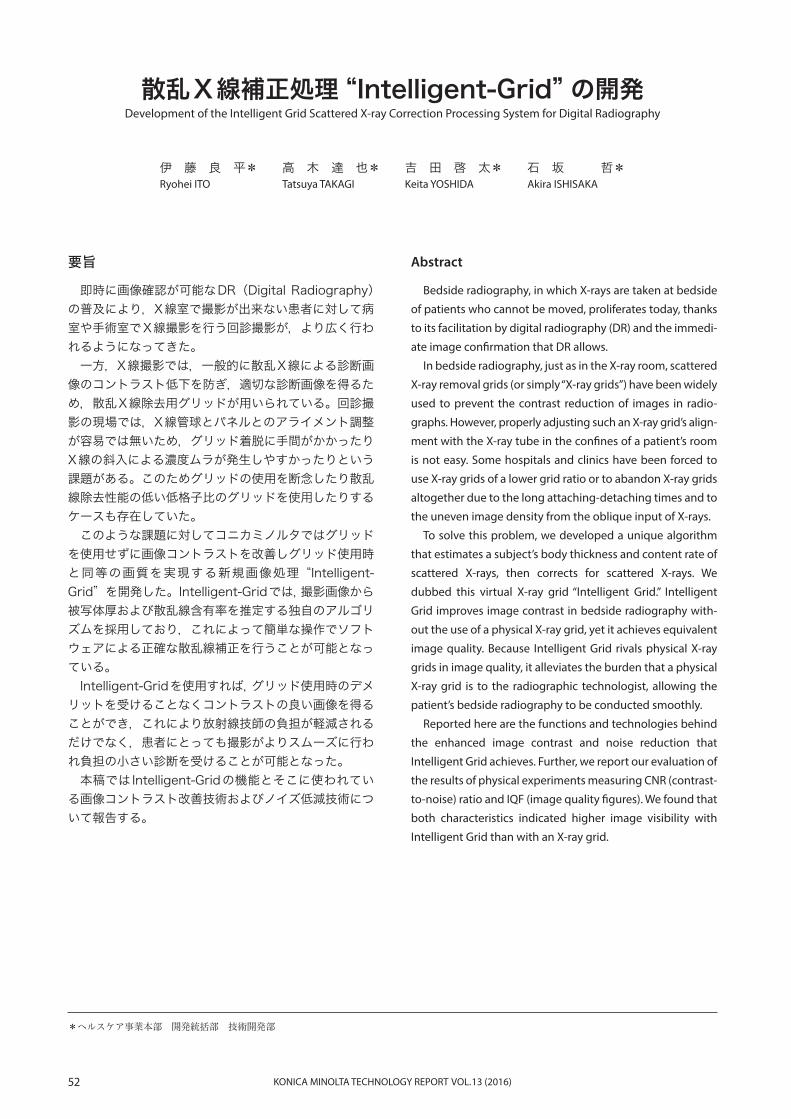
DRパネルに到達する散乱X線を除去してコントラストを高め,高画質の画像を得るための器具であり,X線の吸収率が高い鉛箔と吸収率の小さい中間材とを交互に配置した構造となっている。現在一般的なグリッドは集束グリッドと呼ばれており,鉛箔面の延長が決められた集束距離において一つの点に集束するような構造をしている(Fig. 1)。
そのため撮影時のパネルとX線装置の管球との位置関係が重要であり,正しいアライメントの時に最も散乱X線を除去する効果が発揮出来るように設計されている。
また,鉛箔の間隔を1とした時の鉛箔の高さとの比を格子比と呼ぶ。格子比はグリッドの散乱X線除去効果を表す目安の一つであり,格子比が低いグリッドは散乱X線の除去効果が低いが,その分ポジショニング不良による画質劣化のリスクも低くなる。
2. 2 グリッドのデメリットX線室で撮影される一般撮影と異なり,回診撮影では
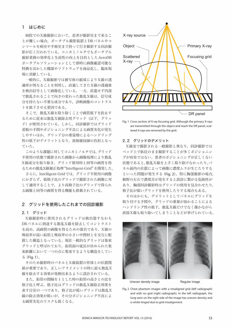
ベッド上で臥位のまま撮影することが多くポジショニングが容易ではない。患者のポジショニングが正しくない状態であると,散乱X線を上手く取り除けなかったり,パネル面内の位置によって画像に濃度ムラが生じたりするといった問題が発生する(Fig. 2)。特に胸部撮影の場合,肺野の左右で濃度差が発生すると誤診に繋がる危険性があり,胸部回診撮影時はグリッドの使用を見合わせたり,格子比が低いグリッドを使用したりする場合もある。
そのほかにも,デメリットとしてパネルにグリッドを取り付ける手間や,グリッドの重量が加わることによるハンドリング性の低下,散乱X線だけでなく僅かながら直接X線も取り除いてしまうことなどが挙げられている。
Fig. 1 Cross section of X-ray focusing grid. Although the primary X-rays are transmitted through the object and reach the DR panel, scat-tered X-rays are removed by the grid.
Fig. 2 Chest phantom images with a misaligned grid (left radiograph) and with no grid (right radiograph). In the left radiograph, the lung seen on the right side of the image has uneven density and is white-tinged due to grid misalignment.
DR panel
Focusing grid
Primary X-ray
ScatteredX-ray
Object
X-ray source
Uneven density image Regular image

54 KONICA MINOLTA TECHNOLOGY REPORT VOL. 13 (2016)
3 散乱X線補正アルゴリズム
3. 1 処理の概要Intelligent-Gridの処理フローは,画像のコントラスト
を改善する部分と散乱X線成分に起因するノイズを低減する部分とに分かれている(Fig. 3)。
画像コントラスト改善部分ではまず被写体厚を推定し,被写体厚に応じた散乱X線含有率を計算し,元画像から散乱X線相当の信号量を取り除く。そして次のプロセスとして,散乱X線成分に起因するノイズを低減し粒状性を改善するためにノイズ低減処理を行う。
画素毎に体厚を求めた後,部位毎,体厚毎,グリッドの有無によって予め作成しておいた体厚と散乱X線含有率のデータベースを使用して画像中の散乱X線成分を推定し,散乱X線画像を作成する(Fig. 4)。注目画素の散乱X線含有率は周囲の様々な構造物から発生する散乱X線の影響を受けているため,当該画素のみならず,周囲の濃度分布情報を利用し,精度を向上させている。最後に元画像から推定した散乱X線相当の信号量を取り除くことで画像のコントラストを改善する。
Fig. 3 Intelligent Grid’s scattered X-ray correction algorithm, consisting of two processes: a contrast improvement process and a noise re-duction process.
Fig. 4 Deriving a scattered X-ray image from an original image so as to improve image contrast. Scattered X-ray components of the im-age are estimated by applying a table of scattered X-ray ratios to object thickness, which is estimated from a histogram of the origi-nal image.
Reduction of noise level
Contrast improvement
Estimation of object thickness
Image output
Image input
Scattered X-ray estimationS
catte
red
X-r
ay ra
tio
Estimated object thickness
Body thicknessimage
Scattered X-ray image
Scattered X-ray ratiomapping table
Image histogram0 4031
3. 2 画像コントラスト改善処理画像コントラスト改善処理部分では元画像の画素毎に
散乱X線成分の量を推定し,元画像から取り除く処理を行っている。散乱X線の割合は患者の体厚が厚いほど多くなるため,まず初めに被写体厚の推定を行う。被写体厚の推定には画像のヒストグラム解析を用いており,ヒストグラムの形状と基準となる信号を基に体厚を推定している。
3. 3 ノイズ低減処理散乱X線成分を取り除いた画像に対し,次にノイズ低
減処理を行う。グリッドは散乱X線成分とそれに起因するノイズ成分を低減させる効果を持つが,先のコントラスト改善処理では散乱X線の低周波成分を除いただけで,ノイズ成分は残存しており,粒状が悪化している。このためグリッド使用時と画像の粒状性を揃えるためにノイズ低減処理が必要となる(Fig. 5)。

55KONICA MINOLTA TECHNOLOGY REPORT VOL. 13 (2016)
画像中から除去すべきノイズの大きさは除去した散乱X線の量によって決まる。そこで除去した散乱X線の量に応じてノイズ低減レベルを変えられるノイズ低減処理を新たに開発した。これによって粒状性を改善し視認性の高い画像を得られるようになった(Fig. 6)。
Fig. 5 Noise levels of images with and without a grid. The noise compo-nent can not be removed even by removing scattered X-rays dur-ing the contrast improvement process.
Fig. 6 Clinical images of a pelvis before and after application of a newly developed noise reduction process.
Fig. 7 Chest images: (a) with neither an X-ray grid nor Intelligent Grid, (b) with only Intelligent Grid, and (c) with only a 6:1 grid ratio X-ray grid. The contrast in chest image (b), with Intelligent Grid, is clear-ly improved over the original chest image (a).
Fig. 8 Abdomen images: (a) with neither an X-ray grid nor Intelligent Grid, (b) with only Intelligent Grid, and (c) with only a 6:1 grid ratio X-ray grid. The contrast in abdomen image (b), with only Intelligent Grid, is clearly improved over the original abdomen image (a).
Removing scattered X-rays
Signal
Sig
nal
leve
l
Primary X-rays
Primary X-rays noise
With X-ray grid
Signal
Scattered X-rays
Primary X-rays
Noise
Signal
Sig
nal
leve
lS
igna
lle
vel
Primary X-rays
Primary X-rays noiseand scattered X-rays noise
Without X-ray grid
Noise
Noise
Before noise reduction After noise reduction
(b)
(c)
(a)
Intelligent-Gridを使用した画像(b)は元画像(a)と比較してコントラストが改善しており,臨床でも使用可能なレベルとの評価を得ている。
(b)
(c)
(a)
3. 4 処理画像の結果Intelligent-Gridはこれらの処理によって構成されて
おり,一例として東海大学医学部附属病院における臨床画像評価1)に使用した画像を掲載する(Fig. 7, Fig. 8)。
これらの画像はグリッドの有無と撮影日時以外,同じ条件で撮影されており,グリッド不使用の画像(a)にIntelligent-Gridを適用した画像(b)を,グリッド使用画像(c)と比較したものである。
56 KONICA MINOLTA TECHNOLOGY REPORT VOL. 13 (2016)
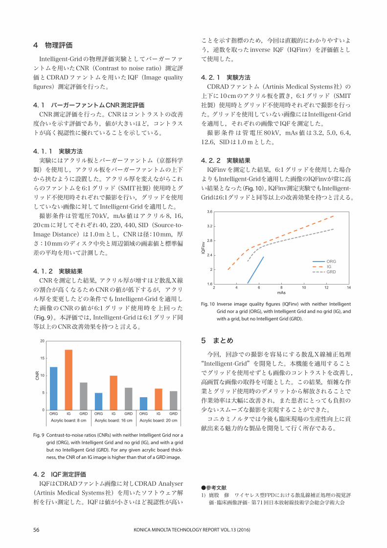
Fig. 9 Contrast-to-noise ratios (CNRs) with neither Intelligent Grid nor a grid (ORG), with Intelligent Grid and no grid (IG), and with a grid but no Intelligent Grid (GRD). For any given acrylic board thick-ness, the CNR of an IG image is higher than that of a GRD image.
Fig. 10 Inverse image quality figures (IQFinv) with neither Intelligent Grid nor a grid (ORG), with Intelligent Grid and no grid (IG), and with a grid, but no Intelligent Grid (GRD).
4 物理評価
Intelligent-Gridの物理評価実験としてバーガーファントムを用いたCNR(Contrast to noise ratio)測定評価とCDRADファントムを用いたIQF(Image quality figures)測定評価を行った。
4. 1 バーガーファントムCNR測定評価CNR測定評価を行った。CNRはコントラストの改善
度合いを示す評価であり,値が大きいほど,コントラストが高く視認性に優れていることを示している。
4. 1. 1 実験方法実験にはアクリル板とバーガーファントム(京都科学
製)を使用し,アクリル板をバーガーファントムの上下から挟むように設置した。アクリル厚を変えながらこれらのファントムを6:1グリッド(SMIT社製)使用時とグリッド不使用時それぞれで撮影を行い,グリッドを使用していない画像に対してIntelligent-Gridを適用した。
撮影条件は管電圧70 kV,mAs値はアクリル8, 16, 20 cmに対してそれぞれ40, 220, 440,SID(Source-to-Image Distance)は1.0 mとし,CNRは径:10 mm,厚さ:10 mmのディスク中央と周辺領域の画素値と標準偏差の平均を用いて計測した。
4. 1. 2 実験結果CNRを測定した結果,アクリル厚が増すほど散乱X線
の割合が高くなるためCNRの値が低下するが,アクリル厚を変更したどの条件でもIntelligent-Gridを適用した画像のCNRの値が6:1グリッド使用時を上回った
(Fig. 9)。本評価では,Intelligent-Gridは6:1グリッド同等以上のCNR改善効果を持つと言える。
ことを示す指標のため,今回は直観的にわかりやすいよう,逆数を取ったinverse IQF(IQFinv)を評価値として使用した。
4. 2. 1 実験方法CDRADファントム(Artinis Medical Systems社)の
上下に10 cmのアクリル板を置き,6:1グリッド(SMIT社製)使用時とグリッド不使用時それぞれで撮影を行った。グリッドを使用していない画像にはIntelligent-Gridを適用し,それぞれの画像でIQFを測定した。
撮 影 条 件 は 管 電 圧 80 kV,mAs 値 は 3.2, 5.0, 6.4, 12.6,SIDは1.0 mとした。
4. 2. 2 実験結果IQFinvを測定した結果,6:1グリッドを使用した場合
よりもIntelligent-Gridを適用した画像のIQFinvが常に高い結果となった(Fig. 10)。IQFinv測定実験でもIntelligent- Gridは6:1グリッドと同等以上の改善効果を持つと言える。
0
5
10
15
20
Acrylic board: 8 cm Acrylic board: 16 cm Acrylic board: 20 cm
CN
R
ORG IG GRD ORG IG GRD ORG IG GRD
1.6
2
2 4 6 8 10 12 14
2.4
2.8
3.2
3.6
IQFi
nv
mAs
ORGIGGRD
5 まとめ
今回,回診での撮影を容易にする散乱X線補正処理“Intelligent-Grid”を開発した。本機能を適用することでグリッドを使用せずとも画像のコントラストを改善し,高画質な画像の取得を可能とした。この結果,煩雑な作業とグリッド使用時のデメリットから解放されることで作業効率は大幅に改善され,また患者にとっても負担の少ないスムーズな撮影を実現することができた。
コニカミノルタでは今後も臨床現場の生産性向上に貢献出来る魅力的な製品を開発して行く所存である。
●参考文献1) 鹿股 修 ワイヤレス型FPDにおける散乱線補正処理の視覚評
価-臨床画像評価- 第71回日本放射線技術学会総会学術大会
4. 2 IQF測定評価IQFはCDRADファントム画像に対しCDRAD Analyser
(Artinis Medical Systems社)を用いたソフトウェア解析を行い測定した。IQFは値が小さいほど視認性が高い