인제대 일산 백병원 장우익. systemic embolism affecting the brain both from cpb and...
TRANSCRIPT
Embolic problems in Cardiac Surgery
- CPB view point
인제대 일산 백병원 장우익

Embolic problems during cardiopul-monary bypass
Systemic embolism affecting the brain
Both from CPB and underlying car-diovascular disease of the patients
Central nervous dysfunction Major stroke - > macroembolism Neuropsychologic problems -> mi-
croembolism
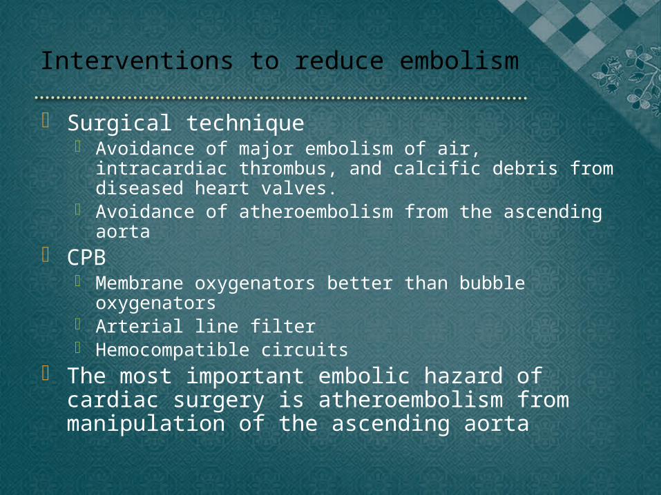
Interventions to reduce embolism
Surgical technique Avoidance of major embolism of air, intracardiac throm-
bus, and calcific debris from diseased heart valves. Avoidance of atheroembolism from the ascending aorta
CPB Membrane oxygenators better than bubble oxygenators Arterial line filter Hemocompatible circuits
The most important embolic hazard of cardiac surgery is atheroembolism from manipulation of the ascending aorta

Macroemboli & Microemboli
Macroemboli ; occluding flow >200um artery -> single macroembolus might re-sult hemiplegia
Microemboli ; smaller arteries, arterioles, and capillaries. Single microembolus, no clinical effect. Numerous emboli can result diffuse pattern of CNS injury
Except perfusion accidents, macroemboli are unlikely to rise from the extracorporeal circuits, but rather from heart and aorta

Type of emboli and tissue effects
Gas bubbles Air, anesthetic gas(esp, nitrous oxide) Dynamic equilibrium with the same gas dis-
solved in the plasma Grow or shrink, dependent on temperature. Small bubbles collapse when less than 10um
Biologic aggregates Thrombus, platelet aggregates, fat
Inorganic debris Fragment of polyvinyl chloride tubing, silicone
antifoam, reduced currently.
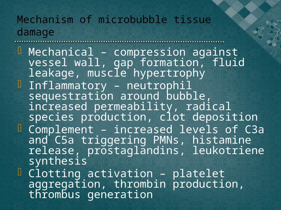
Mechanism of microbubble tissue damage
Mechanical – compression against vessel wall, gap formation, fluid leakage, muscle hypertrophy
Inflammatory – neutrophil sequestration around bubble, increased permeability, radical species production, clot deposition
Complement – increased levels of C3a and C5a triggering PMNs, histamine release, prostaglandins, leukotriene synthesis
Clotting activation – platelet aggregation, throm-bin production, thrombus generation

Entrapment of air into the arterial circulation
Events at the bypass machine Not properly de-aired prior to bypass Inattention to the reservoir level Ruptured arterial pump-head tubing Arterial line separation Unnoticed rotation of the arterial pump head Runaway pump head Reversal of pump-head rotation Reversal of tubing connected to the ventricular vent Inadvertent detachment of oxygenator during CPB Air transmitted through the membrane oxygenator by an
occluded scavenger line Clotted oxygenator Pressurized cardiotomy reservoir
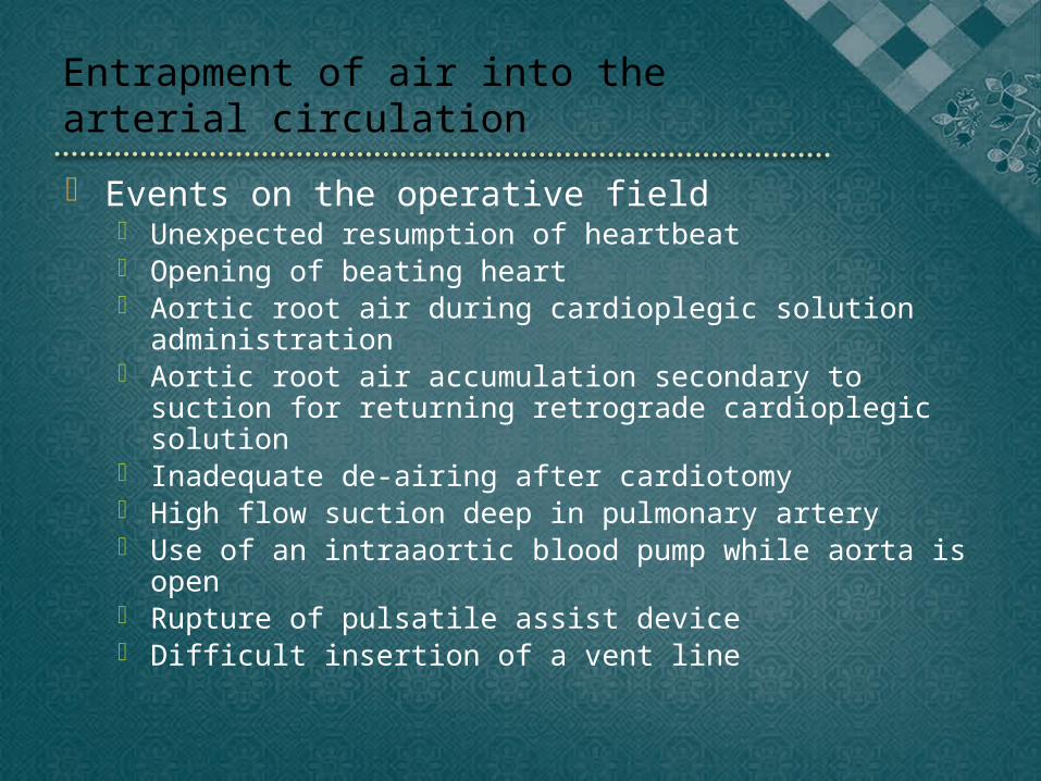
Entrapment of air into the arterial circulation
Events on the operative field Unexpected resumption of heartbeat Opening of beating heart Aortic root air during cardioplegic solution administration Aortic root air accumulation secondary to suction for re-
turning retrograde cardioplegic solution Inadequate de-airing after cardiotomy High flow suction deep in pulmonary artery Use of an intraaortic blood pump while aorta is open Rupture of pulsatile assist device Difficult insertion of a vent line
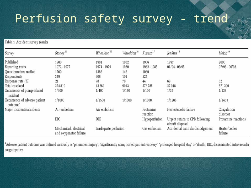
Perfusion safety survey - trend
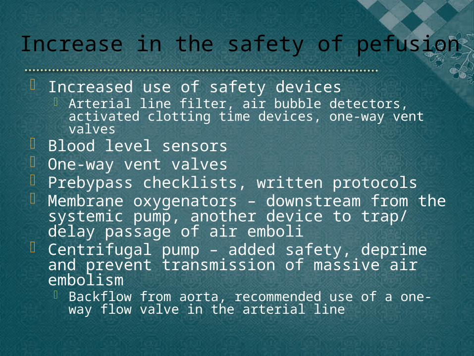
Increase in the safety of pefusion Increased use of safety devices
Arterial line filter, air bubble detectors, activated clotting time devices, one-way vent valves
Blood level sensors One-way vent valves Prebypass checklists, written protocols Membrane oxygenators – downstream from the sys-
temic pump, another device to trap/delay passage of air emboli
Centrifugal pump – added safety, deprime and prevent transmission of massive air embolism Backflow from aorta, recommended use of a one-way flow valve
in the arterial line

Adverse neurologic outcomes
Vary widely, due to multiple other factors, such as cerebral blood flow, systemic inflammation, patient co-morbidities
Cognitive decline, such as memory deterioration ; 60% one week, 25-30% from 2 months to one year postop.
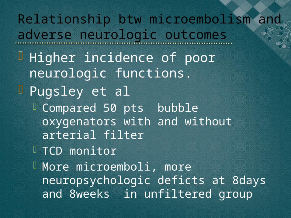
Relationship btw microembolism and adverse neurologic outcomes
Higher incidence of poor neurologic functions.
Pugsley et al Compared 50 pts bubble oxygenators
with and without arterial filter TCD monitor More microemboli, more neuropsycho-
logic deficts at 8days and 8weeks in un-filtered group

Microembolism and neurocognitive functions
Comparisons between OPCAG and on-pump CABG Slight tendency toward decreased per-
formance in neurocognitive tests in the on-pump group. Decreased as the time after surgery increased.
Comparisons between valve and CABG Increased rate of emboli in valve
surgery But no significant difference in neu-
rocognitive test scores
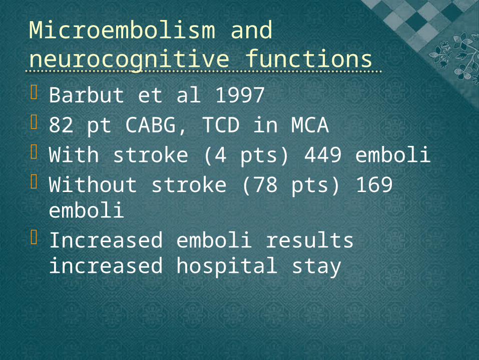
Barbut et al 1997 82 pt CABG, TCD in MCA With stroke (4 pts) 449 emboli Without stroke (78 pts) 169 emboli Increased emboli results increased
hospital stay
Microembolism and neurocognitive functions
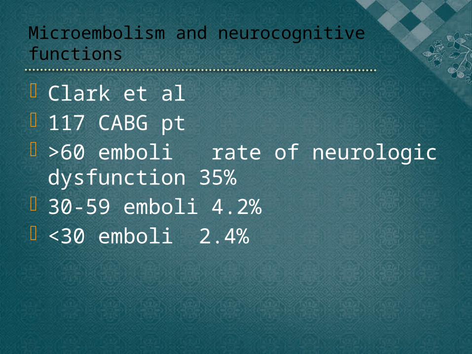
Microembolism and neurocognitive functions
Clark et al 117 CABG pt >60 emboli rate of neurologic dys-
function 35% 30-59 emboli 4.2% <30 emboli 2.4%
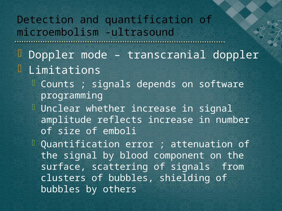
Detection and quantification of microem-bolism -ultrasound
Doppler mode – transcranial doppler Limitations
Counts ; signals depends on software programming Unclear whether increase in signal amplitude reflects
increase in number of size of emboli Quantification error ; attenuation of the signal by
blood component on the surface, scattering of signals from clusters of bubbles, shielding of bubbles by oth-ers
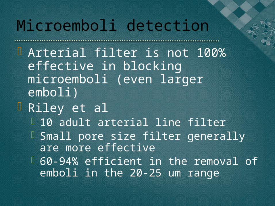
Microemboli detection
Arterial filter is not 100% effective in blocking microemboli (even larger emboli)
Riley et al 10 adult arterial line filter Small pore size filter generally are more
effective 60-94% efficient in the removal of em-
boli in the 20-25 um range

Perfusionist interventions
Borger et al 34 pts 75% of all emboli detected during
perfusionist interventions (drug injec-tion and blood sampling)
Emboli count more higher during per-fusion intervention (6.9/min) than during surgical intervention(1.5/min) or during baseline(0.4/min)
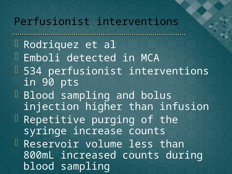
Perfusionist interventions
Rodriquez et al Emboli detected in MCA 534 perfusionist interventions in 90
pts Blood sampling and bolus injection
higher than infusion Repetitive purging of the syringe in-
crease counts Reservoir volume less than 800mL
increased counts during blood sam-pling
Gaseous microemboli
Perfusion intervention ; during drug injection into the venous reservoir. Air in the syringe, source of microemboli
Venous line air ; traversed membrane oxygenator and arterial line filter, possibly d/t bubble deformation or coalescence within or after the filter.

Vacuum assisted venous drainage
Augment drainage of venous blood Smaller venous cannulae Favor formation of gaseous mi-
croemboli > -40 mmHg and high blood flow(6
L/min) ; increased GME
CO2
CO2 flooding of the op site High solubility compared to room air Disadvantage ; hypercarbia and respiratory
acidosis CO2 flooding only during the period of de-air-
ing of the heart CO2 potent cerebral vasodilator
Hypocapnia (PaCO2 30-32mmHg) ; reduce cerebral blood flow and embolization
No significant difference btw hypocarbic gr and normocarbic gr
Potential disadvantage of cerebral hypoperfu-sion
Steep trendelenburg position
Rationale ; buoyancy effects will cause bubbles to rise and minimize cerebral embolization
Study ; did not decrease the cerebral embolic load
GME in flowing blood ejected from the heart respond more as an emul-sion not subject to normal buoyancy effects as would be larger bubbles
Dynamic bubble trap Rotating stream that forced GME to the
center of the flow -> passively vented out to the reservoir by a small tube located midstream and near the exit of the bubble trap.
Volume diverted 400-450mL/min Reduction in the number of bubbles de-
tected in the range 11-40um in MCA Greater efficiency of removal by the bub-
ble trap for the larger-sized GME ( >96% for bubbles >31um)

Dynamic bubble trap
Oxygenators – capability of trapping air
Oxygenator design that provided for rapid blood contact with the membrane material, increased bubble/membrane contact time, avoidance of high blood flow velocities and low pressure drop, and membrane bundle geometry all favored en-trapment of GME
Capability of CPB circuits to remove entrained ve-nous air. Five type oxygenators Air detected after arterial filter in all Statistical different results among different manufac-
tures Contributing factors ; Residence time for blood and bub-
bles within the membrane oxygenator, pressure drop, turbulence in the flowing blood
Avoidance of venous air whenever it is observed.
Cannula type or excessive blood flow velocities
? Increased number of microemboli High blood velocity could contribute to particulate
release fron the aortic wall Theoretically possible for GME to be produced by
high blood flow velocities or abrupt pressure dif-ferences at cannula tips
Banaroia et al ; 32 elective CABG pt No correlation between blood velocities or type of can-
nula and the presence of TCD-detected emboli Conventional cannula under conventional CPB, systemic
flow was not important.
Newer circuits
Minimizing prime volumes Reducing reservoir volumes -> lessen per-
fusionist reaction time in the events Without venous reservoir CPB tubing smaller and shorter ->in-
creased blood flow velocities thru the cir-cuit.
Blood transit time is reduced -> decreased opportunity for GME to be removed prior to its return to the patients
Deairing of the venous drainage-reduction of arterial microbubbles
Deairing ; double clamp and saline filling Connecting venous line without deairing of
the venous line ; incorporation of 15cc air into the circuit
Entrapped air in the venous line is mi-crofragmented while passing through the ECC with subsequent microbubble forma-tion
Microbubbles detected after arterial filter ; once saturated they release captured gas bubbles.
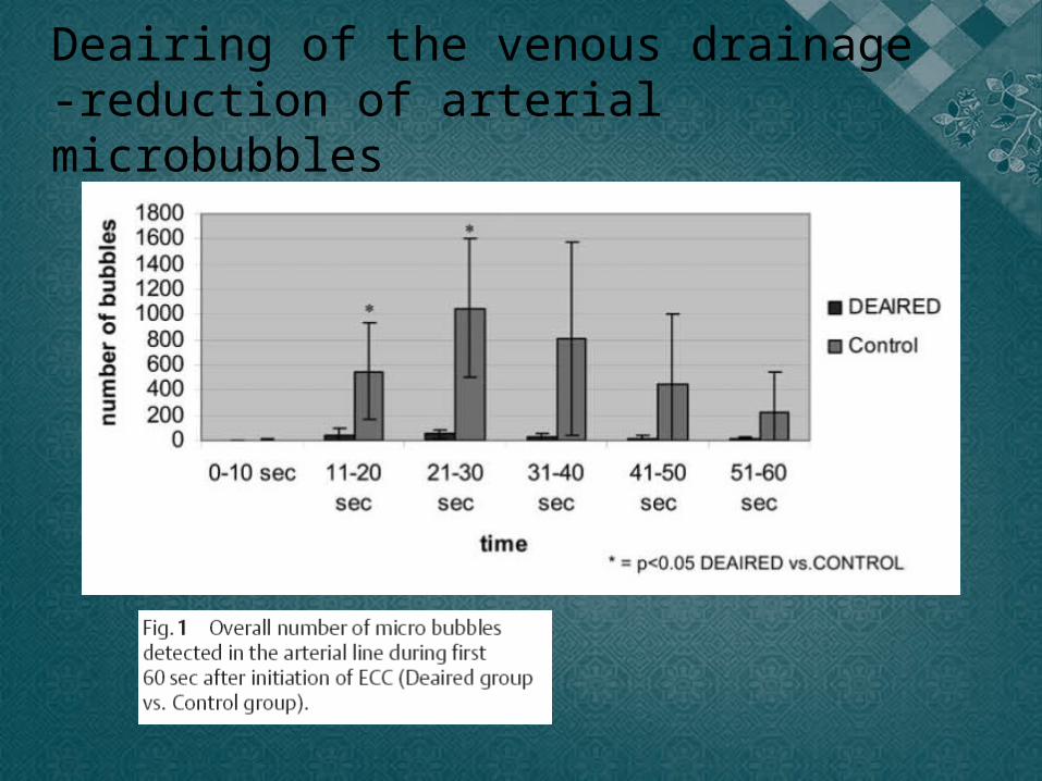
Deairing of the venous drainage-reduction of arterial microbubbles
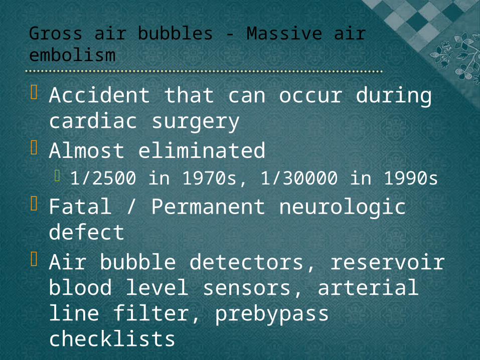
Gross air bubbles - Massive air em-bolism
Accident that can occur during car-diac surgery
Almost eliminated 1/2500 in 1970s, 1/30000 in 1990s
Fatal / Permanent neurologic defect Air bubble detectors, reservoir blood
level sensors, arterial line filter, pre-bypass checklists
Cause
Sudden reduction in the blood level in the venous reservoir that is not noticed by the perfusionist
Inadvertent pressurization of the reservoir. Air from the cardiac chamber Runaway pump head Inversion of left-sided heart vent Reversal of pump head Inadvertent detachment of oxygenator during by-
pass Cardiotomy suction wedged deep into the pul-
monary artery

Treatment
Stop the circulation Steep trendelenburg position De-air the entire pump line Retrograde SVC perfusion Hypothermia Barbiturate and corticosteroid Hyperbaric oxygen therapy
Conclusion Embolic problems during cardiopulmonary bypass
Brain most susceptible. Cause of stroke is mostly from underlying dis-
ease. Especially from atherosclerosis of the aorta. Current CPB circuits itself – low embolic risk Microembolism
Clinical effect ; difficult to notice but has potential risk Efforts to reduce it!!
Gross air – rare incidence but fatal Prevention !!! Prebypass checklist, education, drill Rapid reaction if occurs