a bio‐acoustic levitational (bal) assembly method for ... · optimized for human stem cell...
TRANSCRIPT

© 2015 WILEY-VCH Verlag GmbH & Co. KGaA, Weinheim 161wileyonlinelibrary.com
CO
MM
UN
ICATIO
N
A Bio-Acoustic Levitational (BAL) Assembly Method for Engineering of Multilayered, 3D Brain-Like Constructs, Using Human Embryonic Stem Cell Derived Neuro-Progenitors
Charlène Bouyer , Pu Chen , Sinan Güven , Tugrul Tolga Demirtas , Thomas J. F. Nieland , Frédéric Padilla ,* and Utkan Demirci *
C. Bouyer, Dr. P. Chen, Dr. S. Güven, [+] T. T. Demirtas, Dr. T. J. F. Nieland, Prof. U. Demirci Bio-Acoustic MEMS in Medicine (BAMM) Lab Canary Center for Early Cancer Detection Department of Radiology School of Medicine Stanford University Palo Alto , CA 94304 , USA E-mail: [email protected] C. Bouyer, Prof. F. Padilla Inserm, U1032, LabTau University of Lyon Lyon F-69003 , France E-mail: [email protected] C. Bouyer, Prof. F. Padilla LabEx DEVweCAN University of Lyon Lyon F-69003 , France Prof. U. Demirci Department of Electrical Engineering (By Courtesy) Stanford University Stanford , CA 94305 , USA
DOI: 10.1002/adma.201503916
to create six-layered tissue surrogates of the cerebral cortex. For instance, primary rodent neurons were encapsulated in a layer-by-layer assembly approach in concentric 3D donut-shaped constructs, made from composite hydrogel and silk protein. [ 7 ] Micromolding has also been employed to embed primary neu-rons in 3D networks in collagen. [ 8 ] The generated 3D neural surrogate tissues displayed the characteristic cell density, inter-layer neurite connections, and layer thickness of the cortex, as well as biochemical and electrophysiological functional responses. However, the bioengineering operations involved are complicated, time consuming, and require special exper-tise, hampering their wide implementation in biological labs.
To address these critical challenges in brain bioengineering, we present here a unique, single step bio-acoustic levitational (BAL) assembly technology that is fast and facile to implement ( Figure 1 and Figure S1, Supporting Information). Multilayers are formed in a fi brin 3D microenvironment by the effect of bulk acoustic radiation pressure on mammalian cells. Due to their larger density and lower compressibility than the sur-rounding fl uid, cells are driven to the node planes of acoustic standing waves where there is minimal pressure. [ 9,10 ] The inter-layer distance is proportional to the frequency according to the equation f = c / λ , where c is the sound velocity and λ the wave-length (see the Supporting Information). Thus, the acoustic frequency can be tuned to match the number and spacing of the interlayers to that of native tissues, with an interlayer dis-tance that corresponds to half of the acoustic wavelength. We demonstrate the utility of the BAL method for bioengineering of multilayer 3D tissue surrogates by creating 3D constructs resembling that of the cerebral cortex, using human stem cell derived neuro-progenitor cells (NPCs) as a cell source. During the assembly, the 3D multilayer tissues are permanently stabilized within a single fi brin hydrogel construct, designed to have brain-like mechanical properties. Subsequently, the NPCs are differentiated in situ into neural cells by switching the media type in which the 3D constructs are cultured. Over a period of 30 d, the generated constructs formed a stable inter- and intralayer 3D organization, characteristic of that of the multilayered cortex.
We developed a BAL assembly device for multilayer tissue engineering, comprised of a ceramic transducer driven by a waveform generator, an acoustic resonant chamber that also holds the cell suspension, and a plexiglass acoustic refl ector. A novel, single-step, 3D tissue engineering method was designed
Bioengineering aspires to build biofi delic, 3D surrogates of native organs, such as the brain, liver, or heart. [ 1 ] Bioengi-neering methods that offer the ability to control complex cell patterning, density, and contact in the 3D surrogate microenvi-ronment are invaluable tools to examine the physical properties and functions of these tissues. [ 2 ] This is particularly relevant to neuroscience, where human or animal postmortem brain slices offer the most relevant ex vivo experimental models. They are used to examine, for instance, neural circuit connec-tivity, neuro transmitter mechanism of action, [ 3 ] the dynamics of protein and ion channel signaling, [ 4 ] and the etiology of and therapeutic agents for mental illnesses. [ 5,6 ] 2D culture models have yielded important insights in, for instance, the mole-cular drivers of neural development and activity. However, the random distribution of neurons in culture dishes does not suf-fi ciently recapitulate the 3D aspects of neural connectivity or the microenvironment of the brain.
An alternative choice to these approaches is presented by the in vitro bioengineering of 3D models that recapitulate native brain tissues. Several multistep strategies have been developed
[+]Present address: Izmir Biomedicine and Genome Center, Dokuz Eylul University, Health Campus, 35340, Balcova, Izmir, Turkey
Adv. Mater. 2016, 28, 161–167
www.advmat.dewww.MaterialsViews.com
162 wileyonlinelibrary.com © 2015 WILEY-VCH Verlag GmbH & Co. KGaA, Weinheim
CO
MM
UN
ICATI
ON
and implemented for contactless and rapid acoustic levitational assembly of large populations (up to 4 million cells mL −1 ) of cells into multilayer tissue organizations. Here, the system was optimized for human stem cell derived neuro-progenitors and cells with similar physical properties (size, density, and com-pressibility) (Figure 1 and Figure S1, Supporting Information). In this system, cells are suspended in a fi brin prepolymer solution and placed in a circular levitation chamber, built of a poly(methyl methacrylate) (PMMA) ring (ID: 16 mm; OD: 18 mm) that is sealed to a thin layer of polystyrene fi lm of 0.2 mm thickness. To fi t with the desired application of engi-neering brain cortex-like tissue, the height of the levitation chamber was chosen to be 2.2 mm, which is the average thick-ness for human cortex. [ 11 ] For other tissue engineering appli-cations, the chamber height can be altered to accommodate the designs of 3D constructs of any thickness. The chamber is mounted on a piezoelectric ceramic transducer coupled to the polystyrene fi lm with ultrasound coupling gel, while the top side is covered by a 6 mm thick glass. By applying sine-shaped continuous electric signals to the transducer via an arbitrary waveform generator, bulk acoustic standing waves are formed within the chamber as a result of coherent interfer-ence between incident waves from the transducer and refl ected waves from the glass refl ector. The quick (less than 10 min) crosslinking of fi brin stabilizes the 3D multilayer architecture of the engineered tissues.
Heterogeneous distributions of cells within a node plane, such as radial patterns, may be inadvertently caused by secondary acoustic forces and the associated radial acoustic pressure gra-dients, generated by resonance within the cylindrical levitation chamber. Theoretical modeling of the net forces acting on the cells—acoustic radiation force, viscous drag force arising from acoustic streaming, and vertical force due to gravity and buoy-ancy—can predict the minimal pressure to levitate and maintain cells in parallel horizontal layers. This pressure leads to a null vertical velocity of cells in the bulk acoustic fi eld. Accordingly, the pressure threshold to levitate 10 µm diameter cells, modeled as spherical objects, in homogeneous, multilayered node planes was estimated at 46 kPa (using Matlab software, see the Experi-mental Section in the Supporting Information).
The performance of the complete system, comprised of the air-backed ceramic coupled to the levitation chamber closed
with the plexiglass lid, was then analyzed using Comsol Multiphysics software (Comsol 5.0, Comsol Inc., Palo Alto, CA) ( Figure 2 a). We fi rst identifi ed the input optimal voltage (5 V) needed to obtain the above derived minimal pressure of 46 kPa across the entire antinode planes surface (Figure 2 b,c,e,f). By simulating the acoustic fi eld generated by the piezoelectric ceramic (see the Experimental Section), we confi rmed the for-mation of pressure node layers in the levitation chamber with an interlayer spacing of 250 μm (Figure 2 b,c). This simulation also showed that the 46 kPa minimum acoustic pressure corre-sponds to a ceramic surface displacement of 130 Å (Figure S2a, Supporting Information). Laser interferometry confi rmed this displacement could be achieved experimentally by setting the driving power of the piezoelectric ceramic transducer to 8 mW (Figure S2a,b, Supporting Information). With these settings, the maximum acoustic pressure within the chamber does not exceed 160 kPa (Figure 2 b,c), resulting in low pressure gradi-ents in the radial direction (Figure 2 e), minimizing the genera-tion of a radial secondary acoustic radiation force and resulting radial cell patterns (Figure S3, Supporting Information).
The theoretical operational settings were subsequently used to assess the experimental performance of the acoustic levitation design. First, the pressure fi eld of the bulk acoustic standing waves in the levitation chamber was measured using a fi ber optic hydrophone (Figure 2 b,d). The pressure fi eld showed variations of pressure along vertical direction ( Z -axis), resulting in the expected horizontal planes of pressure nodes (where the pressure is minimal) separated by 250 µm (Figure 2 d,g). Pres-sure amplitudes in the standing wave fi eld were measured above the 46 kPa threshold required for levitation of cells and did not exceed 160 kPa (Figure 2 g) within the entire chamber. Through modeling, numerical simulations and experiments, we thus identifi ed the operational acoustic levitation conditions to engineer multilayers with maximal homogenous distribution of cells in 3D.
With these settings in hands, the performance of the acoustic levitation process was fi rst examined with fl uorescent polystyrene beads (density of 1.06 g cm −3 and compressibility of 0.94/ c 2 ) in aqueous media (density of 0.996 g cm −3 ) ( Figure 3 a). Bead sizes of 8, 49, and 202 µm were tested to evaluate the versatility of the BAL device in levitating a range of objects, ranging from single cells up to cell spheroids (Figure 3 a). For all
Adv. Mater. 2016, 28, 161–167
www.advmat.dewww.MaterialsViews.com
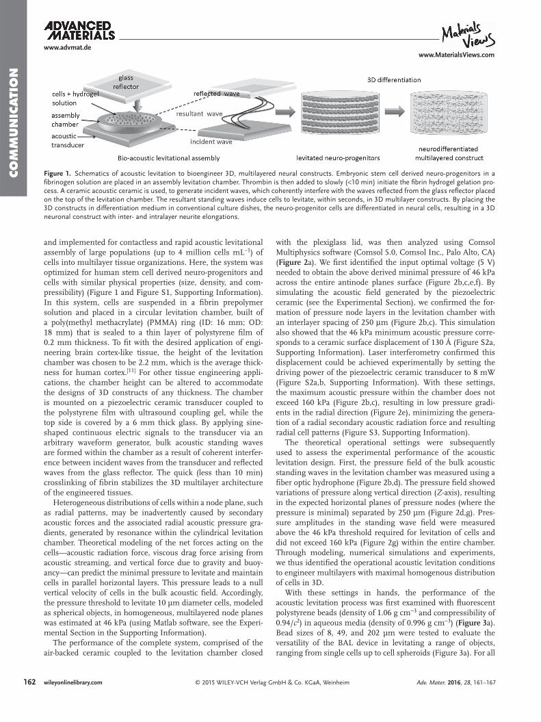
Figure 1. Schematics of acoustic levitation to bioengineer 3D, multilayered neural constructs. Embryonic stem cell derived neuro-progenitors in a fi brinogen solution are placed in an assembly levitation chamber. Thrombin is then added to slowly (<10 min) initiate the fi brin hydrogel gelation pro-cess. A ceramic acoustic ceramic is used, to generate incident waves, which coherently interfere with the waves refl ected from the glass refl ector placed on the top of the levitation chamber. The resultant standing waves induce cells to levitate, within seconds, in 3D multilayer constructs. By placing the 3D constructs in differentiation medium in conventional culture dishes, the neuro-progenitor cells are differentiated in neural cells, resulting in a 3D neuronal construct with inter- and intralayer neurite elongations.

163wileyonlinelibrary.com© 2015 WILEY-VCH Verlag GmbH & Co. KGaA, Weinheim
CO
MM
UN
ICATIO
N
the tested microsized beads, the acoustic levitation system was able to generate multilayers with identical interlayer distances equal to half-wavelength of the ceramic transducer working frequency, confi rming the theoretic calculations. Furthermore, we analyzed the effect of acoustic frequency on the tunability of the interlayer distance. Increasing the acoustic frequency from 340 to 680 kHz decreased the interlayer distance from 2.2 to 1.1 mm (Figure 3 b), and increased the number of layers in the levitation chamber. The number of layers was also tun-able by changing the chamber height (Figure 3 c). The time for the beads to align in a multilayer structure was less than 2 s, recorded by a high speed camera (Figure 3 d and Videos S1a–d, Supporting Information: levitation of particles of 8, 24, 49, and 96 µm diameter). The two features of rapid (within seconds) organization of objects, and the ability to generate multilayered architectures, make BAL a unique tool for microscale bioengi-neering of tissues made of a layered architecture, including that of the cerebral cortex.
We used these properties of BAL to bioengineer in fi brin gel 3D tissue models resembling the multilayer architecture of the cerebral cortex, by using neuro-progenitors as a cell source and
differentiating them into neurons. Robust and reproducible engineering of 3D multilayered tissues was facilitated by opti-mizing the protocol to generate the fi brin extracellular matrix, a biomaterial extensively used in tissue engineering applica-tions [ 12 ] and clinical translations. [ 13,14 ]
One of the advantages of using fi brin as a 3D microenvi-ronment is that by careful tuning of the prepolymer concen-trations (and their ratio) of fi brinogen and thrombin, cells can be levitated rapidly (within seconds) while still in non-crosslinked solution. Fibrin crosslinks slowly, within 10 min, into a stable 3D gel, which then can be removed from the acoustic chamber for culture. Second, the concentration of fi brinogen can be adjusted to create a 3D microenvironment with viscoelastic properties resembling native tissue. To test this, we used a rotational parallel plate rheometer to apply mechanical strains to the 3D fi brin gels. Within the deforma-tion range of 0.1%–10%, the viscoelastic properties measure-ments were not affected (not shown). Reliable elasticity and viscosity properties of the fi brin gel were subsequently meas-ured by applying deformation of 3% and angular frequen-cies from 0.62 to 63 rad s −1 . At a fi brinogen/thrombin ratio
Adv. Mater. 2016, 28, 161–167
www.advmat.dewww.MaterialsViews.com
Figure 2. Characterization of acoustic pressure fi eld generated by the BAL device. a) 3D display of the absolute values of acoustic pressure fi eld from numerical simulation. b) 2D view of the simulated (top) and experimental (bottom) acoustic pressure fi eld along the levitation chamber diameter, obtained with fi ber optic hydrophone for 8 mW driven power. Horizontal plane of pressure nodes (minimal pressure) is present as well as radial pressure variations. The acoustic pressure decreases when moving away from the center of the chamber. Zooms of c) simulated and d) experimental acoustic pressure fi eld, delineated by the dotted squares in (b). e) Simulation of pressure along the horizontal (radial) direction for the node height Z set at 0.945 mm, and antinode height Z set at 0.81 mm, and at a height of 0.8775 mm (dotted lines in (c)). Seven nodes, separated from 1 mm, are present in the horizontal direction. The resultant gradient is about 0.15 MPa in 1 mm in the center of the levitation chamber and 0.02 MPa 2 mm away from the center. f) Simulation of pressure along vertical ( Z ) axis in the center ( R = 0 mm) and 1.3 mm away ( R = 1.3 mm) (dotted lines in (c)), showing that pressure nodes are separated by 250 µm and that the pressure is about twice higher at the center than 1.3 mm away. The pressure gradients are signifi cantly higher (0.14 MPa in 250 μm) in vertical than in horizontal direction because the distance between two nodes is about 250 μm, compared to 1 mm in radial direction (f). g) Experimental pressure along the Z axes (dotted lines in (d)): in the center of the chamber and 1.3 mm away showing that we experimentally obtain what was simulated in (f).

164 wileyonlinelibrary.com © 2015 WILEY-VCH Verlag GmbH & Co. KGaA, Weinheim
CO
MM
UN
ICATI
ON
of 1/0.162 (fi brinogen concentration of 10 mg mL −1 and a thrombin concentration of 10 U mL −1 ), the storage modulus ( G ′ = 474 Pa) was found to be ten times larger than the loss modulus ( G ′′ = 27.8 Pa) (Figure 3 e), similar to the values of the native brain. [ 13,14 ]
To demonstrate that BAL engineered tissues provide a physi-ologically relevant microenvironment for neural cells to grow, we levitated NPCs by setting the ceramic power at 8 mW. Cells were assembled in 2 s and the acoustic power was kept active
for 10 min, during which time the fi brin gel polymerizes. Subsequently, the neural progenitor cells were differentiated into neural cells by placing the 3D multilayer constructs in neuro nal differentiation media (see the Experimental Section in the Supporting Information). We qualitatively confi rmed the viability of 3D tissues immediately after acoustic levitation ( Figure 4 a) and after 7 d of differentiation, using calcein fl uo-rescent dye to mark live cells and ethidium-homodimer to label dead cells (Figure 4 b). We also confi rmed that the BAL assembly
Adv. Mater. 2016, 28, 161–167
www.advmat.dewww.MaterialsViews.com
Figure 3. Characterization of the acoustic levitation process to generate multilayered 3D tissues. a) Multilayer levitation of fl uorescent beads, with a density of 1.06 g cm −3 and a diameter D of 8, 49, or 202 µm, simulating a range of cell sizes and spheroids. The experimental results are shown in black and white, and a schematic depiction of the results is shown in color. The interlayer distance is qualitatively equal when all bead sizes are tested under the identical frequency and power of acoustic waves (shown here for 680 kHz). b) Tuning of the ceramic driven frequency changes the interlayer distance, tested here using 24 µm beads. A 1.13 mm distance was achieved at the ceramic natural frequency (680 kHz). The interlayer distance was double for its fi rst subharmonic (340 kHz). c) Number of layers generated according to ceramic driven frequency. A higher frequency corresponds to an increased number of layers formed in the gel in the levitation chamber. In addition, the number of layers increases with the levitation chamber height. d) Time dependence of levitation in relation to bead size. The larger the beads, the faster they equilibrated their position at the pressure nodes layers. e) Rheometry measurements of the polymerized fi brin gel. Storage and loss modulus evaluation at different frequency indicated that the storage modulus G ′ is equal to 474 Pa and loss modulus G″ to 28.7 Pa at 6.28 rad s −1 . Viscosity and elasticity of the fi brin gel are found to be 4.43 Pa s and 474 Pa s. Scale bar 1 mm.

165wileyonlinelibrary.com© 2015 WILEY-VCH Verlag GmbH & Co. KGaA, Weinheim
CO
MM
UN
ICATIO
N
does not adversely affect the metabolic characteristics of the cells. Indeed, a Presto Blue assay (Life Technologies) performed on 3D encapsulated neurons showed no difference between ultrasound exposed (i.e., multilayer) and nonexposed (i.e., cells randomly encapsulated within the gel volume) samples. This assay revealed that the metabolic activity increased two-fold over a 7 d culture period (Figure S4, Supporting Informa-tion), further suggesting that the generated 3D microenviron-ment is able to support cell metabolism (Figure S4, Supporting Information).
Next, we assessed the ability of the 3D tissue to support neural differentiation. Immediately after assembly in multi-layered constructs immunofl uorescence microscopy analysis showed that NPCs were positive both for the early neural precursor protein DCX (Figure 4 c) and the neuronal marker NeuN (Figure 4 e). The expression of DCX decreased after one week of culture in 3D in neural differentiation medium (Figure 4 d), indicative of neural differentiation. This was
further substantiated by the continued expression of NeuN (Figure 4 f), the emergence of Tuj-1 expression (Figure 4 g and Video S2, Supporting Information), and the formation of neu-rites in every layer of the 3D multilayer assembled structure (Figure 4 h). Continued expression of the neuro-progenitor marker Nestin indicated that neural differentiation was not completed yet (Figure 4 h), as expected given the short differen-tiation span. [ 15 ]
To ascertain the long-term stability of the hydrogel and its ability to support neurite network formation, the 3D multi-layered tissues were cultured for up to 30 d after acoustic levitation ( Figure 5 ). Inter- and intralayer connections started as early as 8 d of culture, when cells were observed to extend Tuj-1 positive neurites (Figure 5 a). After 14 d in culture, the initial network further developed into visible growth of MAP-2 positive dendrites between layers (Figure 5 b). After 30 d, cells coexpressed both the excitatory neuron marker CaMKII and GFAP (Figure 5 c,d and Video S3, Supporting Information), a
Adv. Mater. 2016, 28, 161–167
www.advmat.dewww.MaterialsViews.com
Figure 4. Bioengineering of 3D multilayered neural tissues using bulk acoustic levitation. HUES 64 derived neuro-progenitor cells were exposed to acoustic ultrasound waves to create multilayered constructs in fi brin gels. a) Immediately after acoustic assembly (day 0) and b) after 7 d of culture (day 7), the majority of cells appear positive for the vital dye calcein AM (green: “live”) and negative for the cytotoxicity dye ethidium homodimer-1 (red: “dead”) demonstrating the viability of the cells in the 3D multilayered constructs. Immunofl uorescence characterization of neural differentiation of multilayered constructs c,e) after acoustic assembly (day 0) and d,f) after 7 d of culture in differentiation media. c,d) A decrease in expression of the neural progenitor marker DCX (green) and e,f) a continued expression of the neuronal marker NeuN (green) is observed over time. DAPI (DNA stain: blue) is used to mark all the cells present in the 3D gel. g) The multilayered nature of the neuronal construct is shown by immunofl uorescence staining of neurites (Tuj-1; green) and cells (DAPI: blue) after 7 d in culture. h) Cross section of 3D neuronal construct after 7 d in differentiation media, immunostained with the neuro-progenitor marker Nestin (green) and DAPI (blue). The appearance of the neurite marker Tuj-1 (red) provides an indication that NPCs have initiated their differentiation process. The arrow points to a single cross section ( YX plane with dimension 1 mm × 800 µm) of a single neuronal layer, showing homogenous partition of cells within a layer. Scale bars indicate 250 µm.
Figure 5. Differentiation time course of 3D multilayered neural con-structs. a) Neuro-progenitors were assembled in 3D multilayered tissue constructs by acoustic levitation and differentiated for 8 d in differen-tiation media. The 3D cells immunostained for Nestin (red) and Tuj1 (green) are beginning to show interaction between layers. Scale bars indicate 500 µm (left panel), 250 µm (middle panel), and 100 µm (right panel). b) After 14 d in differentiation media, cells are positive for the mature neuron marker MAP2 (cyan) as well as Nestin and Tuj1. c) After 30 d in 3D differentiation, cells are positive for excitatory neuron marker CaMKII and glial cell marker GFAP. For (b) and (c), scale bars indicate 250 µm (left panel) and 100 µm (middle and right panel). d) A CaMKII and GFAP expressing cell is observed to be differentiated in 3D, multilay-ered constructs for 30 d. Images taken with a 63× objective. Scale bars indicate 30 µm.

166 wileyonlinelibrary.com © 2015 WILEY-VCH Verlag GmbH & Co. KGaA, Weinheim
CO
MM
UN
ICATI
ON
Adv. Mater. 2016, 28, 161–167
www.advmat.dewww.MaterialsViews.com
protein expressed in primates by radial glial cells and immature neurons. [ 16–18 ] Neurons were observed to be heterogeneous in form and shape (Video S3, Supporting Information). These results are indicative of an immature neuronal population, as expected after such a short cultivation period. Thus, by using human stem cell derived neuro-progenitors as a cell source, it is possible with the BAL method to bioengineer long-lived, 3D multilayered neural tissue architectures.
In summary, we demonstrate a novel, acoustic system (BAL), based on near fi eld standing waves, to generate 3D multi-layered neural tissue constructs mimicking the six-layer archi-tecture of the brain. The primary acoustic radiation force and very weak secondary acoustic radiation force generated by the piezoelectric ceramic drive cells in multiple parallel, horizontal planes while limiting the appearance of radial patterns. Cells are then trapped in a stable polymer network by controlling the gelation dynamics of fi brin, which can be achieved through optimization of the prepolymer concentrations. Fibrin was chosen as extracellular matrix of the 3D constructs because its unique, tunable gelation properties and viscoelasticity make it a very suitable substrate for 3D tissue engineering purposes, [ 12 ] as well as clinical applications. [ 13,14 ] For instance, by adjusting the fi brinogen and thrombin concentrations, the stiffness of the engineered 3D tissue can be adjusted to match that of a range of native tissues. Here, we optimized these conditions to create fi brin gels with mechanical properties mimicking that of the brain. [ 14 ]
The fi brin gel based BAL method offers several advantages to bioengineer 3D, multilayered constructs. [ 19,20 ] First, it is a single-step procedure that is relatively easily to use. Second, bio-acoustic levitational assembly does not appear to show adverse effects on cell viability and metabolism. Cells are manipulated rapidly, gently, and contact-free in biocompatible fl uidic envi-ronment during the entire acoustic assembly process and the ensuing, long-term (up to 30 d) neural differentiation. Third, the BAL system provides an excellent platform to create physi-ologically relevant in vitro neuronal models of the brain, such as the multilayered cortex or the cerebellum. The use here of human embryonic stem cell (ESC)-derived neural progenitor cells sets the stage for the bioengineering of 3D models of human brain disease that are based on human induced pluri-potent stem cells (iPSCs). [ 21 ] For instance, defects in iPSC derived neurons from patients with psychiatric illness are well documented. [ 22,23 ] Fourth, by adjusting the size of the levita-tion chamber and acoustic frequency, the interlayer distance, number and thickness of layers can be changed to approxi-mate the layer number and spacing of native organs, such as skin, cardiac tissue, and breast tissue, [ 24 ] suggesting the wide utility of the BAL bioengineering technology in regenerative medicine. However, our method is currently limited to levitate homogenous cell populations or heterogeneous cell popula-tions without spatial difference. More sophisticated strategies are needed to create heterogeneous cellular composition of tis-sues, including the individual build of the six different neocor-tical layers.
We have previously reported diverse assembly technolo-gies for building 3D tissue constructs, in which individually created single gels are combined together to create a macro-scale organization. [ 25–27 ] Here, we signifi cantly improve on
these approaches by introducing the BAL technology for direct assembly of multilayer 3D geometries within one single hydrogel construct. Together, these bioengineering approaches constitute a broad toolbox for the creation of 3D architectures that recapitulate the physiological complexity of native tissues. Their applications include fundamental research, such as the elucidation of the molecules that drive cellular connectivity, and translational applications, such as regenerative medicine, thera-peutic drug discovery, as well as alternative models to animal testing. Their utility includes, but is not limited to, neuro-science, cardiovascular and cancer biology.
Supporting Information Supporting Information is available from the Wiley Online Library or from the author.
Acknowledgements C.B., P.C., and S.G. contributed equally to this work. The authors thank Dr. Jon Madison for providing embryonic stem cells and Dr. Andrew Olson and the Stanford Neuroscience Microscopy Service core facility for confocal imaging resources (NIH NS069375). The authors would like to acknowledge Stanford University Canary Center Research funds, NIH R21-HL112114, 1R01EB015776, and R15HL115556. This material is based in part upon work supported by the National Science Foundation under NSF CAREER Award No. 1150733. Any opinions, fi ndings, and conclusions or recommendations expressed in this material are those of the authors and do not necessarily refl ect the views of the National Science Foundation. The authors would also like to acknowledge the Fulbright program and the “Commission Franco-Américaine” for their support to C. Bouyer through a Fulbright award supporting her work at the BAMM Labs as a visiting Ph.D. student. The authors also thank The Scientifi c and Technical Research Council of Turkey (TUBITAK) for providing fi nancial support (2214A—Abroad research support for Ph.D. students) to T. T. Demirtas supporting his work at the BAMM Labs as a visiting Ph.D. student. This work was performed within the framework of the LABEX DEVweCAN (ANR-10-LABX-0061) of Université de Lyon, within the program “Investissements d’Avenir” (ANR-11-IDEX-0007) operated by the French National Research Agency (ANR). U.D. is a founder of, and has an equity interest in: (i) DxNow Inc., a company that is developing microfl uidic and imaging technologies for point-of-care diagnostic solutions, and (ii) Koek Biotech, a company that is developing microfl uidic IVF technologies for clinical solutions. U.D.’s interests were viewed and managed in accordance with the confl ict of interest policies. Correspondence and requests for materials should be addressed to U.D.
Received: August 11, 2015 Revised: September 14, 2015
Published online: November 10, 2015
[1] S. Guven , P. Chen , F. Inci , S. Tasoglu , B. Erkmen , U. Demirci , Trends Biotechnol. 2015 , 33 , 269 .
[2] S. N. Bhatia , C. S. Chen , Biomed. Microdevices 1999 , 2 , 131 . [3] P. Calabresi , D. Centonze , P. Gubellinib , G. A. Marfi a , A. Pisani ,
G. Sancesario , G. Bernardi , Prog. Neurobiol. 2000 , 61 , 231 . [4] W Müller , J. A. Connor , J. Physiol. 1992 , 86 , 57 . [5] S. M. Fitzjohn , A. J. Doherty , G. L. Collingridge , Eur. J. Pharmacol.
2008 , 585 , 50 .

167wileyonlinelibrary.com© 2015 WILEY-VCH Verlag GmbH & Co. KGaA, Weinheim
CO
MM
UN
ICATIO
N
Adv. Mater. 2016, 28, 161–167
www.advmat.dewww.MaterialsViews.com
[6] S. Cho , A. Wood , M. R. Bowlby , Curr. Neuropharmacol. 2007 , 5 , 19 .
[7] M. D. Tang-Schomer , J. D. White , L. W. Tien , L. I. Schmitt , T. M. Valentin , D. J. Graziano , A. M. Hopkins , F. G. Omenetto , P. G. Haydon , D. L. Kaplan , Proc. Natl. Acad. Sci. USA 2014 , 111 , 13811 .
[8] A. Odawara , M. Gotoh , I. Suzuki , RSC Adv. 2013 , 3 , 23620 . [9] J. F. Spengler , W. T. Coakley , K. T. Christensen , AIChE J. 2003 , 49 ,
2773 . [10] H. Bruus , Lab Chip 2012 , 12 , 20 . [11] B. Fischl , A. M. Dale , Proc. Natl. Acad. Sci. USA 2000 , 97 , 11050 . [12] T. A. E. Ahmed , E. V. Dare , M. Hincke , Tissue Eng., Part B 2008 , 14 , 199 . [13] P. A. Janmey , J. P. Winer , J. W. Weisel , J. R. Soc., Interface 2009 , 6 ,
1 . [14] A. J. Engler , S. Sen , H. L. Sweeney , D. E. Discher , Cell 2006 , 126 , 677 . [15] A. M. Pasca , S. A. Sloan , L. E. Clarke , Y. Tian , C. D. Makinson ,
N. Huber , C. H. Kim , J. Y. Park , N. A. O’Rourke , K. D. Nguyen , S. J. Smith , J. R. Huguenard , D. H. Geschwind , B. A. Barres , S. P. Pasca , Nat. Methods 2015 , 12 , 671 .
[16] N. Zedevic , Glia 2004 , 48 , 27 .
[17] L. S. Honig , K. Herrmann , C. J. Shatz , Cereb. Cortex 1996 , 6 , 794 . [18] P. Levitt , P. Rakic , J. Comp. Neurol. 1980 , 193 , 815 . [19] K. A. Garvin , D. Dalecki , D. C. Hocking , Ultrasound Med. Biol. 2001 ,
37 , 1853 . [20] J. P. Mazzoccoli , D. L. Feke , H. Baskaran , P. N. Pintauro , Biotechnol.
Prog. 2010 , 26 , 600 . [21] R. Dolmetsch , D. H. Geschwind , Cell 2011 , 145 , 831 . [22] I. Ferrer , Prog. Neurobiol. 2009 , 88 , 89 . [23] J. M. Kippe , T. M. Mueller , V. Haroutunian , J. H. Meador-Woodruff ,
Schizophr. Res. 2015 , 166 , 219 . [24] S. R. Shin , B. Aghaei-Ghareh-Bolagh , X. Gao , M. Nikkhah ,
S. M. Jung , A. Dolatshahi-Pirouz , S. B. Kim , S. M. Kim , M. R. Dokmeci , X. S. Tang , A. Khademhosseini , Adv. Funct. Mater. 2014 , 24 , 6136 .
[25] S. Tasoglu , C. H. Yu , H. I. Gungordu , S. Guven , T. Vural , U. Demirci , Nat. Commun. 2014 , 5 , 4702 .
[26] F. Xu , C. A. Wu , V. Rengarajan , T. D. Finley , H. O. Keles , Y. Sung , B. Li , U. A. Gurkan , U. Demirci , Adv. Mater. 2011 , 23 , 4254 .
[27] U. A. Gurkan , Y. Fan , F. Xu , B. Erkmen , E. S. Urkac , G. Parlakgul , J. Bernstein , W. Xing , E. S. Boyden , U. Demirci , Adv. Mater. 2013 , 25 , 1192 .