abcesul pulmonar pulmonar.pdf · –bacterii piogene (staph aur, klebsiella, anaerobi, nocardia)...
TRANSCRIPT

ABCESUL PULMONAR
Dr C Baicus
www.baicus.ro
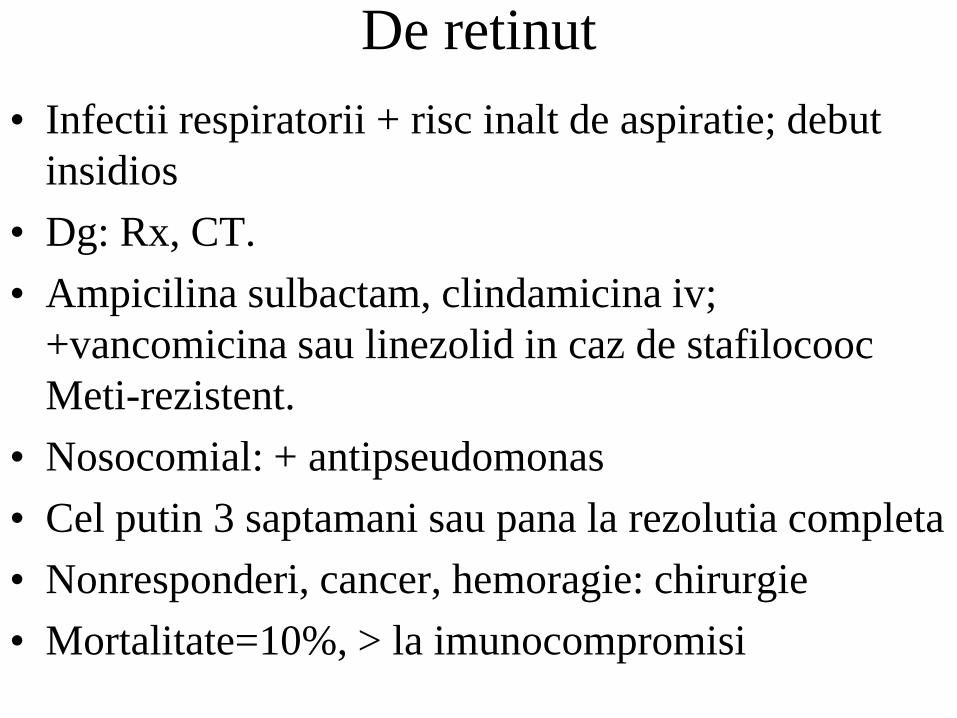
De retinut
• Infectii respiratorii + risc inalt de aspiratie; debut
insidios
• Dg: Rx, CT.
• Ampicilina sulbactam, clindamicina iv;
+vancomicina sau linezolid in caz de stafilocooc
Meti-rezistent.
• Nosocomial: + antipseudomonas
• Cel putin 3 saptamani sau pana la rezolutia completa
• Nonresponderi, cancer, hemoragie: chirurgie
• Mortalitate=10%, > la imunocompromisi

definitie
• Infectie pulmonara cu necroza de parenchim
puroi
– pneumonie necrotizanta
– gangrena pulmonara

clasificare
• acut/cronic - 1 luna
• primar/secundar - conditii asociate
– aspiratie, pacienti anterior sanatosi
– neoplasm, imunodepresie

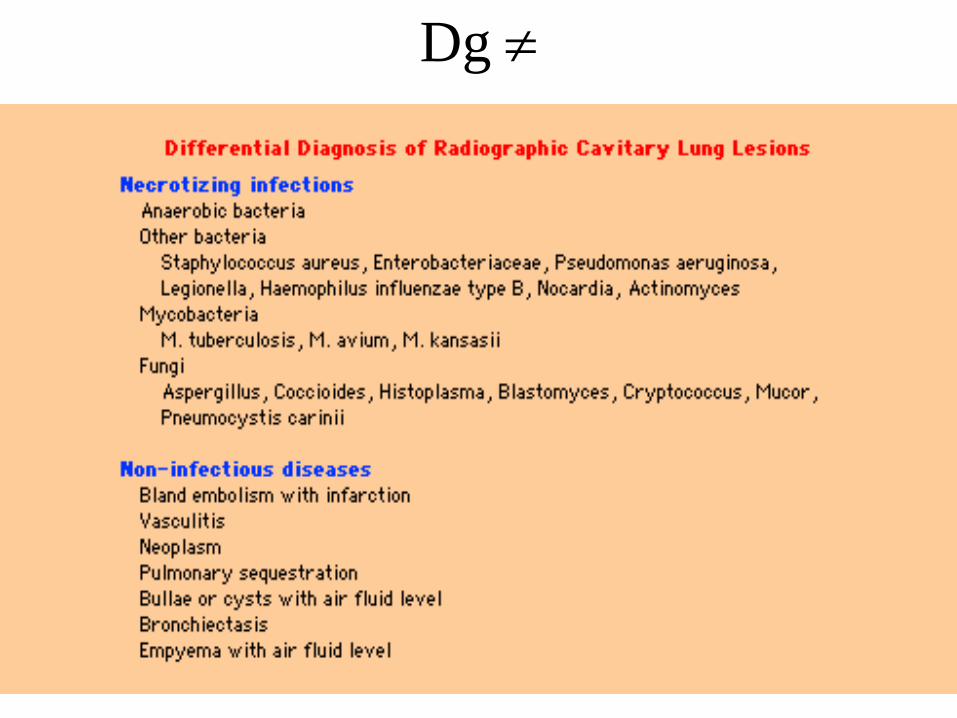
etiologie
• Infectii necrotizante
– bacterii piogene (staph aur, klebsiella, anaerobi,
nocardia)
– mycobacterii
– fungi (coccidioides, histoplasma)
– paraziti (entamoeba hystolitica)

etiologie
• Infarct cavitar
– tromboembolie
– embolie septica (staph aur, candida)
– vasculita (Wegener)
• Neo cavitar
– carcinom bronhogenic
– limfom, metastaze
• Altele
– chist infectat
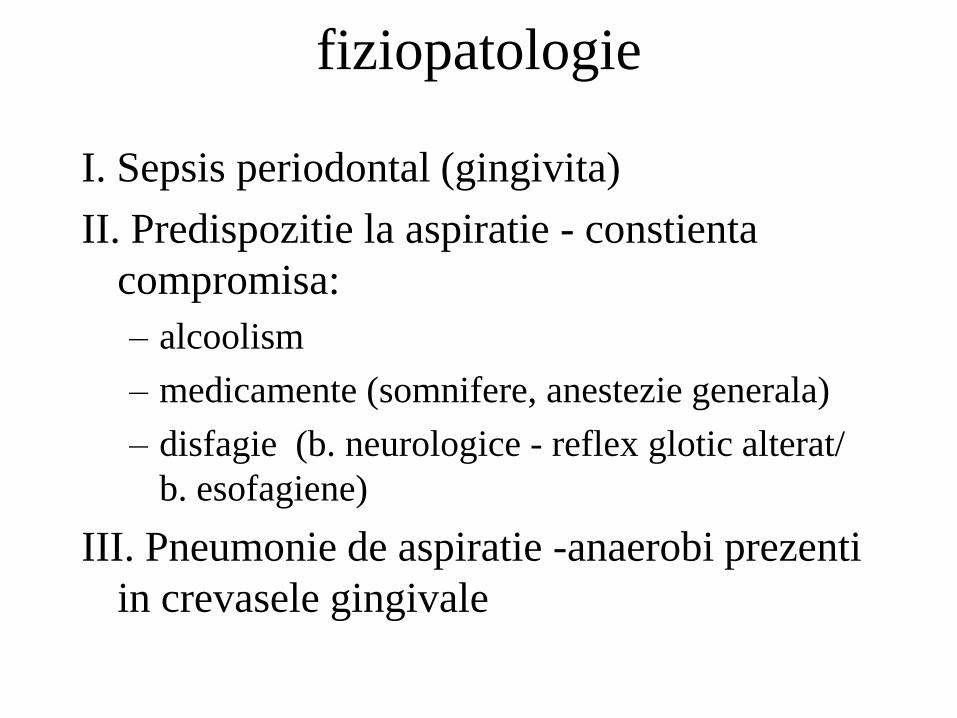
fiziopatologie
I. Sepsis periodontal (gingivita)
II. Predispozitie la aspiratie - constienta
compromisa:
– alcoolism
– medicamente (somnifere, anestezie generala)
– disfagie (b. neurologice - reflex glotic alterat/
b. esofagiene)
III. Pneumonie de aspiratie -anaerobi prezenti
in crevasele gingivale
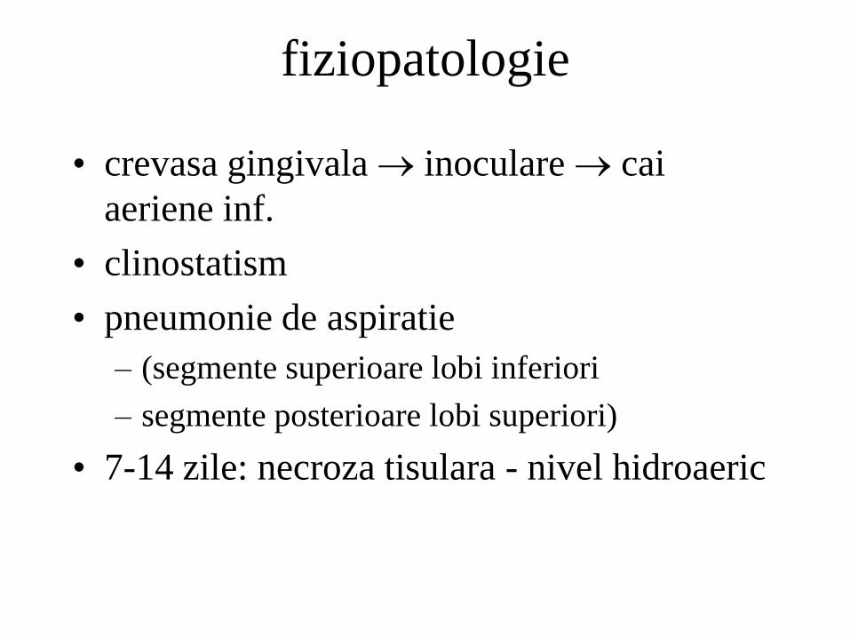
fiziopatologie
• crevasa gingivala inoculare cai
aeriene inf.
• clinostatism
• pneumonie de aspiratie
– (segmente superioare lobi inferiori
– segmente posterioare lobi superiori)
• 7-14 zile: necroza tisulara - nivel hidroaeric

Necroza abces pulmonar
empiem
fistula bronhopleurala
extensie directa a infectiei

Sdr. Lemiérre
Faringe (abces amigdalian/periamigdalian -
Fusobacterium necrophorum)
fuzare gat anterior teaca carotidiana
tromboflebita jugulara interna
emboli septici pulmonari
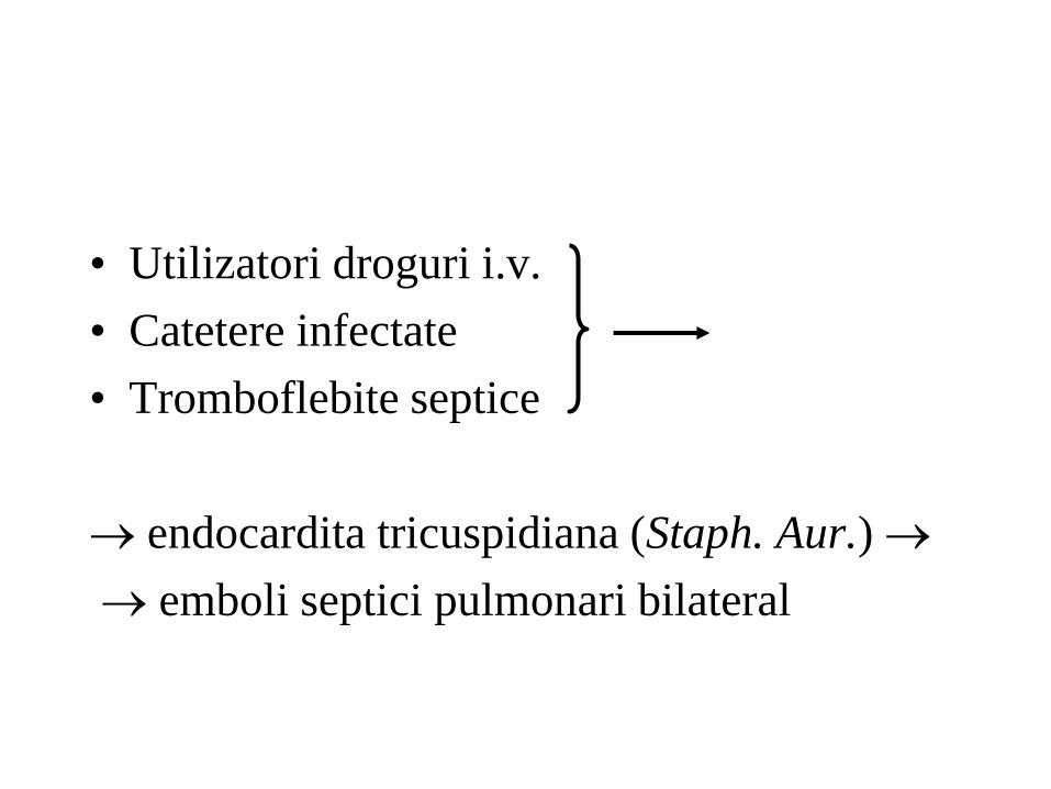
• Utilizatori droguri i.v.
• Catetere infectate
• Tromboflebite septice
endocardita tricuspidiana (Staph. Aur.)
emboli septici pulmonari bilateral

Manifestari clinice
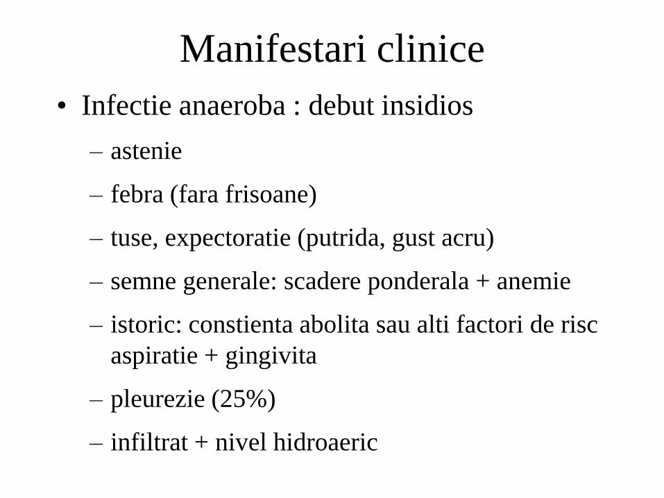
• Infectie anaeroba : debut insidios
– astenie
– febra (fara frisoane)
– tuse, expectoratie (putrida, gust acru)
– semne generale: scadere ponderala + anemie
– istoric: constienta abolita sau alti factori de risc
aspiratie + gingivita
– pleurezie (25%)
– infiltrat + nivel hidroaeric
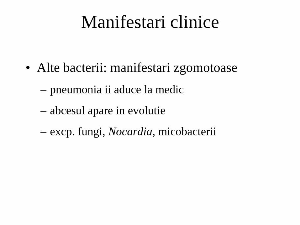
• Alte bacterii: manifestari zgomotoase
– pneumonia ii aduce la medic
– abcesul apare in evolutie
– excp. fungi, Nocardia, micobacterii
Manifestari clinice
Dg
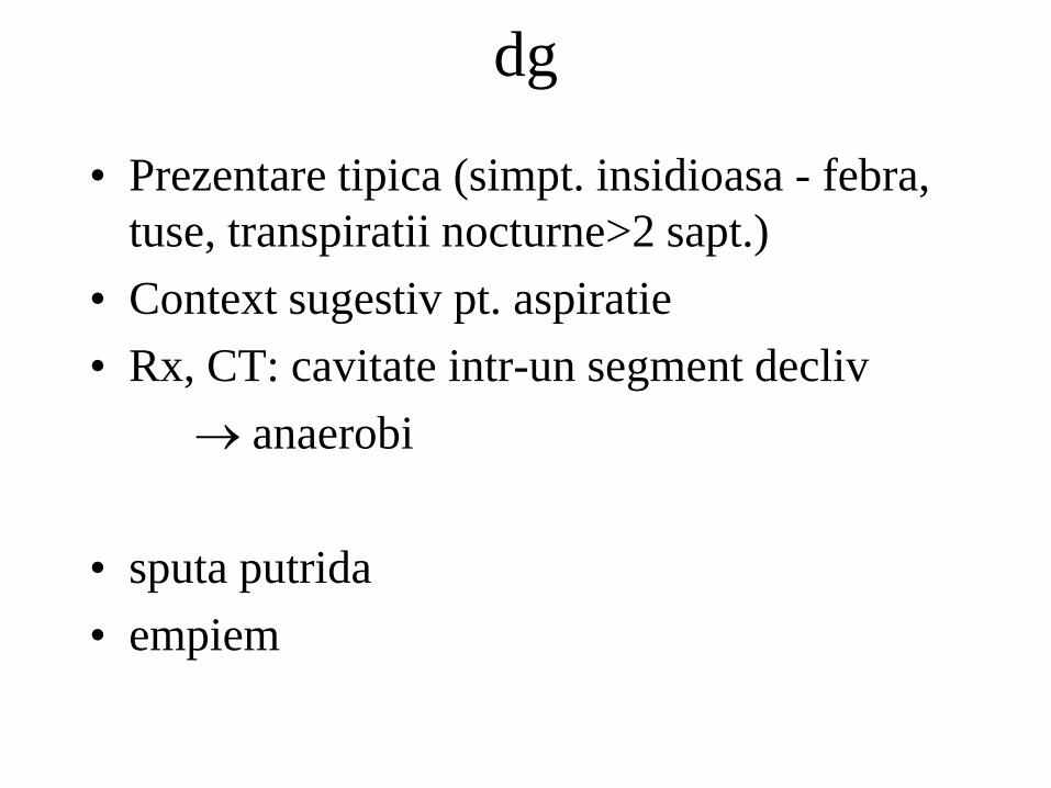
dg
• Prezentare tipica (simpt. insidioasa - febra,
tuse, transpiratii nocturne>2 sapt.)
• Context sugestiv pt. aspiratie
• Rx, CT: cavitate intr-un segment decliv
anaerobi
• sputa putrida
• empiem
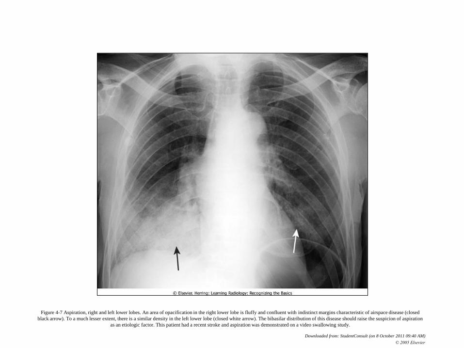
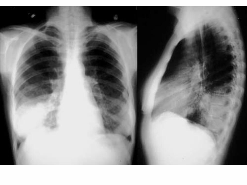
Figure 4-7 Aspiration, right and left lower lobes. An area of opacification in the right lower lobe is fluffy and confluent with indistinct margins characteristic of airspace disease (closed
black arrow). To a much lesser extent, there is a similar density in the left lower lobe (closed white arrow). The bibasilar distribution of this disease should raise the suspicion of aspiration
as an etiologic factor. This patient had a recent stroke and aspiration was demonstrated on a video swallowing study.
Downloaded from: StudentConsult (on 8 October 2011 09:40 AM)
© 2005 Elsevier
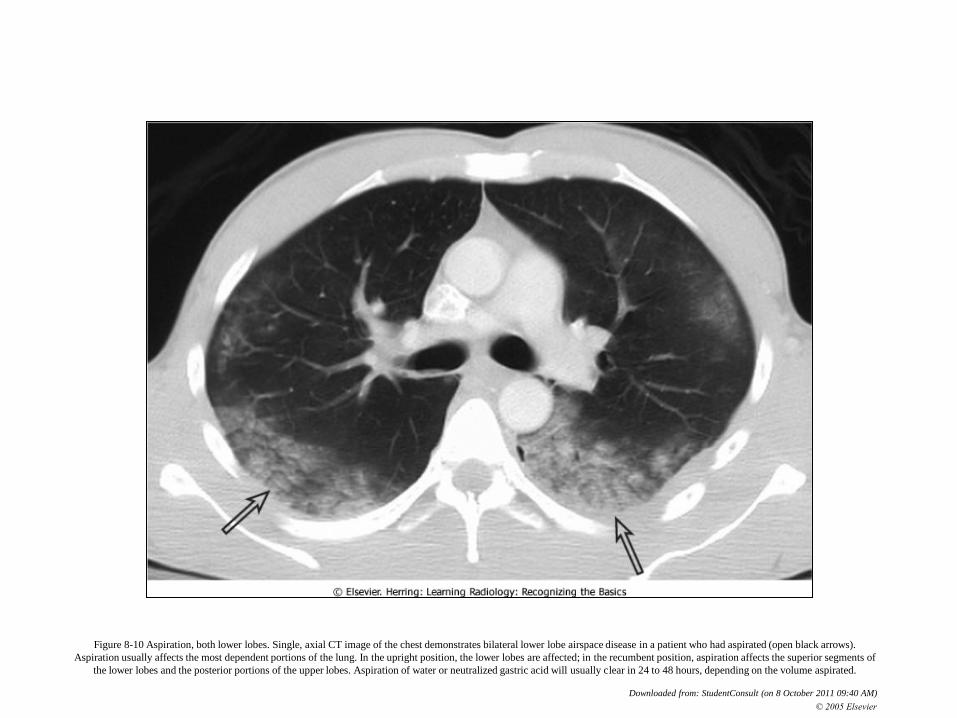
Figure 8-10 Aspiration, both lower lobes. Single, axial CT image of the chest demonstrates bilateral lower lobe airspace disease in a patient who had aspirated (open black arrows).
Aspiration usually affects the most dependent portions of the lung. In the upright position, the lower lobes are affected; in the recumbent position, aspiration affects the superior segments of
the lower lobes and the posterior portions of the upper lobes. Aspiration of water or neutralized gastric acid will usually clear in 24 to 48 hours, depending on the volume aspirated.
Downloaded from: StudentConsult (on 8 October 2011 09:40 AM)
© 2005 Elsevier


• Izolarea bacteriilor anaerobe: dificila
– specimenele din tractul respirator superior sunt
contaminate de flora oro-faringiana
» sputa, aspirate bronhoscopie
– aspirate transtraheale
– aspirate transtoracice
– lichid pleural
– hemoculturi (rar + anaerobi)
– (aspirat bronhoscopic (perie)
– lavaj bronhoalveolar)

• Prezentare mai putin clasica
– excludere TBC
• corp strain aspirat
• neo pulmonar
• stenoza bronsica
bronhoscopie
dg
tratament
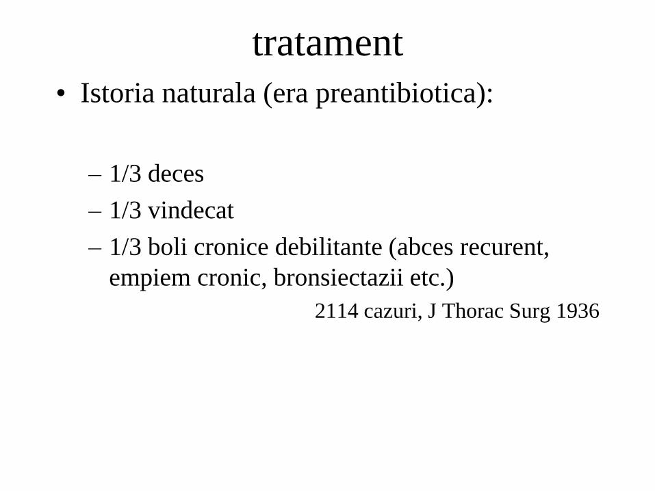
• Istoria naturala (era preantibiotica):
– 1/3 deces
– 1/3 vindecat
– 1/3 boli cronice debilitante (abces recurent,
empiem cronic, bronsiectazii etc.)
2114 cazuri, J Thorac Surg 1936
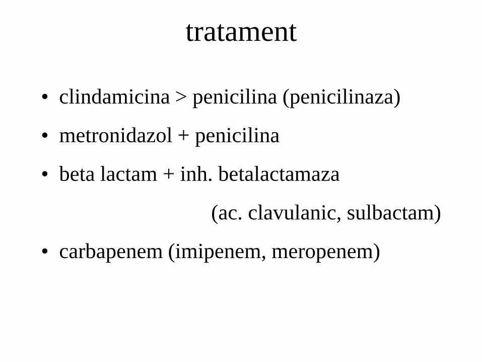
• clindamicina > penicilina (penicilinaza)
• metronidazol + penicilina
• beta lactam + inh. betalactamaza
(ac. clavulanic, sulbactam)
• carbapenem (imipenem, meropenem)
tratament
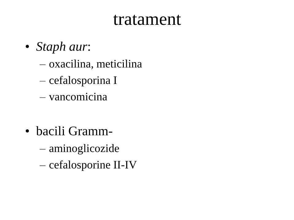
• Staph aur:
– oxacilina, meticilina
– cefalosporina I
– vancomicina
• bacili Gramm-
– aminoglicozide
– cefalosporine II-IV
tratament

Tratament - durata
• Controversata
• 3 sapt. - 6 sapt.
• Pana la disparitia abcesului (2-4 l)

Tratament chirurgical
• neoplasm
• hemoragie importanta
• obstructie bronsica
• refractar la tratament
– obstructie br
– >6cm
– gramm- (P aeruginosa)
– evolutie> 6 sapt. inainte de prezentare
lobectomie, pneumectomie
Risc operator
• Drenaj
– percutan
– endoscopic

Raspuns asteptat
• Imbunatatirea subiectiva a starii generale
• scaderea febrei 3-4 zile
• disparitia febrei 7-14 zile
nu raspunde:
– obstructie br, neoplasm, corp strain
– microb neacoperit cu antibiotic
– cavitate>6 cm, empiem (drenaj)
– cauza neinfectioasa (neoplasm, vasculita)
– febra medicamentoasa
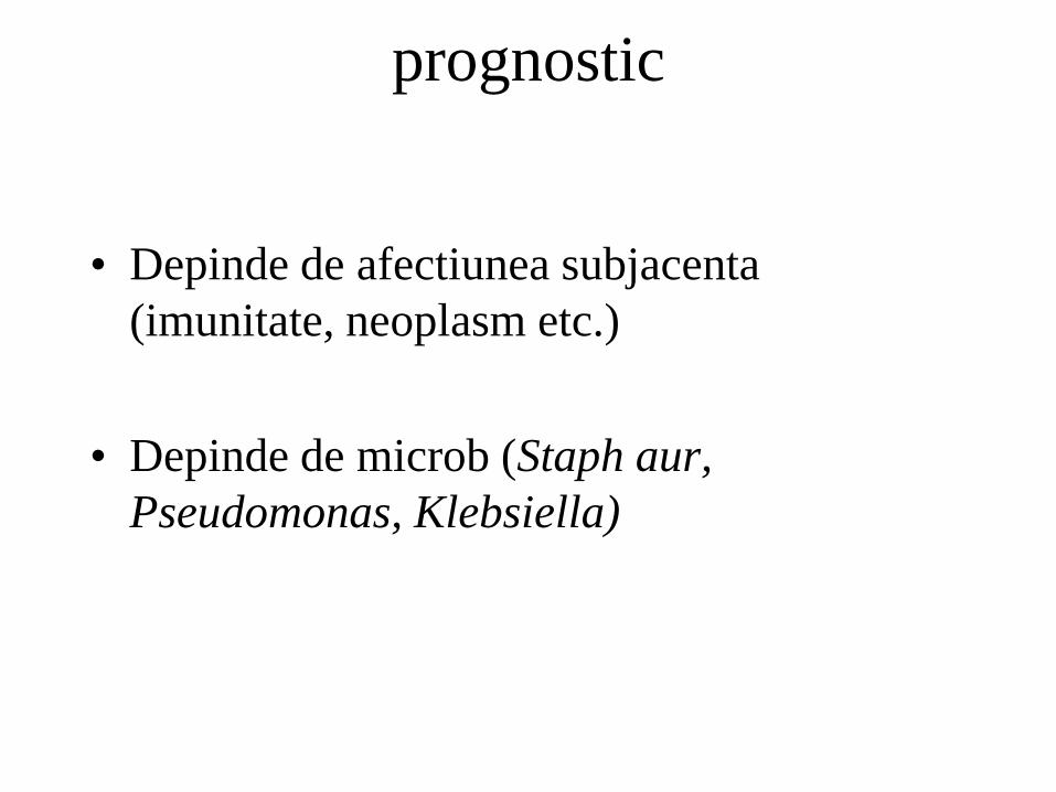
prognostic
• Depinde de afectiunea subjacenta
(imunitate, neoplasm etc.)
• Depinde de microb (Staph aur,
Pseudomonas, Klebsiella)
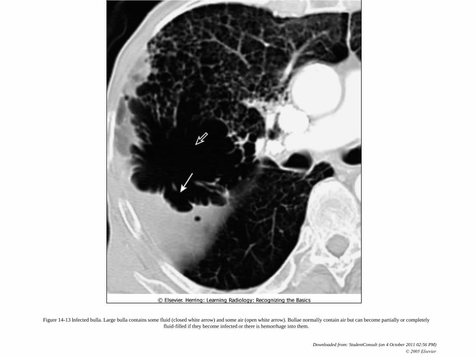
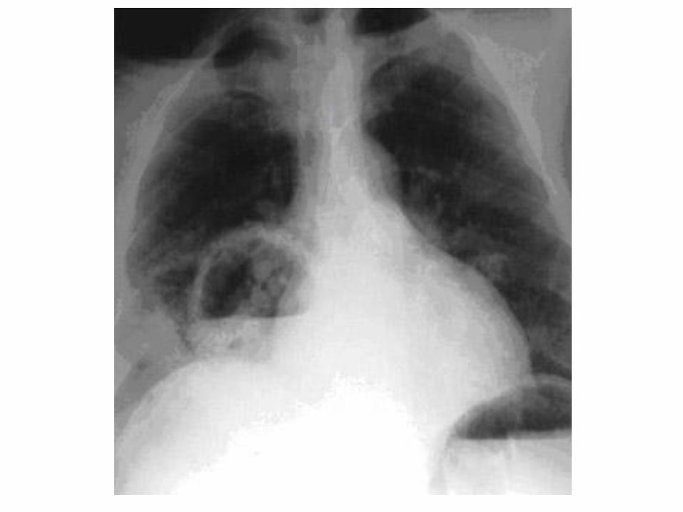
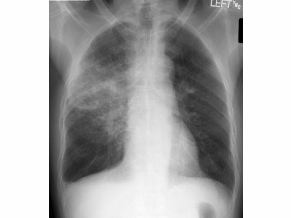
Figure 14-13 Infected bulla. Large bulla contains some fluid (closed white arrow) and some air (open white arrow). Bullae normally contain air but can become partially or completely
fluid-filled if they become infected or there is hemorrhage into them.
Downloaded from: StudentConsult (on 4 October 2011 02:56 PM)
© 2005 Elsevier
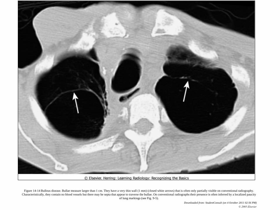
Figure 14-14 Bullous disease. Bullae measure larger than 1 cm. They have a very thin wall (1 mm) (closed white arrows) that is often only partially visible on conventional radiography.
Characteristically, they contain no blood vessels but there may be septa that appear to traverse the bullae. On conventional radiographs their presence is often inferred by a localized paucity
of lung markings (see Fig. 9-5).
Downloaded from: StudentConsult (on 4 October 2011 02:56 PM)
© 2005 Elsevier

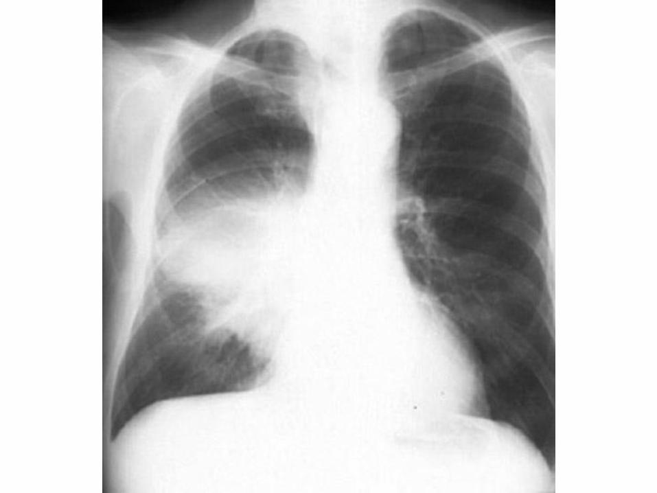
Figure 14-16 Cavitary bronchogenic carcinoma. There is a thick-walled cavitary lesion in the right upper lobe (open white arrow). The inner margin of the cavity is nodular and irregular
(closed black arrow). There is pneumonia surrounding a portion of the mass (closed white arrow). This was a squamous cell carcinoma of the lung.
Downloaded from: StudentConsult (on 4 October 2011 02:56 PM)
© 2005 Elsevier

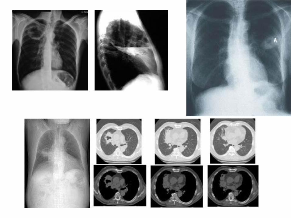
Figure 13-20 Cavitary lesions of the lung. Three of the most common cavitary lesions of the lung can frequently be differentiated from each other by noting the thickness of the wall of the
cavity and the smoothness or nodularity of its inner margin. A, A cavitary squamous cell bronchogenic carcinoma with a thick wall (dotted white arrow) and a nodular inner margin (closed
black arrow); B, upper lobe tuberculosis has a thin-walled cavity with a smooth inner margin (closed white arrow); C, a staphylococcal lung abscess demonstrating a characteristic markedly
thickened wall (open white arrow) and a small but smooth inner margin (open black arrow).
Downloaded from: StudentConsult (on 4 October 2011 02:56 PM)
© 2005 Elsevier

De retinut
• Infectii respiratorii + risc inalt de aspiratie; debut
insidios
• Dg: Rx, CT.
• Ampicilina sulbactam, clindamicina iv;
+vancomicina sau linezolid in caz de stafilocooc
Meti-rezistent.
• Nosocomial: + antipseudomonas
• Cel putin 3 saptamani sau pana la rezolutia completa
• Nonresponderi, cancer, hemoragie: chirurgie
• Mortalitate=10%, > la imunocompromisi

BRONSIECTAZIILE

De retinut
1. Tuse zilnica productiva
2. Dg: CT cu rezolutie inalta
3. In caz de obstructie/ heperreactivitate bronsica:
tratament cu bronhodilatatoare inhalatorii
4. La pacientii cu exacerbari: antibiotice active pe
Haemophilus influenzae si Staphylococcus aureus.
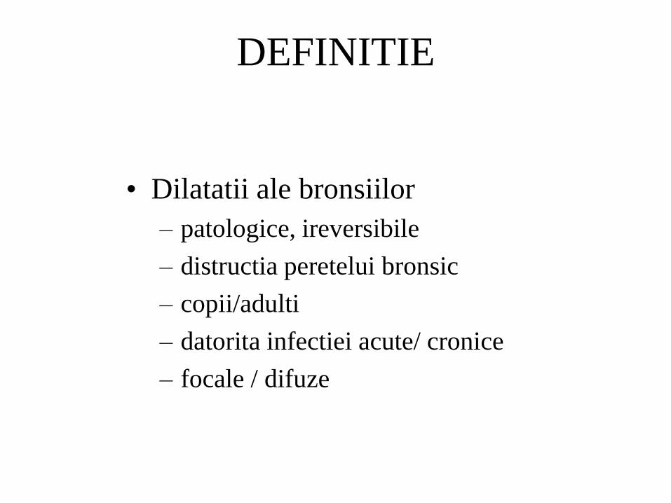
DEFINITIE
• Dilatatii ale bronsiilor
– patologice, ireversibile
– distructia peretelui bronsic
– copii/adulti
– datorita infectiei acute/ cronice
– focale / difuze
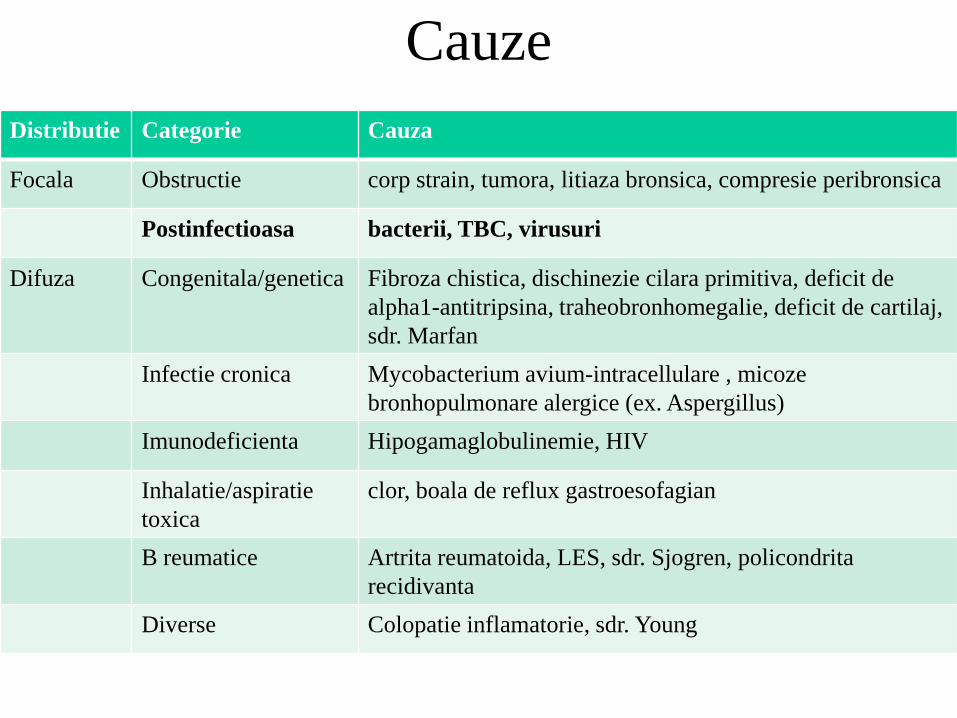
Cauze
Distributie Categorie Cauza
Focala Obstructie corp strain, tumora, litiaza bronsica, compresie peribronsica
Postinfectioasa bacterii, TBC, virusuri
Difuza Congenitala/genetica Fibroza chistica, dischinezie cilara primitiva, deficit de
alpha1-antitripsina, traheobronhomegalie, deficit de cartilaj,
sdr. Marfan
Infectie cronica Mycobacterium avium-intracellulare , micoze
bronhopulmonare alergice (ex. Aspergillus)
Imunodeficienta Hipogamaglobulinemie, HIV
Inhalatie/aspiratie
toxica
clor, boala de reflux gastroesofagian
B reumatice Artrita reumatoida, LES, sdr. Sjogren, policondrita
recidivanta
Diverse Colopatie inflamatorie, sdr. Young

fiziopatologie
Colonizare/infectie bacteriana recurrenta →
activarea mediatorilor inflamatiei (elastaza,
colagenaza) →
distrugerea peretelui bronsic →
proliferarea arteriala + malformatii arteriovenoase →
hemoptizii
prevenire
• Renuntarea la fumat
• Vaccinare antigripala & antipneumococica

Dg clinic
• Tuse cronica zilnica, productiva (purulenta)
– Toti adultii cu bronsiectazii
– 30% au hemoptizii
– Copii: 75% au tuse cu expectoratie, 25% hemoptizie,
50% obstructie br (dispnee)
• Ubeori: ronflante, crackles-uri, degete hipocratice

Dg paraclinic
• RX: ingrosare a peretilor bronsici
• Sn= 13% - 40%, Sp= 95%.
• Spirometria: evaluarea unor afectiuni subjacente
• CT rezolutie inalta: gold standard
• Diametru bronsic marit (bronhia>artera = inel cu
pecete)
• Exacerbari: culturi sputa daca nu apare ameliorare
dupa antibioterapia empirica

Abordarea pacientului
• Pacient cu tuse cronica, productiva:
– De exclus intai BRGE, astmul, picurare postnazala
(postnasal drip).
• Apoi: CT rezolutie inalta.
• Odata dg pus (bronsiectazii), de cautat cauza

tratament
• Exacerbari: atb active pe Haemophilus influenzae,
Staphylococcus aureus si Pseudomonas.
• Obstructie bronsica/hiperreactivitate:
bronhodilatatoare inhalatorii.
• Fizioterapie toracica.
• Bronsiectazii localizate: bronhoscopie / chirurgie.
Tratament de fond
• Bronhodilatatoare.
• Mucolitice? (bromhexin, ACC).
• ATB t lung: reduc vol si purulenta sputei; nu
modifica exacerbarile sau evol naturala a bolii.

Trat. exacerbarilor
• ATB pt 2 saptamani
• Active contra Haemophilus influenzae si
Staphylococcus aureus.
• Fibroza chistica sau ATB recent: risc pt infectie cu
Pseudomonas aeruginosa.
• Raspuns initial inadecvat: culturi sputa, tratament
mai lung.

Trat chirurgical
• Simptome severe, fara raspuns la tratament, afectare
locala.
• Bronsiectazii focale dat obstructiei bronsice:
bronhoscopie, chirurgie.

reabilitare
• Antrenarea mm inspiratori pt a creste capacitatea de
efort.
• Pacientii ce nu pot expectora adecvat: fizioterapie
toracica (percutie, vibratie, drenaj postural).
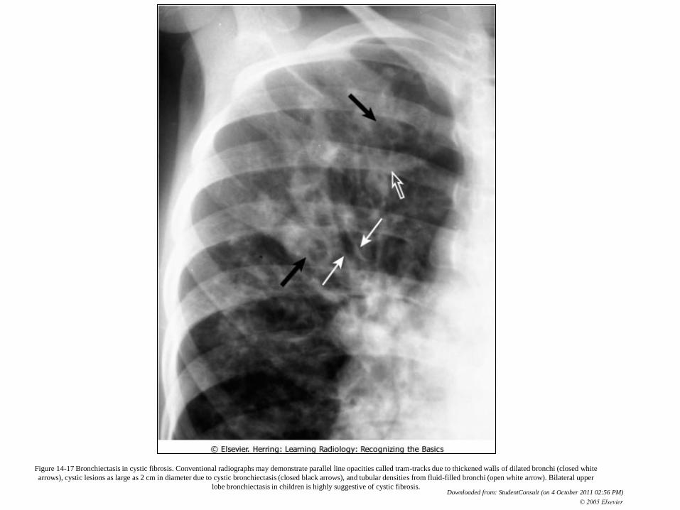
Figure 14-17 Bronchiectasis in cystic fibrosis. Conventional radiographs may demonstrate parallel line opacities called tram-tracks due to thickened walls of dilated bronchi (closed white
arrows), cystic lesions as large as 2 cm in diameter due to cystic bronchiectasis (closed black arrows), and tubular densities from fluid-filled bronchi (open white arrow). Bilateral upper
lobe bronchiectasis in children is highly suggestive of cystic fibrosis. Downloaded from: StudentConsult (on 4 October 2011 02:56 PM)
© 2005 Elsevier
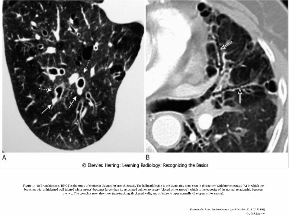
Figure 14-18 Bronchiectasis. HRCT is the study of choice in diagnosing bronchiectasis. The hallmark lesion is the signet ring sign, seen in this patient with bronchiectasis (A) in which the
bronchus with a thickened wall (dotted white arrows) becomes larger than its associated pulmonary artery (closed white arrows), which is the opposite of the normal relationship between
the two. The bronchus may also show tram-tracking, thickened walls, and a failure to taper normally (B) (open white arrows).
Downloaded from: StudentConsult (on 4 October 2011 02:56 PM)
© 2005 Elsevier
De retinut
1. Tuse zilnica productiva
2. Dg: CT cu rezolutie inalta
3. In caz de obstructie/ heperreactivitate bronsica:
tratament cu bronhodilatatoare inhalatorii
4. La pacientii cu exacerbari: antibiotice active pe
Haemophilus influenzae si Staphylococcus aureus.