activation of serum response factor (srf) in the ... · egr-1 in insulinoma cell lines and...
TRANSCRIPT

Activation of Serum Response Factor (SRF) in the Depolarization Induction of Egr-1
Transcription in Pancreatic Islet β-cells
ERNESTO BERNAL-MIZRACHI, BURTON WICE, HIROSHI INOUE, AND M. ALAN PERMUTT
WASHINGTON UNIVERSITY SCHOOL OF MEDICINE
All authors can be contacted at the following address:
Washington University School Of Medicine
Division of Endocrinology, Diabetes and Metabolism
Campus Box 8127
660 S. Euclid Ave.
St. Louis, Missouri 63110
Address correspondence to:
Dr. Ernesto Bernal-Mizrachi
Washington University School Of Medicine
Division of Endocrinology, Diabetes and Metabolism
Campus Box 8127
660 S. Euclid Ave.
St. Louis, Missouri 63110
Telephone: (314) 362-8685
Fax: (314) 747-2692
E-mail: aplab1@ imgate.wustl.edu
RUNNING TITLE
EGR-1 transcription in pancreatic islet β-cells
-cellsβEGR-1 transcription in pancreatic islet
1
Copyright 2000 by The American Society for Biochemistry and Molecular Biology, Inc.
JBC Papers in Press. Published on May 26, 2000 as Manuscript M003424200 by guest on M
arch 26, 2020http://w
ww
.jbc.org/D
ownloaded from
SUMMARY
The results of the current studies define the major elements whereby glucose metabolism in islet
β-cells leads to transcriptional activation of an early response gene in insulinoma cell lines and in rat islets.
Glucose stimulation (2-20 mM) resulted in a 4-fold increase in Egr-1 mRNA at 30 minutes, as
did the depolarizing agents KCl and tolbutamide. This response was inhibited by diazoxide and EGTA,
indicating that β-cell depolarization and Ca2+ influx respectively are essential. Pharmacological
inhibition of the Egr-1 induction by H89 (48%) and calmidazolium (35%), but not by MEK1/MEK2 or
PI3 kinase inhibitors, implied that protein kinase A and Ca2+/calmodulin pathways are involved. Deletion
mapping of the Egr-1 promoter revealed that the proximal -198 base pairs containing two serum
response elements (SREs) and one cAMP response element (CRE) retained the depolarization response.
Depolarization resulted in phosphorylation of CREB, yet partial inhibition by a dominant negative CREB,
along with a robust response of a CRE mutated Egr-1 promoter suggested the presence of a second
Ca2+-responsive element. Depolarization activation of 5XSRE-LUC and serum response factor SRF-GAL4
constructs, along with activation of SRF-GAL4 by co-transfection with constitutively active CAMKIV
and PKA, and binding of Ser103-phosphorylated SRF in nuclear extracts, indicated that the SRE/SRF
complexes contribute to the Ca2+-mediated transcriptional regulation of Egr-1. The results of the current
experiments demonstrate for the first time SRE-dependent transcription and the role of SRF, a
transcription factor known to be a major component of growth responses, in glucose-mediated
transcriptional regulation in insulinoma cells.
-cellsβEGR-1 transcription in pancreatic islet
2
by guest on March 26, 2020
http://ww
w.jbc.org/
Dow
nloaded from

INTRODUCTION
Pancreatic islet β-cell mass can be regulated by multiple stimuli, including growth factors and
nutrients. The ability to produce insulin is proportional to the islet mass that is defined by a balance
between rates of proliferation and apoptosis. Several growth factors have been shown to stimulate
pancreatic β-cell replication (1;2). During pregnancy insulin resistance results in a marked increase in β-cell
mass, and growth hormone and prolactin treatment have been shown to increase β-cell proliferation (5).
Other growth factors, including insulin-like growth factor 1 (IGF-1), hepatocyte growth factor and nerve
growth factor, have also been implicated in β-cell mitogenesis and differentiation (1,6-8). Nutrients
including glucose and certain essential amino acids have been shown to increase β-cell proliferation and
increase islet β-cell mass (1). In insulin resistance states such as obesity for example, impaired insulin
mediated glucose disposal results in increased plasma glucose levels and this mechanism is thought to be
essential for the hyperinsulinemic response of β-cells.
Glucose induces pleiotropic effects in islet β-cells and these effects are potentially mediated by
activation of multiple intracellular signaling pathways. Glucose metabolism results in alteration of
ATP/ADP levels leading to closure of nucleotide regulated K+ channels (KATP). KATP channel
inhibition results in depolarization and activation of voltage dependent Ca2+ channels. Ca2+ influx has
been implicated in the activation of CaM kinases II and IV, protein kinases A (PKA) and C (PKC),
extracellular regulated kinase (ERK) mitogen-activated protein kinase (MAPK), and
phosphatidylinositol 3-kinase (PI3-K) in islet β-cells (12). The relationships between activation of these
pathways and growth responses induced by glucose have not been defined.
Changes in gene expression profiles that result from the activation of these signaling pathways
are responsible for the adaptation of β-cell mass to physiological and pathological states. The specific
expression profile includes the induction of immediate early genes (IEGs). IEGs are rapidly activated by
initiation of signal transduction pathways, and their activation does not require protein synthesis (13). The
-cellsβEGR-1 transcription in pancreatic islet
3
by guest on March 26, 2020
http://ww
w.jbc.org/
Dow
nloaded from
signaling pathways for induction of IEGs exhibit considerable stimulus and tissue specificity and in
general involve activation of kinase/phosphatase cascades (13). Often, IEGs are transcription factors that in
turn activate expression of down-stream target genes generating distinct biological responses by inducing
specific long-term programs of gene expression (13). The IEG responses are not restricted to genes that
control cell cycle reentry but also include genes that participate in other biological processes. IEGs are
universally expressed, and the signal transduction pathways leading to activation of these genes have been
extensively studied in numerous tissues.
Glucose and other nutrients have been shown to transcriptionally activate several IEGs, including
Egr-1 in insulinoma cell lines and pancreatic islets, in a Ca2+-dependent manner (14;15). Egr-1 is a zinc-
finger DNA-binding transcription factor that is expressed in multiple tissues and is induced by diverse
stimuli of which the best studied examples have been the treatment with peptide mitogens (16-18). Egr-1 mRNA
has been shown to be increased by treatment with glucose, cAMP and several gluco-incretins in
insulinoma cells and rat islets (14;15). The promoter elements involved in Egr-1 transcription by depolarization
have not been defined. The genomic 5’-flanking sequence of the murine Egr-1 contains activator
protein-1 (AP-1) sites, a cAMP-responsive element (CRE) and serum response elements (SRE). Of
these elements only the CRE has been implicated in Ca2+ induction of the glucagon gene in pancreatic
β-cells (19). CRE was also essential for the induction of c-fos by the combination of glucose and
glucoincretins in INS1 cells (19). To our knowledge, nothing is known about SRE-dependent transcription of
any gene in pancreatic islet β-cells.
In the current studies, we sought to define the mechanisms whereby glucose induced
depolarization and Ca2+ influx activates Egr-1 transcription in insulinoma cells. We describe the signal
transduction pathways, trans-activating factors, and the cis-regulatory elements involved in this process.
We demonstrate that β-cell depolarization triggers a cascade of events in which Ca2+ influx leads to the
activation of PKA and Ca2+/calmodulin (CaM). The initiation of these signaling pathways leads to
-cellsβEGR-1 transcription in pancreatic islet
4
by guest on March 26, 2020
http://ww
w.jbc.org/
Dow
nloaded from
activation of CREB and SRF, a transcription factor involved in growth responses. This activation results
in Egr-1 induction predominantly through five SRE elements in its proximal promoter. These studies for
the first time link the potential role of SRE-dependent transcription as a mechanism for glucose effect in
pancreatic β-cell mass.
-cellsβEGR-1 transcription in pancreatic islet
5
by guest on March 26, 2020
http://ww
w.jbc.org/
Dow
nloaded from

EXPERIMENTAL PROCEDURES
Chemicals
Phorbol 12-myristate 13-acetate (TPA), potassium chloride, 3-O-methyl glucose, 2-
deoxyglucose, mannose, forskolin (FK), diazoxide, cycloheximide, actinomycin D, EGTA, D-mannitol
and sucrose were obtained from Sigma (St. Louis, MO, USA). PD98059, H89, calmidazolium,
staurosporin, LY294002 and wortmannin were obtained from Biomol (Plymouth Meeting, PA).
Cell culture conditions
The MIN6 insulinoma cell line was obtained from Y. Oka (20) and was maintained in Dulbecco’s
modified Eagle’s medium (DMEM) containing 25mM glucose and supplemented with 15% heat-
inactivated fetal bovine serum (FBS) in humidified 5% CO2-95% air at 37oC. βTC6-F7 insulinoma
cells, obtained from S. Efrat (Albert Einstein College of Medicine) were cultured in DMEM containing
25 mM glucose and supplemented with 15% horse serum and 2.5% FBS. βTC6-F7 and MIN6 cells were
used between passages 16 to 42 and 26 to 35, respectively. For the analysis of expression, cells that were
70-80% confluent were switched to regular DMEM media containing 2mM glucose for 20-24 hours
(time zero of the experiments). The indicated agent was added directly to the media and cells were
incubated as described in the figure legends, followed by a 45-minute incubation in the same media
containing the indicated agents. Pharmacologic inhibitors were added 60 minutes prior to stimulation of
the cells with glucose or other agents, using the concentrations of inhibitors given in the figure legends.
Northern Blot Analysis and RT-PCR
Total cellular RNA was isolated by the acid guanidium thiocyanate-phenol-chloroform method
of Chomczynski and Sacchi (21). Twenty µg of total RNA were fractionated by electrophoresis through a
1.1% agarose gel and transferred to a nylon membrane by the capillary blotting technique. RNA was
cross-linked by UV irradiation. The cDNA probes were labeled with [32P] dCTP using the High-Prime
Labeling System (Boehringer Mannheim). Prehybridization (2-4 hours) and hybridization were carried
-cellsβEGR-1 transcription in pancreatic islet
6
by guest on March 26, 2020
http://ww
w.jbc.org/
Dow
nloaded from
out at 56 °C in Church solution (22). Hybridized blots were washed once with 0.1X SSC and 1% SDS at room
temperature for 15 minutes and twice at 54°C for 15 minutes. The membranes were exposed overnight at
–80°C using intensifying screens. Messenger RNA levels were quantitated by phosphorimager scanning
(Molecular Dynamics) and were normalized to those of β-actin. For RT-PCR, total RNA was reverse
transcribed using the Superscript Preamplification System and random primers (Gibco-BRL). PCR was
done according to the manufacturer’s protocol with addition of [32P] dCTP to the PCR mixture. PCR
products were separated on 5% polyacrylamide gels and exposed to X-ray films. Results were
normalized to those of β-actin. The primers for Egr-1, Egr-3 and NGFI-C have been described
previously (23;24).
Islets of Langerhans mRNA analysis
Primary islets of Langerhans were isolated from adult male Sprague-Dawley rats by standard
collagenase digestion (25). Pancreatic islets were cultured in RPMI media containing 5mM glucose for 48
hours and stimulated by adding glucose to a final concentration of 25mM for 45 minutes.
Plasmids
Plasmids containing the rat Egr-1, Egr-2, Egr-3 and NGFI-C cDNAs were kindly provided by
Dr. J. D. Milbrandt (Washington University School of Medicine). The plasmids containing the murine
Egr-1 promoter linked to the luciferase vector pXP2 constructs were generously provided by Dr. David
Cohen (Oregon Health Sciences University) and have been described in detail previously (26). The pmEgr-
1200 contains 1.2 kb of 5’- flanking region of the mouse Egr-1 gene (EMBL accession number
X12617). The pmEgr-510 construct included sequences -511 to -1013 bp; pmEgrAP1 construct contains
nucleotides -1 to -386 plus -886 to -1000 (from sequence X12617). Plasmids containing nucleotides -
532 to +100 (prEgr-532), -198 to +100 (prEgr-198) and -109 to +100 (prEgr-109) of the rat gene
subcloned into the luciferase reporter plasmid pGL2 (Promega) were obtained from J. Milbrandt. The cis-
reporter plasmid pSRE-luc (5XSRE-luc) contains the luciferase reporter gene driven by a basic promoter
-cellsβEGR-1 transcription in pancreatic islet
7
by guest on March 26, 2020
http://ww
w.jbc.org/
Dow
nloaded from
element (TATA box) joined to five tandem repeats of the c-fos SRE and CRE, respectively (Stratagene).
The CREB-GAL4 system includes a trans-activator plasmid that expresses a fusion protein containing
the activator domain of CREB fused to the DNA binding domain of GAL4 (residues 1-147) (CREB-
GAL4) (Stratagene), cotransfected with a reporter gene containing a synthetic promoter with five tandem
repeats of the yeast GAL4 binding sites that control expression of the luciferase gene pRC-LUC
(Stratagene). The SRF-GAL4 fusion vector and a constitutively active form of CaM kinase IV
(CaMKIVCT) were a gift from Dr. Michael Greenberg and have been described previously (27). The plasmid
containing the catalytic unit of PKA (pFC-PKA) was purchased from Stratagene. The KCREB construct
was kindly provided by Dr. Richard Goodman and has been described previously (28). Briefly, KCREB
(“killer” CREB) contains an amino acid substitution within the leucine zipper that results in KCREB-
CREB heterodimers with no DNA-binding function. The pRL-TK control vector contains the herpes
simplex virus thymidine kinase promoter upstream of Renilla luciferase (Promega).
Transient Transfections
MIN6 cells were transfected by Lipofectamine and Plus Reagent (Gibco-BRL) using the
suggested amounts of DNA according to the manufacturer’s protocol. Briefly, 1X10⁶ cells were plated in
6-well plates 4 days prior to transfection. Cells at about 80% confluence were transfected by mixing the
indicated amount of DNA described in the figure legends and a lipid mixture containing a 1:2 ratio of
Lipofectamine and Plus Reagent in 1 ml of OPTI-MEM media (Gibco-BRL). Following 4 hours of
incubation, 1 ml of DMEM media containing 5mM glucose and 2% serum was added to the cells. After
12 hours, the medium-DNA complexes were replaced by pre-incubation media containing DMEM with
5mM glucose-2% FBS and the cells were left for 24 hours. At the end of the 24 hours the specific
stimulating agent was added to the media and the cells were harvested 6 hours later. For the
overexpression experiments with pFC-PKA and CaMKIVCT, MIN6 cells were transfected as above,
followed by incubation in DMEN containing 5 mM glucose and 2% FBS for 30 hours until harvesting.
Total DNA was maintained constant in all the transfection experiments by using the empty vector of the
-cellsβEGR-1 transcription in pancreatic islet
8
by guest on March 26, 2020
http://ww
w.jbc.org/
Dow
nloaded from

respective cDNA to be overexpressed. To correct for differences in transfection efficiencies, 1 ng of
pRL-TK Renilla luciferase plasmid was simultaneously transfected. All results are normalized for
transfection efficiency and expressed as the ratio of firefly to Renilla luciferase.
Electrophoretic Mobility Shift Assay (EMSA)
The sense strand sequences of the oligonucleotides used are:
c-fos SRE, GGATGTCCATATTAGGACATCTGC;
c-fos mutSRE, ACAGGATGTCCATATTATGACATCTGC;
Egr-1SRE#5 (most distal), GAAACGCCATATAAGGAGCAGGAAGGATCC;
Egr-1SRE#4, GGAAACAGACCTTATTTGGGCAGCGCC;
Egr-1SRE#3, GGGCAGCGCCTTATATGGAGTGGCCCA;
Egr-1SRE#2, CCTTCCTTCCATATAGGGCTTCC;
Egr-1SRE#1, CCTGCTTCCCATATATGGCCATGT;
Egr-1 CRE, GAGGACCCGGAGTGACGTGAAGCCC;
Egr-1 mutCRE, GAGGACCCGGAGTGAAGTGAAGCCC;
Nuclear extracts from MIN6 cells cultured under regular DMEN containing 25mM glucose were prepared
essentially as described previously (29), and protein content was determined using the Bio-Rad Protein Assay
kit with bovine serum albumin as a standard. Lysates (5µg of protein) were incubated in final volume of
20µl containing binding buffer (final concentrations: 4% glycerol, 1mM MgCl2, 0.5 mM dithiothretinol, 50mM
NaCl, 10mM Tris-HCl, pH7.5), 200ng poly(dI-dC) and double stranded γ32P oligonucleotide. Binding
reactions proceeded at room temperature for 30 min and were analyzed by non denaturing polyacrylamide
electrophoresis followed by autoradiography. In assays where antibodies directed against SRF (1:10) (Santa
Cruz Biotechnology, Inc., Santa Cruz, CA) and SRF phosphorylated at Ser-103 (1:10) were tested, these
were pre-incubated with the nuclear extracts 20 min at room temperature (30).
-cellsβEGR-1 transcription in pancreatic islet
9
by guest on March 26, 2020
http://ww
w.jbc.org/
Dow
nloaded from
Luciferase Assay
Cell lysis was performed using 400 µl of Passive Lysis Buffer (Promega). Firefly and Renilla
luciferase were measured by the Dual-Luciferase Reporter Assay System (Promega) using 20 µl of cell
lysate. Luciferase activity was measured in a Monolight 3010 luminometer.
In Vitro Mutagenesis
Because of the absence of an f1 origin in pmEgr-1200, the pmEgr1.2@CRE construct was made
by subcloning the 1.2-kb insert from pmEgr1.2 into pBluescript SK(+). Mutagenesis was performed in
the pBluescript SK(+) construct and the mutated insert was then subcloned back into the original
luciferase reporter vector PXP2. The CRE mutation was performed by oligonucleotide-directed
mutagenesis without the phenotypic selection method (31). In brief, single-stranded uracil-containing DNA
template was obtained by transforming CJ236 and growing overnight in TY media with lambda phage,
followed by PEG/NaCl and ethanol precipitation. Two kinased oligonucleotides that produced a CRE
mutation proven to be effective in other promoters was used: 5’ GGA TGG GAG GGC TCA TCA CAA
CTC CGG GTC CTC (mutated nucleotides are underlined) (32). Double-stranded template was obtained by
primer extension and T4 DNA ligase. Finally, Jm109 competent cells were transformed with 1 µl of the
primer extension reaction. The presence of the mutation was confirmed by sequencing.
Western Blot
Cells were lysed with buffer containing 1X PBS, 0.1% SDS, 0.01 M DTT and ½ tablet of
"Complete" protease inhibitor cocktail (Boehringer Mannheim). After boiling, proteins were separated by
electrophoresis though 10% polyacrylamide, 0.1% SDS gels and transferred to PVDF membranes.
Membranes were incubated overnight at room temperature in blocking buffer containing 0.2% I-Block
(Tropix) and 1:1000 tween 20. Subsequently, the membranes were hybridized at 4oC overnight in
blocking buffer containing the CREB and the phospho-CREB antibody with the dilutions recommended
by the manufacturer (New England Biolabs). After 3 washes at room temperature the membranes were
-cellsβEGR-1 transcription in pancreatic islet
10
by guest on March 26, 2020
http://ww
w.jbc.org/
Dow
nloaded from
incubated in secondary HRP antibody for 1 hour. After washing for 1 hour, immunodetection was
performed with ECL Western Blotting Detection System (Amersham) following the manufacturer’s
protocol.
-cellsβEGR-1 transcription in pancreatic islet
11
by guest on March 26, 2020
http://ww
w.jbc.org/
Dow
nloaded from
RESULTS
Induction of Egr-1 mRNA by glucose in βTC6-F7 and MIN6 insulinoma cell lines and in
rat islets
To establish a model system to study the transcriptional regulation of Egr-1 in pancreatic β-cells,
insulinoma cell lines that have been shown to secrete insulin in response to glucose in the physiological
range were investigated (20). Following pre-incubation of βTC6-F7 cells for 20 hours in 2mM glucose,
addition of glucose to a final concentration of 25 mM resulted in increased Egr-1 mRNA levels between
30 to 60 minutes (4 fold p<0.01) that returned to basal levels by 2 hours (Fig 1A). Similar responses to
glucose stimulation were also noted in MIN6 insulinoma cells and in primary cultures of isolated rat
pancreatic islets (data not shown), and these responses were shown to be glucose dose-dependent (Fig
1B). The half maximum response of Egr-1 transcription is in the same range as that observed for insulin
secretion in this cell line (20). Egr-2 (NGFI-B or Krox 20) mRNA, but not Egr-3 mRNA, was also induced
by glucose under the same conditions (data not shown). Mannose, the metabolizable epimer in position 2
of glucose, increased expression of Egr-1 mRNA, while non-metabolizable sugars 2-deoxyglucose or
3-O-methylglucose had no effect (data not shown). These results are similar to those previously reported
and confirm that glucose metabolism is required for Egr-1 induction (15).
Glucose metabolism is known to have several effects, the major one being depolarization through
inhibition of KATP channels. Egr-1 mRNA was induced in insulinoma cells by tolbutamide and KCl,
agents known to depolarize islet β-cells, with the same time-course as that following glucose stimulation
(data not shown), and this induction was Ca2+-dependent (Fig 1C). To determine whether glucose
induction of Egr-1 resulted solely from inactivation of KATP channels, or additionally through increased
glycolytic intermediates, insulinoma cells (βTC6-F7) were treated with glucose in the presence of
diazoxide, an agent that hyperpolarizes the cell through activation of islet KATP channels. Treatment
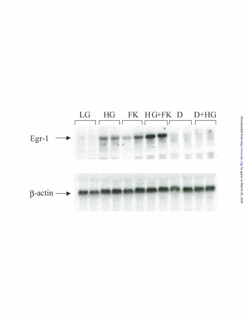
with this agent resulted in complete inhibition of Egr-1 mRNA induction (Fig 1D), indicating that this
-cellsβEGR-1 transcription in pancreatic islet
12
by guest on March 26, 2020
http://ww
w.jbc.org/
Dow
nloaded from
response to glucose was solely dependent on depolarization and subsequent Ca2+ influx. Activation of
PKA by cAMP has been shown to be one of the major regulators of c-fos transcription in INS-1
insulinoma cells (19). In the current experiments, to evaluate the effect of PKA activation on Egr-1, MIN6
cells were treated with forskolin. This treatment resulted in similar induction of Egr-1 mRNA, and the
response to both stimuli was greater than that to either alone (Fig 1D). [INSERT Fig 1A, 1B, 1C, 1D]
Effects of pharmacological agents on glucose induction of Egr-1 mRNA expression
Specific pharmacological inhibitors were utilized to begin to define the signal transduction
pathways involved in glucose and depolarization induction of Egr-1. The CaM and PKA pathways were
assessed by addition of the CaM inhibitor calmidazolium (10 µM) or the PKA inhibitor H89 (20 µM) (33;34).
Calmidazolium and H89 independently reduced the glucose induction of Egr-1 mRNA to 70% (p<0.01)
and to 56% (p<0.01) respectively (Table 1). Simultaneous inhibition of the PKA and CaM pathways
resulted in a combined suppression to 40% of control (p<0.001). Glucose has been shown to activate the
ERK1/ERK2 mitogen activated protein kinase (MAPK) pathway in a Ca2+-dependent manner (9;10). The
specific MEK1 and MEK2 inhibitor PD98059 (100 µM) used at concentrations known to inhibit these
enzymes in other systems had no significant effect on Egr-1 mRNA induction by glucose (34-37) (Table
1). Staurosporin, a known inhibitor of PKC, did not inhibit Egr-1 induction. Interestingly, both PI3 kinase
inhibitors wortmannin and LY294002 resulted in small but significant (p<0.01) enhancements of Egr-1
mRNA induction. Taken together, these results suggested that glucose induction of Egr-1 is a Ca2+-
dependent process and that the Ca2+ effects are mediated at least in part by PKA and Ca2+/CaM
pathways. [INSERT Table 1]
Elements present within –198bp of the Egr-1 promoter maintain Ca2+ activation
To determine whether Egr-1 induction is mediated through transcriptional activation, and
whether the proximal promoter contains the elements required for this response, plasmids containing
deletions of the 5’ flanking region of Egr-1 gene linked to a luciferase reporter were evaluated. The
-cellsβEGR-1 transcription in pancreatic islet
13
by guest on March 26, 2020
http://ww
w.jbc.org/
Dow
nloaded from
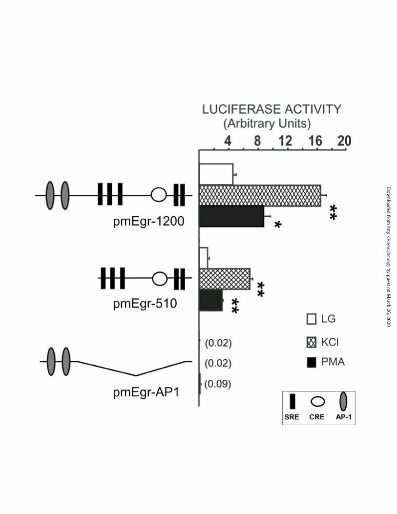
results of transient transfections in MIN6 cells with the indicated constructs is shown in Fig 2A. The
plasmid pmEgr-1200 containing 1200 bp of the 5’ flanking sequence of the mouse Egr-1 promoter was
induced 4-5-fold (p<0.001) by KCl-stimulated depolarization. Treatment of cells with the phorbol ester
PMA, a known activator of SRE-dependent transcription, also resulted in a significant induction
(p<0.01), similar to the results found by Northern blotting (data not shown). No response was obtained by
exogenous insulin treatment at concentrations from 1nM to 1µM (data not shown), suggesting that insulin
signaling through the insulin receptor is not involved in depolarization activation. The plasmid pmEgr-
510 had the 5’ end of the promoter containing the AP-1 elements deleted but still retained five SREs and
a CRE. Transfection of this plasmid resulted in diminished basal activity relative to that of pmEgr-1200,
suggesting that upstream elements may also play a role in the transcriptional regulation of this gene. This
plasmid pmEgr-510 did retain the same fold induction to KCl and PMA however. Basal expression, and
KCl and PMA induction, were all abolished when a construct lacking the SRE and CRE elements
(pmEgrAP1) was employed. The results of these promoter deletion experiments indicated that the
proximal –510 bp of the Egr-1 promoter contains the elements required for depolarization induction.
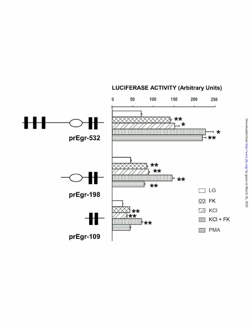
To further define the promoter elements required for induction, several additional deletion
constructs of the –532 bp sequence derived from the rat Egr-1 promoter were tested (the proximal -532
bp of the mouse and rat Egr-1 genes are >93% conserved, with a 100% similarity of the CRE and SREs).
As shown in Fig. 2B, prEgr-532 contains five SREs and one CRE. This plasmid was activated by KCl
and by FK and the effect was additive in the presence of both these agents (2-, 2.3- and 3.3-fold,
p<0.0001, p<0.02 and p<0.005). Treatment of the cells with PMA resulted in a 3.3-fold (p<0.005)
induction. Deletion of the three distal SREs (prEgr-198) resulted in a decrease in the basal activity, but
similar fold responses to FK, KCl (2-2.2-fold respectively, p<0.005), and KCl plus FK (3.3-fold,
p=0.001), although the response to PMA was reduced by 60% (p=0.001). Deletion of the CRE element
(prEgr-109) resulted in a further decrease in basal promoter activity, and a reduction in the fold responses
to KCl and FK, suggesting that the CRE contributes to the depolarization activation of the Egr-1
-cellsβEGR-1 transcription in pancreatic islet
14
by guest on March 26, 2020
http://ww
w.jbc.org/
Dow
nloaded from

promoter. Interestingly, the CRE-deleted promoter containing only the proximal two SREs still retained
significant responses to KCL (1.5-fold, p=0.004), to forskolin (1.6-fold, p=0.004), and to both agents (3-
fold, p<0.01). The conclusions drawn from these promoter deletion experiments are: 1) elements required
for the responses to each of the stimulatory agents are contained within the proximal –532 bp of the
promoter and 2) elements present within –198bp of the promoter respond to depolarization activation, and
3) that both the CRE and the SREs likely contribute to depolarization activation of the promoter.
[INSERT fig 2A, 2B]
Further assessment of the role of the CRE element and CREB phosphorylation in
depolarization induction of Egr-1
Transcriptional activation of the c-fos gene by Ca2+ and cAMP in neuronal cells is mediated by
CREB phosphorylation of Ser-133 (13). To further define the process whereby depolarization activates the
CRE of the Egr-1 promoter, CREB phosphorylation was assessed in MIN6 cells following
depolarization. Western blotting was performed using an antibody specific for CREB phosphorylated at
Ser-133. As shown in Fig. 3A, both KCl and FK treatment induced CREB phosphorylation on Ser-133
within five minutes. High glucose resulted in an increase in CREB phosphorylation after 30 minutes. The
PKA inhibitor H89 diminished both the glucose and the forskolin effect on CREB phosphorylation,
although the magnitude of the inhibitory effect of H89 on forskolin may have been less, as there appeared
to be a lower quantity of CREB protein. The total amount of CREB protein did not differ with any of the
agents tested, as indicated by using an antibody to non-phosphorylated CREB. These results are
consistent with glucose-induced depolarization and Ca2+ activation of CREB, in part by a PKA
dependent mechanism. [INSERT Fig 3]
To assess the transcriptional activity of CREB following depolarization, the induction of Egr-1
was assessed in the presence of a dominant inhibitor of CREB function (KCREB or killer CREB) (28). Co-
transfection of the pmEgr-1200 construct along with KCREB at concentrations previously shown to
-cellsβEGR-1 transcription in pancreatic islet
15
by guest on March 26, 2020
http://ww
w.jbc.org/
Dow
nloaded from
inhibit CREB activation of transcription, resulted in significant inhibition of the responses to glucose ,
KCl and forskolin (66 %, 16%, 60% and p=0.001, p=0.03, p<0.02, respectively) (28;31;38) (Fig 4A). The inhibition
of the response to a constitutively active PKA (100%, p<0.001 data not shown) demonstrated that
KCREB was an effective inhibitor of CREB mediated transcription.
The significant inhibition by KCREB of the glucose and KCl responses further suggested that
CREB-dependent transcriptional activation contributes to Egr-1 regulation. Yet the partial inhibition
strongly suggested that other factors are involved in activation of Egr-1 transcription. To more carefully
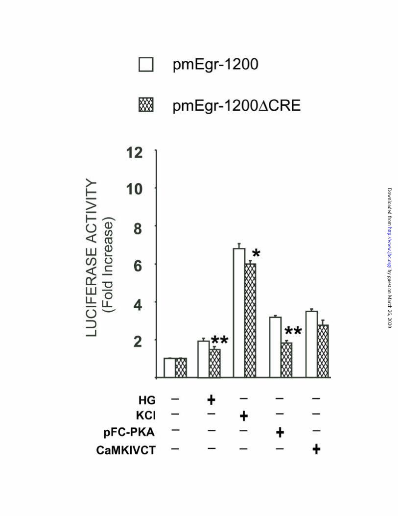
define the role of CRE/CREB in transcriptional induction of Egr-1 by depolarization, a mutant Egr-1
promoter was constructed (pmEgr-1200∆CRE) by altering the CRE element without disturbing
surrounding sequences and relative spacing. The glucose effect was reduced approximately 50% (p<0.02)
in the pmEgr-1200∆CRE relative to the non-mutated promoter. Remarkably the KCL response of the
CRE mutant was still greater than 5 fold (Fig 4B), and inhibited only by 14% (p<0.04), providing further
evidence that other elements are involved in depolarization activation. Co-transfection of pmEgr-
1200∆CRE with a constitutively active PKA resulted in a response that was reduced by 63% relative to co-
transfection with the non-mutated plasmid, indicating that PKA activates other elements in the promoter
in addition to the CRE. The pmEgr-1200∆CRE construct was also activated by cotransfection with a
constitutively active form of CaMKIV, a known activator of both SRE- and CRE-mediated transcription
(27). [INSERT Fig 4A, 4B]
Evaluation of the SRE elements and SRF in depolarization induction of Egr-1 in β-cells
The results of the previous experiments indicated that in addition to CREB activation of the CRE,
other elements of the Egr-1 promoter were involved. To assess the effect of depolarization on SRE-
dependent transcription, a luciferase plasmid containing five tandem repeats of the SRE linked to a
minimal promoter was evaluated (Fig 4). Treatment with KCL resulted in a 3.2-fold response (p<0.0001).
Treatment with forskolin resulted in 1.5-fold (p=0.02) induction, and there was no augmentation of the
KCl response by forskolin. [INSERT Fig 5]
-cellsβEGR-1 transcription in pancreatic islet
16
by guest on March 26, 2020
http://ww
w.jbc.org/
Dow
nloaded from

The SRE is continuously occupied in vivo by SRF and Ets proteins of the ternary complex
subfamily (39). Activated SRF induces transcription by binding to SREs and by recruiting other factors to the
SRE complex. Phosphorylation of SRF at Ser-103 enhances the ability of SRF to bind the SRE (30). To
demonstrate the presence of a nuclear factor binding to the SRE, nuclear extracts from MIN6 cells were
incubated with a c-fos consensus SRE (Fig 6). In the absence of nuclear extracts, band D represents
migration of the free SRE oligonucleotide (lane P). At least three additional bands (A-C) were observed
following incubation with nuclear extracts in the absence of competitors (lanes 1 and 7). They included
two slower migrating bands (A and B) and a diffuse faster migrating complex (C). Bands A and B
migrate similarly to bands identified by others as the SRE-SRF complex (band B), and the ternary
complex (band A) composed of the SRE, SRF, and ELK-1 or another TCF (40-42) (43). The identity of band C is
not known and the intensity varies with cell type (43). Bands A and B disappeared when 10-, 25- and 50-
fold molar excess of oligonucleotide (lanes 2-4) containing the sequence of the most distal Egr-1 SRE
was added. Similar results were obtained by competition with each of the five Egr-1 SREs (data not
shown). Complexes A, B and C were not evident when excess of unlabelled oligonucleotide was present
during the incubation with nuclear extracts (data not shown). In contrast, competition by a mutated SRE
oligonucleotide did not appear to significantly alter bands A and B, suggesting specificity of binding (Fig
5A lanes 1,5 and 6). To identify the identity of the proteins present in the SRE complex (bands A and B),
nuclear extracts from MIN6 cells were pre-incubated with anti-SRF antibody (αSRF). As shown in lane
8, bands A and B disappeared and a more slowly migrating band F appeared when αSRF was added. To
determine whether band F contains Ser-103-phosphorylated SRF, addition of a phospho-Ser-103-
specific αSRF antiserum was used. As shown in lane 9, a fraction of the phospho-SRF-containing
protein-DNA complex in band A and B was supershifted to band F, indicating that under chronic culture
in 25mM glucose a fraction of SRF is in an activated form. No supershift was observed when nonspecific
antibodies were used (lanes 10 and 11). [INSERT Fig 6]
To determine whether depolarization results in transcriptional activation of SRF, a trans-activator
-cellsβEGR-1 transcription in pancreatic islet
17
by guest on March 26, 2020
http://ww
w.jbc.org/
Dow
nloaded from
plasmid SRF-GAL4 containing amino acids 11-508 ligated downstream of the sequence encoding the
DNA-binding and dimerization domain of GAL4 (44) was used. The SRF-GAL4 plasmid was cotransfected
with a reporter gene containing a synthetic promoter with five tandem repeats of the yeast GAL4 binding
site upstream of the luciferase gene. As shown in Fig. 7A, KCl and forskolin treatment activated the
SRF-GAL4 gene (1.8- fold, p=0.002) and the effect was potentiated by the combination of both agents
(8 fold, p<0.001). To assess the transcriptional activation of CREB, a trans-activator plasmid that
expresses a fusion protein containing the activator domain of CREB fused to the DNA-binding
domain of GAL4 (residues 1-147) was tested. This CREB-GAL4 construct responded approximately
2-fold to forskolin and more that 10-fold to a constitutively active PKA or CaMKIV. Yet, in contrast to
the activation of SRF-GAL4, there was no significant response of CREB-GAL4 to KCl (Fig. 7B).
[INSERT Fig 7A and 7B]
DISCUSSION
The results of the current studies have now elucidated mechanisms whereby glucose induced
depolarization and Ca2+ influx regulates Egr-1 transcription in insulinoma cells. Failure of glucose to
induce Egr-1 in the presence of diazoxide, an inhibitor of depolarization, conclusively showed that
glucose metabolism in the absence of depolarization was not sufficient to activate Egr-1 transcription.
Pharmacological inhibitor studies suggested that CaM and PKA pathways are involved in the glucose
response. In addition, augmentation of the response by PI3 kinase inhibitors suggested that this pathway
may also be involved, perhaps in an inhibitory fashion. Examination of the Egr-1 promoter indicated that:
1) the elements required for the response to depolarization are contained in the proximal –532bp of the
promoter containing five SREs and a CRE, 2) the distal three SREs in the –532bp promoter contribute,
but are not essential for depolarization induced transcriptional activation, and 3) the CRE also
contributed, but was not necessary for the depolarization response. Further promoter deletion
experiments, transactivation assays, and gel shifts assays supported these conclusions, and also
-cellsβEGR-1 transcription in pancreatic islet
18
by guest on March 26, 2020
http://ww
w.jbc.org/
Dow
nloaded from

demonstrated for the first time the activation of SRF in the depolarization response in pancreatic islet β-
cells.
We considered whether the glucose induced rapid transcriptional activation of Egr-1 in
insulinoma cells could be secondary to glucose-induced insulin secretion, i.e. an autocrine/paracrine
effect. A prominent anabolic property of insulin is its effect on gene transcription. Acting through the
insulin receptor, insulin has been shown to activate IEG transcription through a ras/raf/MAPK-dependent
pathway (45-47). This appears to be an unlikely mechanism for glucose induction of Egr-1 transcription in
insulinoma cells however, as KCl activation of Egr-1 transcription was not inhibited by inhibitors of
MAPK or PI-3K (Table 1), known mediators of insulin signaling pathways for gene transcription.
Further, experiments evaluating directly the effects of insulin on MIN6 cells showed no activation of
Egr-1 transcription when exogenous insulin was added at concentrations as high as 1uM. That this insulin
signaling pathway is intact in these insulinoma cells has been demonstrated by activation of Akt/PkB in
response to exogenous insulin in this concentration range (S. Srinivasan, E. Bernal-Mizrachi, and MA
Permutt, unpublished observations).
Because the depolarization-induced activation of Egr-1 transcription occurred as early as 30
minutes and was independent of new protein synthesis, we assessed the role of phosphorylation of
transcription factors known to activate early gene transcription in other cells. For example, in neuronal
cells, depolarization induction of c-fos results for the most part through Ca2+ activation of CREB (48;49). We
initially suspected that depolarization induction of Egr-1 in pancreatic islet β-cells would be due to a
similar Ca2+ activation of CREB. In fact, earlier studies of depolarization-induced gene transcription in
hamster insulinoma (HIT) cells emphasized the role of activated CREB interacting with the CRE of the
glucagon gene promoter (50). The current observations demonstrated that, like depolarization activation of the
glucagon gene, CRE/CREB is also involved in depolarization mediated transcriptional induction of Egr-
1. The results of the promoter deletions and mutation of the CRE element supported this idea.
-cellsβEGR-1 transcription in pancreatic islet
19
by guest on March 26, 2020
http://ww
w.jbc.org/
Dow
nloaded from

Depolarization resulted in phosphorylation of CREB, and transfection of insulinoma cells with a
dominant negative CREB had a significant inhibitory effect. It should be noted, however, that the small
inhibition of the depolarization response by the dominant negative KCREB, and the robust response of
the mutated CRE construct, is not surprising since the Egr-1 promoter contains five SREs in addition to
the CRE. These results diverge to those described for the c-fos gene in which the CRE is a potent
mediator of transcriptional activation by Ca2+ signaling(48;49). The sum of these findings, along with the
demonstration of binding of CREB to the Egr-1 promoter CRE (data not shown), indicated that Ca2+-
mediated CREB dependent transcriptional activation contributes to depolarization induction of Egr-1,
although other elements in the promoter can mediate this response.
The results of the current experiments demonstrate for the first time the role of the SRE/SRF in
depolarization induction of transcription in insulinoma cells. Susini et al evaluated glucose and cAMP
induction of the c-fos gene in INS-1 insulinoma cells. In their study induction of the c-fos promoter was
dependent on an intact CRE, and not altered following mutation of the SRE. This study differed from the
present one in that c-fos was not induced by glucose in the absence of cAMP, nor was the response to
KCl-induced depolarization assessed. In addition, the c-fos promoter has only one SRE, while the Egr-1
promoter has five SREs. In the present studies, the responses of the mutated CRE plasmid pmEgr-
1200∆CRE and 5XSRE-LUC to depolarization demonstrated that the SREs also serve as Ca2+-response
elements in this model. Similar conclusions have been made for the c-fos promoter in PC12 cells, where
membrane depolarization rapidly induces the phosphorylation of SRF at Ser-103, and this
phosphorylation enhances the ability of SRF to bind the SRE (30). The SRE consensus sequence,
CC(A/T)6GG, is bound in vitro and in vivo by SRF protein. The demonstration of transcriptional activation of SRF by
the SRF-GAL4 assay, as well as that of phosphorylated SRF bound to the SRE (Fig 6), indicated that a
similar mechanism exists in insulinoma cells. The present studies do not define how Ca2+ /CaM activates
SRF. While both CaMKII and CaMKIV can phosphorylate SRF in vitro, CaMKIV is the best candidate
-cellsβEGR-1 transcription in pancreatic islet
20
by guest on March 26, 2020
http://ww
w.jbc.org/
Dow
nloaded from
because of its nuclear localization (51). Demonstration that transfection of insulinoma cells with a
constitutively active CaMKIV also induced the activation of SRF (see Fig.7) suggested that this kinase is
likely to mediate Ca2+ /CaM activation of transcription.
These findings demonstrate the effect of glucose on SRE/SRF dependent transcription in
pancreatic islet β-cells. The SRF is a transcription factor early defined as one of many mediating
mitogenic responses and regulating fibroblast proliferation in response to growth factors (52). Activated SRF
induces transcription by binding to SREs and by recruiting accessory proteins (ternary complex factors).
These accessory proteins can potentiate the transcriptional response of the SRF. However, the
contribution of transcription factors that interact with SRF in SRE-dependent transcription by glucose in
β-cells remains undefined. Prolonged exposure to glucose is a well-recognized stimulus to β-cell
hypertrophy and hyperplasia (1). The current results suggest that following exposure to glucose, ensuing β-
cell depolarization and Ca2+ induction of SRE-mediated transcription, as well as CRE-mediated
transcription, could represent important mechanisms that regulate the normal morphological and
physiological changes of pancreatic islets and the changes occurring in pathological conditions such as
diabetes."
-cellsβEGR-1 transcription in pancreatic islet
21
by guest on March 26, 2020
http://ww
w.jbc.org/
Dow
nloaded from

Acknowledgements:
We gratefully acknowledge Jeffrey Milbrandt, David Cohen, Stuart Adler, Thiruvamoor
Ramkumar, Mike McDaniel and Stephen Giddings for their careful comments and suggestions and
Michael D. Shornick, Cris M. Welling and Jon Wasson for technical assistance. The authors would like to
thank Mr. Gary Skolnick for preparation of the manuscript. This work was supported in part by NIH grant
DK16746 (MAP) and the DRTC for technical support.
-cellsβEGR-1 transcription in pancreatic islet
22
by guest on March 26, 2020
http://ww
w.jbc.org/
Dow
nloaded from
Reference List
1. Swenne, I. (1992) Diabetologia 35, 193-201
2. Welsh, M., Mares, J., Oberg, C., and Karlsson, T. (1993) Diabetes. Metab. Rev. 9, 25-36
3. Nielsen, J.H., Linde, S., Welinder, B.S., Billestrup, N., and Madsen, O.D. (1989) Mol. Endocrinol. 3,
165-173
4. Billestrup, N. and Nielsen, J.H. (1991) Endocrinology 129, 883-888
5. Brelje, T.C. and Sorenson, R.L. (1991) Endocrinology 128, 45-57
6. Swenne, I., Hill, D.J., Strain, A.J., and Milner, R.D. (1987) Diabetes 36, 288-294
7. Beattie, G.M., Rubin, J.S., Mally, M.I., Otonkoski, T., and Hayek, A. (1996) Diabetes 45, 1223-1228
8. Polak, M., Scharfmann, R., Seilheimer, B., Eisenbarth, G., Dressler, D., Verma, I.M., and Potter, H.
(1993) Proc. Natl. Acad. Sci. U.S.A. 90, 5781-5785
9. Khoo, S. and Cobb, M.H. .(1997) Proc. Natl. Acad. Sci. U.S.A. 94, 5599-5604
10. Frodin, M., Sekine, N., Roche, E., Filloux, C., and Pretki, M. (1995) J. Biol. Chem. 270, 7882-7889
11. Persaud, S.J., Wheller-Jones, C.P.D., and Jones, P.M. (1996) Biochem. J. 313, 119-124
12. MacFarlane, W.M., Smith, S.B., James, R.F.L., Clifton, A.D., Doza, Y.N., Cohen, P., and Docherty, K.
(1997) J. Biol. Chem. 272, 20936-20944
13. Sheng, M. and Greenberg, M.E. (1990) Neuron 4, 477-485
14. Susini, S., Roche, E., Prentki, M., and Schlegel, W. (1998) FASEB Journal 12, 1173-1182
15. Josefsen, K., Sorensen, L.R., Buschard, K., and Birkenbach, M. (1999) Diabetologia 42, 195-203
16. Christy, B.A., Lau, L.F., and Nathans, D) (1988) Proc. Natl. Acad. Sci. U.S.A. 85, 7857-7861
17. Lemaire, P., Revelant, O., Bravo, R., and Charnay, P. (1988) Proc. Natl. Acad. Sci. U.S.A. 85, 4691-
4695
18. Milbrandt, J. (1987) Science 238, 797-799
19. Susini, S., van Haasteren, G., Li, S.L., Prentki, M., and Schlegel, W. (2000) FASEB J. 14, 128-136
20. Ishihara, H., Asano, T., Tsukuda, K., Katagiri, H., Inukai, K., Anai, M. , Kikuchi, M., Yazaki, Y.,
Miyazaki, J.I., and Oka, Y. (1993) Diabetologia 36, 1139-1145
21. Chomczynski, P. and Sacchi, N. (1987) Analytical Biochem. 162, 156-9
-cellsβEGR-1 transcription in pancreatic islet
23
by guest on March 26, 2020
http://ww
w.jbc.org/
Dow
nloaded from
22. Church GM and Gilbert W (1984) Proc. Natl. Acad. Sci. U.S.A. 81, 1991-1995
23. Mack, K.J., Yi, S.D., Chang, S., Millan, N., and Mack, P. .(1995) Mol. Brain. Res. 29, 140-146
24. Shao, H., Kono, D.H., Chen, L.Y., Rubin, E.M., and Kaye, J. .(1997) J. Exp. Med. 185, 731-744
25. McDaniel, M.L., Colca, J.R., Kotagal, N., and Lacy, P.E. (1983) Methods Enzymol. 98, 182-200
26. Cohen, D.M., Gullans, S.R., and Chin, W.W. .(1996) J. Biol. Chem. 271, 12903-12908
27. Miranti, C.K., Ginty, D.D., Huang, G., Chatila, T., and Greenberg, M.E.(1995) Mol. Cell. Biol. 15,
3672-3684
28. Walton, K.M., Rehfuss, R.P., Chrivia, J.C., Lochner, J.E., and Goodman, R.H. (1992) Mol.
Endocrinol. 6, 647-655
29. Dignam, J.D., Lebovitz, R.M., and Roeder, R.G. .(1983) Nucleic Acids. Res. 11, 1475-1489
30. Rivera, V.M., Miranti, C.K., Misra, R.P., Ginty, D.D., Chen, R.H., Blenis, J., and Greenberg, M.E.
.(1993) Mol. Cell. Biol. 13, 6260-6273
31. Ausbel, F.M., Brent, R., Kingston, R.E., Moore, D.D., Seidman, J.G., Smith, J.A., and Struhl, K. (1989)
Current Protocols in Molecular Biology., Wiley Interscience, New York
32. Taupenot, L., Mahata, S.K., Wu, H., and O’Connor, D.T. .(1998) J. Clin. Invest. 101, 863-876
33. Chijiwa, T., Mishima, A., Hagiwara, M., Sano, M., Hayashi, K., Inoue, T., Naito, K., Toshioka, T., and
Hidaka, H. .(1990) J. Biol. Chem. 265, 5267-5272
34. Kuwada, M., Teramoto, T., Kumagaye, K.Y., Nakajima, K., Watanabe, T., Kawai, T., Kawakami, Y.,
Niidome, T., Sawada, K., and Nishizawa, Y. .(1994) Mol. Pharmacol. 46, 587-593
35. Alessi, D.R., Cuenda, A., Cohen, P., Dudley, D.T., and Salteil, A.R. (1995) J. Biol. Chem. 270,
27489-27494
36. Burgering, B.M. and Coffer, P.J. (1995) Nature 376, 599-602
37. Vlahos, C.J., Matter, W.F., Hui, K.Y., and Brown, R.F. .(1994) J. Biol. Chem. 269, 5241-5248
38. Pearman, A.T., Chou, W.Y., Bergman, K.D., Pulumati, M.R., and Partridge, N.C. .(1996) J. Biol.
Chem. 271, 25715-25721
39. Treisman, R. (1992) TIBS. 17, 423-426
40. Treisman, R. .(1994) Curr. Opin. Genetics Dev. 4, 96-101
-cellsβEGR-1 transcription in pancreatic islet
24
by guest on March 26, 2020
http://ww
w.jbc.org/
Dow
nloaded from
41. Hipskind, R.A., Rao, V.N., Mueller, C.G., Reddy, E.S., and Nordheim, A. (1991) Nature 354, 531-534
42. Shaw, P.E., Schroter, H., and Nordheim, A. (1989) Cell 56, 563-572
43. Liao, J., Hodge, C., Meyer, D., Ho, P.S. , Rosenspire, K., and Schwartz, J. (1997) J. Biol. Chem. 272,
25951-25958(Abstract)
44. Misra, R.P., Bonni, A., Miranti, C.K., Rivera, V.M., Sheng, M., and Greenberg, M.E. .(1994) J. Biol.
Chem. 269, 25483-25493
45. Bruning, J.C., Winnay, J., Cheatham, B., and Kahn, C.R. .(1997) Mol. Cell. Biol. 17, 1513-1521
46. Harada, S., Smith, R.M., Smith, J.A., White, M.F., and Jarett, L. .(1996) J. Biol. Chem. 271, 30222-
30226
47. Jhun, B.H., Haruta, T., Meinkoth, J.L., Leitner, W., Draznin, B., Saltiel, A.R., Pang, L., Sasaoka, T.,
and Olefsky, J.M. (1995) Biochemistry 34, 7996-8004
48. Sheng, M., McFadden, G., and Greenberg, M.E. (1990) Neuron 4, 571-582
49. Ginty, D.D., Kornhauser, J.M., Thompson, M.A., Bading, H., Mayo, K.E., Takahashi, J.S., and
Greenberg, M.E. (1993) Science 260, 238-241
50. Knepel, W., Chafitz, J., and Habener, J.F. .(1990) Mol. Cell. Biol. 10, 6799-6804
51. Hardingham, G.E., Cruzalegui, F.H., Chawla, S., and Bading, H. (1998) Cell Calcium 23, 131-134
52. Vandromme, M., Gauthier-Rouviere, C., Carnac, G., Lamb, N., and Fernandez, A. (1992) J. Cell.
Biol. 118, 1489-1500
-cellsβEGR-1 transcription in pancreatic islet
25
by guest on March 26, 2020
http://ww
w.jbc.org/
Dow
nloaded from
FOOTNOTES
*The abbreviations used are: IEGs, immediate early genes; AP-1, activator protein-1; SRE,
serum response element; CRE, cAMP response element; SRF, serum response factor; CREB, cAMP
response element binding protein; PKA, protein kinase A; CaM, calmodulin; CaMK, Ca2+/calmodulin-
dependent kinase; and FK, forskolin.
-cellsβEGR-1 transcription in pancreatic islet
26
by guest on March 26, 2020
http://ww
w.jbc.org/
Dow
nloaded from
FIGURE LEGENDS
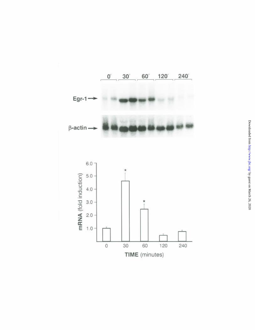
Fig. 1
Effects of glucose, KCl, tolbutamide, and forskolin on Egr-1 mRNA expression in
insulinoma cells .
A. Time course for glucose-induced expression of Egr-1 mRNA in βTC6-F7 cells. Glucose
stimulation of βTC6-F7 cells was obtained by adding glucose to a final concentration of 25 mM after 24-
hour pre-incubation (time 0) in regular DMEM media containing 2 mM glucose. Messenger RNA was
isolated as described in “Experimental Procedures” at the indicated time points. The upper panel shows a
representative Northern blot hybridization for Egr-1 and β-actin. The lower panel shows quantitative
analysis performed by phosphorimager scanning (Molecular Dynamics) that was normalized to β-actin
levels. The data is expressed as mean + S.E. of the fold induction over the RNA levels at time 0 (end of
the pre-incubation period). The results are expressed as mean values + S.E. of three independent
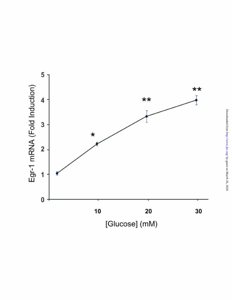
experiments done in triplicates (*p<0.001). B. Dose dependence of glucose induction of Egr-1 mRNA in
βTC6-F7 cells. Cells were pre-incubated as described in panel A and stimulated for 45 minutes by adding
glucose to the media to the final concentrations indicated. Total RNA, Northern hybridization and
quantitation was obtained as described above. Results are expressed as a fold induction over the levels at
the end of 20 hours preincubation. Data is shown as mean (+ S.E.) of three independent experiments done
in duplicate (*p<0.02) (**p<0.001). C. Depolarizing agent induction of Egr-1 mRNA. βTC6-F7 cells
were pre-incubated as described in panel A, followed by incubation for 45 minutes with KCl and
tolbutamide (TOLB). EGTA was added to the media where indicated 5 minutes prior to stimulation. All
the panels show a Northern hybridization for Egr-1 and β-actin representative of three experiments done
in duplicate. D. K-ATP inward rectifying channel activators (Diazoxide) and PKA effects on Egr-1
mRNA induction. βTC6-F7 cells were pre-incubated as described followed by a 45-minute stimulation
with 10 µM forskolin (FK), 25 mM glucose (HG), or both (HG+FK). Diazoxide (0.6 mM) (D) was added
-cellsβEGR-1 transcription in pancreatic islet
27
by guest on March 26, 2020
http://ww
w.jbc.org/
Dow
nloaded from

to the culture media 30 minutes prior to glucose. Northern hybridization for Egr-1 and β-actin is
representative of three experiments done in duplicate.
Fig. 2
Identification of the promoter sequences and cis regulatory elements implicated in
transcriptional induction of the Egr-1 gene by depolarization.
A. Depolarization and PMA effects on Egr-1 promoter activity. A diagram of the of the Egr-1 luciferase
promoter 5’ deletion constructs is shown in the insert. MIN6 cells were transiently transfected as
described in “Experimental Procedures” with 0.25 µg of the indicated constructs. To control for
transfection efficiency 2 ng of the pRL-TK luciferase construct was used. After 24 hours pre-incubation
in regular media containing 5 mM glucose and 2% serum, cells were continued in pre-incubation media
containing 5mM glucose (LG) or stimulated with 45 mM KCl or 100 nM PMA for 6 hours. Values are
expressed as the ratio of firefly to Renilla luciferase. The responses of the pmEgr-AP1 construct are not
visible on this scale, and are indicated in brackets for each stimulus. B. Effects of forskolin,
depolarization and PMA on additional Egr-1 promoter deletion constructs shown schematically in the
insert. Following transient transfection with 0.5 µg of the indicated prEgr-luciferase constructs and
culture as described in panel A, cells were kept in the same pre-incubation media or treated with 10 µM
forskolin (FK), 45 mM KCl, FK and KCl together, or 100 nM PMA. Results are shown as mean + S.E. of
three independent experiments done in triplicate (*p<0.01) (**p<0.001).
Fig. 3
Glucose and KCl induced Ser-133 CREB Phosphorylation
Western blotting hybridization for phospho-CREB. MIN6 cells were pre-cultured for 24 hours
in DMEN media containing 2 mM glucose and then stimulated with 25 mM glucose (HG), 45 mM KCl
(K) and 10 µM forskolin (F-5 minutes, FK-30 min) for the indicated periods of time. The protein kinase
A inhibitor H89 was added to the culture media to a final concentration of 20 µM one hour prior to
-cellsβEGR-1 transcription in pancreatic islet
28
by guest on March 26, 2020
http://ww
w.jbc.org/
Dow
nloaded from
stimulation. Protein was subjected to Western blot analysis using anti-phosphoCREB (PCREB) and anti-
CREB antibodies (CREB).
Fig. 4
The CRE elements contribute to the transcriptional activation of Egr-1 by
depolarization.
A. Effects of a dominant negative mutant of CREB on depolarization induction of the Egr-1 promoter.
MIN6 cells were cotransfected with KCREB (sub-cloned in pRC-RSV) (0.3 µg) or equimolar vector and
pmEgr-1200 (0.3 µg). DNA concentration was maintained constant by addition of pRC-RSV DNA.
After culturing in regular media containing 5 mM glucose and 2% FBS for 24 hours, the cells were
continued in the same media or stimulated for 6 hours with 25 mM glucose (HG), 10 µM forskolin (FK)
and 45 mM KCl (KCl). The data is expressed as mean + S.E. of the fold induction over the luciferase
activity at 5 mM glucose. The results are representative of three independent experiments done in
triplicate (*p=0.04), (**p=0.02), (***p<0.0001). B. Transfection with the mutated CRE Egr-1 promoter.
MIN6 insulinoma cells were transfected with 0.5 µg of pmEgr-1200 and 0.5 µg of pmEgr-1200∆CRE as
described in “Experimental Procedures”. Constitutively active PKA (pFC-PKA) (50 ng) and CaMKIV
(CaMKIVCT) (50 ng) were cotransfected where indicated. Pre-culture and stimulation were done as
described in panel A. The data is expressed as mean + S.E. of the fold induction over the luciferase
activity at 5 mM glucose. Values are representative of three independent experiments done in triplicate.
Significance was calculated by comparing the response to the wild type (*p=0.04), (**p<0.02). To control
for transfection efficiency 2 ng of pRL-TK luciferase construct was used in all the experiments.
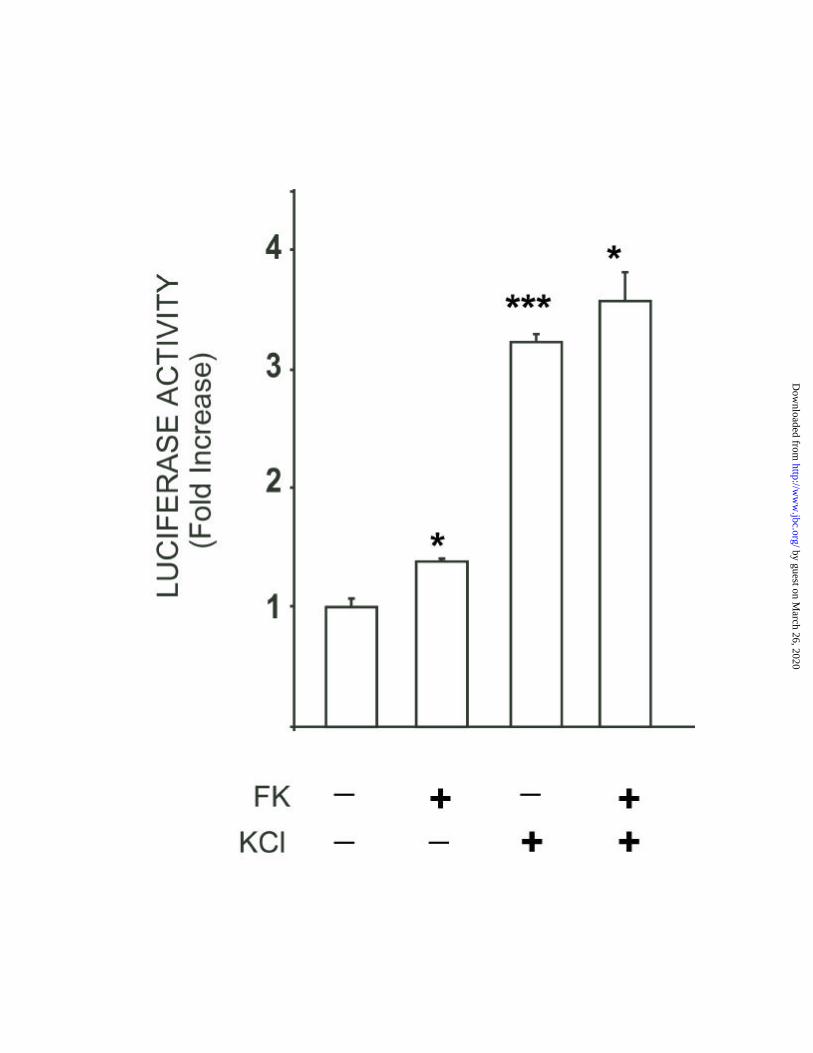
Fig. 5
The SREs are major elements responsible for depolarization induction of Egr-1.
Depolarization and forskolin effects on transfected SRE elements. MIN6 insulinoma cells were
transfected with 0.5 µg of SRE-LUC as described in “Experimental Procedures”. To control for
-cellsβEGR-1 transcription in pancreatic islet
29
by guest on March 26, 2020
http://ww
w.jbc.org/
Dow
nloaded from
transfection efficiency 2 ng of pRL-TK luciferase construct was used. After culturing in regular media
containing 5 mM glucose and 2% FBS for 24 hours, the cells were continued in the same media or
stimulated for 6 hours with 10 µM forskolin (FK) or 45 mM KCl, either alone or in the combinations
shown. The data are expressed as mean + S.E. of the fold induction over the luciferase activity at 5 mM
glucose. The results are representative of three independent experiments done in triplicate. (*p<0.02),
(**p<0.002),(***p<0.0003).
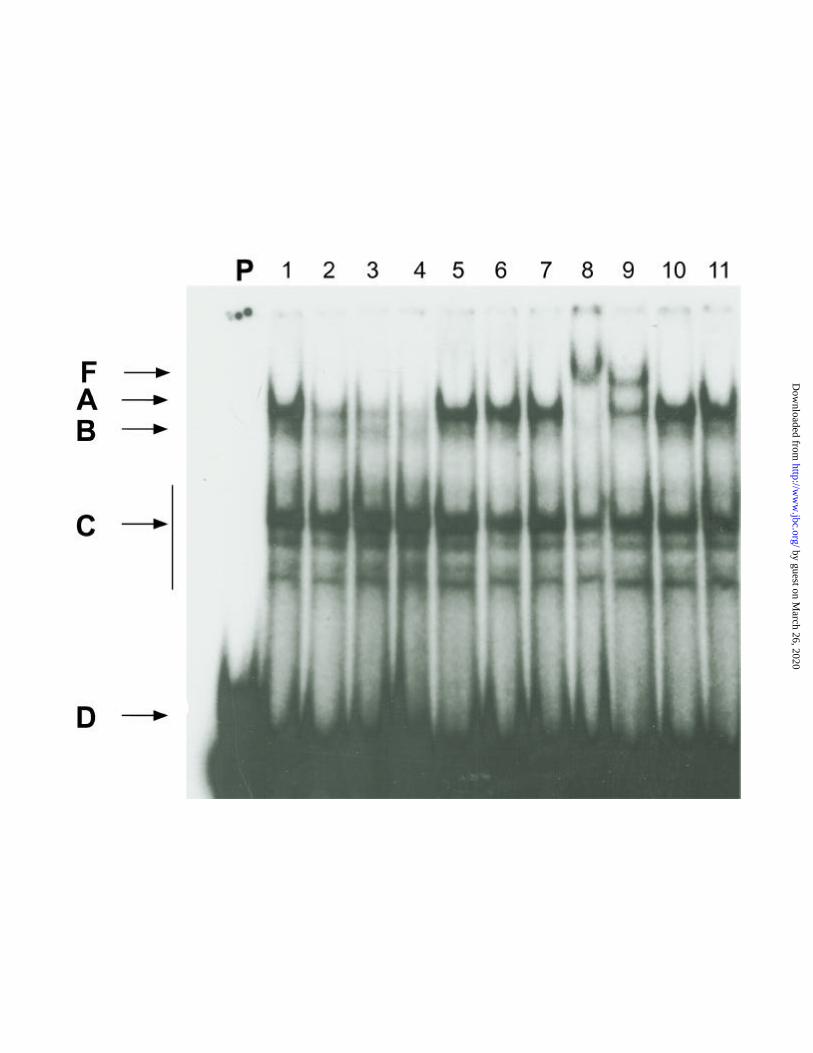
Fig. 6SRF and phosphorylated Ser-103 SRF bind the Egr-1 SREs.
Nuclear extracts from MIN6 cells cultured in 25mM glucose were used in EMSAs with a probe
containing the c-fos SRE sequence (lane 1 and 7). The individual bands representing SRE-binding
complexes are labeled A, B, and C. D indicates migration of free probe. Unlabeled Egr-1 SRE#5 (most
distal) competitor nucleotide was either not added (lane 1) or added at 10-, 25- or 50-fold molar excess
as indicated (lane 2,3,4 respectively). Competition with unlabelled mutated c-fos SRE (See Experimental
Procedures) at 25- or 50-fold molar excess is shown in lanes 5 and 6. Pre-incubation of nuclear extracts
from MIN6 cells with antibodies against SRF (lane 8) or phosphorylated Ser-103 specific αSRF (lane 9)
were used. Lanes 10 and 11 demonstrate the addition of two non-specific antibodies. Similar results
were obtained in three different experiments.
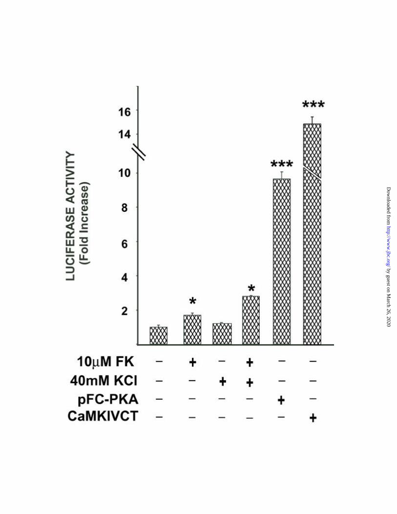
Fig. 7
Activation of SRF and CREB by depolarization in β-cells.
Effects of depolarization induction and PKA activation on transcriptional activation of the
transcription factors SRF and CREB. MIN6 insulinoma cells were transfected with 0.1 ng of SRF-GAL4
(panel A), 12 ng of CREB-GAL4 (panel B), and 0.25 µg of a reporter plasmid pFR-LUC, as described in
“Experimental Procedures”. After culturing in regular media containing 5 mM glucose and 2% FBS for
24 hours the cells were continued in the same media (5 mM gluc) or stimulated for 6 hours with forskolin
(FK) and KCl at the indicated concentrations, either alone or in the combinations shown in the figure.
-cellsβEGR-1 transcription in pancreatic islet
30
by guest on March 26, 2020
http://ww
w.jbc.org/
Dow
nloaded from
Constitutively active PKA (pFC-PKA) (50 ng) and CaMKIV (CaMKIVCT) (50 ng) were cotransfected
where indicated. To control for transfection efficiency 2 ng of pRL-TK luciferase construct was used.
The data is expressed as mean + S.E. of the fold induction over the luciferase activity at 5 mM glucose.
The results are representative of two independent experiments done in triplicate (*p<0.01), (**p<0.001),
(***p<0.0001).
-cellsβEGR-1 transcription in pancreatic islet
31
by guest on March 26, 2020
http://ww
w.jbc.org/
Dow
nloaded from
TABLE 1
Effects of pharmacological agents on glucose induction of Egr-1 mRNA expression.
βTC6-F7 cells were pre-incubated for 20 hours in regular media containing 2.0 mM glucose,
then stimulated for 45 minutes in the presence of regular medium containing 25 mM glucose and the
indicated pharmacological inhibitors . Pharmacological inhibitors were added to the media one hour prior
to stimulation with glucose. Total RNA was isolated as described in “Experimental Procedures”. Egr-1
mRNA levels were obtained by phosphorimager scanning (Molecular Dynamics) and were optimized to
those of β-actin. The results are expressed as mean (+ S.E.) of 6 independent experiments (*p<0.02)
(**p<0.01) (***p<0.005).
-cellsβEGR-1 transcription in pancreatic islet
32
by guest on March 26, 2020
http://ww
w.jbc.org/
Dow
nloaded from


% of the responseto 25mM Glucose +SD
25mM Glucose 100 + 1.710µ Calmidazolium 71.9 + 7 **20µM H89 56 + 11.1 **20µM H89 + 10µMCalmidazolium
41.1 + 1 ***
100µM PD98059 131.3 + 15.10.3nM Staurosporin 125.4 + 15.1 *0.2µM Wortmannin 156.7 + 12.8 ***50µM LY294002 223.5 + 18.2 ***
by guest on March 26, 2020
http://ww
w.jbc.org/
Dow
nloaded from
Ernesto Bernal-Mizrachi, Burton Wice, Hiroshi Inoue and M. Alan Permutt-cellsβtranscription in Pancreatic Islet
Activation of Serum Response Factor (SRF) in the Depolarization Induction of Egr-1
published online May 26, 2000J. Biol. Chem.
10.1074/jbc.M003424200Access the most updated version of this article at doi:
Alerts:
When a correction for this article is posted•
When this article is cited•
to choose from all of JBC's e-mail alertsClick here
by guest on March 26, 2020
http://ww
w.jbc.org/
Dow
nloaded from