an improved solution structure for ψ-conotoxin p iiie†,‡
TRANSCRIPT

An Improved Solution Structure forψ-Conotoxin PIIIE†,‡
Ryan M. Van Wagoner§ and Chris M. Ireland*
Department of Medicinal Chemistry, UniVersity of Utah, Salt Lake City, Utah 84112
ReceiVed December 2, 2002; ReVised Manuscript ReceiVed March 25, 2003
ABSTRACT: A revised, high-resolution structure ofψ-conotoxin PIIIE (ψ-PIIIE), a noncompetitive inhibitorof the nicotinic acetylcholine receptor (nAChR), produced through the use of NMR and molecular modelingcalculations is presented. The original structures ofψ-PIIIE had a relatively high degree of disorder,particularly in the conformation of the disulfide bridges. Our studies utilized13C-labeling of selectedcysteine residues allowing the resolution of all problems of resonance overlap for the cysteine residues.The improved data were used to produce a new set of structures by a molecular modeling processincorporating relaxation matrix methods for the determination of interproton distance restraints and acombination of distance geometry and simulated annealing for structure generation. The structures producedare very well converged with the RMSD of backbone atom positions of the main body of the peptideimproving from 0.73 to 0.13 Å. Other indicators of correlation with the experimental data and quality ofcovalent geometry showed significant improvement in the new structures. The overall conformation ofthe peptide backbone is similar between the two determinations with the exception of the N-terminus.This difference leads to a significant effect on the predicted distribution of positive charge withinψ-PIIIE,a property likely to influence interpretation of future mutational studies.
ψ-Conotoxin PIIIE (ψ-PIIIE; Figure 1)1 is a noncompetitiveantagonist of skeletal nicotinic acetylcholine receptors (nAChR)isolated from a Gulf of California specimen of the pisciverouscone snailConus purpurascens. We have recently determinedthe three-dimensional structure ofψ-PIIIE through the useof NMR in the determination of distance restraints (1). Theresults of the calculations were an ensemble of 14 structures
(PDB ID 1AS5, Figure 2) that had the same three-dimensional disulfide bridge topology as the voltage sensitivesodium channel inhibitoryµ-conotoxins, peptides having asimilar pattern of occurrence of cysteine residues anddisulfide bridge pairing. The models exhibited a significantdegree of disorder associated not only with the terminal endsof ψ-PIIIE, but also with many areas of the backbone withinthe main body of the peptide. We raised the question as towhether the disorder was indicative of structural dynamicsfor ψ-PIIIE or whether it resulted from a paucity of NMRdata caused by resonance overlap. One interesting aspect ofthe structures calculated forψ-PIIIE was the amount ofdisorder in disulfide bridge conformation. The one bridgethat was somewhat well-converged (Cys4-16) was the onlybridge for which cross-disulfide distance restraints had beendetermined. For most other cysteine residues, overlap ofcrucial cross-peaks indicative of3JHRHâ or of intraresidue
† This work was supported by an NIH fellowship (GM08573) toR.M.V., by an American Foundation for Pharmaceutical Educationpredoctoral fellowship to R.M.V., and by an American ChemicalSociety Division of Medicinal Chemistry predoctoral fellowship fundedby Pfizer to R.M.V.
‡ Atomic coordinates for the 13 converged structures ofψ-PIIIE havebeen deposited in the Protein Data Bank for release upon publication(accession code 1JLO).
* Corresponding author. Tel: (801) 581-8305. Fax: (801) 581-6208. E-mail: [email protected].
§ Current Address: Department of Biological Chemistry and Mo-lecular Pharmacology, Harvard Medical School, 250 Longwood Ave.,Boston, MA 02115, USA.
1 Abbreviations:ψ-PIIIE, ψ-conotoxin PIIIE; O, trans-4-hydroxypro-line; nAChR, nicotinic acetylcholine receptor; NMR, nuclear magneticresonance; fmoc, fluorenylmethoxycarbonyl; GSH, reduced form ofglutathione; GSSG, oxidized form of glutathione; C18 HPLC, octade-cylsilyl reversed-phase high-performance liquid chromatography; CH3-CN, acetonitrile; TFA, trifluoroacetic acid; MALDI-MS, matrix-assistedlaser desorption ionization mass spectrometry; D2O, deuterium oxide;TOCSY, total correlation spectroscopy, DQF-COSY, double quantumfiltered correlation spectroscopy; NOESY-HSQC, three-dimensionalnuclear Overhauser effect spectroscopy/heteronuclear single quantumcoherence spectroscopy; fHSQC, fast heteronuclear single quantumcoherence spectroscopy; NOESY, nuclear Overhauser effect spectros-copy; PECOSY, primitive exclusive correlation spectroscopy; WET,water suppression enhanced through T1 effects; WATERGATE, watersuppression by gradient-tailored excitation; I-BURP, inverting band-selective uniform response pure phase pulse; FID, free induction decay;DGII, distance geometry II; IRMA, iterative relaxation matrix approach;CVFF, constant valence force field; nOe, nuclear Overhauser effect;RMSD, root-mean-squared deviation.
FIGURE 1: Sequence ofψ-conotoxin PIIIE. O ) trans-4-hydroxy-proline; -NH2 ) amidated C-terminus.
FIGURE 2: Original structures calculated forψ-conotoxin PIIIE byMitchell et al. Defocused stereoview showing the backbone as aribbon with the heavy atoms of side chains shown as well. Theside chains of basic residues are colored blue.
6347Biochemistry2003,42, 6347-6352
10.1021/bi027274e CCC: $25.00 © 2003 American Chemical SocietyPublished on Web 05/07/2003

distance restraints prevented determination of restraints onø1. Thus, it was impossible to determine whether the sidechains of the cysteine residues are rotationally static ordynamic.
We postulated that greater resolution of the molecularmodel could be achieved if restraints onø1 were determinedfor all cysteine residues, thus potentially elucidating whetherthe side chains of the cysteine residues are rotationally staticor dynamic. Interestingly, based on analysis of depositedconotoxin structures in the Protein Data Bank (2) for whichmultiple independent structural determinations have beenundertaken, the appearance of heterogeneity in disulfidebridge conformation often results from having insufficientdata to distinguish between the alternate conformers ratherthan from any actual dynamic motion of the disulfide bridges(3). There have been cases, however, where experimentalobservations indicate that dynamic fluctuation of disulfidebridge conformation may indeed be taking place under theexperimental conditions (4-6). To explore the conformationof cysteine residues inψ-PIIIE, we used chemical synthesisto produce ψ-PIIIE containing 3-13C-labeled cysteine atselected positions in the sequence in such a way as to allowresolution of all1H NMR overlap problems for the cysteineresidues ofψ-PIIIE. We were able to collect an improveddata set containing a large number of restraints on interprotondistances and torsion angles that were applied to molecularmodeling calculations to produce a new set of structures ofψ-PIIIE. The use of a more extensive data set led to significantimprovement over the structures originally reported forψ-PIIIE in a number of parameters describing structuralquality.
EXPERIMENTAL PROCEDURES
Peptide Synthesis.99% [3-13C]-N-(9-fluorenylmethoxy-carbonyl)-S-trityl- L-cysteine was synthesized as previouslydescribed (7, 8) using 99% [3-13C]-L-cysteine (CambridgeIsotope Laboratories, CLM-1868). The labeled amino acidwas incorporated into positions C4 and C21 ofψ-PIIIE usingfmoc-based solid-phase chemical synthesis (50µmole scale)(9). The13C-labeled residues were reacted with the resin ata 5-fold excess and the natural isotopic abundance residueswere reacted at a 10-fold excess. All cysteine residues wereincorporated as trityl thioether derivatives and the side chainsof other residues were protected as previously described (10).Deprotection and purification of the peptide were achievedusing a procedure similar to that originally used for thesynthesis ofψ-PIIIE (11). Disulfide bridge formation wasachieved by incubation of linear peptide in a 0.1 M pH 7.7phosphate buffer solution containing 1.0 mM GSH and 0.5mM GSSG for 12-16 h at room temperature. The majorfolded product isolated by preparative C18 HPLC was foundto coelute with authenticψ-PIIIE using a 10-60% gradientof 60% CH3CN, 0.092% TFA in 0.1% TFA over 50 min onanalytical C18 HPLC. MALDI-MS indicated an averagemolecular mass of 2720.77 Da (calculated 2720.2 Da). Thetotal yield from 200 mg of resin was 14.1 mg of peptide(4.14 µmol of the hexa-TFA salt ofψ-PIIIE).
NMR Data Acquisition.For NMR studies, 14.1 mg ofpeptide were dissolved in 260µL of a 9:1 water/D2O solutionto provide a solution roughly 16 mM in concentration. ThepH of the solution was adjusted to 3.48 with dilute TFA
prior to addition of the peptide. For studies requiringobservation of peaks with resonance frequencies similar towater, 14.1 mg of lyophilized peptide were dissolved in 260µL of D2O. The solution conditions for NMR data acquisitionwere chosen to be similar to those used for the originalstructure determination ofψ-PIIIE.
NMR spectra were acquired on a Varian INOVA 600 MHzNMR spectrometer. Experiments were acquired using aNalorac triple-resonance gradient-capable probe. A 5-mmNMR microtube with insert matched to D2O (Shigemi Co.,Ltd.) was used for all experiments. The pulse sequences usedfor TOCSY (12), DQF-COSY (13), NOESY-HSQC (14),and fHSQC (15) were similar to those previously described.Pulse sequences for NOESY (16) and PECOSY (17) experi-ments were converted to doubly13C-filtered variants throughinsertion of appropriate sequences prior to each detectionperiod (18). One-dimensional13C-edited1H NMR spectrawere obtained by using a modified double pulsed fieldgradient selective excitation (19) strategy. A modified formof WET (20) water suppression was used for the TOCSY,DQF-COSY, and doubly13C-filtered NOESY. Water sup-pression was applied prior to frequency labeling for TOCSYand DQF-COSY and during the mixing time for NOESYspectra. A WATERGATE based methodology (21) utilizinga 3919 sequence was used for water suppression of theNOESY-HSQC, and fHSQC experiments. PECOSY and13C-edited1H NMR spectra were acquired in D2O, negatingthe need for water suppression. Quadrature discriminationof frequencies in the indirectly detected dimension wasachieved in all experiments through use of States hyper-complex acquisition (22). Unless noted otherwise, thespectral width was 6600 Hz for1H dimensions and 2000 Hzfor 13C dimensions. For the TOCSY and DQF-COSYexperiments, a total recycle time of 2.31 s was used with 16scans each consisting of 2048 complex points. A total of512 complex points int1 were acquired and the acquisitiontemperature was 4°C. A mixing time of 80 ms was usedfor the TOCSY experiment. Parameters were similar for thedoubly 13C-filtered NOESY spectra except 32 scans wereacquired, the recycle time was 1.510 s, and mixing times of100, 200, and 400 ms were used. For the NOESY-HSQC,64 points were acquired int1, 128 points int2, and 1024points int3. The recycle time was 0.605 s and four transientswere acquired. The spectra were acquired at 4°C with amixing time of 150 ms. For the fHSQC 128 complex pointsin t1 and 2048 complex points int2 were acquired with arecycle time of 1.01 s. Four transients were acquired andthe temperature of acquisition was 4°C. The PECOSYexperiment was acquired at a temperature of 22°C with arecycle time of 2.62 s. A total of 512 complex points wereacquired int1 while 4096 points were acquired int2. Thirty-two scans were acquired for eacht1 time point. The13C-edited1H NMR spectrum was acquired on a Varian UNITY500 MHz spectrometer using a Nalorac triple resonancegradient-capable probe. A shaped I-BURP (23) pulse patternhaving 8.3 ms duration was used for selective inversion ofthe carbon whose protons were to be observed. A spectralwidth of 4629 Hz was used and 8192 complex points werecollected. The data were acquired at 4°C.
Data Processing.All NMR data sets were analyzed usingeither VNMR (Varian, Inc.) or FELIX (Accelrys, Inc).TOCSY, DQF-COSY, and NOESY experiments were zero-
6348 Biochemistry, Vol. 42, No. 21, 2003 Van Wagoner and Ireland

filled once in D1 and three times in D2 to give final matrixesconsisting of 4096 by 2048 real points with resolution valuesof 1.4 and 2.8 Hz/pt for the D1 and D2 vectors, respectively.For NOESY and TOCSY spectra,t2 FIDs andt1 interfero-grams were processed by convolution with 90° phase-shiftedsinebell squared curves (with widths of 2048 and 1024 points,respectively) prior to complex fast Fourier transformation.DQF-COSY spectra were processed with 60° phase-shiftedsinebell squared curves int2 (2048 point width) andt1 (1024point width) prior to transformation. The PECOSY data setwas processed by zero-filling once in D1 and three times inD2 to give a final matrix of 8192 by 2048 real points andresolution values of 0.7 and 2.8 Hz/pt for the D1 and D2vectors. Apodization and resolution enhancement wereachieved by convolution with 60° phase-shifted sinebellwindow functions with widths of 4096 points (int2) and 512points (int1) prior to transformation. Baseline correction wasapplied in both dimensions of NOESY and TOCSY experi-ments to enhance detection of cross correlations and increasethe accuracy of volume measurement. Automatic baselinedetection was achieved by use of the FLATT algorithm (24)with default parameters as provided by FELIX.
Determination of Restraints and Molecular Modeling.Allcalculations were performed using the Insight II 97.0 suiteof programs (Accelrys, Inc). Molecular dynamics calculationswere performed using DISCOVER 2.98 (Accelrys, Inc),while DGII 97.0 (Accelrys, Inc.) was used for distancegeometry calculations. Distance restraints for the highestquality NOESY cross-peaks were calculated using standardIRMA (Accelrys, Inc.) methodology (25, 26). Lower andupper bound distance restraints from lower quality cross-peaks in NOESY spectra forψ-PIIIE were respectively setat 90 and 120% of the distance calculated from the isolatedspin pair approximation for data collected with a mixing timeof 0.100 s. The dihedral angleφ was restrained to values of-90° to -40° for 3JHNHR < 5 Hz and to the range-160° to-80° for 3JHNHR > 8 Hz (27). Restraints onø1 were restrictedto be(15° of the values determined according to the methodof Hyberts et al. (28). The pairing of disulfide bonds hadbeen determined experimentally previously (11) and wereenforced as explicit covalent bonds prior to metric matrixcalculations within Insight II. The final structures weregenerated by distance geometry calculations followed bysimulated annealing and energy minimization using standardprotocols (1, 29). CVFF Provided all empirical potentialenergy terms (30). The 50 structures generated by thismethodology were analyzed on the basis of residual viola-tions of dihedral angle and distance restraints and by potentialenergy calculations. The force constants were set to valuesof 20.00 kcal (mol Å2)-1 for all distance restraints and 30.00kcal (mol radian2)-1 for dihedral angle restraints. Electrostaticpotentials were disregarded for all modeling calculations dueto the use of in vacuo conditions. The figures were generatedusing the program MOLMOL 2k.2 (31) in conjunction withthe POV-Ray rendering program.
RESULTS
Resolution of Data OVerlap for Cysteine Residues.Forψ-PIIIE a selective13C-labeling approach was taken as ameans of resolving overlap problems. The two cysteineresidues withâ proton resonance signals involved in mostof the overlap problems in the original data set ofψ-PIIIE
(C4 and C21) were labeled, thus removing all overlapproblems for C5, C10, and C22 by using isotopic filtering.The use of13C-labeling also allowed the application of13C-editing approaches to further resolve the overlap problemsbetween C4 and C21, providing that the13C chemical shiftswere sufficiently well resolved between the two residues.Incorporation of cysteine labeled with13C at Câ by solidphase chemical synthesis was selected as the most straight-forward approach likely to provide the desired informationgiven reactant availability and the specific overlap problemspresent. Selective13C-labeling of C4 and C21 providedadditional advantages over selective13C-labeling of allcysteine residues. The likelihood of overlap for13C chemicalshift was greatly reduced with only two residues labeledcompared to five or six. Also, smaller amounts of theexpensive13C-labeled cysteine reagent were required forlabeling of only two residues.
Measurement of intraresidue nOe coupling interactionsfor C4 and C21 was achieved through the use of a three-dimensional NOESY-HSQC. Separate NOESY spectra forC4 and for C21 were produced by taking two-dimensionalslices at the chemical shift of the respective carbons. Themethod decided upon for determination of3JHRHâ for C4 andC21 was direct measurement of3JHRHâ from the multipletstructure observed in a13C-selected one-dimensional1HNMR spectrum. Fortunately, the line widths ofψ-PIIIE arenarrow enough to allow measurement of3JHRHâ as small as3-4 Hz. Because of the overlap of C4HâH and C21HâL, how-ever, it was necessary to develop an approach that allowedselective observation of Hâ for C4 and C21 separately. Thiswas achieved using a gradient-selected13C to1H polarizationtransfer experiment (see Supporting Information for a pulsediagram). Variants of NOESY and PECOSY experimentswere designed to incorporate13C-filtering elements at eitheror both of the1H detection periods, thus allowing13C-filteringof signals in either or both dimensions of the experiments(see Supporting Information). Measurement of3JHRHâ andthe intraresidue HN:Hâ and HR:Hâ nOe cross-peak volumesfrom these filtered experiments provided information on boththe conformation and dynamics associated withø1 for thenonlabeled cysteine residues.
Assignment of1H Resonances, Determination of Restraintson Dihedral Angle, and Stereospecific Assignment.Assign-ments were made for all observed1H resonances accordingto the methodology of Wu¨thrich (32). Restraints onø1 weredetermined for H1, C4, C5, L6, Y7, C10, Y13, C16, S18,S20, C21, C22, and Q23 based on3JHRHâ and on intraresidueHN:Hâ and HR:Hâ nOe interactions (28). This analysis alsoallowed the determination of stereospecific restraints for theprochiral side chain hydrogen atoms of residues C4, C5, L6,Y7, C10, R12, Y13, C16, S18, S20, C21, C22, and Q23.Additionally, analysis of the pattern of intraresidue nOeinteractions for hydroxyproline residues (1) led to stereospe-cific assignment of all prochiral hydrogen atoms of residuesO2, O3, and O14. Restraints onφ were determined frommeasurement of3JHNHR according to previously describedmethods (33). The intraresidue and sequential nOe interac-tions measured were not indicative of regularR-helical orâ-strand structures inψ-PIIIE.
Structure Calculations and EValuation forψ-PIIIE. A totalof 524 distance restraints were derived from the NOESYdata through either relaxation matrix calculations (25, 26)
Improved Structure ofψ-Conotoxin PIIIE BIOCHEMISTRY, VOL. 42, NO. 21, 2003 6349

or estimations based on the isolated spin pair approximation(34) for cross-peaks in the 100 ms NOESY spectrum (Figure3). The restraints on interproton distances and dihedral angleswere used as input for molecular modeling calculations basedon combined use (29) of distance geometry II (DGII) (35)and simulated annealing to generate a set of 50 structures.Thirteen of the 50 structures were found to converge byanalysis of residual restraint violations and energy calcula-tions. The ensemble of structures calculated forψ-PIIIE showsgood convergence of backbone atom coordinates (see Table1). An overall RMSD of 0.22 Å is found for all residueswith that figure decreasing to 0.13 Å for the main body ofthe peptide between residues 4 and 22. The side chains ofmost residues are also reasonably well-converged with onlyK9, R12, and R24 showing high degrees of disorder andR11 showing a somewhat smaller amount of heterogeneityof structure. The stereochemistry for the models ofψ-PIIIE
is reasonably consistent with expected peptide geometry.Overall, 78% of residues occur in the most favored regionsidentified by Ramachandran analysis (36) while 22% occurin additionally allowed regions of the plot as determined bythe program PROCHECK_NMR (37). One recent report hasnoted that small disulfide-rich proteins tend to score poorlyby stereochemical analysis of the type reported above,possibly due to strain associated with the tightly constrainedstructures (38). The structures also showed low RMSD fromideal bond lengths and bond angles, indicating that satisfac-
tion of the experimental restraints on structure does notrequire significant distortion of covalent geometry.
Three-Dimensional Structure.The converged structures ofψ-PIIIE are illustrated in Figure 4. Like many conotoxins,ψ-PIIIE consists of a compact disulfide core with mostnoncysteine side chains oriented away from the interior ofthe peptide. Residues likely to constitute the hydrophobiccore of the peptide (indicated by>80% buried surface areaas calculated by MOLMOL 2k.2) include C4, C5, L6, Y13,C16, A19, C21, and C22. Residues with>60% solventexposure include R12 and R24.ψ-PIIIE does not appear tocontain anyâ-sheets longer than two residues. The regionbetween residues 19 and 23 has a roughlyR-helicalconformation though secondary structure detection byMOLMOL2k.2 indicated helicity for only 1 of the 13structures. There are, however, a number of reverse turnsindicated by the backbone torsion angles and predictedH-bonds in the calculated structures (Table 2 and SupportingInformation).
DISCUSSION
ReVised Structure ofψ-PIIIE. Overall the structure ofψ-PIIIE is compact and well defined. The main body of thepeptide has the rough shape of a shallow trigonal prism; whenviewed from the “side” the molecule has a smaller profilethan when viewed from the “front” or “back” (see Figure5). The N-terminus is well-converged with the aromatic sidechain of H1 aligned along a face of the peptide. Each of thethree disulfide bridges are very well-converged with the soleexception being a single alternate disulfide conformation forthe bridge Cys4-16. The structures calculated forψ-PIIIE
do not have either the intercysteine loop spacings nor thethree-dimensional disulfide bridge arrangement expected foran inhibitor cystine knot fold (39). As was the case for theoriginal structures ofψ-PIIIE, the overall fold bears littlesimilarity to other conotoxins aside from the overall three-
FIGURE 3: Distribution of distance restraints across the sequenceof ψ-PIIIE. Black bars indicate intraresidue restraints, crosshatchedbars indicate sequential restraints, gray bars indicate restraintsbetween residues separated by 2-4 amino acids, and white barsindicate long-range distance restraints.
Table 1: Structural Statistics for the Revised and OriginalStructures ofψ-Conotoxin PIIIE
revisedstructures
originalstructures
RMSD for CR, N, C atoms fromaverage molecule coordinates (Å)
residues 4-22 0.13 0.73all residues 0.22 0.91
RMSD from distance restraints (Å) 0.140 0.130RMSD from dihedral restraints (deg) 2.34 3.03R1 for ensemble 0.350 0.597RMSD from idealized geometry
bond lengths (Å) 0.0142( 0.002 0.0185( 0.004bond angles (deg) 2.67( 0.02 3.6( 0.2
energies (kcal mol-1)Erestraint 224( 5 190( 20EL-J 189( 3 300( 20Ebond+ Eangle+ Eimproper 298( 5 460( 30
Ramachandran plot statistics (%)residues in most favored region 78 55residues in additionally allowed region 22 37residues in generously allowed region 0 8residues in nonallowed region 0 0.4
FIGURE 4: Defocused stereoviews of the 13 converged structuresof ψ-PIIIE produced by simulated annealing calculations. Allstructures are aligned with the N-terminus toward the top of thefigure and the C-terminus toward the bottom. Only the heavy atomsof the side chains are shown. The side chains of basic residues arecolored blue.
Table 2: Observed Elements of Secondary Structure in the NewStructures ofψ-PIIIE
residues structural element
H1-C4 Type VII â turnO2-C5 Type VIaâ turnO3-L6 Type VIII â turnL6-K9 Type I′ â turnR11-Y13 inverseγ turnY13-C16 Type IIâ turnC16-A19 Type Iâ turnA19-C22 Type IIIâ turnS20-Q23 Type IIIâ turnC21-R24 Type IIIâ turn
6350 Biochemistry, Vol. 42, No. 21, 2003 Van Wagoner and Ireland

dimensional arrangement of disulfide bridges in theµ-cono-toxins.
There are six positive charges present onψ-PIIIE at thepH used for NMR studies and these are distributed amongthree main sites of positive charge density: near residue 1,near residues 9-12, and at residue 24. The positive chargesassociated with H1 are both located on the rear face of themolecule near the top. Both positively charged moieties arewell defined in the structures. The three positive chargesoccurring at K9, R11, and R12 are located in the upper halfof the front of the peptide. Of the three residues, the sidechain of R11 appears to be best defined by the data. Theside chain of R11 is also the only one of the three to alignalong the face of the molecule; the side chains of K9 andR12 project perpendicular to the body of the peptide. Theside chains of K9 and R12 have a significantly higher degreeof solvent exposure than R11 (see Figure 4). The third regionof positive charge density occurs at the C-terminus of thepeptide. The conformation of the side chain of R24 is morepoorly defined than that of any other residue in the peptide.
Comparison with the Original Structures ofψ-PIIIE. Beforecomparisons between the original and new structures deter-mined for ψ-PIIIE are made, it should be noted that therewere a number of improvements made to the restraint set inaddition to specification ofø1 restraints for all cysteineresidues. A higher concentration of peptide was used in thenew studies (16 mM) compared to the old studies (4.3 mM).There were additional improvements in the quality of watersuppression and in magnetic field strength that increased thequality and quantity of restraints on structure that could bedetermined from the data. A comparison of the restraint setsis given in Table 3. It is therefore not surprising to note thatthere was significant improvement in a number of parametersindicative of structure quality for the revised structures ofψ-PIIIE (see Table 1). Taken together, these statistics indicatethat the new structures ofψ-PIIIE are more precise, lessstrained, and a better fit to the experimental data than werethe original structures. Additionally, the occurrence ofapparent shielding effects in some protons is better explainedby the arrangement of aromatic rings in the new structuresthan in the old structures.
The backbone conformations in the original and newstructures are very similar to each other (compare Figures 2and 4). The overall paths traversed by the backbones arenearly identical over most of the length of the toxin. Bothsets of structures include reverse turns at similar positions(O3-L6, L6-K9, R11-Y13, Y13-C16, C16-A19, andA19-C22). In the original structure determination, however,the N-terminus projects toward the front of the peptidewhereas in the new structure determination it projects towardthe back. All six of the positive charges in the originalstructure ofψ-PIIIE are located on the front face of thepeptide (see Figure 2), a circumstance indicating that thisface of the molecule is important in interacting with thenAChR as the channel contains both cation-binding sites anda cation-conductive channel. In particular, sinceψ-PIIIE actsas a noncompetitive inhibitor, it is possible that the toxin-binding site could be near the ion channel where the peptidecan sterically block ion conductance. In the new set ofstructures, however, the two positive charges associated withH1 are actually located on the back face of the peptide (seeFigure 5).
The two sets of structures differ in the degree of definitionof the conformations of the three disulfide bridges. As wasmentioned previously, all three disulfide bridges were poorlydefined in the original structure determination ofψ-PIIIE. Thedisulfide bridges of the new structures, on the other hand,are very well-converged (see Figure 6). This convergence
FIGURE 5: The electrostatic surface potential of a representative model ofψ-PIIIE is shown from three different vantage points. Blueregions indicate a positive electric potential. The regions of most intense color correspond to a charge of+1 or greater. Potential calculationswere performed using the SimpleCharge algorithm of MOLMOL 2k.2. (a) View from the “front” side of the peptide, or the side of thepeptide containing all of the positive charges in the original structure determination ofψ-PIIIE. (b) View from the side ofψ-PIIIE. The frontof the peptide corresponds to the right side of the molecule where the side chain of R12 is visible in the upper half of the molecule. (c) Theback face ofψ-PIIIE. The side chain of H1 is packed against the top of the molecule in this vantage point.
Table 3: Comparison of Restraints Used for the StructureDeterminations ofψ-PIIIE
originalstructures
newstructures
distance restraintsintraresidue 142 154sequential 99 156medium 19 131long 20 83
total 280 524
dihedral restraintsφ 10 9ø1 6 13
total 16 22
Improved Structure ofψ-Conotoxin PIIIE BIOCHEMISTRY, VOL. 42, NO. 21, 2003 6351
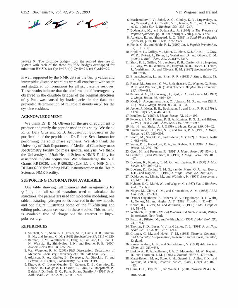
is well supported by the NMR data as the3JHRHâ values andintraresidue distance restraints were all consistent with staticand staggered conformations for all six cysteine residues.These results indicate that the conformational heterogeneityobserved in the disulfide bridges of the original structuresof ψ-PIIIE was caused by inadequacies in the data thatprevented determination of reliable restraints onø1 for thecysteine residues.
ACKNOWLEDGMENT
We thank Dr. B. M. Olivera for the use of equipment toproduce and purify the peptide used in this study. We thankR. G. Dela Cruz and R. B. Jacobsen for guidance in thepurification of the peptide and Dr. Robert Schackmann forperforming the solid phase synthesis. We also thank theUniversity of Utah Department of Medicinal Chemistry massspectrometry facility for mass spectral analysis. We thankthe University of Utah Health Sciences NMR Facility forassistance in data acquisition. We acknowledge the NIHGrants RR13030, and RR06262 (C.M.I.), and NSF GrantDBI-0002806 for funding NMR instrumentation in the HealthSciences NMR Facility.
SUPPORTING INFORMATION AVAILABLE
One table showing full chemical shift assignments forψ-PIIIE, the full set of restraints used to calculate thestructures, the parameters used for molecular modeling, onetable illustrating hydrogen bonds observed in the new models,and one figure illustrating some of the13C-filtering andediting pulse sequences used in these studies. This materialis available free of charge via the Internet at http://pubs.acs.org.
REFERENCES
1. Mitchell, S. S., Shon, K. J., Foster, M. P., Davis, D. R., Olivera,B. M., and Ireland, C. M. (1998)Biochemistry 37, 1215-1220.
2. Berman, H. M., Westbrook, J., Feng, Z., Gilliland, G., Bhat, T.N., Weissig, H., Shindyalov, I. N., and Bourne, P. E. (2000)Nucleic Acids Res. 28, 235-242.
3. Van Wagoner, R. M. (2001) PhD Dissertation, Department ofMedicinal Chemistry, University of Utah, Salt Lake City.
4. Atkinson, R. A., Kieffer, B., Dejaegere, A., Sirockin, F., andLefevre, J. F. (2000)Biochemistry 39, 3908-3919.
5. Rigby, A. C., Lucas-Meunier, E., Kalume, D. E., Czerwiec, E.,Hambe, B., Dahlqvist, I., Fossier, P., Baux, G., Roepstorff, P.,Baleja, J. D., Furie, B. C., Furie, B., and Stenflo, J. (1999)Proc.Natl. Acad. Sci. U.S.A. 96, 5758-5763.
6. Maslennikov, I. V., Sobol, A. G., Gladky, K. V., Lugovskoy, A.A., Ostrovsky, A. G., Tsetlin, V. I., Ivanov, V. T., and Arseniev,A. S. (1998)Eur. J. Biochem. 254, 238-247.
7. Bodanszky, M., and Bodanszky, A. (1994) inThe Practice ofPeptide Synthesis, pp 68-69, Springer-Verlag, New York.
8. Atherton, E., and Sheppard, R. C. (1989) inSolid-Phase PeptideSynthesis, p 60, IRL Press, New York.
9. Fields, G. B., and Noble, R. L. (1990)Int. J. Peptide Protein Res.35, 161-214.
10. Hopkins, C., Grilley, M., Miller, C., Shon, K. J., Cruz, L. J., Gray,W. R., Dykert, J., Rivier, J., Yoshikami, D., and Olivera, B. M.(1995)J. Biol. Chem. 270, 22361-22367.
11. Shon, K. J., Grilley, M., Jacobsen, R. B., Cartier, G. E., Hopkins,C., Gray, W. R., Watkins, M., Hillyard, D. R., Rivier, J., Torres,J., Yoshikami, D., and Olivera, B. M. (1997)Biochemistry 36,9581-9587.
12. Braunschweiler, L., and Ernst, R. R. (1983)J. Magn. Reson. 53,521-528.
13. Rance, M., Sørensen, O. W., Bodenhausen, G., Wagner, G., Ernst,R. R., and Wu¨thrich, K. (1983)Biochem. Biophys. Res. Commun.117, 479-485.
14. Palmer, A. G., III, Cavanagh, J., Byrd, R. A., and Rance, M. (1992)J. Magn. Reson. 96, 416-424.
15. Mori, S., Abeygunawardana, C., Johnson, M. O., and van Zijl, P.C. (1995)J. Magn. Reson. B 108, 94-98.
16. Jeener, J., Meier, B. H., Bachmann, P., and Ernst, R. R. (1979)J.Chem. Phys. 71, 4546-4553.
17. Mueller, L. (1987)J. Magn. Reson. 72, 191-196.18. Folkers, P. J. M., Folmer, R. H. A., Konings, R. N. H., and Hilbers,
C. W. (1993)J. Am. Chem. Soc. 115, 3798-3799.19. Hu, H., and Shaka, A. J. (1999)J. Magn. Reson. 136, 54-62.20. Smallcombe, S. H., Patt, S. L., and Kiefer, P. A. (1995)J. Magn.
Reson. A 117, 295-303.21. Piotto, M., Saudek, V., and Sklenar, V. (1992)J. Biomol. NMR
2, 661-665.22. States, D. J., Haberkorn, R. A., and Ruben, D. J. (1982)J. Magn.
Reson. 48, 286-292.23. Geen, H., and Freeman, R. (1991)J. Magn. Reson. 93, 93-141.24. Guntert, P., and Wu¨thrich, K. (1992)J. Magn. Reson. 96, 403-
407.25. Boelens, R., Koning, T. M. G., and Kaptein, R. (1988)J. Mol.
Struct. 173, 299-311.26. Boelens, R., Koning, T. M. G., van der Marel, G. A., van Boom,
J. H., and Kaptein, R. (1989)J. Magn. Reson. 82, 290-308.27. DeMarco, A., Llina´s, M., and Wu¨thrich, K. (1978)Biopolymers
17, 617-636.28. Hyberts, S. G., Marki, W., and Wagner, G. (1987)Eur. J. Biochem.
164, 625-635.29. Nilges, M., Clore, G. M., and Gronenborn, A. M. (1988)FEBS
Lett. 229, 317-324.30. Dauber-Osguthorpe, P., Roberts, V. A., Osguthorpe, D. J., Wolff,
J., Genest, M., and Hagler, A. T. (1988)Proteins 4, 31-47.31. Koradi, R., Billeter, M., and Wu¨thrich, K. (1996)J. Mol. Graphics
14, 51-55.32. Wuthrich, K. (1986)NMR of Proteins and Nucleic Acids, Wiley-
Interscience, New York.33. Pardi, A., Billeter, M., and Wu¨thrich, K. (1984)J. Mol. Biol. 180,
741-751.34. Thomas, P. D., Basus, V. J., and James, T. L. (1991)Proc. Natl.
Acad. Sci. U.S.A. 88, 1237-1241.35. Crippen, G. M., and Havel, T. M. (1988)Distance Geometry
and Molecular Conformation, Research Studies Press, Taunton,England.
36. Ramachandran, G. N., and Sasisekharan, V. (1968)AdV. ProteinChem. 23, 283-438.
37. Laskowski, R. A., Rullmann, J. A. C., MacArthur, M. W., Kaptein,R., and Thornton, J. M. (1996)J. Biomol. NMR 8, 477-486.
38. Martı-Renom, M. A., Stote, R. H., Querol, E., Aviles, F. X., andKarplus, M. (2000)Proteins: Struct., Funct., Genet. 40, 482-493.
39. Craik, D. J., Daly, N. L., and Waine, C. (2001)Toxicon 39, 43-60.
BI027274E
FIGURE 6: The disulfide bridges from the revised structure ofψ-PIIIE with each of the three disulfide bridges overlapped forminimum RMSD. (a) Cys4-16, (b) Cys5-21, (c) Cys10-22.
6352 Biochemistry, Vol. 42, No. 21, 2003 Van Wagoner and Ireland