an isocratic liquid chromatographic method with diode-array detection for the simultaneous...
TRANSCRIPT
69
An isocratic high-performance liquid chromatography (HPLC)method for the simultaneous determination of α-tocopherol,retinol, and five carotenoids (lutein–zeaxanthin, β-cryptoxanthin,lycopene, and α- and β-carotene) in human serum is described.Serum samples are deproteinized with ethanol and extracted oncewith n-hexane. Resulting extracts are injected onto a C18 reversed-phase column eluted with methanol–acetonitrile–tetrahydrofuran(75:20:5, v/v/v), and full elution of all the analytes is realizedisocratically within 20 min. The detection is operated using threechannels of a diode-array spectrophotometer at 290, 325, and450 nm for tocopherol, retinol, and the carotenoids, respectively.An internal standard is used for each channel, which improvesprecision. The choice of internal standards is discussed, as well asthe extraction protocol and the need for adding an antioxidantduring the extraction and chromatographic steps. The analyticalrecoveries for liposoluble vitamins and carotenoids are more than85%. Intra-assay relative standard deviation (RSD) values (n = 20)for measured concentrations in serum range from 3.3% (retinol)to 9.5% (lycopene), and interassay RSDs (n = 5) range from 3.8%(α-tocopherol) to 13.7% (β-cryptoxanthin). The present method isused to quantitate the cited vitamins in healthy subjects (n = 168)from ages 9 to 55 years old.
Introduction
Retinol (vitamin A) and α-tocopherol (vitamin E) are nonen-zymatic antioxidants (1). Vitamin A acts as a direct “scavenger”of reactive oxygen species (ROS) and is also thought to inhibitfree radical synthesis via increasing the activity of detoxifyingsystems (2).
Vitamin E protects unsaturated fatty acids located in both celland organelle membranes against endo- and exogenous free rad-icals and ROS, which are involved in the initiation and extent ofmembrane damages caused by nonenzymatic lipid peroxidation(3,4). Carotenoids act as ROS and free radical scavengers (5),stimulants of immune response (6), and anticarcinogenic agents(7). Because of their wide variety of functions and biologicalroles, clinical interest in the evaluation of retinol, α-tocopherol,and carotenoids has increased in recent years owing to their roleas antioxidants, which may be important in reducing the risk ofnumerous diseases including cancer (8–11), coronary heart dis-ease (12,13), and diabetes mellitus (14–18).Thus, rapid, simple, sensitive, and selective methods for the
simultaneous determination of these antioxidants in biologicalfluids are needed. As a matter of fact, the measurement of anindividual class of antioxidants such as thiols (19), hydrophilic,or liposoluble vitamins providesmore information for themech-anistic evaluation of a clinical disease linked to oxidative stressthan a total antioxidant status assay (20).Numerous spectroscopic and separative methods have already
been reported for the assay of retinol, α-tocopherol, andcarotenoids in plasma or serum, and among them high-perfor-mance liquid chromatography (HPLC) is one of the most pow-erful analytical tools for this purpose (21–25).Both normal-phase (26–28) and reversed-phase (29–35) HPLC
conditions have been widely used. However, many of thesemethods include gradient elution (36–39), flow rate (34,36),wavelength time-programmation (36,40), a switching devicebetween coupled columns (41,42), and the use of two differentdetectors in series (43,44). All of these approaches are time-con-suming because of their long-equilibration period between eachrun and troublesome because of the hyphenated systems needed.Indeed, themain difficulty for the simultaneous determination
of liposoluble vitamins and carotenoids results from their dif-ferent spectral characteristics (absorption maxima vary in the
Abstract
An Isocratic Liquid Chromatographic Method withDiode-Array Detection for the SimultaneousDetermination of µ-Tocopherol, Retinol, andFive Carotenoids in Human Serum
Sonia Gueguen1, Bernard Herbeth1, Gérard Siest1, and Pierre Leroy21Inserm U525, Centre de Médecine Préventive, 2 rue du Doyen Jacques Parisot, 54500 Vandoeuvre-lès-Nancy, France and 2Thiols andCellular Functions, Faculté de Pharmacie, Université Henri Poincaré Nancy 1, 30 rue Lionnois, 54000 Nancy, France
Reproduction (photocopying) of editorial content of this journal is prohibited without publisher’s permission.
Journal of Chromatographic Science, Vol. 40, February 2002
* Author to whom correspondence should be addressed: email [email protected].
Journal of Chromatographic Science, Vol. 40, February 2002
70
range of 292 to 450 nm). This problem has been solved by usingmultichannel UV–vis spectrophotometric detectors (31,37,40,45–47). More recently, a technique combining both isocratic elu-tion in reversed-phase mode and diode-array detection wasreported, providing selectivity between the three classes ofliposoluble vitamins and thus a convenient way for their simul-taneous measurements (32).For all these methods, the preanalytical treatments, especially
the extraction procedure relying upon either liquid–liquid(26–28,30–35,39,43,47,48) or solid–liquid (38,49,50) partition,are critical steps to obtain reliable data.This study deals with some improvements of a previously
reported method (32); the full validation of the optimized assay;and its use to quantitate retinol, α-tocopherol,lutein–zeaxanthin, β-cryptoxanthin, lycopene, and α- and β-carotene in healthy subjects.
Experimental
Chemicals, reagents, and standardsAll solvents and reagents used were of analytical- or HPLC-
grade. Ultrapure water was prepared using a Milli-Q system
(Millipore Milford, MA). Tert-butylated hydroxytoluene (BHT)was purchased from Sigma-Aldrich (St. Quentin Fallavier,France).All-trans retinol (henceforth simply referred to as retinol),
retinol acetate, α-tocopherol, α-tocopherol acetate, andβ-carotene standards were obtained from Fluka (Buchs,Switzerland). Zeaxanthin and β-cryptoxanthin were a generousgift from Hoffman-Laroche (Basle, Switzerland). Lycopene andechinenone were purchased from CaroteNature (Lupsingen,Switzerland). Stock solutions of retinol, α-tocopherol, and theircorresponding internal standards (acetate form) were preparedin ethanol (EtOH) added with 0.01% (w/v) BHT. Carotenoidswere prepared in tetrahydrofuran (THF) added with 0.01% BHT.Stock solutions were protected from light in ambered glass bot-tles, titrated by spectrophotometry using their specificabsorbance (Table I), and stored under nitrogen at –80°C for upto 2 mo. The concentrations of stock solutions were 0.25–0.5mg/mL for retinol and retinol acetate, 3–4 mg/mL for α-toco-pherol and α-tocopherol acetate, and 0.1–0.2 mg/mL forcarotenoids.Daily working solutions for calibration curves were prepared
by diluting stock solutions in EtOH containing 0.01% BHT. Theranges of tested concentrations are indicated in Table II. Aninternal standard mixture containing retinol acetate, α-toco-
pherol acetate, and echinenone was also prepareddaily following a similar procedure (combining100 µL of each stock solution of internal standardand diluting the volume to 20 mL withEtOH–0.01% BHT). All the operations were per-formed by handling solutions in darkness and ice.The standards of β-carotene and zeaxanthin
were used to quantitate α-carotene and bothlutein and zeaxanthin, respectively.
Blood collection and storage conditionsBlood was collected at the antecubital vein of
168 healthy control subjects from ages 9 to 55years old (informed consent was obtained, andthe research protocol was in agreement with theHelsinki Declaration) in a reclined position in drytubes (Vacutainer Tube, Becton Dickinson,Grenoble, France). Blood samples were cen-
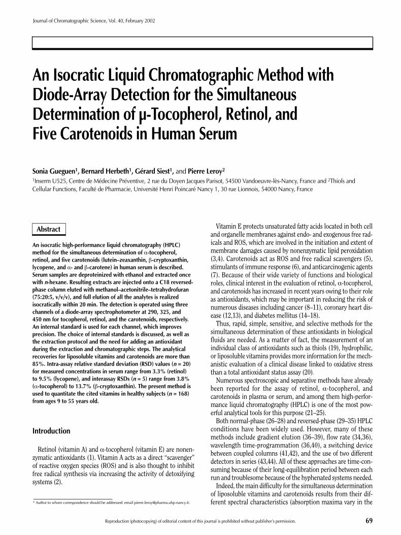
Table I. Characteristics of Standards Used
MaximumMolecular weight wavelength
Compounds (g/mol) (nm) A1%1 cm* ε (mol–1/L/cm–1)
Retinol 286.5 325 1835 (32,61) 52573Retinol acetate 328.5 326 1550 (32,61) 50912α-Tocopherol 430.7 292 75.8 (45) 3265α-Tocopherol acetate 472.8 290 40 (32) 1891Echinenone 550.9 458 2244 123622
(Hoffmann-Laroche data source)
Lutein–zeaxanthin 568.9 452 2765/2416 (45) 157301/137446β-Cryptoxanthin 552.9 452 2486 (45) 137451Lycopene 536.9 472 3450 (32,61) 185231β-Carotene 536.9 450 2620 (35) 140667
* In EtOH as the solvent. Data references appear in the parentheses.
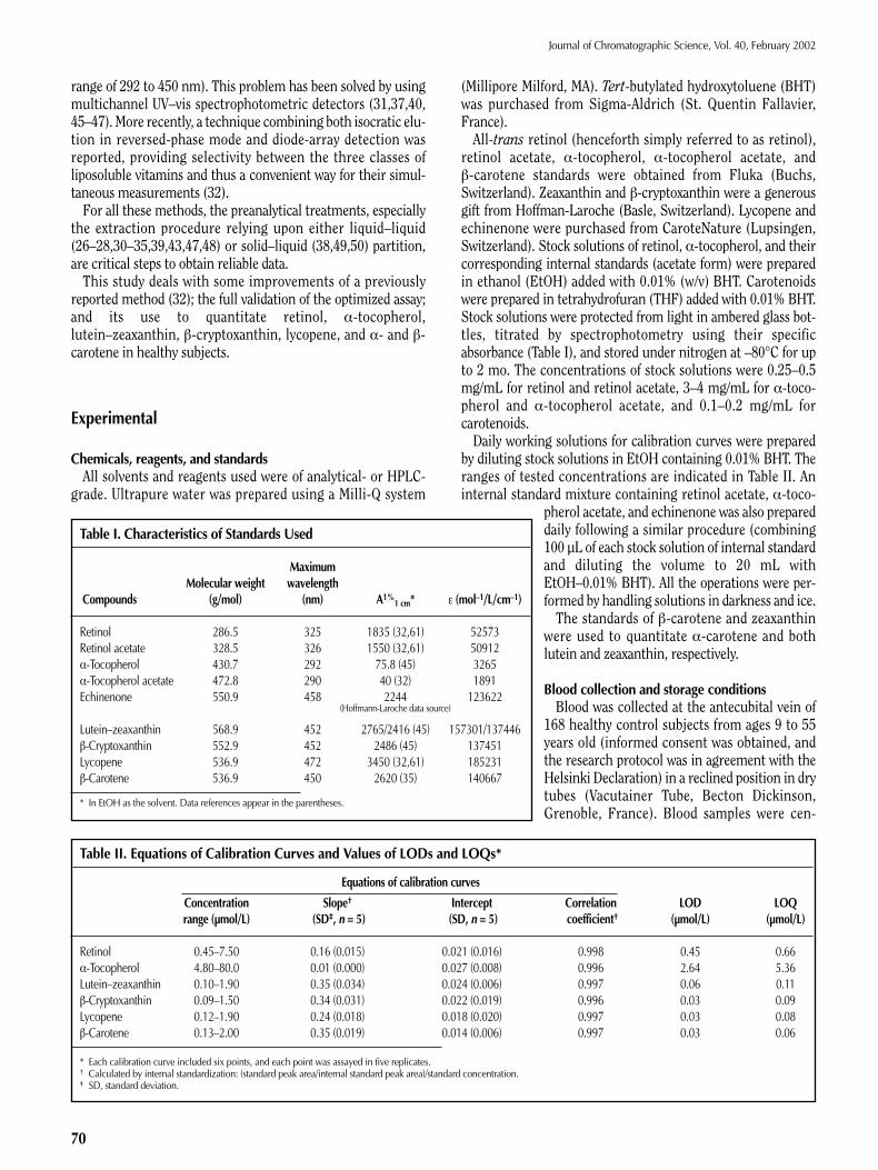
Table II. Equations of Calibration Curves and Values of LODs and LOQs*
Equations of calibration curves
Concentration Slope† Intercept Correlation LOD LOQrange (µmol/L) (SD‡, n = 5) (SD, n = 5) coefficient† (µmol/L) (µmol/L)
Retinol 0.45–7.50 0.16 (0.015) 0.021 (0.016) 0.998 0.45 0.66α-Tocopherol 4.80–80.0 0.01 (0.000) 0.027 (0.008) 0.996 2.64 5.36Lutein–zeaxanthin 0.10–1.90 0.35 (0.034) 0.024 (0.006) 0.997 0.06 0.11β-Cryptoxanthin 0.09–1.50 0.34 (0.031) 0.022 (0.019) 0.996 0.03 0.09Lycopene 0.12–1.90 0.24 (0.018) 0.018 (0.020) 0.997 0.03 0.08β-Carotene 0.13–2.00 0.35 (0.019) 0.014 (0.006) 0.997 0.03 0.06
* Each calibration curve included six points, and each point was assayed in five replicates.† Calculated by internal standardization: (standard peak area/internal standard peak area)/standard concentration.‡ SD, standard deviation.
Journal of Chromatographic Science, Vol. 40, February 2002
71
trifuged (1500 g for 15 min at 4°C) within 2 h after collection,and resulting serum samples were frozen in liquid nitrogen untilHPLC analysis.
Serum sample treatmentAll the handling operations were carried out in darkness. The
serum samples were rapidly thawed at room temperature,homogenized, and 200 µL was transferred into a borosilicateglass tube kept on ice and 300 µL of the internal standard mix-ture added. Aftermixing with a vortex for 20 s, proteins were pre-cipitated by adding 200 µL of EtOH–0.01% BHT, and the volumewas diluted to 1 mL with ultrapure water. After mixing with anorbital shaker at 2500 rpm for 1 min, 2 mL of n-hexane–0.01%BHT was added. The samples were shaken for 1 min and cen-trifuged at 2700 g for 20 min at 4°C.The organic layer was carefully transferred into a glass tube
and evaporated to dryness under a stream of nitrogen at roomtemperature. The dried residue was redissolved in 25 µL ofTHF–0.01% BHT and vortexed for 30 s. A 75-µL amount ofmobile phase was added, and the resulting mixture was vortexedfor another 30 s. Samples were then transferred to 200-µL insertvials and placed into the HPLC autosampler.
HPLC system and operating conditionsThe HPLC system consisted of an isocratic solvent delivery
pump (Model Kontron Instruments 422), an autosamplerequipped with a 20-µL injection loop, a cooling sample tray anda column oven (Model AS-300, ThermoQuest, Les Ulis, France),a UV–vis diode-array detector (Model Gold LC-168, BeckmanCoulter, Fullerton, CA), and data-processing software (Gold New,Beckman).A guard column (8- × 3-mm i.d.) packed with Nucleosil C18
(5 µm) (Macherey Nagel, Duren, Germany) and an analyticalcolumn (250- × 3-mm i.d.) packed with Nucleosil 100 C18 (5 µm)(Macherey Nagel) were eluted with a mobile phase consisting ofa mixture of methanol–acetonitrile–tetrahydrofuran (75:20:5,v/v/v) containing 0.01% (w/v) BHT. Themobile phase was filteredthrough a 0.45-µm Nylon membrane and was used at a columntemperature of 35°C and a flow rate of 0.6 mL/min. Three chan-nels corresponding with different wavelength values were usedto acquire data for the selective monitoring of α-tocopherol (290nm), retinol (325 nm), and carotenoids (450 nm) and theirrespective internal standard. During analysis, the tray compart-ment containing sample vials was cooled at 5°C. After eachworking period (approximately 50 samples), it was necessary torinse the column with methanol at a flow rate of 0.6 mL/min for20min to eliminate highly hydrophobic compounds and preventthe loss of column efficiency.
CalculationThe vitamin concentrations were determined from a standard
curve of the peak-area ratio of the analyte–internal standardplotted against the concentration of analyte (expressed in micro-moles per liter). A linear least-square regression analysis wasperformed for each analyte, and the standard curve was repeatedif the correlation coefficient was below 0.990.The detection limit (LOD) and the quantitation limit (LOQ)
were expressed, respectively, as:
LOD = (a0 + 3sa0) / a1 Eq. 1
and
LOQ = (a0 +10sa0) / a1 Eq. 2
where a1 is the slope, a0 the intercept, and sa0 the standard devi-ation of the intercept (51).
Quality controlA human serum pool made with 1mL of fresh serum from 100
healthy subjects and stored at –80°C was used for the routinequality control. Aliquots were extracted and analyzed accordingto the same procedure that was described previously. Evaluationof the method performance was assessed by comparing theresults of the quality control with the means and relative stan-dard deviations (RSDs) calculated using results from several pre-liminary runs (n = 20 per day for five days).
Results and Discussion
Optimization of sample treatment and HPLC techniqueThe basic method used in this study has been described by
Talwar et al. (32). Some modifications relating to the internalstandards, the sample preparation procedure, and the use of anantioxidant during both the extraction and chromatography pro-cesses have been made. We chose this method because it allowsin a fast and easy way the simultaneous separation of the twoclasses of lipophilic vitamins (namely retinol, α-tocopherol, andcarotenoids). Ourmain objective was tomeasure simultaneouslylipophilic vitamins and carotenoids, which are the most abun-dant in human serum. Thus, the separation of the isomers ofretinol, α-tocopherol, and carotenoids did not appear relevantfor our present epidemiological studies.In most methods, the use of an antioxidant during sample
treatment was demonstrated to be necessary to prevent a signif-icant loss in carotenoid contents, especially lycopene andβ-carotene (32,37,39,40,47). Thus, we initially added 0.01%ascorbic acid to the organic solvents used for the standardspreparation (EtOH and THF) and to the mobile phase, as indi-cated by Talwar et al. (32). After analyzing the same sampleseveral times, we observed a decrease of the carotenoid concen-trations, indicating degradation as a function of time. We testedanother antioxidant (BHT) that is widely used in other methods(37,39,47) and added it to the mobile phase and all the solvents(EtOH, THF, and hexane) used for the standard and samplepreparation. Indeed, hexane containing BHT efficiently pro-tected the carotenoids from degradation during the evaporationof the extractive organic layers, and the addition of BHT to themobile phase also prevented any loss of these analytes and prob-ably helped increase the longevity of the column by neutralizingperoxides present in THF. Moreover, we observed that decreasingthe evaporation temperature from 40°C to room temperaturesignificantly increased carotenoid recoveries, as already noted bydifferent authors (39,43).Other parameters have to be optimized in order to provide the
best conditions for the extraction of liposoluble vitamins and
Journal of Chromatographic Science, Vol. 40, February 2002
72
carotenoids. The addition of ultrapure water to the deproteinizedserum with EtOH has been noted to improve the recoveries ofcarotenoids and liposoluble vitamins (37,52). We tested severalEtOH–water proportions in the 1:4 to 1:1 range (v/v) in order toobtain the highest recoveries, and we selected the 1:1 (v/v) pro-portion. Single and double extraction steps with n-hexane (anincrease of the shaking period) were tested, but no significantimprovement of recoveries was observed.The method previously described (32) used two internal stan-
dards: retinol acetate as an internal standard for retinol, andα-tocopherol acetate as an internal standard for both α-toco-pherol and carotenoid. We used a third internal standard (echi-
nenone) for the quantitation of the carotenoids. Echinenone is asynthetic carotenoid and has a structure and chemical propertiesvery similar to the naturally occurring carotenoids in serum.Thus, the use of echinenone is preferable to the use of retinolacetate andα-tocopherol acetate or tocol currently used in othermethods (34,43,47), because it is detected at the same wave-length as the other carotenoids and is light- and temperature-sensitive as other carotenoids. Thus, the use of three internalstandards allows for a better quality control and helps to correctanalytical variations occurring for each liposoluble vitamin andcarotenoid during the extraction and chromatography pro-cesses.Because no loss of analytes was observed in serum extracts
kept in darkness for at least 24 h at 5°C, as already reported (39),the automation of the technique was possible with a highthroughput of samples (approximately 30 per day).Several methods have been developed to measure the main
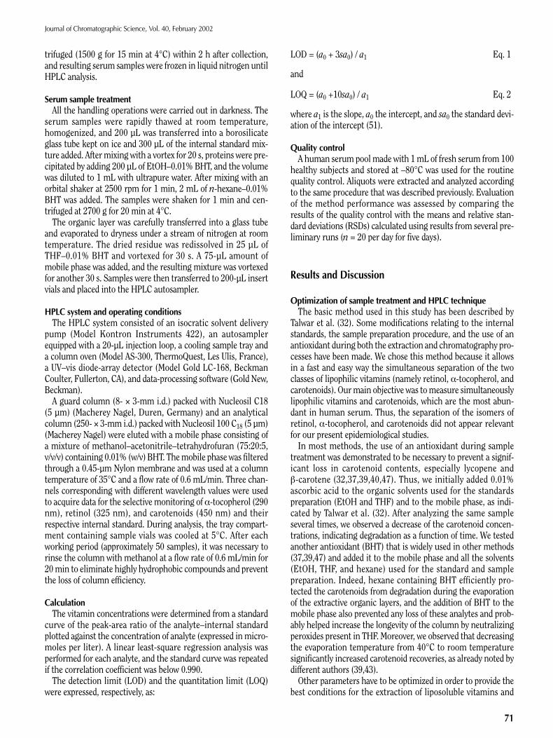
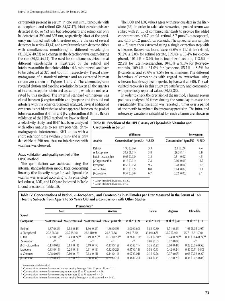
Figure 1. Typical chromatograms corresponding with a mixture of retinol,α-tocopherol, and carotenoid standards: (A) channel 1, diode-array detectionat 290 nm for α-tocopherol and α-tocopherol acetate; (B) channel 2, diode-array detection at 325 nm for retinol and retinol acetate; and (C) channel 3,diode-array detection at 450 nm for carotenoids and echinenone. The peaknumbers are as follows: (1) 26 µmol/L α-tocopherol, (2) α-tocopherol acetate(the internal standard), (3) 2.43 µmol/L retinol, (4) retinol acetate (internal stan-dard), (5) 0.62 µmol/L lutein–zeaxanthin, (6) 0.49 µmol/L β-cryptoxanthin, (7)echinenone (internal standard), (8) 0.62 µmol/L lycopene, (9) α-carotene, and(10) 0.65 µmol/L β-carotene.
A
B
C
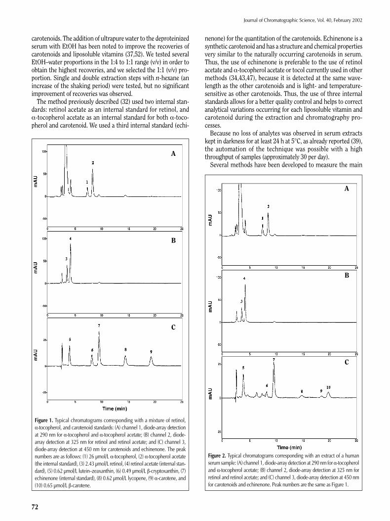
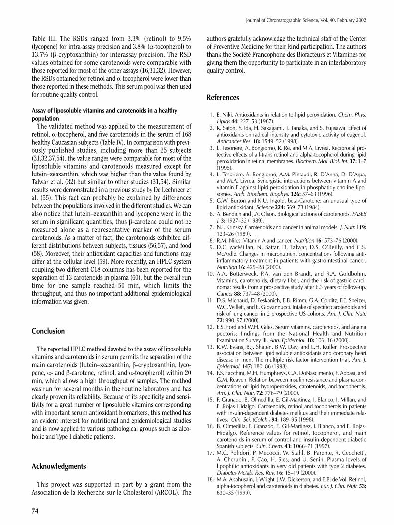
Figure 2. Typical chromatograms corresponding with an extract of a humanserum sample: (A) channel 1, diode-array detection at 290 nm for α-tocopheroland α-tocopherol acetate; (B) channel 2, diode-array detection at 325 nm forretinol and retinol acetate; and (C) channel 3, diode-array detection at 450 nmfor carotenoids and echinenone. Peak numbers are the same as Figure 1.
A
B
C
Journal of Chromatographic Science, Vol. 40, February 2002
73
carotenoids present in serum in one run simultaneously withα-tocopherol and retinol (30–34,37,47). Most carotenoids aredetected at 450 or 473 nm, butα-tocopherol and retinol can onlybe detected at 290 and 325 nm, respectively. Most of the previ-ously mentioned methods therefore require the use of severaldetectors in series (43,44) and amultiwavelength detector eitherwith simultaneous monitoring at different wavelengths(31,36,37,40,53) or a change in the detection wavelength duringthe run (30,32,44,47). The need for simultaneous detection atdifferent wavelengths is illustrated by the retinol andlutein–zeaxanthin that elute within a 0.3-min interval and haveto be detected at 325 and 450 nm, respectively. Typical chro-matograms of a standard mixture and an extracted humanserum are shown in Figures 1 and 2. The chromatogramsrevealed elution and baseline resolution between all the analytesof interest except for lutein and zeaxanthin, which are not sepa-rated by this method. The internal standard echinenone waseluted between β-cryptoxanthin and lycopene and thus did notinterfere with the other carotenoids analyzed. Several additionalcarotenoids not identified as of yet appeared between the peak oflutein–zeaxanthin at 4min and β-cryptoxanthin at 8min. Beforevalidation of the HPLC method, we have realizeda selectivity study, and BHT has been analyzedwith other analytes to see any potential chro-matographic interference. BHT elutes with ashort retention time (within 3 min) and is onlydetectable at 290 nm, thus no interference withvitamins was observed.
Assay validation and quality control of theHPLC methodThe quantitation was achieved using the
internal standardization mode. Data concerninglinearity (the linearity range for each liposolublevitamin was selected according to its physiolog-ical values), LOD, and LOQ are indicated in TableII (and precision in Table III).
The LOD and LOQ values agree with previous data in the liter-ature (32). In order to calculate recoveries, a pooled serum wasspiked with 20 µL of combined standards to provide the addedconcentrations of 0.7 µmol/L retinol, 8.7 µmol/L α-tocopherol,and 0.15 to 0.2 µmol/L carotenoids. The spiked serum samples(n = 5) were then extracted using a single extraction step withn-hexane. Recoveries found were 99.6% ± 11.1% for retinol,91.2% ± 2.0% for retinol acetate, 109.4% ± 13.4% for α-toco-pherol, 101.2% ± 3.0% for α-tocopherol acetate, 112.6% ±22.2% for lutein–zeaxanthin, 104.3% ± 9.1% for β-crypto-xanthin, 109.4% ± 31.0% for lycopene, 85.1% ± 8.5% forβ-carotene, and 95.6% ± 9.5% for echinenone. The differentbehaviors of carotenoids with regard to extraction usingn-hexane has already been reported by Barua et al. (48). The cal-culated recoveries in this study are satisfactory and comparablewith previously reported values (30,32,33).In order to check the precision of the method, a human serum
pool was analyzed 20 times during the same day to assess therepeatability. This operation was repeated 5 times over a periodof onemonth to evaluate the interassay precision. The intra- andinterassay variations calculated for each vitamin are shown in
Table III. Precision of the HPLC Assay of Liposoluble Vitamins andCarotenoids in Serum
Within run Between run
Analyte Concentration* (µmol/L) %RSD Concentration† (µmol/L) %RSD
Retinol 1.90 (0.06) 3.3 2.1 (0.09) 4.4α-Tocopherol 34.9 (1.31) 3.8 29.3 (1.1) 3.8Lutein–zeaxanthin 0.65 (0.02) 3.8 0.51 (0.02) 4.5β-Cryptoxanthin 0.13 (0.01) 7.8 0.10 (0.01) 13.7Lycopene 0.53 (0.05) 9.5 0.28 (0.04) 12.5α-Carotene 0.18 (0.02) 8.8 0.14 (0.02) 12.1β-Carotene 0.57 (0.04) 6.7 0.52 (0.05) 9.1
* Mean (standard deviation), n = 20.† Mean (standard deviation), n = 5.
Table IV. Concentrations of Retinol, α-Tocopherol, and Carotenoids in Millimoles per Liter Measured in the Serum of 168Healthy Subjects from Ages 9 to 55 Years Old and a Comparison with Other Studies
Present study*Men Women Talwar Steghens Olmedilla
Sowell
Compound 9–20 years old 21–55 years old 9–20 years old 21–55 years old et al.*,† (32) et al.*,‡ (37) et al.*,§ (54) et al.**,†† (31)
Retinol 1.37 (0.36) 2.18 (0.43) 1.36 (0.31) 1.86 (0.53) 2.00 (0.60) 1.84 (0.80) 1.71 (0.39) 1.91 (1.05–2.97)α-Tocopherol 20.6 (4.08) 29.7 (8.16) 23.6 (10.9) 26.6 (6.38) 29.6 (7.60) 33.0 (6.67) 32.7 (7.40) 25.7 (13.9–47.0)Lutein 0.42 (0.12)‡‡ 0.43 (0.24)‡‡ 0.49 (0.23)‡‡ 0.52 (0.25)‡‡ 0.26 (0.11)‡‡ 0.71 (0.30)‡‡ 0.24 (0.21)‡‡ 0.36 (0.14–0.74)‡‡
Zeaxanthin –‡‡ –‡‡ –‡‡ –‡‡ –‡‡ 0.09 (0.05) 0.07 (0.04) –‡‡
β-Cryptoxanthin 0.13 (0.08) 0.13 (0.11) 0.19 (0.14) 0.17 (0.12) 0.55 (0.11) 0.35 (0.27) 0.60 (0.47) 0.22 (0.05–0.52)Lycopene 0.33 (0.16) 0.28 (0.16) 0.31 (0.16) 0.32 (0.22) 0.37 (0.18) 0.56 (0.43) 0.42 (0.24) 0.40 (0.11–0.80)α-Carotene 0.08 (0.06) 0.10 (0.13) 0.13 (0.11) 0.14 (0.14) 0.07 (0.04) 0.36 (0.26) 0.07 (0.05) 0.08 (0.02–0.22)β-Carotene 0.49 (0.43) 0.42 (0.29) 0.60 (0.37) 0.64 (0.72) 0.38 (0.20) 0.81 (0.45) 0.37 (0.23) 0.34 (0.07–0.88)
* Means (standard deviation).† Concentrations in serum for men and women ranging from ages 19 to 62 years old, n = 111.‡ Concentrations in serum for women ranging from ages 35 to 50 years old, n = 96.§ Concentrations in serum for women ranging from ages 25 to 59 years old, n = 54.
** Concentrations in serum for men and women ranging from ages 4 to 93 years old, n = 3480.
Journal of Chromatographic Science, Vol. 40, February 2002
74
Table III. The RSDs ranged from 3.3% (retinol) to 9.5%(lycopene) for intra-assay precision and 3.8% (α-tocopherol) to13.7% (β-cryptoxanthin) for interassay precision. The RSDvalues obtained for some carotenoids were comparable withthose reported for most of the other assays (16,31,32). However,the RSDs obtained for retinol and α-tocopherol were lower thanthose reported in these methods. This serum pool was then usedfor routine quality control.
Assay of liposoluble vitamins and carotenoids in a healthypopulationThe validated method was applied to the measurement of
retinol, α-tocopherol, and five carotenoids in the serum of 168healthy Caucasian subjects (Table IV). In comparison with previ-ously published studies, including more than 25 subjects(31,32,37,54), the value ranges were comparable for most of theliposoluble vitamins and carotenoids measured except forlutein–zeaxanthin, which was higher than the value found byTalwar et al. (32) but similar to other studies (31,54). Similarresults were demonstrated in a previous study by De Leehneer etal. (55). This fact can probably be explained by differencesbetween the populations involved in the different studies. We canalso notice that lutein–zeaxanthin and lycopene were in theserum in significant quantities, thus β-carotene could not bemeasured alone as a representative marker of the serumcarotenoids. As a matter of fact, the carotenoids exhibited dif-ferent distributions between subjects, tissues (56,57), and food(58). Moreover, their antioxidant capacities and functions maydiffer at the cellular level (59). More recently, an HPLC systemcoupling two different C18 columns has been reported for theseparation of 13 carotenoids in plasma (60), but the overall runtime for one sample reached 50 min, which limits thethroughput, and thus no important additional epidemiologicalinformation was given.
Conclusion
The reported HPLCmethod devoted to the assay of liposolublevitamins and carotenoids in serum permits the separation of themain carotenoids (lutein–zeaxanthin, β-cryptoxanthin, lyco-pene, α- and β-carotene, retinol, and α-tocopherol) within 20min, which allows a high throughput of samples. The methodwas run for several months in the routine laboratory and hasclearly proven its reliability. Because of its specificity and sensi-tivity for a great number of liposoluble vitamins correspondingwith important serum antioxidant biomarkers, this method hasan evident interest for nutritional and epidemiological studiesand is now applied to various pathological groups such as alco-holic and Type I diabetic patients.
Acknowledgments
This project was supported in part by a grant from theAssociation de la Recherche sur le Cholesterol (ARCOL). The
authors gratefully acknowledge the technical staff of the Centerof Preventive Medicine for their kind participation. The authorsthank the Société Francophone des Biofacteurs et Vitamines forgiving them the opportunity to participate in an interlaboratoryquality control.
References
1. E. Niki. Antioxidants in relation to lipid peroxidation. Chem. Phys.Lipids 44: 227–53 (1987).
2. K. Satoh, Y. Ida, H. Sakagami, T. Tanaka, and S. Fujisawa. Effect ofantioxidants on radical intensity and cytotoxic activity of eugenol.Anticancer Res. 18: 1549–52 (1998).
3. L. Tesoriere, A. Bongiorno, R. Re, and M.A. Livrea. Reciprocal pro-tective effects of all-trans retinol and alpha-tocopherol during lipidperoxidation in retinal membranes. Biochem.Mol. Biol. Int. 37: 1–7(1995).
4. L. Tesoriere, A. Bongiorno, A.M. Pintaudi, R. D’Anna, D. D’Arpa,and M.A. Livrea. Synergistic interactions between vitamin A andvitamin E against lipid peroxidation in phosphatidylcholine lipo-somes. Arch. Biochem. Biophys. 326: 57–63 (1996).
5. G.W. Burton and K.U. Ingold. beta-Carotene: an unusual type oflipid antioxidant. Science 224: 569–73 (1984).
6. A. Bendich and J.A. Olson. Biological actions of carotenoids. FASEBJ. 3: 1927–32 (1989).
7. N.I. Krinsky. Carotenoids and cancer in animal models. J. Nutr. 119:123–26 (1989).
8. R.M. Niles. Vitamin A and cancer. Nutrition 16: 573–76 (2000).9. D.C. McMillan, N. Sattar, D. Talwar, D.S. O’Reilly, and C.S.
McArdle. Changes in micronutrient concentrations following anti-inflammatory treatment in patients with gastrointestinal cancer.Nutrition 16: 425–28 (2000).
10. A.A. Botterweck, P.A. van den Brandt, and R.A. Goldbohm.Vitamins, carotenoids, dietary fiber, and the risk of gastric carci-noma: results from a prospective study after 6.3 years of follow-up.Cancer 88: 737–48 (2000).
11. D.S. Michaud, D. Feskanich, E.B. Rimm, G.A. Colditz, F.E. Speizer,W.C. Willett, and E. Giovannucci. Intake of specific carotenoids andrisk of lung cancer in 2 prospective US cohorts. Am. J. Clin. Nutr.72: 990–97 (2000).
12. E.S. Ford and W.H. Giles. Serum vitamins, carotenoids, and anginapectoris: findings from the National Health and NutritionExamination Survey III. Ann. Epidemiol. 10: 106–16 (2000).
13. R.W. Evans, B.J. Shaten, B.W. Day, and L.H. Kuller. Prospectiveassociation between lipid soluble antioxidants and coronary heartdisease in men. The multiple risk factor intervention trial. Am. J.Epidemiol. 147: 180–86 (1998).
14. F.S. Facchini, M.H. Humphreys, C.A. DoNascimento, F. Abbasi, andG.M. Reaven. Relation between insulin resistance and plasma con-centrations of lipid hydroperoxides, carotenoids, and tocopherols.Am. J. Clin. Nutr. 72: 776–79 (2000).
15. F. Granado, B. Olmedilla, E. Gil-Martinez, I. Blanco, I. Millan, andE. Rojas-Hidalgo. Carotenoids, retinol and tocopherols in patientswith insulin-dependent diabetes mellitus and their immediate rela-tives. Clin. Sci. (Colch.) 94: 189–95 (1998).
16. B. Olmedilla, F. Granado, E. Gil-Martinez, I. Blanco, and E. Rojas-Hidalgo. Reference values for retinol, tocopherol, and maincarotenoids in serum of control and insulin-dependent diabeticSpanish subjects. Clin. Chem. 43: 1066–71 (1997).
17. M.C. Polidori, P. Mecocci, W. Stahl, B. Parente, R. Cecchetti,A. Cherubini, P. Cao, H. Sies, and U. Senin. Plasma levels oflipophilic antioxidants in very old patients with type 2 diabetes.Diabetes Metab. Res. Rev. 16: 15–19 (2000).
18. M.A. Abahusain, J. Wright, J.W. Dickerson, and E.B. de Vol. Retinol,alpha-tocopherol and carotenoids in diabetes. Eur. J. Clin. Nutr. 53:630–35 (1999).
Journal of Chromatographic Science, Vol. 40, February 2002
75
19. J.F. Salazar, H. Schorr, W. Herrmann, B. Herbeth, G. Siest, andP. Leroy. Measurement of thiols in human plasma using liquid chro-matography with precolumn derivatization and fluorescence detec-tion. J. Chromatogr. Sci. 37: 469–76 (1999).
20. G. Cao and R.L. Prior. Comparison of different analytical methodsfor assessing total antioxidant capacity of human serum. Clin.Chem. 44: 1309–15 (1998).
21. T. Arnhold and H. Nau. “Vitamin A”. In Modern AnalyticalMethodologies in Fat- and Water-Soluble Vitamins. John Wiley &Sons, Inc., New York, NY, 2000, pp. 3–49.
22. H.J. Nelis, E. D’Haese, and K. Vermis. “Vitamin E”. In ModernChromatographic Analysis of Vitamins, 3rd ed. Marcel Dekker, Inc.New York, NY, 2000, pp. 143–228.
23. R.R. Eitenmiller and W.O. Landen. “Simultaneous Determination ofFat-Soluble Vitamins in Food, Feed, and Serum”. In ModernAnalytical Methodologies in Fat- and Water-Soluble Vitamins. JohnWiley & Sons, Inc, New York, 2000, pp. 171–221.
24. A.B. Barua, H.C. Furr, J.A. Olson, and R.B. van Breemen. “VitaminA and Carotenoids”. In Modern Chromatographic Analysis ofVitamins, 3rd ed. Marcel Dekker, Inc., New York, NY, 2000, 1–74.
25. R.R. Eitenmiller and W. Landen. Vitamin Analysis for the Health andFood Sciences. CRC Press, Boca Raton, FL, 1998, 1–501.
26. R. Ohmacht, G. Toth, and G. Voigt. Separation of serum carotenoidsand vitamin A on chromsil-amino and -cyano phases by a bi-direc-tional gradient elution technique. J. Chromatogr. 395: 609–12(1987).
27. G. Panfili, P. Manzi, and L. Pizzoferrato. High-performance liquidchromatographic method for the simultaneous determination oftocopherols, carotenes, and retinol and its geometric isomers inItalian cheeses. Analyst 119: 1161–65 (1994).
28. R.B. McGeachin and C.A. Bailey. Determination of carotenoid pig-ments, retinol, and alpha-tocopherol in feeds, tissues, and bloodserum by normal phase high performance liquid chromatography.Poult. Sci. 74: 407–11 (1995).
29. P. Grolier. “Dosages des Caroténoïdes Sanguins”. In Le statut vita-minique. G. Le Moël, A. Saverot-Dauvergne, T. Gousson, and J.L.Guéant, Eds. EM Int., Cachan., France, 1998, pp. 42–48.
30. Z. Zaman, P. Fielden, and P.G. Frost. Simultaneous determination ofvitamins A and E and carotenoids in plasma by reversed-phaseHPLC in elderly and younger subjects. Clin. Chem. 39: 2229–34(1993).
31. A.L. Sowell, D.L. Huff, P.R. Yeager, S.P. Caudill, and E.W. Gunter.Retinol, alpha-tocopherol, lutein/zeaxanthin, beta-cryptoxanthin,lycopene, alpha-carotene, trans-beta-carotene, and four retinylesters in serum determined simultaneously by reversed-phase HPLCwith multiwavelength detection. Clin. Chem. 40: 411–16 (1994).
32. D. Talwar, T.K. Ha, J. Cooney, C. Brownlee, and D.S. O’Reilly.A routine method for the simultaneous measurement of retinol,alpha- tocopherol and five carotenoids in human plasma by reversephase HPLC. Clin. Chim. Acta 270: 85–100 (1998).
33. D. Hess, H.E. Keller, B. Oberlin, R. Bonfanti, and W. Schuep.Simultaneous determination of retinol, tocopherols, carotenes andlycopene in plasma by means of high-performance liquid chro-matography on reversed phase. Int. J. Vitam. Nutr. Res. 61: 232–38(1991).
34. M.A. Abahusain, J. Wright, J.W. Dickerson, M.A. el Hazmi, andH.Y. Aboul Enein. Determination of retinol, alpha-tocopherol,alpha- and beta-carotene by direct extraction of human serum usinghigh performance liquid chromatography. Biomed. Chromatogr. 12:89–93 (1998).
35. J.P. Steghens, B. Lyan, G. Le Moël, C. Galabert, V. Fayol, H. Faure,P. Grolier, N. Cheribi, F. Dubois, and F. Nabet. Measurement ofcarotenoids by high pressure liquid chromatography: from difficul-ties to solutions. Ann. Biol. Clin. (Paris) 58: 327–35 (2000).
36. O. Sommerburg, L.Y. Zang, and F.J. van Kuijk. Simultaneous detec-tion of carotenoids and vitamin E in human plasma. J. Chromatogr.B Biomed. Sci. Appl. 695: 209–15 (1997).
37. J.P. Steghens, A.L. van Kappel, E. Riboli, and C. Collombel.Simultaneous measurement of seven carotenoids, retinol and alpha-
tocopherol in serum by high-performance liquid chromatography.J. Chromatogr. B Biomed. Sci. Appl. 694: 71–81 (1997).
38. H.M. Arafa, F.M. Hamada, M.M. Elmazar, and H. Nau. Fully auto-mated determination of selective retinoic acid receptor ligands inmouse plasma and tissue by reversed-phase liquid chromatographycoupled on-line with solid-phase extraction. J. Chromatogr. A 729:125–36 (1996).
39. Q. Su, K.G. Rowley, and K. O’Dea. Stability of individualcarotenoids, retinol and tocopherols in human plasma during expo-sure to light and after extraction. J. Chromatogr. B Biomed. Sci.Appl. 729: 191–98 (1999).
40. D.W. Nierenberg and S.L. Nann. A method for determining con-centrations of retinol, tocopherol, and five carotenoids in humanplasma and tissue samples. Am. J. Clin. Nutr. 56: 417–26 (1992).
41. A. Sobczak, B. Skop, and B. Kula. Simultaneous determination ofserum retinol and alpha- and gamma-tocopherol levels in type IIdiabetic patients using high-performance liquid chromatographywith fluorescence detection. J. Chromatogr. B Biomed. Sci. Appl.730: 265–71 (1999).
42. H. Moriyama, H. Yamasaki, S. Masumoto, K. Adachi, N. Katsura,and T. Onimaru. Rapid determination of vitamins A and E in serumwith surfactant as a diluent by column-switching high-performanceliquid chromatography. J. Chromatogr. A 798: 125–30 (1998).
43. L. Yakushina and A. Taranova. Rapid HPLC simultaneous determi-nation of fat-soluble vitamins, including carotenoids, in humanserum. J. Pharm. Biomed. Anal. 13: 715–18 (1995).
44. K.S. Epler, R.G. Ziegler, and N.E. Craft. Liquid chromatographicmethod for the determination of carotenoids, retinoids and toco-pherols in human serum and in food. J. Chromatogr. 619: 37–48(1993).
45. R.W. Browne and D. Armstrong. Simultaneous determination ofserum retinol, tocopherols, and carotenoids by HPLC. MethodsMol. Biol. 108: 269–75 (1998).
46. J.R. Lane, L.W. Webb, and R.V. Acuff. Concurrent liquid chromato-graphic separation and photodiode array detection of retinol, toco-pherols, all-trans-alpha-carotene, all-trans-beta-carotene and themono-cis isomers of beta-carotene in extracts of human plasma.J. Chromatogr. A 787: 111–18 (1997).
47. M.H. Bui. Simple determination of retinol, alpha-tocopherol andcarotenoids (lutein, all-trans-lycopene, alpha- and beta-carotenes)in human plasma by isocratic liquid chromatography.J. Chromatogr. B Biomed. Appl. 654: 129–33 (1994).
48. S. Barua, S. Tarannum, L. Nahar, and M. Mohiduzzaman. Retinoland alpha-tocopherol content in breast milk of Bangladeshi mothersunder low socio-economic status. Int. J. Food Sci. Nutr. 48: 13–18(1997).
49. K.E. Savolainen, K.M. Pynnonen, S.P. Lapinjoki, and M.T. Vidgren.Determination of fat-soluble vitamins in a pharmaceutical dosageform by solid-phase extraction and reversed-phase liquid chro-matography. J. Pharm. Sci. 77: 802–803 (1988).
50. H. Berg, C. Turner, L. Dahlberg, and L. Mathiasson. Determinationof food constituents based on SFE: applications to vitamins A and Ein meat and milk. J. Biochem. Biophys. Methods 43: 391–401(2000).
51. M. Feinberg. La Validation des Méthodes d’Analyse. Une ApprocheChimiométrique de l’Assurance Qualité au Laboratoire. Masson,Paris, France, 1996.
52. M. Jezequel-Cuer, G. Le Moël, J. Mounie, J. Peynet, C. Le Bizec,M.H. Vernet, Y. Artur, A. Laschi-Loquerie, and S. Troupel.Determination of serum or plasma alpha-tocopherol by high perfor-mance liquid chromatography: optimization of operative models.Ann. Biol. Clin. (Paris) 53: 343–52 (1995).
53. J. Arnaud, I. Fortis, S. Blachier, D. Kia, and A. Favier. Simultaneousdetermination of retinol, alpha-tocopherol and beta-carotene inserum by isocratic high-performance liquid chromatography.J. Chromatogr. 572: 103–16 (1991).
54. B. Olmedilla, F. Granado, I. Blanco, and E. Rojas-Hidalgo. Seasonaland sex-related variations in six serum carotenoids, retinol, andalpha-tocopherol. Am. J. Clin. Nutr. 60: 106–10 (1994).
Journal of Chromatographic Science, Vol. 40, February 2002
76
55. A.P. De Leenheer, H.J. Nelis, W.E. Lambert, and R.M. Bauwens.Chromatography of fat-soluble vitamins in clinical chemistry.J. Chromatogr. 429: 3–58 (1988).
56. W. Stahl, W. Schwarz, A.R. Sundquist, and H. Sies. cis-trans Isomersof lycopene and beta-carotene in human serum and tissues. Arch.Biochem. Biophys. 294: 173–77 (1992).
57. W. Stahl and H. Sies. Uptake of lycopene and its geometrical iso-mers is greater from heat-processed than from unprocessed tomatojuice in humans. J. Nutr. 122: 2161–66 (1992).
58. A.R. Mangels, J.M. Holden, G.R. Beecher, M.R. Forman, andE. Lanza. Carotenoid content of fruits and vegetables: an evaluationof analytic data. J. Am. Diet. Assoc. 93: 284–96 (1993).
59. F. Khachik, G.R. Beecher, M.B. Goli, W.R. Lusby, and J.C. Smith.
Separation and identification of carotenoids and their oxidationproducts in the extracts of human plasma. Anal. Chem. 64: 2111–22(1992).
60. B. Lyan, V. Azais-Braesco, N. Cardinault, V. Tyssandier, P. Borel,M.C. Alexandre-Gouabau, and P. Grolier. Simple method for clin-ical determination of 13 carotenoids in human plasma using an iso-cratic high-performance liquid chromatographic method.J. Chromatogr. B Biomed. Sci. Appl. 751: 297–303 (2001).
61. The Merck Index. An Encyclopedia of Chemicals, Drugs andBiologicals. Centennial Edition, 11th ed. Merck & Co., Inc., Rahway,NY, 1989.
Manuscript accepted December 7, 2001.