anticancerofpalladium-dopedmagnesia nanoparticles
TRANSCRIPT
Short Communication
For reprint orders, please contact: [email protected]
Anticancer of palladium-doped magnesiananoparticles: synthesis, characterization,and in vitro studyMohamed Qasim Al-Fahdawi1, Abdullah Rasedee*,1,2, Faris AJ Al-Doghachi3, Rozita Rosli1,Yun Hun Taufiq-Yap4,5 & Mothanna Sadiq Al-Qubaisi11Institute of Bioscience, Universiti Putra Malaysia, Serdang, Selangor 43400 UPM, Malaysia2Department of Veterinary Pathology & Microbiology, Faculty of Veterinary Medicine, Universiti Putra Malaysia, Serdang, Selangor43400 UPM, Malaysia3Department of Chemistry, Faculty of Science, University of Basra, Basra, Iraq4Catalysis Science & Technology Research Centre, Faculty of Science, Universiti Putra Malaysia, Serdang, Selangor 43400 UPM,Malaysia5Department of Chemistry, Faculty of Science; Universiti Putra Malaysia, Serdang, Selangor 43400 UPM, Malaysia*Author for correspondence: Tel.: +603 8946 3455; Fax: +603 8946 1971; [email protected]
Aim: To prepare, physicochemically characterize and determine the anticancer of palladium-doped mag-nesia (Pd/MgO) nanoparticles. Materials & methods: Pd/MgO nanoparticles were prepared by the co-precipitation method from the aqueous solution of Mg(NO3)2.6H2O using K2CO3 and the impregnationof MgO into palladium acetylacetonate. Results: Pd/MgO nanoparticles were between 47 and 70 nmin size, cuboid in shape and tended to form aggregates. Nanoparticles were more antiproliferative to-ward cancer than the normal cells. In cancer cells, Pd/MgO nanoparticles induced apoptosis by increasingcaspase activities and stimulating cytochrome C release. The anticancer effects of Pd/MgO nanoparticleswere accentuated by the upregulation of Bax and p53 and downregulation of Bcl-2 protein expressions.Conclusion: Pd/MgO nanoparticles have potential to be developed as an anticancer compound.
First draft submitted: 1 May 2019; Accepted for publication: 25 July 2019; Published online: TBC
Keywords: apoptosis • cytotoxicity • mitochondrial pathway • noble metals • palladium-doped magnesia nanopar-ticles
Cancer, or malignant tumor, is a disease caused by abnormal cell growth. Excessive proliferation of cancer tissue cellseventually damages neighboring healthy cells and tissues with lethal outcomes. Cancer is in fact, one of the deadliestand most complicated diseases in medicine; the result of mutations in DNA responsible for the synthesis of cellularproteins. The DNA abnormalities cause production of abnormal proteins that are harmful to the organism [1,2]
Cancer treatments have evolved from surgical removal, chemotherapies, radiotherapy and immunotherapy to theuse of innovative nanoparticulated drug delivery systems with sustained effect and controlled release characteristics.These delivery systems have several advantages over pure chemotherapeutics by protecting the loaded compoundsor drugs from degradation while improving their solubility, stability, and targeted delivery [3,4].
The use of noble metals in the treatment of cancers began approximately four decades ago [5,6]. Noble metalsshown to have anticancer effects include platinum, ruthenium, rhodium, iridium, gold , and silver [7]. Amongnoble metal therapeutic compounds already marketed for treatment of cancers are the platinum-based derivatives,cisplatin, carboplatin and oxaliplatin. Cisplatin, for example was shown to target DNA [8], RNA [9], mitochondria [10]
and functional proteins [11,12]. Cisplatin can also passively or actively penetrate tumor cells, and interacts with DNAto inhibit cell proliferation [13].
The microenvironment of cancer tissues is relatively more acidic than that of normal tissues [14]. Since palladium-doped magnesia (Pd/MgO) dissociates at low pH [15], resulting in the release of palladium from the complex, theacidic cancer tissue microenvironment would facilitate delivery of the metal to exert its therapeutic effect. Pd/MgOnanoparticles can also offer an improved and targeted tumor therapeutic strategy while showing less toxic effects onnormal tissues than conventional chemotherapeutics. However, at this juncture, the full potential of the Pd/MgO
Nanomedicine (Lond.) (Epub ahead of print) ISSN 1743-588910.2217/nnm-2019-0178 C© 2019 Future Medicine Ltd
Short Communication Al-Fahdawi, Rasedee, Al-Doghachi, Rosli, Taufiq-Yap & Al-Qubaisi
nanoparticles as an anticancer agent is yet to be determined. In this study, Pd/MgO nanoparticles were developed,characterized and their anticancer properties determined in vitro.
Materials & methodsChemicals & reagentsTrypsin/ethylenediamine tetracetic acid (EDTA) solution was purchased from Invitrogen (CA, USA). Dimethylsulfoxide (DMSO), phosphate-buffered saline (PBS), 3-(4,5-dimethylthiazol-2-yl)-2,5-diphenyltetrazolium bro-mide (MTT), Dulbecco’s modified Eagle’s medium (DMEM), diphenylamine (DPA) reagent (100 ml glacial aceticacid 1.5 g diphenylamine, 1.5 ml concentrated sulfuric acid 0.5 ml and 16 mg/ml acetaldehyde stock) and trypanblue dye were purchased from Sigma Chemical Company (Perth, WA, Australia).
Preparation of Pd/MgO nanoparticlesThe Pd/MgO nanoparticles were prepared using the precipitation method as described previously [16]. MgO wasprepared using a 0.1 M aqueous solution of Mg(NO3)2.6H2O (Merck; >99.0%) and 1 M K2CO3 (Merck,Darmstadt, Germany; >99.7%) as precipitants. The sample (precipitant) was filtered and washed with hot waterand dried at 120◦C for 12 h. Subsequently, the dried samples were precalcined in air at 500◦C for 5 h to removeCO2. The sample was then pressed into discs at 600 kg/m2 (Hydrolic for KBr disc) and calcined at 1150◦C for 20h for the enhancement of mechanical properties.
The sample was impregnated with 5% Pd using Pd(C5H7O2)2. H2O, (Acros Chemicals; >99%), dissolved withdichloromethane for 5 h to produce Pd(acac)2/MgO. After impregnation in air, the samples were dried for 12 hat 120◦C, crushed and sieved (250 μm) to obtain particles of 80–150 or 150–250 μm in diameter. Finally, thePd2+ phase nanoparticles were reduced to metal Pd◦ phase at the active site of the nanoparticles using 5% H2/Arto produce Pd/MgO nanoparticles.
Physicochemical properties of nanoparticlesScanning electron microscopy & energy dispersive x-ray spectroscopy
Scanning electron microscopy (SEM, Model LEO 1450VP [LEO Electron Microscopy Ltd. Cambridge, UK]),with an accelerating voltage of 30 kV, and energy dispersive x-ray spectroscopy (EDX) were used to determine themorphology and elemental composition of powdered nanoparticles, respectively. The samples were degassed in anevacuated heated chamber at 100◦C overnight. Prior to SEM scanning, dried samples were spread over double-sidedconductive tape adhered to the specimen stub.
Transmission electron microscopy
Transmission electron microscopy (TEM, Hitachi H-7100, Japan) was used to determine the fine structure of thecrystals. The nanoparticle powder was disseminated in deionized water, placed onto carbon-cover copper grids onfilter paper and dried at room temperature before viewing.
X-ray diffraction
The x-ray diffraction (XRD) characterization of nanoparticle powder was performed using the Shimadzu diffrac-tometer model XRD 6000, Japan. The analysis employed the Cu-Kα radiation generated by a Philips glass diffractionx-ray tube broad focus 2.7 kW type at ambient temperature. The crystallite size D of the samples was calculated bythe Debye–Scherrer’s relationship according to standard procedures [17,18] using the following formula:
T = 0.9 λ / (β cosθ)
where T is the crystallite size, λ is the incident x-ray wavelength, β is the full width at half-maximum, and θ isthe diffraction angle.
Fourier transform infrared
Fourier transform infrared (FTIR) spectra for powdered nanoparticles were recorded over the range of 400–4000cm-1 on a Smart Orbit spectrometer (Thermo Nicolet Nexus, USA) using a sample of approximately 1% in 200mg of spectroscopic-grade potassium bromide (KBr) with 10 tons of pressure.
10.2217/nnm-2019-0178 Nanomedicine (Lond.) (Epub ahead of print) future science group

Anticancer of palladium-doped magnesia nanoparticles: synthesis, characterization & in vitro study Short Communication
Thermogravimetric analysis
The thermal strength of the nanoparticles was investigated using the Mettler Toledo TG-SDTA apparatus (Ptcrucibles, Pt/Pt– Rh thermocouple) (Switzerland), with a purge gas (nitrogen) flow rate of 30 ml min-1 and aheating rate of 10◦C min-1 from room temperature to 1000◦C.
Brunauer, Emmett & Teller analysis
The total surface area of the nanoparticles was evaluated via nitrogen adsorption at -196◦C. The evaluation wasperformed using a nitrogen adsorption-desorption analyser (Surfer Analyser, Milan, Italy).
Cell culture
Four human cell lines, colorectal adenocarcinoma (HT29), lung carcinoma (A549), normal colon (CCD-18Co)and normal lung (MRC-5) cells, all virus-negative, were obtained from the American Type Culture Collection(ATCC; MD, USA). The cells were cultured in DMEM (Sigma Aldrich, MO, USA) supplemented with 10% FBSand 1% penicillin (100 U/ml) (Isocillin, Aventis, Germany) in an incubator at 37◦C in the presence of 5% CO2.
3-(4,5-dimethylthiazol-2-yl)-2,5-diphenyltetrazolium bromide assay
The Pd/MgO nanoparticles were mixed well in DMEM medium (Sigma Aldrich) containing fresh 10% heat-inactivated FBS and nanoparticle colloidal suspension was obtained using ultrasound method [17,18]. Two hundredmicroliters of 5 × 10³ cells/ml suspension were added to each well of a 96-well cell culture plate to a finalconcentration 1 × 10³ cells/well. The media was aspirated and replaced with 200 μl fresh media containingnanoparticles of concentrations ranging from 1.56 to 100 μg/ml, and chemotherapeutic agents (oxaliplatin forHT29 and CCD-18Co cells, and paclitaxel for A549 and MRC-5 cells) ranging from 0.156 to 10.0 μg/ml. Thelast row was used for the nontreated control. The plate was then incubated at 37◦C under 5% CO2, for 24 h. Themedium was aspirated and the cells washed with PBS buffer trice to remove the test compounds, and fresh mediumadded. Then, 200 μl of 5 mg/ml 3-(4,5-dimethylthiazol-2-yl)-2,5-diphenyltetrazolium bromide (MTT) solutionwas added to each well and the plate incubated for 4–6 h at 37◦C under 5% CO2. The MTT-containing mediumwas then carefully removed and replaced with 200 μl DMSO/well, to dissolve the formazan crystals. The plateswere read on automated spectrophotometric EL 340 multiplate reader (Bio-Tek Instruments, Inc., VT, USA) at570 nm. The viability percentage is calculated by:
(ODtreated well) / (ODnontreated well) × 100.
OD refers to optical densityFor each test compound and cell line, the 24 h IC50 values (50% cell growth inhibition concentration) were
determined from the dose–response curves.
Caspases
The effect of treatment on HT29 and A549 cell caspase-3, -8 and -9 activities was determined using commercialcolorimetric assay kits (Promega, WI, USA) in accordance with recommended protocol.
Quantification of Bax, p53, Bcl-2 & cytochrome C protein assay
The expressions of p53, Bax, Bcl-2 and cytochrome C proteins in HT29 and A549 cells treated with Pd/MgOnanoparticles were determined using the ELISA assay kits (R&D Systems, MN, USA).
Microscopic examination of cell morphology
Two milliliters of cell suspension were seeded into six-well plates to contain 1 × 10⁴ HT29 or A549 cells/well andtreated with Pd/MgO nanoparticles at the predetermined IC50 concentrations. The cells were examined under alight inverted microscope for morphological assessment.
Statistical analysis
All experiments were done in three triplicates. Data were expressed as means ± S.D. All statistical analyses wereperformed using Minitab statistical software (Minitab, Inc., PA, USA). Treatment effects were determined using
future science group 10.2217/nnm-2019-0178
Short Communication Al-Fahdawi, Rasedee, Al-Doghachi, Rosli, Taufiq-Yap & Al-Qubaisi
one-way analysis of variance (ANOVA) followed by Tukey’s post hoc analysis. A value of p < 0.05 was consideredsignificant unless otherwise indicated.
ResultsMaterial characterizationMorphological & elemental analysis of nanoparticles
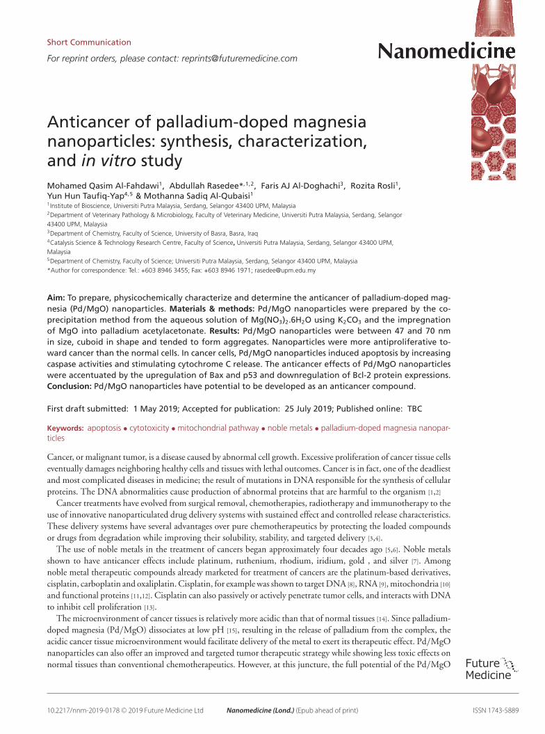
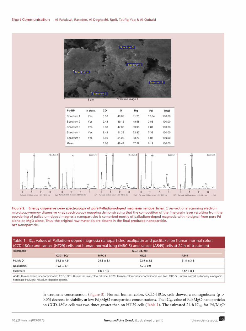
Scanning electron microscopy & EDXThe SEM and EDX micrographs for the pure Pd/MgO samples are shown in Figures 1A and 2, respectively. Thestrong interaction among nanoparticles caused the Pd/MgO nanoparticles to form aggregates.
Transmission electron microscopyThe TEM images of Pd/MgO nanoparticles are shown in Figure 2B. It seemed that the Pd/MgO nanoparticleslike the Pt/MgO are cuboid but with nanosize range of 60–79 nm and averaging at 70 nm. Most of the Pd/MgOnanoparticles were in aggregates, which is attributed to the electrostatic interaction between nanoparticles. Thisbehavior indicates that there were strong interactions among nanoparticles, which is consistent with the SEMfindings.
Crystallinity
The patterns generated by XRD instrument for Pd/MgO nanoparticles are shown in Figure 2C. High temperaturecalcination is essential for the development of the catalytically active cuboid phase of magnesium oxide. Magnesiumoxide crystallized at 500◦C. According to data obtained from powder XRD for the Pd/MgO nanoparticles calcinedat 600◦C, the magnesium oxide comprises of a cuboid system. The sharp peaks located at about 2θ = 37.0◦ (111),43.1◦ (200), 62.3◦ (220), 74.8◦ (311) and 78.9◦ (222) are attributed to the cuboid crystalline form of magnesiumoxide (JSPDS file no.: 01-081-1545). The x-ray diffraction pattern for palladium that has been incorporated ontothe surface of magnesium oxide showed stable cuboid Pd, located at 2θ = 2θ = 40.0◦ (111), 46.5◦ (200) and 68.5◦
(220) (JSPDS file no.: 01-081-1545). The size of the Pd/MgO crystals determined with the Debye–Scherrer’sequation was 47.3 nm. The BET specific surface area (SBET), pore volume and pore radius of the Pd/MgOnanoparticles were 15.7 m2/g, 0.2122 Cm3/g and 11.4◦A, respectively. The calculated pore volume-to-SBET ratiowas 13.5 10-9 m.
Infrared absorption bands
In Figure 2D, the peaks at 2936 and 1733 cm-1 in the FTIR spectrum represent a weak C–H and a strong C=Ostretching, respectively, which are attributed to chemical residues from nanoparticle preparation. On the otherhand, the peak at 3562 cm-1 represents vibrational bands, which is due to moisture in the Pg/MgO nanoparticlesample. The peaks at approximately 3154 and 1583 cm-1 represent the stretching and bending modes, respectively,of hydroxyl groups. Although the sample was subjected to high temperature calcination, the appearance of thesemodes signifies that the Bronsted acid-active sites were present on the surface of Pd/MgO nanoparticles. The bandat 3780 and 1442 cm-1 corresponds to the stretching vibration and bending, respectively, of -OH bonded to Mg.The bands at 1369, 1238, and 1165 cm-1 correspond to bending of C-H bonds in CH3, stretching of C–C, andbending of C–H, respectively. The band at 1005 and 855 cm-1 corresponds to C–O bond.
Thermogravimetric analysis
Figure 2E shows the thermogravimetric curve for Pd/MgO nanoparticles. From the curve, the weight loss wasestimated at 2%, occurring in the temperature range of 100–300◦C. This weight loss may be attributed to loss ofmoisture from the Pd/MgO nanoparticle sample. The study shows that calcination of Pd/MgO nanoparticles at atemperature range of 25–400◦C caused elimination of not only physically adsorbed water, but also loss of nitrogenoxide gases, and the transformation of MgO(OH)2 to palladium. The second mass loss began at approximately680 and ended at 1000◦C. This loss of mass is attributed to the degradation of carbon residue to CO2 gas.
Anticancer of Pd/MgO nanoparticlesCytotoxicity
The toxic effect of Pd/MgO nanoparticles on the CCD-18Co, MRC-5, HT29 and A549, was determined by MTTassay. The toxic effect of Pd/MgO nanoparticles on the HT29 and A549 cells increased exponentially with increase
10.2217/nnm-2019-0178 Nanomedicine (Lond.) (Epub ahead of print) future science group
Anticancer of palladium-doped magnesia nanoparticles: synthesis, characterization & in vitro study Short Communication
a.u
4000
2000
00
2θ
20 40 60 80
a.u
100
99
98
974000
CM-1
3500 3000 2500
3780
3562 31
54
2936
1733
2000 1500 100015
83
1005
855
116512
381369
1442
We
igh
t (%
)
100
98
0
Temp (°C)
200 400 600 800 1000
1000 nm
76.3 nm
44.5 nm
85.4 nm
78.3 nm
x 100,000 5.0 kv SEI SEM WD 4.6 mm100 nm EMUPM
Figure 1. Characterization of Pd/MgO nanoparticles by: (A) Scanning ultramicroscopic structure of pure palladium-dopedmagnesia (Pd/MgO) nanoparticles. The nanoparticles were well dispersed and sizes ranged from 41 to 65 nm. (B) Transmissionultramicroscopic structure of pure Pd/MgO nanoparticles. (C) X-ray diffraction pattern of Pd/MgO nanoparticles. The data shows thatPd/MgO nanoparticles were well-formed with definitive structural and phase purity. (D) Fourier transform infrared spectrum of Pd/MgOnanoparticles. (E) Thermograph of the Pd/MgO nanoparticles showing phase with thermal treatments.
Short Communication Al-Fahdawi, Rasedee, Al-Doghachi, Rosli, Taufiq-Yap & Al-Qubaisi
Spectrum 1
Spectrum 2
Spectrum 3
Spectrum 4
Spectrum 5
Mean
Pd-NP In stats. CO O Mg Pd Total
Yes
Yes
Yes
Yes
Yes
6.10
9.43
9.33
8.42
6.96
8.06
Spectrum 1
Spectrum 5
Spectrum 4
Spectrum 3
Spectrum 2
49.85
39.16
47.82
51.28
54.23
48.47
31.21
48.58
39.99
32.97
33.72
37.29
12.84
2.83
2.87
7.33
5.08
6.19
8 µm Electron image 1
100.00
100.00
100.00
100.00
100.00
100.00
0 1 2Full scale 11993 cts cursor: 1.227 (13139 cts) KeV
3
O
Mg
Pd
Pd PdC
Spectrum 1
0 1 2Full scale 5568 cts cursor: 2.349 (92 cts) KeV
3
O
Mg
PdPdC
Spectrum 2
0 1 2Full scale 5049 cts cursor: 1.227 (13162 cts) KeV
3
O Mg
Pd
Pd PdC
Spectrum 3
0 1 2Full scale 5140 cts cursor: 1.317 (2491 cts) KeV
3
O Mg
Pd
Pd PdC
Spectrum 4
0 1 2Full scale 18655 cts cursor: 1.167 (1316 cts) KeV
3
O
Mg
Pd
Pd PdC
Spectrum 5
Figure 2. Energy dispersive x-ray spectroscopy of pure Palladium-doped magnesia nanoparticles. Cross-sectional scanning electronmicroscopy-energy dispersive x-ray spectroscopy mapping demonstrating that the composition of the fine-grain layer resulting from thepowdering of palladium-doped magnesia nanoparticles is comprised mostly of palladium-doped magnesia with no signal from pure Pdalone or, MgO alone. Thus, the original raw materials are absent in the final produced nanoparticle.NP: Nanoparticle.
Table 1. IC50 values of Palladium-doped magnesia nanoparticles, oxalipatin and paclitaxel on human normal colon(CCD-18Co) and cancer (HT29) cells and human normal lung (MRC-5) and cancer (A549) cells at 24 h of treatment.Treatment IC50 (μg/ml)
CCD-18Co MRC-5 HT29 A549
Pd/MgO 51.6 ± 4.9 24.8 ± 3.1 22.9 ± 3.6 21.8 ± 3.8
Oxaliplatin 10.5 ± 8.1 - 4.7 ± 0.0 -
Paclitaxel - 0.8 ± 1.6 - 0.12 ± 0.1
A549: Human breast adenocarcinoma; CCD-18Co: Human normal colon cell line; HT29: Human colorectal adenocarcinoma cell line; MRC-5: Human normal pulmonary embryonicfibroblast; Pd/MgO: Palladium-doped magnesia.
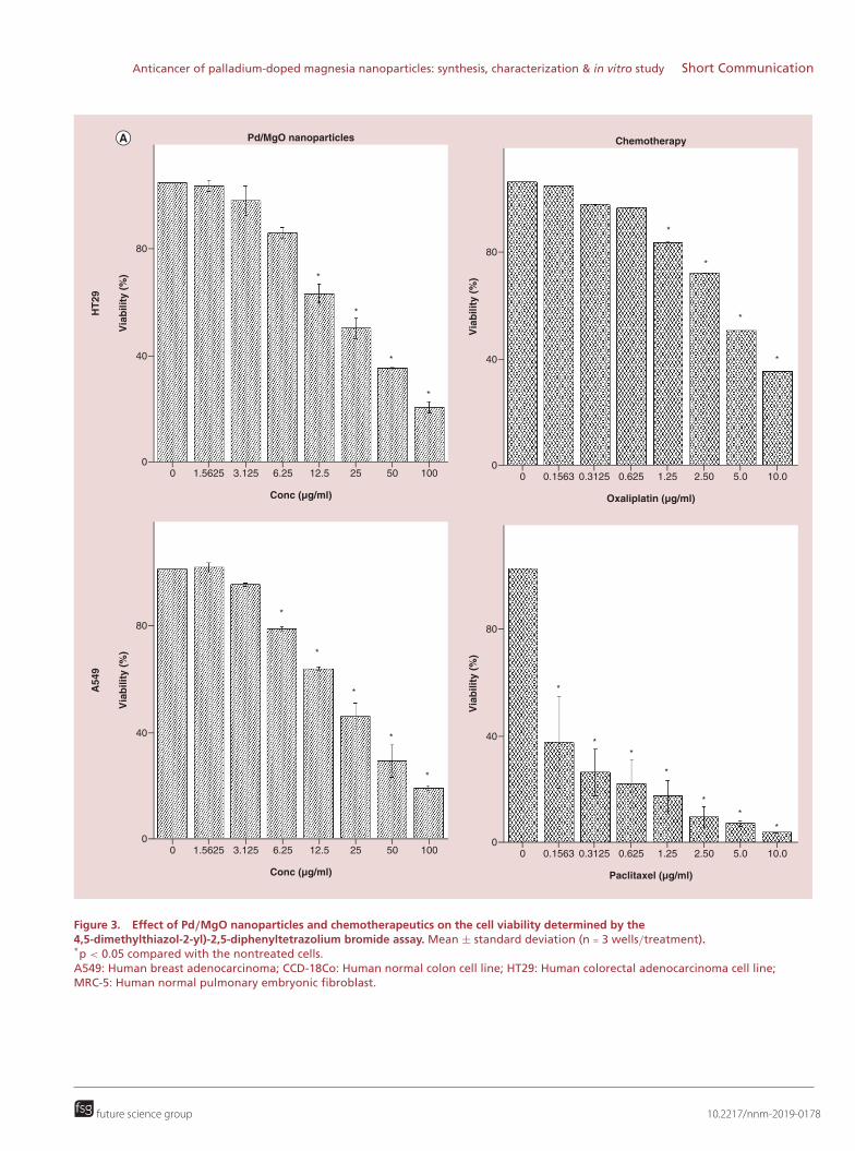
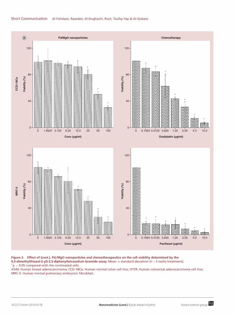
in treatment concentration (Figure 3). Normal human colon, CCD-18Co, cells showed a nonsignificant (p >
0.05) decrease in viability at low Pd/MgO nanoparticle concentrations. The IC50 value of Pd/MgO nanoparticleson CCD-18Co cells was two-times greater than on HT29 cells (Table 1). The estimated 24-h IC50 for Pd/MgO
10.2217/nnm-2019-0178 Nanomedicine (Lond.) (Epub ahead of print) future science group
Anticancer of palladium-doped magnesia nanoparticles: synthesis, characterization & in vitro study Short Communication
Via
bilit
y (
%)
HT
29
A549
80
40
00
Conc (µg/ml)
Pd/MgO nanoparticles
1.5625 3.125 6.25 12.5 25 50 100V
iab
ilit
y (
%)
80
40
00
Oxaliplatin (µg/ml)
Chemotherapy
0.1563 0.3125 0.625 1.25 2.50 5.0 10.0
Via
bilit
y (
%)
80
40
00
Conc (µg/ml)
1.5625 3.125 6.25 12.5 25 50 100
Via
bilit
y (
%)
80
40
00
Paclitaxel (µg/ml)
0.1563 0.3125 0.625 1.25 2.50 5.0 10.0
*
*
*
*
*
*
*
*
*
*
*
*
*
*
**
*
**
*
Figure 3. Effect of Pd/MgO nanoparticles and chemotherapeutics on the cell viability determined by the4,5-dimethylthiazol-2-yl)-2,5-diphenyltetrazolium bromide assay. Mean ± standard deviation (n = 3 wells/treatment).*p < 0.05 compared with the nontreated cells.A549: Human breast adenocarcinoma; CCD-18Co: Human normal colon cell line; HT29: Human colorectal adenocarcinoma cell line;MRC-5: Human normal pulmonary embryonic fibroblast.
future science group 10.2217/nnm-2019-0178
Short Communication Al-Fahdawi, Rasedee, Al-Doghachi, Rosli, Taufiq-Yap & Al-Qubaisi
Via
bilit
y (
%)
80
120
40
00
Conc (µg/ml)
Pd/MgO nanoparticles
1.5625 3.125 6.25 12.5 25 50 100
Via
bilit
y (
%)
80
120
40
00
Oxaliplatin (µg/ml)
Chemotherapy
0.1563 0.3125 0.625 1.25 2.50 5.0 10.0
Via
bilit
y (
%)
MR
C-5
CC
D-1
8C
o
80
120
40
00
Conc (µg/ml)
1.5625 3.125 6.25 12.5 25 50 100
Via
bilit
y (
%)
80
120
40
00
Paclitaxel (µg/ml)
0.1563 0.3125 0.625 1.25 2.50 5.0 10.0
*
*
*
*
*
*
*
*
*
*
*
*
*
* *
**
** *
Figure 3. Effect of (cont.). Pd/MgO nanoparticles and chemotherapeutics on the cell viability determined by the4,5-dimethylthiazol-2-yl)-2,5-diphenyltetrazolium bromide assay. Mean ± standard deviation (n = 3 wells/treatment).*p < 0.05 compared with the nontreated cells.A549: Human breast adenocarcinoma; CCD-18Co: Human normal colon cell line; HT29: Human colorectal adenocarcinoma cell line;MRC-5: Human normal pulmonary embryonic fibroblast.
10.2217/nnm-2019-0178 Nanomedicine (Lond.) (Epub ahead of print) future science group

Anticancer of palladium-doped magnesia nanoparticles: synthesis, characterization & in vitro study Short Communication
nanoparticles on the cancer cells ranged between 21 and 23 μg/ml. The Pd/MgO nanoparticles were toxic tothe normal lung, MRC-5, cells. Treatment with 12.5 μg/ml Pd/MgO nanoparticles caused approximately 8 and34% lower rate of CCD-18Co and MRC-5 cell growth than the nontreated control cells, respectively, after 24-hincubation. However, under the same conditions, incubation of these cells with 25 μg/ml Pd/MgO nanoparticles,resulted in 19 and 50% lower rate cell growth, respectively, at the same time points. The A549 cells were the mostsensitive to antiproliferative effect of paclitaxel. Overall, the effects of Pd/MgO nanoparticles and oxaliplatin onHT29 cells were similar, decreasing cell viability with increasing treatment concentrations (Figure 3).
Caspase-3, -8 & -9
Caspase-3The HT29 and A549 cells after treatment with Pd/MgO nanoparticles, at all concentrations, for 12 and 24 h,showed significantly (p < 0.05) greater caspase-3 activities than the nontreated cells (Figure 4). The activity of thisenzyme increased with increase in treatment concentrations. However, after 12 h of treatment, the anticancer effectsof Pd/MgO nanoparticles were more prominent on the A549 than HT29 cells. At the same treatment period,the caspase-3 activities in the HT29 (696%) and A549 (692%) cells lines treated with 12.5 μg/ml Ag/MgOnanoparticles, were markedly higher than in nontreated cells.
Caspase-8Unlike caspase-3, the caspase-8 activity in the tumor cells did not change significantly ( p > 0.05) with treatment(Figure 4). However, the activity of caspase-8 in the lung cancer, A549, cells was shown to be higher than innontreated cells by 1.34-fold after 24 h. Similar effect was not observed in the HT29 cells, suggesting thatPd/MgO nanoparticles is a more effective antiproliferative agent for A549 than HT29 cells.
Caspase-9The activity of caspase-9 in colon cancer, HT29, cell line after treatment with 12.5 μg/ml Pd/MgO nanoparticlewas approximately threefold higher than in nontreated cells after 12 and 24 h (Figure 4). In the A549 cellstreated with Pd/MgO nanoparticles, the activity of caspase-9 rose quickly after 12-h exposure, to approximatelyfivefold higher than in nontreated cells. By 24 h, the caspase-9 activity in treated A549 cell decreased again toonly approximate twofold higher than nontreated cells. The study shows that Pd/MgO nanoparticles increasedcaspase-9 activity in A549 cells more rapidly than in HT29 cells.
Bcl-2, Bax, p53 & cytochrome C proteins
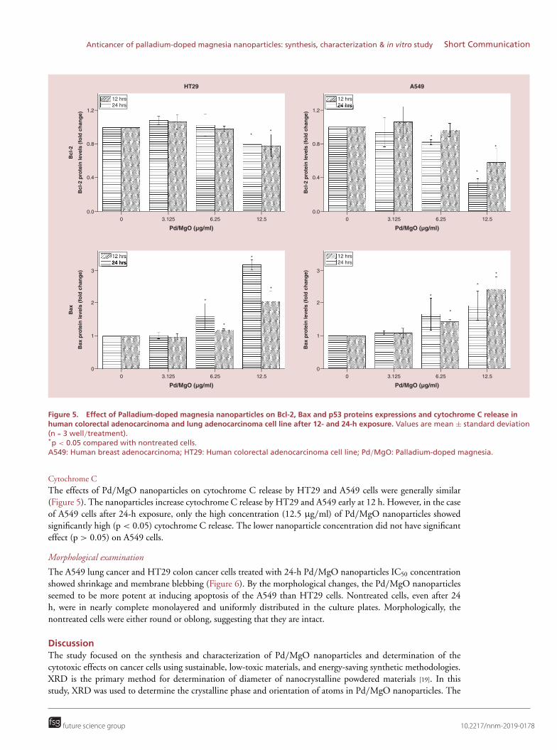
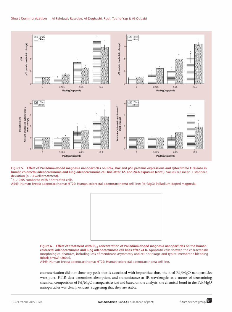
Bcl-2Pd/MgO nanoparticles at 12.5 μg/ml were more effective at downregulating the Bcl-2 protein expression in A549than HT29 cells. This was observed at both the 12- and 24-h exposure periods. The A549 cell Bcl-2 expressiondecreased to approximately 34% of the nontreated cells. In the Pd/MgO nanoparticle-treated HT29, the expressionof Bcl-2 decreased to between 78 and 80% of the nontreated cells (Figure 5).
BaxThe HT29 cell Bax protein expression after treatment with 12.5 μg/ml Pd/MgO nanoparticles increased by morethan 316%, which was between two- to threefold higher than that produced by 6.25 and 3.125 μg/ml Pd/MgOnanoparticles (Figure 5). The upregulation of Bcl-2 protein in Pd/MgO nanoparticle-treated A549 cells was lesssignificant than in the HT29 cells. These effects of Pd/MgO nanoparticle on cancer cells Bax expression weregradual and concentration-dependent.
p53P53 protein expression in HT29 and A549 cancer cells treated with 12.5 μg/ml Pd/MgO nanoparticles was four-to sevenfold higher that in nontreated cells (Figure 5). However, in the HT29 cells, at 12 h, the p53 proteinexpression became significant higher (p < 0.05) even after treatment with 3.125 μg/ml Pd/MgO nanoparticles. Itappears that the effect of Pd/MgO nanoparticles is concentration-dependent and there is no observable differencein effect between HT29 and A549 cells.
future science group 10.2217/nnm-2019-0178

Short Communication Al-Fahdawi, Rasedee, Al-Doghachi, Rosli, Taufiq-Yap & Al-Qubaisi
12 hrs24 hrs
12 hrs24 hrs
12 hrs24 hrs
12 hrs24 hrs
12 hrs24 hrs
12 hrs24 hrs
Casp
ase-3
(O
D)
Casp
ase-3
0.6
0.4
0.2
0.00
Pd/MgO (µg/ml)
HT29
3.125 6.25 12.5
Casp
ase-3
(O
D)
0.6
0.4
0.2
0.00
Pd/MgO (µg/ml)
A549
3.125 6.25 12.5
Casp
ase-8
(O
D)
Casp
ase-8
0.20
0.15
0.10
0.05
0.000
Pd/MgO (µg/ml)
3.125 6.25 12.5
Casp
ase-8
(O
D)
0.20
0.15
0.10
0.05
0.000
Pd/MgO (µg/ml)
3.125 6.25 12.5
Casp
ase-9
(O
D)
Casp
ase-9
0.4
0.2
0.00
Pd/MgO (µg/ml)
3.125 6.25 12.5
Casp
ase-9
(O
D)
0.4
0.2
0.00
Pd/MgO (µg/ml)
3.125 6.25 12.5
** *
*
**
*
*
*
* *
*
*
*
*
*
*
* *
*
*
*
Figure 4. Effect of Palladium-doped magnesia nanoparticles on human colorectal adenocarcinoma and lung adenocarcinoma cell linecaspase-3, -8 and -9 activities after 12- and 24-h exposure. Values are mean ± standard deviation (n = 3 well/treatment).*p < 0.05 compared with nontreated cells.A549: Human breast adenocarcinoma; HT29: Human colorectal adenocarcinoma cell line; OD: Optical density; Pd/MgO: Palladium-dopedmagnesia.
10.2217/nnm-2019-0178 Nanomedicine (Lond.) (Epub ahead of print) future science group
Anticancer of palladium-doped magnesia nanoparticles: synthesis, characterization & in vitro study Short Communication
12 hrs24 hrs
12 hrs24 hrs
12 hrs24 hrs
12 hrs24 hrs
Bc
l-2
pro
tein
le
ve
ls (
fold
ch
an
ge
)
Bc
l-2
1.2
0.8
0.4
0.00
Pd/MgO (µg/ml)
HT29
3.125 6.25 12.5
Ba
x p
rote
in l
ev
els
(fo
ld c
ha
ng
e)
Ba
x
3
2
1
00
Pd/MgO (µg/ml)
3.125 6.25 12.5
Ba
x p
rote
in l
ev
els
(fo
ld c
ha
ng
e) 3
2
1
00
Pd/MgO (µg/ml)
3.125 6.25 12.5
Bc
l-2
pro
tein
le
ve
ls (
fold
ch
an
ge
) 1.2
0.8
0.4
0.00
Pd/MgO (µg/ml)
A549
3.125 6.25 12.5
**
*
*
*
*
*
*
*
*
*
*
*
Figure 5. Effect of Palladium-doped magnesia nanoparticles on Bcl-2, Bax and p53 proteins expressions and cytochrome C release inhuman colorectal adenocarcinoma and lung adenocarcinoma cell line after 12- and 24-h exposure. Values are mean ± standard deviation(n = 3 well/treatment).*p < 0.05 compared with nontreated cells.A549: Human breast adenocarcinoma; HT29: Human colorectal adenocarcinoma cell line; Pd/MgO: Palladium-doped magnesia.
Cytochrome CThe effects of Pd/MgO nanoparticles on cytochrome C release by HT29 and A549 cells were generally similar(Figure 5). The nanoparticles increase cytochrome C release by HT29 and A549 early at 12 h. However, in the caseof A549 cells after 24-h exposure, only the high concentration (12.5 μg/ml) of Pd/MgO nanoparticles showedsignificantly high (p < 0.05) cytochrome C release. The lower nanoparticle concentration did not have significanteffect (p > 0.05) on A549 cells.
Morphological examination
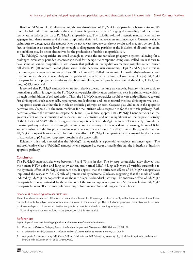
The A549 lung cancer and HT29 colon cancer cells treated with 24-h Pd/MgO nanoparticles IC50 concentrationshowed shrinkage and membrane blebbing (Figure 6). By the morphological changes, the Pd/MgO nanoparticlesseemed to be more potent at inducing apoptosis of the A549 than HT29 cells. Nontreated cells, even after 24h, were in nearly complete monolayered and uniformly distributed in the culture plates. Morphologically, thenontreated cells were either round or oblong, suggesting that they are intact.
DiscussionThe study focused on the synthesis and characterization of Pd/MgO nanoparticles and determination of thecytotoxic effects on cancer cells using sustainable, low-toxic materials, and energy-saving synthetic methodologies.XRD is the primary method for determination of diameter of nanocrystalline powdered materials [19]. In thisstudy, XRD was used to determine the crystalline phase and orientation of atoms in Pd/MgO nanoparticles. The
future science group 10.2217/nnm-2019-0178
Short Communication Al-Fahdawi, Rasedee, Al-Doghachi, Rosli, Taufiq-Yap & Al-Qubaisi
12 hrs24 hrs
12 hrs24 hrs
12 hrs24 hrs
12 hrs24 hrs
p5
3 p
rote
in l
ev
els
(fo
ld c
ha
ng
e)
p5
3
6
4
2
00
Pd/MgO (µg/ml)
3.125 6.25 12.5
Am
ou
nt
of
rele
as
ed
cy
toc
hro
me
C
(fo
ld c
ha
ng
e)
Cy
toc
hro
me
C
4
3
2
1
00
Pd/MgO (µg/ml)
3.125 6.25 12.5
p5
3 p
rote
in l
ev
els
(fo
ld c
ha
ng
e)
6
4
2
00
Pd/MgO (µg/ml)
3.125 6.25 12.5
Am
ou
nt
of
rele
as
ed
cy
toc
hro
me
C
(fo
ld c
ha
ng
e)
4
3
2
1
00
Pd/MgO (µg/ml)
3.125 6.25 12.5
*
*
*
*
*
* *
* *
*
*
*
**
*
*
*
Figure 5. Effect of Palladium-doped magnesia nanoparticles on Bcl-2, Bax and p53 proteins expressions and cytochrome C release inhuman colorectal adenocarcinoma and lung adenocarcinoma cell line after 12- and 24-h exposure (cont.). Values are mean ± standarddeviation (n = 3 well/treatment).*p < 0.05 compared with nontreated cells.A549: Human breast adenocarcinoma; HT29: Human colorectal adenocarcinoma cell line; Pd/MgO: Palladium-doped magnesia.
Figure 6. Effect of treatment with IC50 concentration of Palladium-doped magnesia nanoparticles on the humancolorectal adenocarcinoma and lung adenocarcinoma cell lines after 24 h. Apoptotic cells showed the characteristicmorphological features, including loss of membrane asymmetry and cell shrinkage and typical membrane blebbing(Black arrow) (200×).A549: Human breast adenocarcinoma; HT29: Human colorectal adenocarcinoma cell line.
characterization did not show any peak that is associated with impurities; thus, the final Pd/MgO nanoparticleswere pure. FTIR data determines absorption, and transmittance at IR wavelengths as a means of determiningchemical composition of Pd/MgO nanoparticles [20] and based on the analysis, the chemical bond in the Pd/MgOnanoparticles was clearly evident, suggesting that they are stable.
10.2217/nnm-2019-0178 Nanomedicine (Lond.) (Epub ahead of print) future science group
Anticancer of palladium-doped magnesia nanoparticles: synthesis, characterization & in vitro study Short Communication
Based on SEM and TEM ultrastructure, the size distribution of Pd/MgO nanoparticles is between 44 and 85nm. The ball mill is used to reduce the size of metallic particles [21,22]. Changing the annealing and calcinationtemperatures reduces the size of Pd/MgO nanoparticles [23]. The palladium-doped magnesia nanoparticles tend toaggregate into dense masses and this can compromise their performance as an anticancer agent. Current availabletechniques to disaggregate the nanoparticles do not always produce consistent results and may not be useful. Infact, sonication at an energy level high enough to disaggregate the particles or the inclusion of albumin or serumas a stabilizer may be better alternatives for the production of usable nanoparticles [24].
The Pd/MgO nanoparticles are small enough to evade the mononuclear phagocytic system, allowing for aprolonged circulatory period, a characteristic ideal for therapeutic compound complexes. Palladium is shown tohave some anticancer properties. It was shown that palladium–diethyldithiocarbamate complex caused cancercell death. Pd (II) induced G2/M phase arrest in the hepatocellular carcinoma, HepG2 and S phase arrest inthe esophageal squamous carcinoma, Kyse-30, cell lines [25]. Palladium in complex with ethylenediamine andpyridine content show effects similarly to that produced by cisplatin on the human leukemia cell line [26]. Pd/MgOnanoparticles with properties similar to the above complexes, are antiproliferative toward the colon, HT29, andlung, A549, cancer cells.
It seemed that Pd/MgO nanoparticles are not selective toward the lung cancer cells, because it is also toxic tonormal lung cells. It is suggested the Pd/MgO nanoparticles affect cancer and normal cells in a similar way, which isthrough the inhibition of cell replication. Thus, the Pd/MgO nanoparticles would be very antiproliferative towardfast-dividing cells such cancer cells, hepatocytes, and leukocytes and less so toward the slow-dividing normal cells.
Apoptosis occurs via either the intrinsic or extrinsic pathways, or both. Caspases play vital roles in the apoptoticpathways [27]. Caspase-9 is the main mediator for the intrinsic while caspase-8 is for the extrinsic pathway. Bothpathways activate the executioner caspase-3, -6 and -7 to induce apoptosis [28]. Pd/MgO nanoparticles had thegreatest effect on the stimulation of caspases-3 and -9 activities and not as significant on the caspase-8 activityof the HT29 and A549 cells. This suggests the apoptotic effect of Pd/MgO nanoparticles is mainly through theintrinsic pathway and mediated through the mitochondrial activity. This was evident by downregulation of Bcl-2and upregulation of the Bax protein and increase in release of cytochrome C in these cancer cells [29], as the result ofPd/MgO nanoparticle treatments. The anticancer effect of Pd/MgO nanoparticles is accentuated by the increasein expression of p53 tumor suppressor protein in the cancer cells.
Evidently, this study showed that the Pd/MgO nanoparticle is a potential efficacious anticancer agent. Theantiproliferative effect of Pd/MgO nanoparticles is suggested to occur primarily through the induction of intrinsicapoptosis pathway.
ConclusionThe Pd/MgO nanoparticles were between 47 and 70 nm in size. The in vitro cytotoxicity assay showed thatthe human HT29 colon and lung A549 cancer, and normal MRC-5 lung cells were all variably susceptible tothe cytotoxic effect of Pd/MgO nanoparticles. It appears that the anticancer effects of Pd/MgO nanoparticlesimplicated the caspase-9, Bcl-2 family of proteins and cytochrome C release, suggesting that the mode of deathinduced by Pd/MgO nanoparticles is via the intrinsic/mitochondrial pathway. The anticancer effect of Pd/MgOnanoparticles was accentuated by the activation of the tumor suppressor protein, p53. In conclusion, Pd/MgOnanoparticles is an effective antiproliferative agent for human colon and lung cancer cell lines.
Financial & competing interests disclosure
The authors have no relevant affiliations or financial involvement with any organization or entity with a financial interest in or finan-
cial conflict with the subject matter or materials discussed in the manuscript. This includes employment, consultancies, honoraria,
stock ownership or options, expert testimony, grants or patents received or pending, or royalties.
No writing assistance was utilized in the production of this manuscript.
ReferencesPapers of special note have been highlighted as: • of interest; •• of considerable interest
1. Pecorino L. Molecular Biology of Cancer: Mechanisms, Targets, and Therapeutics. OUP Oxford, UK (2012).
2. Macdonald F, Ford C, Casson A. Molecular Biology of Cancer. Taylor & Francis, London, UK (2004).
3. Al-Qubaisi M, Rozita R, Yeap S-K, Omar A-R, Ali A-M, Alitheen NB. Selective cytotoxicity of goniothalamin against hepatoblastomaHepG2 cells. Molecules 16(4), 2944–2959 (2011).
future science group 10.2217/nnm-2019-0178
Short Communication Al-Fahdawi, Rasedee, Al-Doghachi, Rosli, Taufiq-Yap & Al-Qubaisi
Summary points
• Co-precipitation method from aqueous solution is a promising approach in the synthesis of palladium-dopedmagnesia (Pd/MgO) nanoparticles.
• Scanning electron microscopy images showed that the Pd/MgO nanoparticle had uniform size distribution andcuboid in structure.
• X-ray diffraction and Thermogravimetric analysis (TGA) showed that the nanoparticles were pure and thermallystable.
• Treatment with our nanoparticles resulted in death of cancer cells.• Pd/MgO nanoparticles are selective toward the colon cancer cells when compared with normal colon cells.• Nanoparticles induced apoptosis primarily via caspase-9-dependent mitochondrial signaling pathway.• Downregulation of Bcl-2, upregulation of Bax and p53 proteins and increase in release of cytochrome C in cancer
cells occurred as the result of Pd/MgO nanoparticle treatments.• Cancer cells treated with Pd/MgO nanoparticles showed shrinkage, membrane blebbing and loss of cell
membrane integrity.
4. Al-Qubaisi M, Rasedee A, Flaifel M et al. Characterization of thymoquinone/hydroxypropyl-β-cyclodextrin inclusion complex:application to anti-allergy properties. Eur. J. Pharm. Sci. 133, 167–182 (2019).
•• Shows serious issue of poor aqueous solubility of drugs in medical application and development of anticancer compounds.
5. Dabrowiak JC. Metals in Medicine. Wiley, West Sussex, UK (2013).
6. Prestayko AW. Cisplatin: Current Status and New Developments. Elsevier Science, NY, USA 445–458 (2013).
7. Markowska A, Kasprzak B, Jaszczynska-Nowinka K, Lubin J, Markowska J. Noble metals in oncology. Contemp. Oncol. 19(4), 271(2015).
• Determines the role and mechanism of noble metals in cancer treatment and their limitations.
8. Jamieson ER, Lippard SJ. Structure, recognition, and processing of cisplatin− DNA adducts. Chem. Rev. 99(9), 2467–2498 (1999).
9. Rosell R, Danenberg KD, Alberola V et al. Ribonucleotide reductase messenger RNA expression and survival ingemcitabine/cisplatin-treated advanced non-small cell lung cancer patients. Clin. Cancer Res. 10(4), 1318–1325 (2004).
10. Cullen KJ, Yang Z, Schumaker L, Guo Z. Mitochondria as a critical target of the chemotheraputic agent cisplatin in head and neckcancer. J. Bioenerg. Biomembr. 39(1), 43–50 (2007).
11. Campbell KC, Rybak LP, Meech RP, Hughes L. D-methionine provides excellent protection from cisplatin ototoxicity in the rat. Hear.Res. 102(1-2), 90–98 (1996).
12. Kroning R, Lichtenstein A, Nagami G. Sulfur-containing amino acids decrease cisplatin cytotoxicity and uptake in renal tubule epithelialcell lines. Cancer Chemother. Pharmacol. 45(1), 43–49 (2000).
• Reveals the serious side effects of chemotherapies on human health, which in turn, encourage ongoing search for more selectiveanticancer drugs with minimal side effects.
13. Zhen X, Wang X, Xie C, Wu W, Jiang X. Cellular uptake, antitumor response and tumor penetration of cisplatin-loaded milk proteinnanoparticles. Biomaterials 34(4), 1372–1382 (2013).
•• Nanotechnology and nanomedicine are promising strategies employed in the development of anticancer drugs.
14. Kundu M, Sadhukhan P, Ghosh N et al. pH-responsive and targeted delivery of curcumin via phenylboronic acid-functionalized ZnOnanoparticles for breast cancer therapy. J. Adv. Res. 18, 161–172 (2019).
•• Using transition metal oxide as drug carrier offers an improved, targeted tumor therapy strategy for cancer treatment withoutless side effect.
15. Soudee E, Pera J. Mechanism of setting reaction in magnesia-phosphate cements. Cement Concrete Res. 30(2), 315–321 (2000).
16. Al-Doghachi FJA. Effects of platinum and palladium metals on Ni/Mg1-xZrxO catalysts in the CO2 reforming of methane. Bull. Chem.React. Eng. Catalysis 13(2), 295–310 (2018).
•• The loading of transition metal oxide with Palladium exhibit more active site on the nanoparticles surface.
17. Al-Qubaisi MS, Rasedee A, Flaifel MH et al. Induction of apoptosis in cancer cells by NiZn ferrite nanoparticles through mitochondrialcytochrome C release. Int. J. Nanomed. 8, 4115–4130 (2013).
18. Al-Qubaisi MS, Rasedee A, Flaifel MH et al. Cytotoxicity of nickel zinc ferrite nanoparticles on cancer cells of epithelial origin. Int. J.Nanomed. 8, 2497–2508 (2013).
19. Suryanarayana C, Norton MG. X-Ray Diffraction: A Practical Approach. Springer Science and Business Media, NY, USA (2013).
20. Moore E. Fourier Transform Infrared Spectroscopy (FTIR): Methods, Analysis and Research Insights. Nova Science Publishers,Incorporated, NY, USA (2016).
21. Tsuzuki T, Mccormick PG. Mechanochemical synthesis of metal sulphide nanoparticles. Nanostruct. Mater. 12(1-4), 75–78 (1999).
10.2217/nnm-2019-0178 Nanomedicine (Lond.) (Epub ahead of print) future science group
Anticancer of palladium-doped magnesia nanoparticles: synthesis, characterization & in vitro study Short Communication
22. US20060153728 (2006)
23. Mohammadi FM, Ghasemi N. Influence of temperature and concentration on biosynthesis and characterization of zinc oxidenanoparticles using cherry extract. J. Nanostruct. Chem. 8(1), 93–102 (2018).
24. Bihari P, Vippola M, Schultes S et al. Optimized dispersion of nanoparticles for biological in vitro and in vivo studies. Particle FibreToxicol. 5(1), 14 (2008).
• Recommendation to reduce nanoparticle aggregation, which leads to increase drug efficacy.
25. Hadizadeh S, Najafzadeh N, Mazani M, Amani M, Mansouri-Torshizi H, Niapour A. Cytotoxic effects of newly synthesized palladium(II) complexes of diethyldithiocarbamate on gastrointestinal cancer cell lines. Biochem. Res. Int. 2014, 813457–813457 (2014).
26. Pranczk J, Jacewicz D, Wyrzykowski D, Chmurzynski L. Platinum (II) and palladium (II) complex compounds as anti-cancer drugs.Methods of cytotoxicity determination. Curr. Pharm. Analysis 10(1), 2–9 (2014).
•• Good introduction for using palladium in cancer treatment and its side effects.
27. Cohen GM. Caspases: the executioners of apoptosis. Biochem. J. 326(1), 1–16 (1997).
28. Nicholson DW, Thornberry NA. Caspases: killer proteases. Trends Biochem. Sci. 22(8), 299–306 (1997).
29. Reed JC, Green DR. Apoptosis: Physiology and Pathology. Cambridge University Press, NY, USA (2011).
future science group 10.2217/nnm-2019-0178
