aortic stenosis
TRANSCRIPT

AORTIC STENOSIS
(AS)

Đinh Trần Xuân Trường
Nguyễn Ngọc Tú Quỳnh
Trần Thanh Tuấn
Trần Triển
Ngô Trung
Huỳnh Trọng Ân
Huỳnh An Khang
Đỗ Nhật Minh Trực
Nguyễn Hoàng Phi Yến
Members

1. Anatomy and Physiology
2. Pathophysiology
3. Signs and Symptoms
4. Pharmacology
5. Investigation
6. Management
7. Discussion
Layout


Anatomy and Physiology

The abnormal narrowing of the aortic valve
Anatomy and Physiology

Congenital heart defect : 50% of cases
Aortic valve calcification : 30 -40% of cases
Rheumatic fever : less than 10% of cases
Pathophysiology

Congenital heart defect One leaflet (unicuspid) Two leaflets (bicuspid 27 - 50%) Four leaflets (quadricuspid)
Pathophysiology

Unicuspid
Quadricuspid
Bicuspid

The buildup of calcium deposits narrow the valve(30-40% of cases).
Aortic valve calcification
Pathophysiology

Streptococcus Pyogenes
PathophysiologyRheumatic fever : less than 10% of cases

Streptococcus infection Strep bacterium present M
protein (virulence factor) on cell surface.
Immune response to strep antigens
Buildup of fibrotic connective tissue in valves.
Fibrous thickening/calcification of valves leaflets fusion of
commissures
Aortic Stenosis
Inflammatory response

In infants and children:
• Become easily tiredwith exertion (in mildcases)
• Serious breathingproblems that developwithin days or weeks ofbirth (in severe cases)
Symptoms

SymptomsFailure to gain weight Poor feeding

Symptoms
ASC (Aortic Stenosis Complications)
The early : Angina
More seriously : Syncope
Finally : Congestive Heart Failure
Sudden Death

SymptomsAngina

FaintingWeakness Dizziness with exertion activity
SymptomsSyncope
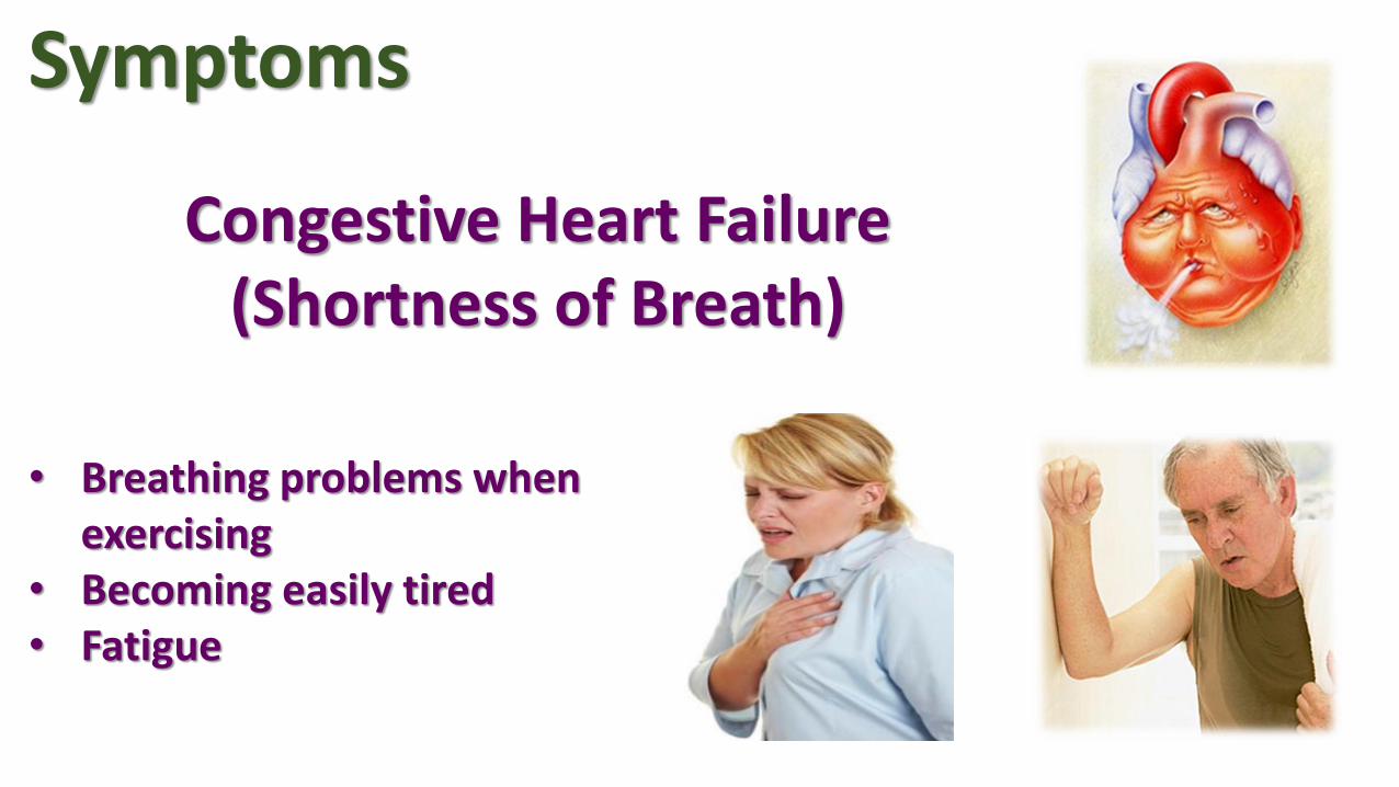
• Breathing problems when exercising
• Becoming easily tired• Fatigue
Symptoms
Congestive Heart Failure(Shortness of Breath)

Normal Heart Sound Aortic Stenosis Heart Sound
Signs

Signs


Pharmacology Beta-Adrenergic Receptor Blockers
Cardiac Glycoside
Loop Diuretics
Angiotensin-converting Enzyme (ace) Inhibitor
Opioid Analgesics

Dichloroisoprenaline - first beta blocker Propranolol - first beta blocker There are three types of beta receptors : Β1 receptors are located in the heart, eye, and kidneys. β2 receptors are found in the lungs, gastrointestinal tract, liver, uterus, blood vessels,
and skeletal muscle. Β3 receptors are located in fat cells.
PharmacologyBeta – Adrenergic Receptor Blockers

PharmacologyBeta – Adrenergic Receptor Blockers

PharmacologyCardiac Glycoside

PharmacologyLoop Diuretics

PharmacologyLoop Diuretics

PharmacologyAngiotensin – converting Enzyme (ACE) Inhibitor

PharmacologyPharmacologyOpioid Analgesics

PharmacologyOpioid Analgesics
Common and short term
Itch
Nausea
Vomiting
Constipation
Drowsiness
Dry mouth
Other
Opioid dependence
Dizziness
Decreased sex drive
Impaired sexual function
Decreased testosterone levels
Depression
Immunodeficiency
Opioid-induced abnormal pain sensitivity
Irregular menstruation
Increased risk of falls
Slowed breathing

PharmacologyPhysical Exam

PharmacologyEchocardiogram
Degree of ASMean gradient
(mmHg)Aortic valve area
(cm2)
Mild < 25 > 1.5
Moderate 25 – 40 1.0 – 1.5
Severe > 40 < 1.0
Critical > 70 < 0.6

Figure 1.Transesophageal echocardiograms of a normal aortic valve.(A) Axial view. (B) Horizontal four-chamber view.
PharmacologyEchocardiogram
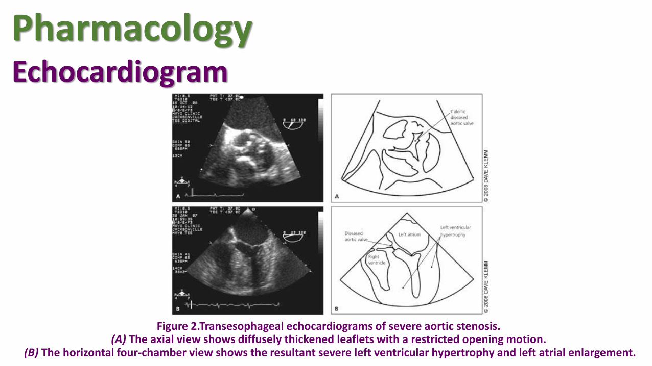
Figure 2.Transesophageal echocardiograms of severe aortic stenosis.(A) The axial view shows diffusely thickened leaflets with a restricted opening motion.
(B) The horizontal four-chamber view shows the resultant severe left ventricular hypertrophy and left atrial enlargement.
PharmacologyEchocardiogram

PharmacologyOther tests for aortic valve stenosis
ECG
Chest X-ray
Cardiac catheterization
Exercise tests
Computerized tomography (CT)
Magnetic resonance imaging (MRI)

Monitoring
Hospitalization
Lifestyle patterns
Should stop smoking and be
tested for high cholesterol.
Dental health - Good oral and
dental hygiene
Management

Severity of aortic valve stenosis
How often you should have an echocardiogram
MILD Every 3 to 5 years
MODERATE Every 1 to 2 years
SEVERE Every 6 to 12 months
Management

ManagementBalloon valvuloplasty

ManagementTranscatheter aortic valve replacement

ManagementAortic valve replacement

ManagementSurgical valvuloplasty

Discussion
