aptameter biosensor
TRANSCRIPT
8/17/2019 Aptameter Biosensor
http://slidepdf.com/reader/full/aptameter-biosensor 1/11
4 IEEE TRANSACTIONS ON BIOMEDICAL CIRCUITS AND SYSTEMS, VOL. 8, NO. 1, FEBRUARY 2014
Developing Trends in Aptamer-Based Biosensor
Devices and Their ApplicationsScott MacKay, David Wishart, James Z. Xing, and Jie Chen
Abstract— Aptamers are, in general, easier to produce, easierto store and are able to bind to a wider variety of targets thanantibodies. For these reasons, aptamers are gaining increasingpopularity in environmental monitoring as well as disease detec-
tion and disease management applications. This review article
examinesthe research and design of RNA and DNA aptamerbasedbiosensor systems and applications as well as their potential forintegration in effective biosensor devices. As single stranded DNAor RNA molecules that can bind to specific targets, aptamers are
well suited for biomolecular recognition and sensing applications.Beyond being able to be designed for a near endless number of
specific targets, aptamers can also be made which change theirconformation in a predictable and consistent way upon binding.
This can lead to many unique and effective detection methodsusing a variety of optical and electrochemical means.
Index Terms— Antibody, aptamer, biosensor, carbon nanotube,electrochemical detection, mass based detection, optical detection,RNA/DNA, SELEX.
I. I NTRODUCTION
A CCURATELY detecting, identifying and quantifying
biomolecules and other biological substances is of great
importance in many fields of life science. For instance, the
detection of bacteria, viruses and other pathogens is critical for
environmental and food safety monitoring. The detection of
abnormal proteins or abnormal levels of proteins, such as tro-
ponin I or C-reactive protein is critical to diagnosing a number
of diseases [1]. Likewise the measurement of metabolites
such as glucose or creatinine is important for the monitoring
Manuscript received September 03, 2013; revised January 02, 2014; acceptedJanuary 24, 2014. Date of publication February19, 2014; date of current version
March 25, 2014. This work was supported by the Canadian National ResearchCouncil/National Institute for Nanotechnology (NRC/NINT), Natural Sciences
and Engineering Research Council of Canada (NSERC), Alberta Innovates-Health Solutions (AIHS), Alberta Innovates—Technology Futures (AITF), and
Alberta Enterprise and Advanced Education. This paper was recommended by
Associate Editor S. T. C. Wong.S. MacKay is with the Electrical and Computer Engineering Depart-
ment, University of Alberta, Edmonton, AB T6G 2V4, Canada (e-mail:[email protected]).
D. Wishart is with the Biological Sciences Department and Computing Sci-ence Department, University of Alberta, Edmonton, AB T6G 2V4, Canada, and
also with the National Research Council/National Institute for Nanotechnology,Edmonton, AB T6G 2M9, Canada (e-mail: [email protected]).
J. Z. Xingis with Departmentof Laboratory Medicineand Pathology, Univer-
sity of Alberta, Edmonton, AB T6G 2V4, Canada (e-mail: [email protected]).J. Chen is with the Electrical and Computer Engineering Department and
Biomedical Engineering Department, University of Alberta, Edmonton, ABT6G 2V4, Canada, and also with the National Research Council/National
Institute for Nanotechnology, Edmonton, AB T6G 2M9, Canada (e-mail: [email protected]).
Color versions of one or more of the figures in this paper are available online
at http://ieeexplore.ieee.org.
Digital Object Identifier 10.1109/TBCAS.2014.2304718
of organ function and metabolic stability [2], [3]. Bacteria,
proteins and metabolites are examples of biological materials
or biopolymers that need to be routinely detected. While many
biological detection methods utilize large instruments such as
chemical analyzers, mass spectrometers or nuclear magnetic
resonance (NMR) spectrometers, there are a growing number of
biological and chemical detection methods that rely on smaller
biosensors [4], [5]. A biosensor is a device that can be used for
the detection of a molecule, which combines a “soft” biological
component with a “hard” physicochemical detector. The bio-
logical component is usually the recognition element, the part of
the biosensor which specifically recognizes the presence of thetarget molecule; while the physiochemical detection element
is the part of the biosensor which translates the recognition of
the target into a measurable and reportable signal [6]. There are
many variations of recognition and detection elements leading
to a number of different biosensor designs, each of which suited
to specific targets and applications [7]–[15]. Perhaps the most
widespread biosensor is blood glucose monitor, a device that is
used on a daily basis by millions of diabetics worldwide [2],
[16]. The modern glucose biosensor uses the enzyme glucose
oxidase to break down blood glucose leading to the release of
electrons and a detectable current [17]. In this case, the elec-
trode is the detection element and the enzyme is the biologicalrecognition element. Enzymes need not be the only possible
bio-recognition element for biosensors, in fact the biological
recognition elements of biosensors can vary from enzymes to
antibodies to peptides to aptamers.
Aptamers (from the Latin aptus, meaning fit, and Greek
meros, meaning region) are literally polymers designed to fit
specific substrates. Aptamers can be composed of nucleotides
or amino acids, although most often the term is reserved for
RNA or DNA oligonucleotides [18]. Aptamers have proven to
be an effective tool in the creation of novel, accurate and very
specific biosensors for a variety of applications ranging from
food and environmental testing [19]–[22], to toxin detection
[22], to protein monitoring [12], [13], [23], [24], to a range
of metabolomic and chemical sensing applications [11], [25],
[26]. Aptamer-based sensors take advantage of the ability of
aptamers to bind strongly to specific targets as well as their
ability to undergo substantial conformational changes upon
binding [15], [27], [28]. There are many detection methods
which utilize aptamers, including fluorescent and optical de-
tection [7], [8], [10], [21], [28], electrochemical and electrical
detection [11]–[13], [19], [25], [29]–[31], as well as mechanical
(mass-based) detection [9], [24].
In some respects, aptamers are very similar to antibodies
as they can both bind to biomolecules with great specificity.
U.S. Government work not protected by U.S. copyright.
8/17/2019 Aptameter Biosensor
http://slidepdf.com/reader/full/aptameter-biosensor 2/11
MACKAY et al.: DEVELOPING TRENDS IN APTAMER-BASED BIOSENSOR DEVICES AND THEIR APPLICATIONS 5
Although antibodies have also been used in the creation of
biosensors, aptamers present detection opportunities that were
not previously available through other biosensing modalities
due to their size (being smaller than antibodies), stability, and
structure switching capabilities [32]. This review provides a
brief overview of aptamers, their associated detection modal-
ities, their applications in biosensing, and their potential for
integration with other design elements, such as microfabricated
devices, CMOS electronics and microfluidics, for the creation
of effective complete biosensors.
As with similar research in detecting biomolecules, the final
goal of much aptamer-based biodetection research is the even-
tual creation of complete biosensing systems which utilize ap-
tamers for the recognition of specific biological molecules. It
therefore becomes necessary to not only consider the properties
of aptamers to fit target biomolecules, but also how they can
best be incorporated into biosensor systems and the other de-
sign aspects that must be considered in creating a biosensor that
can take advantage of many of the favorable properties aptamers
provide in biosensing applications.
II. BACKGROUND
Aptamers are double-stranded DNA or single-stranded
RNA/DNA molecules that bind specific molecular targets. The
first aptamers were discovered or developed independently by
two different laboratories in 1990. In particular, Larry Gold
at the University of Colorado in Boulder, designed aptamers
to bind to T4 DNA polymerase [33] while Jack Szostak at
the Massachusetts General Hospital in Boston, created RNA
aptamers to bind to various organic dyes [34]. It was Szostak
who actually coined the term ‘aptamer’. Depending on their
sequence, aptamers can be made to bind a wide variety of targets ranging from metal ions, to metabolites, to specific pro-
teins, to subcellular organelles—even to full sized cells [7], [8],
[27], [34]. Aptamers are similar to antibodies in their selective
binding, but also offer a number of distinct advantages over
antibodies. Aptamers are, in general, easier to produce, easier
to store and maintain and are able to bind to a wider variety of
targets than antibodies [9]. While most aptamers in use today
are synthetic or “designer” molecules, natural aptamers also
exist. These naturally occurring aptamers or “riboswtiches”
were first discovered in 2002 and were found to possess similar
molecular recognition properties to synthetic aptamers [35]. A
riboswitch is a part of an mRNA molecule that directly bindsto a small target molecule, usually a metabolite, to change
the gene’s activity. Riboswitches may also be RNA enzymes
(ribozymes) that cleave themselves in the presence of a small
molecule metabolite [36]. Many of the earliest riboswitches
were found in the 5 untranslated regions of mRNAs. To date,
most known riboswitches have been found in viruses and
eubacteria, but they have also been discovered in plants and
certain fungi [36]. The first human riboswitch was identified in
2009 in human vascular endothelial growth factor (VEGF) [37].
The most widely used technique for the production of syn-
thetic aptamers is SELEX [7], [33] (Systematic Evolution of
Ligands by EXponential enrichment). The process begins with a
random set of single stranded nucleic acids (ssRNA or ssDNA).
Next, a sample of the desired target is added to the nucleic
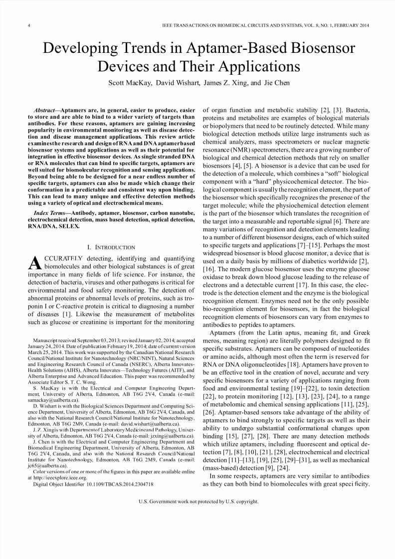
Fig. 1. A simplified process flow for the SELEX method of making aptamers.
acids where some of those random sequences bind with the tar-
gets (approximately one strand in every strands will bind
[7]). The unbound targets and nucleic acids are then removed,
leaving only the desired strands. The bound targets are then re-
moved and the selected strands are amplified using PCR (poly-
merase chain reaction). This process is repeated five to six times
using the selected strands as the starting nucleic acids in order tofurther refine the aptamers. Unlike the production of antibodies
or enzymes, this is an entirely in-vitro process. This means that
aptamers can be easily made to target molecules that are toxic
to cells or targets which would present complications to in-vivo
production methods [9]. Although first developed for protein
targets, the SELEX technique can be used for potentially any
target molecule [33].
III. DETECTION METHODS
As mentioned previously, there are several different methods
by which the presence of a target can be reported by aptamers.
These generally fall into three categories: optical detection,electrical and electrochemical detection, and mechanical de-
tection. Although all of these techniques have been and are
currently being studied, many researchers prefer electrochem-
ical techniques as they tend to be simpler, more accurate,
more sensitive and generally cheaper than other detection
methods [30].
A. Optical Detection
Although the individual techniques can vary, most op-
tical detection methods take advantage of the conformational
changes occurring in aptamers to either generate or quench
fluorescence. Optical detection methods have signi
ficant appeal because of their ease of detection and their exquisite sensitivity
[8], [10], [38].
The simplest route to achieve optical detection in aptamer-
based biosensors is to chemically attach a fluorophore to a spe-
cific site on the aptamer. The conformational change induced in
the aptamer by the target molecule almost always changes the
interactions between the fluorescent nucleotides of the aptamer
and the fluorophore, causing a change in the fluorescent output
[10], [39]. Therefore, if a change influorescence is detected, this
indicates that the target molecule is present. This particular type
of detection has been shown to detect ATP at concentrations as
low as 1 mM [39].
Another optical detection method that can be used is to chem-
ically attach both a fluorophore and a quencher molecule to the
8/17/2019 Aptameter Biosensor
http://slidepdf.com/reader/full/aptameter-biosensor 3/11
6 IEEE TRANSACTIONS ON BIOMEDICAL CIRCUITS AND SYSTEMS, VOL. 8, NO. 1, FEBRUARY 2014
Fig.2. (a) An optical detection method where the conformational change in the
aptamerupon bindingchangesthe fluorescence produced by thefluorophore[7].(b) In the unbound state, the quencher (black square) is in close proximity to the
fluorophore (green square), but when the aptamer is bound to the target, theymove apart and fluorescence can be measured [7], [40].
aptamer. These molecules are placed in such a way that in the
aptamer’s unbound state the quencher is close enough to the
fluorophore to eliminate any fluorescence from being gener-
ated. However, when the aptamer is bound to the target mol-
ecule, the quencher and fluorophore are separated, and fluores-
cence can be detected [10]. The reverse is also possible, where
fluorescence is detected when the aptamer is ligand-free and
is quenched when the ligand is bound [10]. The preparation
of these double labeled aptamers is somewhat more complex
than just using one fluorophore, but double-labeled aptamers
also generate a greater change in fluorescence, making detec-
tion easier. This fluorophore-quencher aptamer biosensor has
been used to detect thrombin with a detection limit of about0.3 nM [40]. Another similar technique is to use a second fluo-
rophore rather than a quencher [10]. In this case, the change in
proximity of the fluorophores causesfluorescence resonance en-
ergy transfer (FRET), which can be measured [6], [10]. Carbon
nanotubes [41] and graphene [42], [43] have also been utilized
as quenchers, blocking fluorescence from fluorophores on ap-
tamers when they are not bound to their target ligand. Graphene
quenching has been used to detect oxytetracycline with a limit
of detection of 10 nM [43]. When bound to the target ligand, the
conformational change in the aptamer makes quenching from
the carbon nanotube or graphene impossible [41]–[43]. Other
variations on this design use separate strands of DNA labeledwith quenchers or fluorophores [38], [44]. The quencher strand
and fluorophore labeled aptamer are complementary enough to
bind when no target is present, thus quenching the fluorescence,
but when the target is present, the favorable interaction is for the
aptamer to bind to the target, thus preventing quenching [45].
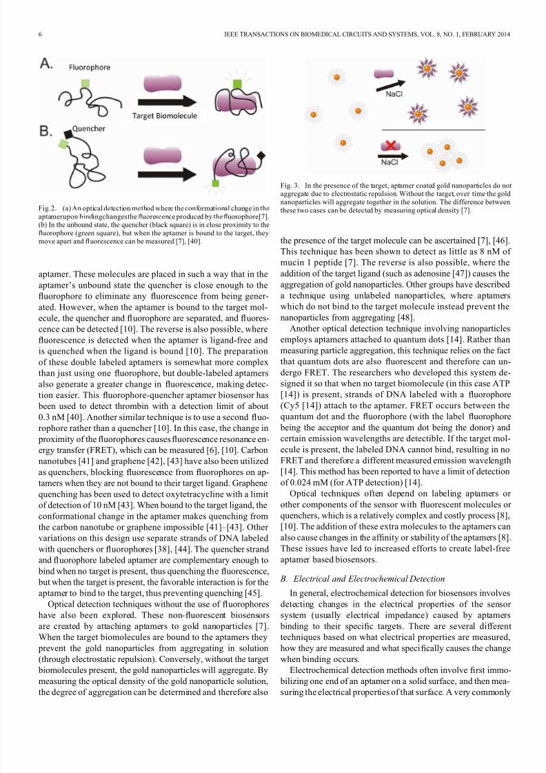
Optical detection techniques without the use of fluorophores
have also been explored. These non-fluorescent biosensors
are created by attaching aptamers to gold nanoparticles [7].
When the target biomolecules are bound to the aptamers they
prevent the gold nanoparticles from aggregating in solution
(through electrostatic repulsion). Conversely, without the target
biomolecules present, the gold nanoparticles will aggregate. By
measuring the optical density of the gold nanoparticle solution,
the degree of aggregation can be determined and therefore also
Fig. 3. In the presence of the target, aptamer coated gold nanoparticles do notaggregate due to electrostatic repulsion. Without the target, over time the gold
nanoparticles will aggregate together in the solution. The difference between
these two cases can be detected by measuring optical density [7].
the presence of the target molecule can be ascertained [7], [46].
This technique has been shown to detect as little as 8 nM of
mucin 1 peptide [7]. The reverse is also possible, where the
addition of the target ligand (such as adenosine [47]) causes theaggregation of gold nanoparticles. Other groups have described
a technique using unlabeled nanoparticles, where aptamers
which do not bind to the target molecule instead prevent the
nanoparticles from aggregating [48].
Another optical detection technique involving nanoparticles
employs aptamers attached to quantum dots [14]. Rather than
measuring particle aggregation, this technique relies on the fact
that quantum dots are also fluorescent and therefore can un-
dergo FRET. The researchers who developed this system de-
signed it so that when no target biomolecule (in this case ATP
[14]) is present, strands of DNA labeled with a fluorophore
(Cy5 [14]) attach to the aptamer. FRET occurs between thequantum dot and the fluorophore (with the label fluorophore
being the acceptor and the quantum dot being the donor) and
certain emission wavelengths are detectible. If the target mol-
ecule is present, the labeled DNA cannot bind, resulting in no
FRET and therefore a different measured emission wavelength
[14]. This method has been reported to have a limit of detection
of 0.024 mM (for ATP detection) [14].
Optical techniques often depend on labeling aptamers or
other components of the sensor with fluorescent molecules or
quenchers, which is a relatively complex and costly process [8],
[10]. The addition of these extra molecules to the aptamers can
also cause changes in the af fi
nity or stability of the aptamers [8].These issues have led to increased efforts to create label-free
aptamer based biosensors.
B. Electrical and Electrochemical Detection
In general, electrochemical detection for biosensors involves
detecting changes in the electrical properties of the sensor
system (usually electrical impedance) caused by aptamers
binding to their specific targets. There are several different
techniques based on what electrical properties are measured,
how they are measured and what specifically causes the change
when binding occurs.
Electrochemical detection methods often involve first immo-
bilizing one end of an aptamer on a solid surface, and then mea-
suring the electrical properties of that surface. A very commonly
8/17/2019 Aptameter Biosensor
http://slidepdf.com/reader/full/aptameter-biosensor 4/11
MACKAY et al.: DEVELOPING TRENDS IN APTAMER-BASED BIOSENSOR DEVICES AND THEIR APPLICATIONS 7
Fig. 4. Without the target present, the single strands of DNA with attachedfluorophores (red lines with attached green squares in this figure) attach to
the unbound aptamers on the quantum dots. When the excitation wavelengthis added , FRET occurs between the quantum dot and the fluorophore,
producing two emission wavelengths ( and ). With the target present,the af finity of the aptamer for the target is greater than for the second DNA
sequence, thus the aptamers bind with the targets only, no FRET occurs and
there is only one emission wavelength [14].
used material for these surfaces is gold [29] as it is generally
nonreactive, has well established electrical properties and co-
valent linkage to gold is easily accomplished through the use of
a thiol bond [49].
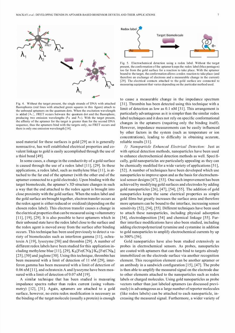
In some cases, a change in the conductivity of a gold surface
is caused through the use of a redox label [11], [29]. In these
applications, a redox label, such as methylene blue [11], is at-
tached to the far end of the aptamer (with the other end of theaptamer connected to a gold electrode). Upon binding with the
target biomolecule, the aptamer’s 3D structure changes in such
a way that the end attached to the redox agent is brought into
close proximity with the gold surface. When the redox label and
the gold surface are brought together, electron transfer occurs as
the redox agent is either reduced or oxidized (depending on the
chosen redox label). This electron transfer causes a change in
the electrical properties that can be measured using voltammetry
[11], [19], [29]. It is also possible to have aptamers which in
their unbound state have the redox label close to the surface and
the redox agent is moved away from the surface after binding
occurs. This technique has been used previously to detect a va-riety of biomolecules such as interferon gamma [11], ochra-
toxin A [19], lysozyme [50] and thrombin [29]. A number of
different redox labels have been studied for this application in-
cluding methylene blue [11], [29], K [Fe(CN) ]/K [Fe(CN) ]
[25], [50] and juglone [30]. Using this technique, thrombin has
been measured with a limit of detection of 11 nM [29], inter-
feron gamma has been measured with a limit of detection of
0.06 nM [11], and ochratoxin A and lysozyme have been mea-
sured with a limit of detection of 0.07 nM [19].
A similar technique that has been studied is measuring
impedance spectra rather than redox current (using voltam-
metry) [12], [31]. Again, aptamers are attached to a gold
surface, however, no extra redox modification is necessary as
the binding of the target molecule (usually a protein) is enough
Fig. 5. Electrochemical detection using a redox label. Without the target
present, the confo rmation o f the aptamer k eeps the redox label (blu e pentago n)
too far from the gold surface for a reaction to take place. With the aptamer bound to the target, the confor mation allows a redox reaction to take place (and
therefore an exchange of electrons and a measurable change in the current)[29]. The electrical contacts attached to the gold surface are connected to
measuring equipment that varies depending on the particular method used.
to cause a measurable change in the impedance spectrum
[31]. Thrombin has been detected using this technique with a
limit of detection as low as 0.1 nM [31]. This arrangement is
particularly advantageous as it is simpler than the similar redox
label techniques and it does not rely on specific conformational
changes in the aptamers (requiring only the binding itself).
However, impedance measurements can be easily influenced
by other factors in the system (such as temperature or ion
concentrations), leading to dif ficulty in obtaining accurate,
reliable results [11].
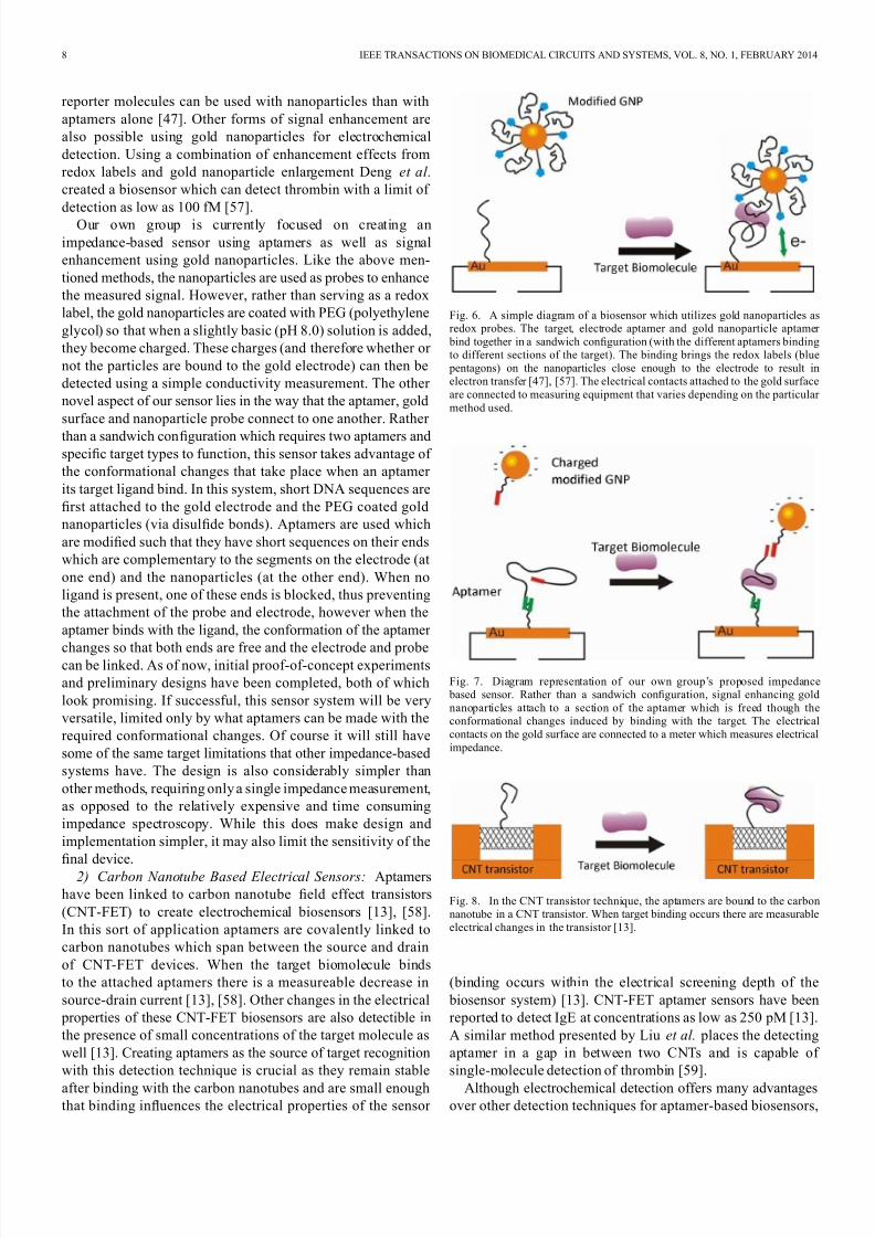
1) Nanoparticle Enhanced Electrical Detection: Just as
with optical detection methods, nanoparticles have been used
to enhance electrochemical detection methods as well. Specifi-
cally, gold nanoparticles are particularly appealing as they can
be chemically modified for a wide variety of applications [51],
[52]. A number of techniques have been developed which use
nanoparticles to improve upon and as the basis for electrochem-ical sensor designs [47], [53]. One such improvement has been
achieved by modifying gold surfaces and electrodes by adding
gold nanoparticles [26], [47], [54], [55]. The addition of gold
nanoparticles keeps the same electrical properties as simple
gold films but greatly increases the surface area and therefore
more aptamers can be bound to the interface, increasing sensor
sensitivity [52], [54], [55]. Different techniques have been used
to attach these nanoparticles, including physical adsorption
[54], electrodeposition [54] and chemical linkage [55]. Fur-
ther interface modifications have also been studied, including
adding electropolymerized tyramine and cystamine in addition
to gold nanoparticles to amplify electrochemical currents by upto 300% [56].
Gold nanoparticles have also been studied extensively as
probes in electrochemical sensors. As probes, nanoparticles
are coated with aptamers that can then bind to targets already
immobilized on the electrode surface via another recognition
element. This recognition element can be another aptamer or
an antibody in a sandwich configuration [15], [47]. The probe
is then able to amplify the measured signal on the electrode due
to other elements attached to the nanoparticles such as redox
labels or charged molecules. Using gold nanoparticles as probe
vectors rather than just labeled aptamers (as discussed previ-
ously) is advantageous as a large number of reporter molecules
(like redox labels) can be attached to each nanoparticle, in-
creasing the measured signal. Furthermore, a wider variety of
8/17/2019 Aptameter Biosensor
http://slidepdf.com/reader/full/aptameter-biosensor 5/11
8 IEEE TRANSACTIONS ON BIOMEDICAL CIRCUITS AND SYSTEMS, VOL. 8, NO. 1, FEBRUARY 2014
reporter molecules can be used with nanoparticles than with
aptamers alone [47]. Other forms of signal enhancement are
also possible using gold nanoparticles for electrochemical
detection. Using a combination of enhancement effects from
redox labels and gold nanoparticle enlargement Deng et al.
created a biosensor which can detect thrombin with a limit of
detection as low as 100 fM [57].
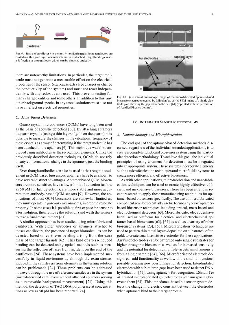
Our own group is currently focused on creating an
impedance-based sensor using aptamers as well as signal
enhancement using gold nanoparticles. Like the above men-
tioned methods, the nanoparticles are used as probes to enhance
the measured signal. However, rather than serving as a redox
label, the gold nanoparticles are coated with PEG (polyethylene
glycol) so that when a slightly basic (pH 8.0) solution is added,
they become charged. These charges (and therefore whether or
not the particles are bound to the gold electrode) can then be
detected using a simple conductivity measurement. The other
novel aspect of our sensor lies in the way that the aptamer, gold
surface and nanoparticle probe connect to one another. Rather
than a sandwich configuration which requires two aptamers andspecific target types to function, this sensor takes advantage of
the conformational changes that take place when an aptamer
its target ligand bind. In this system, short DNA sequences are
first attached to the gold electrode and the PEG coated gold
nanoparticles (via disulfide bonds). Aptamers are used which
are modified such that they have short sequences on their ends
which are complementary to the segments on the electrode (at
one end) and the nanoparticles (at the other end). When no
ligand is present, one of these ends is blocked, thus preventing
the attachment of the probe and electrode, however when the
aptamer binds with the ligand, the conformation of the aptamer
changes so that both ends are free and the electrode and probecan be linked. As of now, initial proof-of-concept experiments
and preliminary designs have been completed, both of which
look promising. If successful, this sensor system will be very
versatile, limited only by what aptamers can be made with the
required conformational changes. Of course it will still have
some of the same target limitations that other impedance-based
systems have. The design is also considerably simpler than
other methods, requiring only a single impedance measurement,
as opposed to the relatively expensive and time consuming
impedance spectroscopy. While this does make design and
implementation simpler, it may also limit the sensitivity of the
final device.
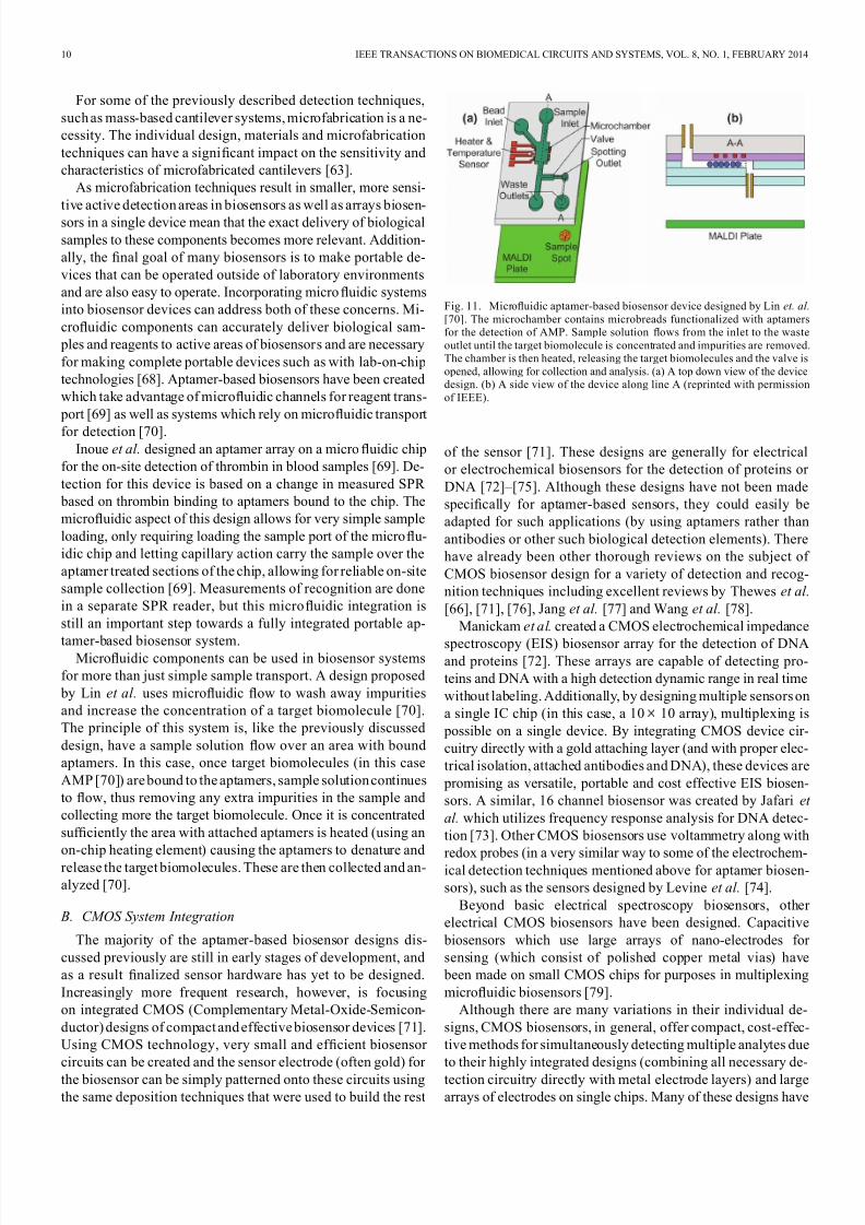
2) Carbon Nanotube Based Electrical Sensors: Aptamers
have been linked to carbon nanotube field effect transistors
(CNT-FET) to create electrochemical biosensors [13], [58].
In this sort of application aptamers are covalently linked to
carbon nanotubes which span between the source and drain
of CNT-FET devices. When the target biomolecule binds
to the attached aptamers there is a measureable decrease in
source-drain current [13], [58]. Other changes in the electrical
properties of these CNT-FET biosensors are also detectible in
the presence of small concentrations of the target molecule as
well [13]. Creating aptamers as the source of target recognition
with this detection technique is crucial as they remain stable
after binding with the carbon nanotubes and are small enoughthat binding influences the electrical properties of the sensor
Fig. 6. A simple diagram of a biosensor which utilizes gold nanoparticles asredox probes. The target, electrode aptamer and gold nanoparticle aptamer
bind together in a sandwich configuration (with the different aptamers bindingto different sections of the target). The binding brings the redox labels (blue
pentagons) on the nanoparticles close enough to the electrode to result inelectron transfer [47], [57]. The electrical contacts attached to the gold surface
are connected to measuring equipment that varies depending on the particular
method used.
Fig. 7. Diagram representation of our own group’s proposed impedance based sensor. Rather than a sandwich configuration, signal enhancing gold
nanoparticles attach to a section of the aptamer which is freed though theconformational changes induced by binding with the target. The electrical
contacts on the gold surface are connected to a meter which measures electrical
impedance.
Fig. 8. In the CNT transistor technique, the aptamers are bound to the carbon
nanotube in a CNT transistor. When target binding occurs there are measurable
electrical changes in the transistor [13].
(binding occurs within the electrical screening depth of the
biosensor system) [13]. CNT-FET aptamer sensors have been
reported to detect IgE at concentrations as low as 250 pM [13].
A similar method presented by Liu et al. places the detecting
aptamer in a gap in between two CNTs and is capable of
single-molecule detection of thrombin [59].
Although electrochemical detection offers many advantagesover other detection techniques for aptamer-based biosensors,
8/17/2019 Aptameter Biosensor
http://slidepdf.com/reader/full/aptameter-biosensor 6/11
MACKAY et al.: DEVELOPING TRENDS IN APTAMER-BASED BIOSENSOR DEVICES AND THEIR APPLICATIONS 9
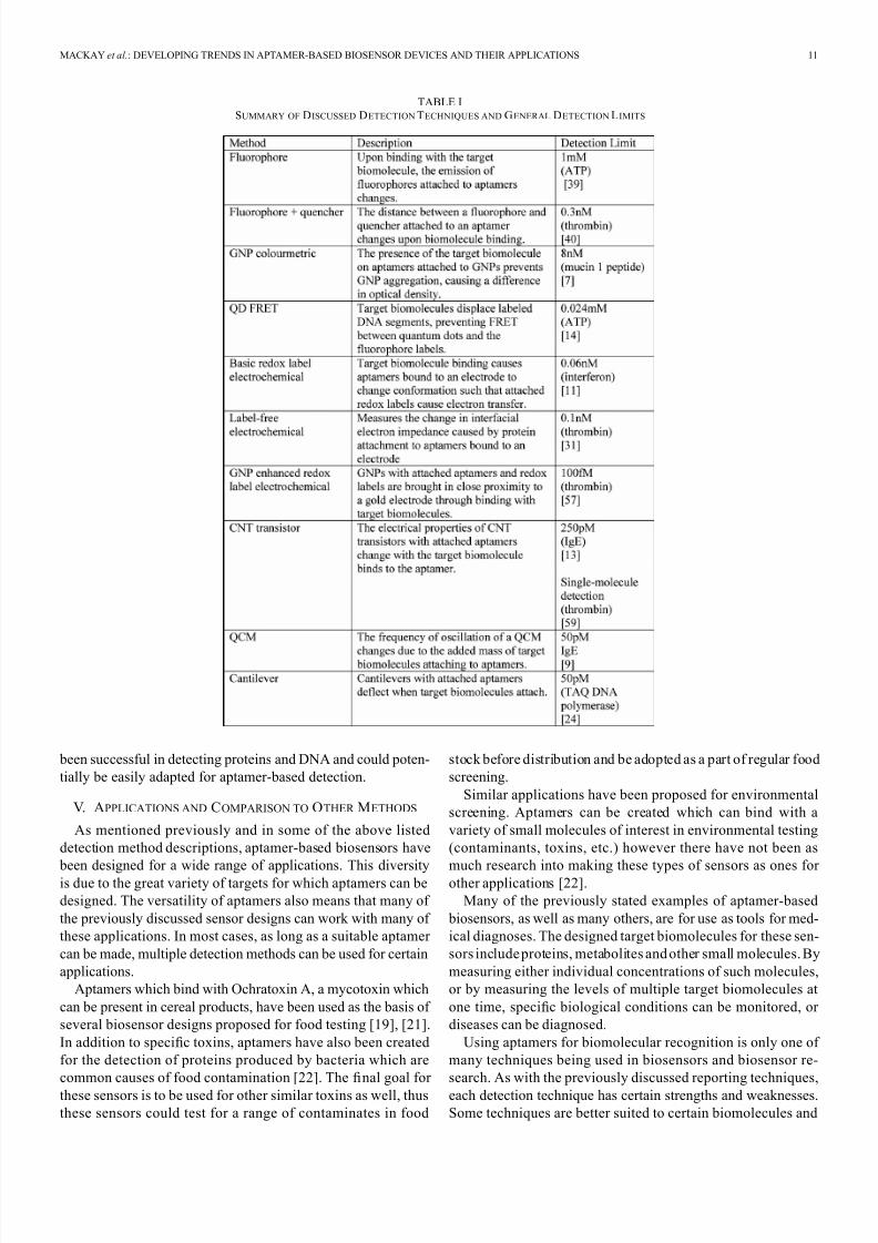
Fig. 9. Basis of cantilever biosensors. Microfabricated silicon cantilevers are
coated in a thin gold layer to which aptamers are attached. Target binding causesa deflection in the cantilever, which can be detected optically.
there are noteworthy limitations. In particular, the target mol-
ecule must not generate a measurable effect on the electrical
properties of the sensor (e.g., cause extra free charges or change
the conductivity of the system) and must not react indepen-
dently with any redox agents used. This prevents testing for
many charged entities and some others. In addition to this, any
other background species in any tested solutions must also not
have an effect on electrical properties.
C. Mass Based Detection
Quartz crystal microbalances (QCMs) have long been used
as the basis of acoustic detection [60]. By attaching aptamers
to quartz crystals (using a thin layer of gold on the quartz), it is
possible to measure the changes in the vibrational frequency of
these crystals as a way of determining if the target molecule has
been attached to the aptamers [9]. This technique was first em-
ployed using antibodies as the recognition elements. Unlike the
previously described detection techniques, QCMs do not rely
on any conformational change in the aptamers, just the binding
itself.
Even though antibodies can also be used as the recognitionel-ement in QCM based biosensors, aptamers have been shown to
have several distinct advantages. Aptamer based QCM biosen-
sors are more sensitive, have a lower limit of detection (as low
as 50 pM for IgE detection), are more stable and more accu-
rate than antibody-based QCM sensors [9]. However, the ap-
plications of most QCM biosensors are somewhat limited as,
they must operate in gaseous environments, in order to resonate
properly. In some cases it is possible to first expose the sensor to
a test solution, then remove the solution (and wash the sensor)
to take a final measurement [61].
A similar approach has been studied using microfabricated
cantilevers. With either antibodies or aptamers attached totheses cantilevers, the presence of target biomolecules can be
detected based on cantilever bending arising from the extra
mass of the target ligands [62]. This kind of stress-induced
bending can be detected using optical methods such as mea-
suring the reflection of laser light incident on the end of the
cantilevers [24]. These systems have been implemented suc-
cessfully in liquid environments, although the extra stresses
induced in the cantilevers from elements in the testing solution
can be problematic [24]. These problems can be addressed
however, through the use of reference cantilevers in the system
(microfabricated cantilevers without attached aptamers serving
as a removable background measurement) [24]. Using this
method, the detection of TAQ DNA polymerase at concentra-
tions as low as 50 pM has been reported [24].
Fig. 10. (a) Optical microscope image of the microfabricated aptamer-based
biosensor electr odes created by Löhndorf et. al. (b) SEM image of a single elec-
trode pair, showing the gap between the pair [64] (reprinted with the permissionof Applied Physics Letters).
IV. I NTEGRATED SENSOR MICROSYSTEMS
A. Nanotechnology and Microfabrication
The end goal of the aptamer-based detection methods dis-
cussed, regardless of the individual intended applications, is to
create a complete functional biosensor system using that partic-
ular detection methodology. To achieve this goal, the individual
principles of using aptamers for detection must be integrated
into an appropriate system. These systems incorporate elements
such as microfabrication techniques and microfluidic systems tocreate more ef ficient and effective biosensors.
As with other applications, microfabrication and nanofabri-
cation techniques can be used to create highly effective, ef fi-
cient and inexpensive biosensors. There has been a trend in re-
cent research to apply these manufacturing techniques for ap-
tamer-based biosensors specifically. The use of microfabricated
components can be potentially useful for most types of aptamer-
based biosensor techniques including optical, mass-based and
electrochemical detection [63]. Microfabricated electrodes have
been used as platforms for electrical and electrochemical ap-
tamer-based biosensors [63], [64] as well as a variety of other
biosensor systems [23], [65]. Microfabrication techniques areused to pattern thin metal layers deposited on substrates, often
gold, to create small, sensitive electrodes for these applications.
Arrays of electrodes can be patterned onto single substrates for
higher throughput biosensors as well as for increased sensitivity
and the potential for detecting multiple targets simultaneously
from a single sample [64], [66]. Microfabricated electrode de-
signs can add functionality as well, with the small dimensions
possible opening new possibilities for detection. Interdigitated
electrodes with sub-micron gaps have been used to detect DNA
hybridization [67]. Using aptamers for recognition, Löhndorf et
al. created microfabricated gold electrodes with nm spacing be-
tween them [64]. This impedance-based biosensor system de-
tects the change in dielectric constant between the electrodes
when aptamers bind to their target protein.
8/17/2019 Aptameter Biosensor
http://slidepdf.com/reader/full/aptameter-biosensor 7/11
10 IEEE TRANSACTIONS ON BIOMEDICAL CIRCUITS AND SYSTEMS, VOL. 8, NO. 1, FEBRUARY 2014
For some of the previously described detection techniques,
such as mass-based cantilever systems, microfabrication is a ne-
cessity. The individual design, materials and microfabrication
techniques can have a significant impact on the sensitivity and
characteristics of microfabricated cantilevers [63].
As microfabrication techniques result in smaller, more sensi-
tive active detection areas in biosensors as well as arrays biosen-
sors in a single device mean that the exact delivery of biological
samples to these components becomes more relevant. Addition-
ally, the final goal of many biosensors is to make portable de-
vices that can be operated outside of laboratory environments
and are also easy to operate. Incorporating microfluidic systems
into biosensor devices can address both of these concerns. Mi-
crofluidic components can accurately deliver biological sam-
ples and reagents to active areas of biosensors and are necessary
for making complete portable devices such as with lab-on-chip
technologies [68]. Aptamer-based biosensors have been created
which take advantage of microfluidic channels for reagent trans-
port [69] as well as systems which rely on microfluidic transport
for detection [70].Inoue et al. designed an aptamer array on a microfluidic chip
for the on-site detection of thrombin in blood samples [69]. De-
tection for this device is based on a change in measured SPR
based on thrombin binding to aptamers bound to the chip. The
microfluidic aspect of this design allows for very simple sample
loading, only requiring loading the sample port of the microflu-
idic chip and letting capillary action carry the sample over the
aptamer treated sections of the chip, allowing for reliable on-site
sample collection [69]. Measurements of recognition are done
in a separate SPR reader, but this microfluidic integration is
still an important step towards a fully integrated portable ap-
tamer-based biosensor system.Microfluidic components can be used in biosensor systems
for more than just simple sample transport. A design proposed
by Lin et al. uses microfluidic flow to wash away impurities
and increase the concentration of a target biomolecule [70].
The principle of this system is, like the previously discussed
design, have a sample solution flow over an area with bound
aptamers. In this case, once target biomolecules (in this case
AMP [70]) are bound to the aptamers, sample solution continues
to flow, thus removing any extra impurities in the sample and
collecting more the target biomolecule. Once it is concentrated
suf ficiently the area with attached aptamers is heated (using an
on-chip heating element) causing the aptamers to denature and
release the target biomolecules. These are then collected and an-
alyzed [70].
B. CMOS System Integration
The majority of the aptamer-based biosensor designs dis-
cussed previously are still in early stages of development, and
as a result finalized sensor hardware has yet to be designed.
Increasingly more frequent research, however, is focusing
on integrated CMOS (Complementary Metal-Oxide-Semicon-
ductor) designs of compact and effective biosensor devices [71].
Using CMOS technology, very small and ef ficient biosensor
circuits can be created and the sensor electrode (often gold) for
the biosensor can be simply patterned onto these circuits using
the same deposition techniques that were used to build the rest
Fig. 11. Microfluidic aptamer-based biosensor device designed by Lin et. al.
[70]. The microchamber contains microbreads functionalized with aptamersfor the detection of AMP. Sample solution flows from the inlet to the waste
outlet until the target biomolecule is concentrated and impurities are removed.The chamber is then heated, releasing the target biomolecules and the valve is
opened, allowing for collection and analysis. (a) A top down view of the devicedesign. (b) A side view of the device along line A (reprinted with permission
of IEEE).
of the sensor [71]. These designs are generally for electrical
or electrochemical biosensors for the detection of proteins or
DNA [72]–[75]. Although these designs have not been made
specifically for aptamer-based sensors, they could easily be
adapted for such applications (by using aptamers rather than
antibodies or other such biological detection elements). There
have already been other thorough reviews on the subject of
CMOS biosensor design for a variety of detection and recog-
nition techniques including excellent reviews by Thewes et al.
[66], [71], [76], Jang et al. [77] and Wang et al. [78].
Manickam et al. created a CMOS electrochemical impedance
spectroscopy (EIS) biosensor array for the detection of DNAand proteins [72]. These arrays are capable of detecting pro-
teins and DNA with a high detection dynamic range in real time
without labeling. Additionally, by designing multiple sensors on
a single IC chip (in this case, a 10 10 array), multiplexing is
possible on a single device. By integrating CMOS device cir-
cuitry directly with a gold attaching layer (and with proper elec-
trical isolation, attached antibodies and DNA), these devices are
promising as versatile, portable and cost effective EIS biosen-
sors. A similar, 16 channel biosensor was created by Jafari et
al. which utilizes frequency response analysis for DNA detec-
tion [73]. Other CMOS biosensors use voltammetry along with
redox probes (in a very similar way to some of the electrochem-ical detection techniques mentioned above for aptamer biosen-
sors), such as the sensors designed by Levine et al. [74].
Beyond basic electrical spectroscopy biosensors, other
electrical CMOS biosensors have been designed. Capacitive
biosensors which use large arrays of nano-electrodes for
sensing (which consist of polished copper metal vias) have
been made on small CMOS chips for purposes in multiplexing
microfluidic biosensors [79].
Although there are many variations in their individual de-
signs, CMOS biosensors, in general, offer compact, cost-effec-
tive methods for simultaneously detecting multiple analytes due
to their highly integrated designs (combining all necessary de-
tection circuitry directly with metal electrode layers) and large
arrays of electrodes on single chips. Many of these designs have
8/17/2019 Aptameter Biosensor
http://slidepdf.com/reader/full/aptameter-biosensor 8/11
MACKAY et al.: DEVELOPING TRENDS IN APTAMER-BASED BIOSENSOR DEVICES AND THEIR APPLICATIONS 11
TABLE ISUMMARY OF DISCUSSED DETECTION TECHNIQUES AND GENERAL DETECTION LIMITS
been successful in detecting proteins and DNA and could poten-
tially be easily adapted for aptamer-based detection.
V. APPLICATIONS AND COMPARISON TO OTHER METHODS
As mentioned previously and in some of the above listed
detection method descriptions, aptamer-based biosensors have
been designed for a wide range of applications. This diversityis due to the great variety of targets for which aptamers can be
designed. The versatility of aptamers also means that many of
the previously discussed sensor designs can work with many of
these applications. In most cases, as long as a suitable aptamer
can be made, multiple detection methods can be used for certain
applications.
Aptamers which bind with Ochratoxin A, a mycotoxin which
can be present in cereal products, have been used as the basis of
several biosensor designs proposed for food testing [19], [21].
In addition to specific toxins, aptamers have also been created
for the detection of proteins produced by bacteria which are
common causes of food contamination [22]. The final goal for
these sensors is to be used for other similar toxins as well, thus
these sensors could test for a range of contaminates in food
stock before distribution and be adopted as a part of regular food
screening.
Similar applications have been proposed for environmental
screening. Aptamers can be created which can bind with a
variety of small molecules of interest in environmental testing
(contaminants, toxins, etc.) however there have not been as
much research into making these types of sensors as ones for other applications [22].
Many of the previously stated examples of aptamer-based
biosensors, as well as many others, are for use as tools for med-
ical diagnoses. The designed target biomolecules for these sen-
sors include proteins, metabolites and other small molecules. By
measuring either individual concentrations of such molecules,
or by measuring the levels of multiple target biomolecules at
one time, specific biological conditions can be monitored, or
diseases can be diagnosed.
Using aptamers for biomolecular recognition is only one of
many techniques being used in biosensors and biosensor re-
search. As with the previously discussed reporting techniques,
each detection technique has certain strengths and weaknesses.
Some techniques are better suited to certain biomolecules and
8/17/2019 Aptameter Biosensor
http://slidepdf.com/reader/full/aptameter-biosensor 9/11
12 IEEE TRANSACTIONS ON BIOMEDICAL CIRCUITS AND SYSTEMS, VOL. 8, NO. 1, FEBRUARY 2014
testing constraints such as required testing time, cost, detection
limit and sample background. Unfortunately, the most accurate
techniques are often impractical and too expensive for many
biosensing applications [6], [58].
In terms of recognition elements, it is easiest to compare
the relative merits of aptamers and antibodies. Although they
are very similar, in that they both selectively bind to specific
molecules, there are some key differences. These differences
and their differing implementations are worth further discus-
sion. In terms of production, aptamers are easy to prepare in
large quantities (using PCR) with very little variation (less than
in antibody production) [23], [47]. Also, unlike the standard
procedures used in making antibodies, the SELEX process for
making aptamers is a fully chemical process that does not in-
volve the use of animals or bacteria (again leading to more con-
sistent production). Aptamers are generally more stable than
antibodies. In particular, they bond well to surfaces and are
not easily or permanently damaged by temperature fluctuations
[23]. At around 100 base pairs in length, most aptamers are
smaller than antibodies [6], [47]. This fact can be exploitedin biosensor designs by allowing a larger, denser concentra-
tion of recognition elements to adhere to surfaces, or for target
biomolecules to be brought in closer proximity to sensor devices
(such as in the CNT-FET sensor described above) [13]. Perhaps
the most significant advantage of aptamers over antibodies lies
in the versatility of aptamers; not only in the variety of possible
targets, but also in the variety of possible applications. Anti-
bodies are only suited for targeting immunogenic compounds
such as proteins, whereas there is almost no limit to what ap-
tamers can be designed to target [47]. The fact that some ap-
tamers not only bind specifically with targets, but undergo pre-
dictable, consistent conformational changes when they do leadsto a number of new possibilities.
Many of the above-mentioned detection methods rely on con-
formational changes to function and therefore can only work
with these structure switching aptamers. Despite all of these ad-
vantages, there are still some important limitations preventing
aptamers from being more widely utilized. Being a relatively
new field of research, aptamers are generally not as well estab-
lished as antibodies. Although aptamers can be made to bind to a
wide variety of targets, there are still many aptamer-target pairs
which have not yet been characterized or produced [15] and
in some cases with smaller molecular targets, aptamers have a
lower detection limit than other recognition elements [80]. Also,
even though aptamers can be made which bind to a wide variety
of targets, not all of these aptamers will have the desired struc-
ture switching properties. Therefore, just because an aptamer
can bind to a specific target this does not mean that the aptamer
can be used in all biosensor applications. This can be a serious
limitation to the implementation of certain aptamer biosensor
designs.
The following table briefly summarizes most of the biosensor
designs discussed. It is important to note that although some
of these techniques have clearly superior detection limits, this
does not mean that they are superior in all aspects. Other, less
sensitive biosensor designs may be less complex, less expensive
and be suitable for different applications than more sensitivedesigns. The reported detection limits are based on one example
of specific research for specific targets only. They are meant
to give a general idea of the detection limits for that type of
detection design, not a finalized limit. The detection limits for
different designed targets can vary as well as the type of sample
tested.
VI. CONCLUSION
Aptamers offer many promising opportunities in applications
in biosensor circuits and systems. Not only do they lend them-
selves well to biosensing, but also because of their stability, ease
of production and high specificity, they have many unique prop-
erties that can be exploited in a number of novel detection tech-
niques. Aptamers have been shown to be well suited for biosen-
sors that utilize a variety of optical, electrochemical and mass
based detection techniques. Even when used along with de-
tection techniques compatible with other recognition elements,
such as antibodies, aptamers have been shown to create biosen-
sors that are superior in terms of sensitivity, stability and limit
of detection. CMOS based biosensor designs have been studiedfor protein and DNA detection. These offer compact highly ef-
ficient multiplexing designs that could be easily adapted for use
with aptamers as recognition elements. One of the main prob-
lems limiting aptamers from being used in more applications
lies in the fact that not all aptamers are compatible with cer-
tain standard detection techniques. Further research is therefore
required to identify or design aptamers that are better suited
to not only a wide variety of targets, but also to a variety of
detection methods. Future integration of aptamer-based detec-
tion with microfluidic systems and microfabrication techniques
could overcome this and lead to portable, effective, versatile
biosensors for numerous potential applications.Incorporating microfluidic systems with aptamer-based
biosensor designs would further simplify and miniaturize de-
vices. Microfluidic channels could carry sample solutions from
a large input port to the active area (or areas) of the sensor,
eliminating the need for laboratory technicians to process
samples. In-device electronics or other miniaturized devices
could be used to detect target biomolecules (through electronic
detection, electrochemical detection, optical detection etc.).
The resulting biosensors could be operated directly by doctors
in hospital or non-clinic settings for health monitoring, for
on-site environmental testing or even by people to monitor
their own health.
ACKNOWLEDGMENT
The authors would like to thank Dr. Y. Hao, M. Huang, and
R. Yang for their work on the lab group’s sensor device.
R EFERENCES
[1] M. Mascini and S. Tombelli, “Biosensors for biomarkers in medical
diagnostics,” Biomarkers, vol. 13, no. 7, pp. 637–657, Nov. 2008.[2] J. Wang, “Glucose biosensors: 40 years of advances and challenges,”
Electroanal., vol. 13, no. 12, pp. 983–988, Aug. 2001.[3] A. J. Killard and M. R. Smyth, “Creatinine biosensors: Principles and
designs,” Trends Biotechnol., vol. 18, no. 10, pp. 433–437, Oct. 2000.
[4] I. R. Lauks, “Microfabricated biosensors and microanalytical systemsfor blood analysis,” Accounts Chem. Res., vol. 31, no. 5, pp. 317–324,
May 1998.
8/17/2019 Aptameter Biosensor
http://slidepdf.com/reader/full/aptameter-biosensor 10/11
MACKAY et al.: DEVELOPING TRENDS IN APTAMER-BASED BIOSENSOR DEVICES AND THEIR APPLICATIONS 13
[5] A. Hierlemann, O. Brand, C. Hagleitner, and H. Baltes, “Microfabri-cation techniques for chemical/biosensors,” Proc. IEEE , vol. 91, no. 6,
pp. 839–863, Jun. 2003.
[6] Y. Liu, Z. Matharu, and M. Howland, “Af finity and enzyme-based biosensors: Recent advances and emerging applications in cell anal-
ysis and point-of-care testing,” Anal. Bioanal. Chem., vol. 404, pp.1181–1196, 2012.
[7] L. Tan, K. Neoh, E. Kang, W. Choe, and X. Su, “Af finity analysisof DNA aptamer-peptide interactions using gold nanoparticles,” Anal.
Biochem., vol. 421, no. 2, pp. 725–731, 2011.[8] D. Zheng, R. Zou, and X. Lou, “Label-free fluorescent detection of
ions, proteins,and small molecules using structure-switching aptamers,
SYBR gold, and exonuclease I,” Anal. Chem., vol. 84, pp. 3554–3560,2012.
[9] C. Yao, T. Zhu, Y. Qi, Y. Zhao, H. Xia, and W. Fu, “Development of aquartz crystalmicrobalance biosensorwith aptamers as bio-recognition
element,” Sensors, vol. 10, pp. 5859–5871, 2010.[10] R. Nutiu and Y.Li, “Aptamers withfluorescence-signaling properties,”
Methods, vol. 37, pp. 16–25, 2005.
[11] Y. Liu, N. Tuleouva, E. Ramanculov, and A. Revzin, “Aptamer-basedelectrochemical biosensor for interferon gamma detection,” Anal.
Chem., vol. 82, no. 19, pp. 8131–8136, 2010.[12] H. Cai, T. Lee, and I. Hsing, “Label-free protein recognition using
an aptamer-based impedance measurement assay,” Sens. Actuators B,Chem., vol. 114, pp. 433–437, 2006.
[13] K. Maehashi, T. Katsura,and K. Kerman,“Label-free proteinbiosensor based on aptamer-modified carbon nanotube field-effect transistors,” Anal. Chem., vol. 79, no. 2, pp. 782–787, 2007.
[14] Z. Chen, G. Li, L. Zhang, J. Jiang, and Z. Li, “A new method for thedetection of ATP using a quantum-dot-tagged aptamer,” Anal. Bioanal.
Chem., vol. 392, no. 6, pp. 1185–1188, 2008.[15] S. Song, L. Wang, J. Li, C. Fan, and J. Zhao, “Aptamer-based biosen-
sors,” TrAC Trends Anal. Chem., vol. 27, no. 2, pp. 108–117, 2008.
[16] E. Yoo and S. Lee, “Glucose biosensors: An overview of use in clinical practice,” Sensors, vol. 10, pp. 4558–4576, 2010.
[17] J. Wang, “Electrochemical glucose biosensors,” Chem. Rev., vol. 108,no. 2, pp. 814–825, Feb. 2008.
[18] D. H. J. Bunka and P. G. Stockley, “Aptamers come of age—At last,” Nat. Rev. Microbiol., vol. 4, no. 8, pp. 588–596, Aug. 2006.
[19] L. Bonel, J. Vidal, P. Duato, and J. Castillo, “An electrochemical com- petitive biosensor for ochratoxin A based on a DNA biotinylated ap-
tamer,” Biosens. Bioelectron., vol. 26, no. 7, pp. 3254–3259, 2011.
[20] S. Rodriguez-Mozaz, “Biosensors as useful tools for environmentalanalysis and monitoring,” Anal. Bioanal. Chem., vol. 386, pp.
1025–1041, 2006.[21] J. Cruz-Aguado and G. Penner, “Fluorescence polarization based dis-
placement assay for the determination of small molecules with ap-tamers,” Anal. Chem., vol. 80, no. 22, pp. 8853–8855, 2008.
[22] S. Tombelli, M. Minunni, and M. Mascini, “Aptamers-based assays for
diagnostics, environmental and food analysis,” Biomol. Eng., vol. 24, pp. 191–200, 2007.
[23] T. Lee, “Over-the-counter biosensors: Past, present, and future,” Sen- sors, vol. 8, pp. 5535–5559, 2008.
[24] C. Savran and S. Knudsen, “Micromechanical detection of proteinsusing aptamer-based receptor molecules,” Anal. Chem., vol. 76, no. 11,
pp. 3194–3198, 2004.[25] Y. Yun, A. Bhattacharya, N. Watts, and M. Schulz, “A label-free elec-
tronic biosensor for detection of bone turnover markers,” Sensors, vol.
9, pp. 7957–7969, 2009.[26] X. Wang, P. Dong, P. He, and Y. Fang, “A solid-state electrochemi-
luminescence sensing platform for detection of adenosine based onferrocene-labeled structure-switching signaling aptamer,” Anal. Chim.
Acta., vol. 658, pp. 128–132, 2010.[27] T. Hermann and D. Patel, “Adaptive recognition by nucleic acid ap-
tamers,” Science, vol. 287, pp. 820–825, 2000.
[28] M. Choi, M. Yoon, J. Baeg, and J. Kim, “Label-free dual assay of DNAsequences and potassium ions using an aptamer probe and a molecular
light switch complex,” Chem. Commun., pp. 7419–7421, 2009.[29] G. Bang, S. Cho, and B. Kim, “A novel electrochemical detection
method for aptamer biosensors,” Biosens. Bioelectron., vol. 21, pp.863–870, 2005.
[30] M. Saberian, H. Hamzeiy, A. Aghanejad, and D. Asgari, “Aptamer- based nanosensors: Juglone as an attached-redox molecule for detec-
tion of small molecules,” Bioimpacts, vol. 1, no. 1, pp. 31–36, 2011.
[31] D. Xu, X. Yu, Z. Liu, W. He, and Z. Ma, “Label-free electrochemicaldetection for aptamer-based array electrodes,” Anal. Chem., vol. 77,
no. 16, pp. 5107–5113, 2005.
[32] J.-O. Lee, H.-M. So, E.-K. Jeon, H. Chang, K. Won, and Y.H. Kim, “Aptamers as molecular recognition elements for elec-
trical nanobiosensors,” Anal. Bioanal. Chem., vol. 390, no. 4, pp.
1023–1032, Feb. 2008.[33] C. Tuerk and L. Gold, “Systematic evolution of ligands by exponen-
tial enrichment: RNA ligands to bacteriophage T4 DNA polymerase,”Science, vol. 249, pp. 505–510, 1990.
[34] A. Ellington and J. Szostak, “In vitro selection of RNA moleculesthat bind specific ligands,” Nature, vol. 346, no. 30, pp. 818–822,
1990.
[35] W. Winkler, A. Nahvi, and R. Breaker, “Thiamine derivatives bindmessenger RNAs directly to regulate bacterial gene expression,” Na-
ture, vol. 419, pp. 952–956, 2002.[36] A. Serganov and D. J. Patel, “Ribozymes, riboswitches and beyond:
Regulation of gene expression without proteins,” Nat. Rev. Genet., vol.8, no. 10, pp. 776–790, Oct. 2007.
[37] P. Ray, J. Jia, P. Yao, and M. Majumder, “A stress-responsive RNA
switch regulates VEGF expression,” Nature, vol. 457, no. 7231, pp.915–919, 2009.
[38] N. Li and C. Ho, “Aptamer-based optical probes with separatedmolecular recognition and signal transduction modules,” J. Amer.
Chem. Soc., vol. 130, pp. 2380–2381, 2008.[39] S. D. Jhaveri, R. Kirby, R. Conrad, E. J. Maglott, M. Bowser, R. T.
Kennedy, G. Glick, and A. D. Ellington, “Designed signaling aptamersthat transduce molecular recognition to changes in fluorescence inten-
sity,” J. Amer. Chem. Soc., no. 15, pp. 2469–2473, 2000.[40] W. Wang, C. Chen, M. X. Qian, and X. S. Zhao, “Aptamer biosensor
for protein detection based on guanine-quenching,” Sens. Actuators B,
Chem., vol. 129, no. 1, pp. 211–217, Jan. 2008.[41] Z. Guo, J. Ren, J. Wang, and E. Wang, “Single-walled carbon
nanotubes based quenching of free FAM-aptamer for selective de-termination of ochratoxin A,” Talanta, vol. 85, no. 5, pp. 2517–2521,
2011.
[42] L. Sheng, J. Ren, Y. Miao, J. Wang, and E. Wang, “PVP-coatedgraphene oxide for selective determination of ochratoxin A via
quenching fluorescence of free aptamer,” Biosens. Bioelectron., vol.26, no. 8, pp. 3494–3499, 2011.
[43] H. Zhao, S. Gao, M. Liu, Y. Chang, X. Fan, and X. Quan, “Fluores-cent assay for oxytetracycline based on a long-chain aptamer assem-
bled onto reduced graphene oxide,” Microchim. Acta., vol. 180, no.9–10, pp. 829–835, Apr. 2013.
[44] R. Nutiu and Y. Li, “Structure-switching signaling aptamers,” J. Amer.
Chem. Soc., vol. 18, no. 11, pp. 4771–4778, 2003.[45] J. Chen, Z. Fang,J. Liu, and L. Zeng, “A simple andrapid biosensorfor
ochratoxin A based on a structure-switching signaling aptamer,” Food Control , vol. 25, no. 2, pp. 555–560, 2012.
[46] Z. Liu, J. Ge, and X. Zhao, “Quantitative detection of adenosinein urine using silver enhancement of aptamer-gold nanoparticle
aggregation and progressive dilution,” Chem. Commun., vol. 47, pp.4956–4958, 2011.
[47] Y. Du, B. Li, and E. Wang, “Analytical potential of gold nanoparti-
cles in functional aptamer-based biosensors,” Bioanal. Rev., vol. 1, pp.187–208, 2010.
[48] C. Yang,Y. Wang,J. Marty, andX. Yang,“Aptamer-basedcolorimetric biosensing of ochratoxin A using unmodified gold nanoparticles indi-
cator,” Biosens. Bioelectron., vol. 26, no. 5, pp. 2724–2727, 2011.[49] T. Herne and M. Tarlov, “Characterization of DNA probes immobi-
lized on gold surfaces,” J. Amer. Chem. Soc., vol. 7863, no. 13, pp.
8916–8920, 1997.[50] Y. Peng, D. Zhang, Y. Li, and H. Qi, “Label-free and sensitive faradic
impedance aptasensor for the determination of lysozyme based ontarget-induced aptamer displacement,” Biosens. Bioelectron., vol. 25,
pp. 94–99, 2009.[51] R. DeLong and C. Reynolds, “Functionalized gold nanoparticles for
the binding, stabilization, and delivery of therapeutic DNA, RNA, and
other biological macromolecules,” Nanotechnol. Sci. Appl., vol. 3, pp.53–63, 2010.
[52] P. Baptista, E. Pereira, and P. Eaton, “Gold nanoparticles for the de-velopment of clinical diagnosis methods,” Anal. Bioanal. Chem., vol.
391, pp. 943–950, 2008.[53] S. Guo and S. Dong, “Biomolecule-nanoparticle hybrids for electro-
chemical biosensors,” TrAC Trends Anal. Chem., vol. 28, no. 1, pp.
96–109, 2009.[54] Z. Chen, L. Li, H. Zhao, L. Guo, and X. Mu, “Electrochemical
impedance spectroscopy detection of lysozyme based on electrode- posited gold nanoparticles,” Talanta, vol. 83, no. 5, pp. 1501–1506,
2011.
8/17/2019 Aptameter Biosensor
http://slidepdf.com/reader/full/aptameter-biosensor 11/11
14 IEEE TRANSACTIONS ON BIOMEDICAL CIRCUITS AND SYSTEMS, VOL. 8, NO. 1, FEBRUARY 2014
[55] Y. Li, H. Qi, Q. Gao, J. Yang, and C. Zhang, “Nanomaterial-ampli-fied signal off/on electrogenerated chemiluminescence aptasensors for
the detection of thrombin,” Biosens. Bioelectron., vol. 26, no. 2, pp.
754–759, 2010.[56] Z. Wu, M. Guo, and S. Zhang, “Reusable electrochemical sensing plat-
form for highly sensitive detection of small molecules based on struc-ture-switching signaling aptamers,” Anal. Chem., vol. 79, no. 7, pp.
2933–2939, 2007.[57] C. Deng, J. Chen, Z. Nie, M. Wang, X. Chu, X. Chen, X. Xiao, C.
Lei, and S. Yao, “Impedimetric aptasensor with femtomolar sensitivity based on the enlargement of surface-charged gold nanoparticles,” Anal.Chem., vol. 81, no. 2, pp. 739–745, Jan. 2009.
[58] J. Lee, M. Jo, T. Kim, J. Ahn, D. Lee, S. Kim, and S. Hong, “Aptamer sandwich-based carbon nanotube sensors for single-carbon-atomic-
resolution detection of non-polar small molecular species,” Lab. Chip,vol. 11, no. 1, pp. 52–56, 2011.
[59] S. Liu, X. Zhang, W. Luo, Z. Wang, X. Guo, M. L. Steigerwald, andX. Fang, “Single-molecule detection of proteins using aptamer-func-
tionalized molecular electronic devices,” Angew. Chem. Int. Ed. Engl.,
vol. 50, no. 11, pp. 2496–2502, Mar. 2011.[60] K. Marx, “Quartz crystal microbalance: A useful tool for studying thin
polymer films and complex biomolecular systems at the solution-sur-face interface,” Biomacromol., vol. 4, no. 5, 2003.
[61] S. Tombelli, M. Minunni, E. Luzi, and M. T. Mascini, “Aptamer-based biosensors for the detection of HIV-1 tat protein,” Bioelectrochem.,
vol. 67, pp. 135–141, 2005.[62] N. Navani and Y. Li, “Nucleic acid aptamers and enzymes as sensors,”
Curr. Opin. Chem. Biol., vol. 10, pp. 272–281, 2006.
[63] T. Nguyen, J. P. Hilton, and Q. Lin, “Emerging applications of ap-tamers to micro- and nanoscale biosensing,” Micro fl uid. Nano fl uidics,
vol. 6, no. 3, pp. 347–362, Jan. 2009.[64] M. Löhndorf, U. Schlecht, T. M. A. Gronewold, A. Malavé, and M.
Tewes, “Microfabricated high-performance microwave impedance
biosensors for detection of aptamer-protein interactions,” Appl. Phys. Lett., vol. 87, no. 24, p. 243902, 2005.
[65] M. Varshney, Y. Li, B. Srinivasan, and S. Tung, “A label-free, mi-crofluidics and interdigitated array microelectrode-based impedance
biosensor in combination with n anoparticles immunos eparation for de-tection of Escherichia coli O157:H7 in food samples,” Sens. Actuators
B, C hem., vol. 128, no. 1, pp. 99–107, Dec. 2007.[66] R. Thewes and F. Hofmann, “Sensor arrays for fully-electronic DNA
detection on CMOS,” IEEE J. Solid-State Circuits, vol. 1, pp.350–473,
2002.[67] A. Bonanni, I. Fernández-Cuesta, X. Borrisé, F. Pérez-Murano, S. Ale-
gret, and M. Valle, “DNA hybridization detection by electrochemicalimpedance spectroscopy using interdigitatedgold nanoelectrodes,” Mi-
crochim. Acta., vol. 170, pp. 275–281, Apr. 2010.[68] J. Mairhofer, K. Roppert, and P. Ertl, “Microfluidic systems for
pathogen sensing: A review,” Sensors, vol. 9, no. 6, pp. 4804–4823,
Jan. 2009.[69] S. Inoue, M. Seyama, T. Miura, and T. Horiuchi, “Multi-layered ap-
tamer array integrated in microfluidic chip for on-site blood analysis,”in Proc. 15th Int. Conf. Eon Miniatureized Syst. Chem. Life Sci., Seattle,
WA, USA, Oct. 2–6, 2011, pp. 1876–1878.[70] Q. Lin and T. Nguyen, “Aptamer-based microfluidic biosensors,” in
Proc. 9th IEEE C onf. Nanotechnology, 2009, vol. 8, pp. 812–814.[71] R. Thewes, C. Paulus, M. Schienle, F. Hofmann, A. Frey, R. Bred-
erlow, M. Augustyniak, M. Jenkner, B. Eversmann, P. Schindler-Bauer,
M. Atzesberger, B. Holzapfl, G. Beer, T. Haneder, and H.-C. Hanke,“CMOS-based biosensor arrays,” in Proc. Des. Autom. Test Eur. Conf.
Exhib., 2005, vol. 2, pp. 1222–1223.[72] A. Manickam, A. Chevalier, M. McDermott, A. D. Ellington, and A.
Hassibi, “A CMOS electrochemical impedance spectroscopy (EIS) biosensor array,” IEEE Trans. Biomed. Circuits Syst., vol. 4, no. 6, pp.
379–390, Dec. 2010.
[73] H. Jafari, L. Soleymani, and R. Genov, “16-channel CMOS impedancespectroscopy DNA analyzer with dual-slope multiplying ADCs,” IEEE
Trans. Biomed. Circuits Syst., vol. 6, no. 5, pp. 468–478, Oct. 2012.
[74] P. Levine and P. Gong, “Active CMOS sensor array for electrochem-ical biomolecular detection,” IEEE J. Solid-State Circuits, vol. 43, no.
8, pp. 1859–1871, 2008.[75] S.-J. Kim and E. Yoon, “Label-free CMOS bio sensor with on-chip
noise reduction scheme for real-time quantitative monitoring of biomolecules,” IEEE Trans. Biomed. Circuits Syst., vol. 6, no. 3, pp.
189–196, Jun. 2012.[76] R. Thewes and C. Paulus, “Integrated circuits for the biology-to-sil-
icon interface,” in Proc. 30th Eur. Solid-State Circuits Conf. , 2004, pp.
19–28.[77] B. Jang and A. Hassibi, “Biosensor systems in standard CMOS pro-
cesses: Fact or fiction?,” IEEE Trans. Ind. Electron., vol. 56, no. 4, pp.979–985, 2009.
[78] H. Wang, “Intergrated biosensors in CMOS,” in Proc. IEEE 54th Int. Midwest Symp. Circuits Syst., 2011, pp. 1,4,7–11.
[79] F. Widdershoven, “CMOS biosensor platform,” in Proc. IEEE Int.
Electron Devices Meeting , 2010, pp. 36.1.1–36.1.4.[80] W. Li, Z. Nie, X. Xu, Q. Shen, C. Deng, J. Chen, and S. Yao, “A sensi-
tive, label free electrochemical aptasensor for ATP detection,” Talanta,vol. 78, pp. 954–958, 2009.
Scott MacKay received the B.Sc. degree in en-gineering physics from the University of Alberta,
Edmonton, AB, Canada.Currently, he is working toward the Ph.D. degree
in electrical engineering at the University of Alberta.
His research interests include biomedical applica-tions of nanotechnology and biosensors.
David Wishart Please refer to the biography in the guest editorial.
James Z. Xing received the B.S. degree in medicalsciences from Medical College, Sun Yat-Sen Univer-
sity, Guangzhou,China, andthe Ph.D. degree in phar-
maceutical sciences from Robert Gordon University,Aberdeen, U.K.
Currently, he is a Senior Research Scientist at In-telligentNano Inc., Edmonton, AB, Canada, where
he developselectronic sensors with nanotechnologiesfor biological and medical applications. His interests
include research on the development of novel micro-
electronic molecular, proteomics, metabolomics, andcellular sensor systems for environmental risk assessment, clinical diagnosis
and biological researches. He is an adjunct Associate Professor in the Depart-ment of Laboratory Medicine and Pathology, University of Alberta, Edmonton,
AB, Canada. Previously, he was an Assistant Professor in the Department of Laboratory Medicine and Pathology, and an adjunct Assistant Professor in the
Department of Public Health, University of Alberta. He has a number of patents
and scientific publications in the area of electronic sensors.
Jie Chen Please refer to the biography in the guest editorial.