cad-cam systems vs conventional design. quality evaluation
TRANSCRIPT

CAD-CAM systems VS conventional design.
Quality evaluation.
Thomas BLANC
ADVERTIMENT: L'accés als continguts d'aquest document i la seva utilització ha de respectar els drets de
la persona autora. Pot ser utilitzada per a consulta o estudi personal, així com en activitats o materials d'investigació i docència en els termes establerts a l'art. 32 del Text Refós de la Llei de Propietat Intel·lectual (RDL 1/1996). Per altres utilitzacions es requereix l'autorització prèvia i expressa de la persona autora. En qualsevol cas, en la utilització dels seus continguts caldrà indicar de forma clara el nom i cognoms de la persona autora i el títol. No s'autoritza la seva reproducció o altres formes d'explotació efectuades amb finalitats de lucre ni la seva comunicació pública des d'un lloc aliè. Tampoc s'autoritza la presentació del seu contingut en una finestra o marc aliè a RECERCAT (framing)
ADVERTENCIA:
El acceso a los contenidos de este documento y su utilización debe respetar los
derechos de la persona autora. Puede ser utilizada para consulta o estudio personal, así
como en actividades o materiales de investigación y docencia en los términos
establecidos en el art. 32 del Texto Refundido de la Ley de Propiedad Intelectual (RDL
1/1996). Para otros usos se requiere la autorización previa y expresa de la persona
autora. En cualquier caso, en la utilización de sus contenidos se deberá indicar de forma
clara el nombre y apellidos de la persona autora y título. No se autoriza su reproducción
u otras formas de explotación efectuadas con fines lucrativos ni su comunicación pública
desde un sitio ajeno. Tampoco se autoriza la presentación de su contenido en una
ventana o marco ajeno a RECERCAT (framing).
!
! !
CAD-CAM systems VS conventional design. Quality evaluation.
Master : Master in Aesthetic Restorative Dentistry
Main researcher/Director (IP) : Dr. MIGUEL ROIG
Tutor : Dr. Juan Ricardo Mayoral
Secondary researcher (student) : Thomas BLANC
Study code : REST-ELM-2015-14
Sant Cugat del Valles, 6/05/2016.
! 1

!
! 2
!
INDEX
Abstract ………………….………………………………………………………..…5
1. Introduction :……………….………….………………………….…………….…6
2. Objectives : …………………………………….…….….….….………….………8
2.1. Main Objective ………………………………….……….….….….………8
2.2. Secondary Objectives ………….…………….……………………..……..8
3. Hypothesis : ………………………….………………………………………..…..9
3.1. Null (H0) : ………………….…….…..…………………………………….9
3.2. Alternative (H1) : ……………….………….…………………………..…..9
4. Material and Methods : ….……………….…………….……….……………..…10
5. Results : …………………………………………….…………………………….19
6. Discussion : …………………………….…………………….………….……..…24
7. Conclusions : ……………… ……….……………………………………………26
8. Future expectations : ………………………………….…………………………..26
8. References : …………………………..….……….………………………………27
9. Annexes
! 3

!
! 4
!
Abstract
OBJECTIVE : The aim of this study was to determine, by using a comparative
scale, the quality of an indirect inlay done by two types of methods. Conventional
hand-maid inlay (group control) and CAD-CAM inlay. MATERIAL AND
METHODS : three groups were used to design both types of restoration : Graduate
Students (GS) Master students (MS) and teachers (T). The Conventional hand-maid
inlay (CHM) was done with with FILTEK composite, and the CAD-CAM inlay (CC)
was done with Lava ultimate. Five criteria were analyzed in this study : surface lustre
(SL), esthetic anatomical form (AF), marginal adaptation (MA), occlusal form
contact point (OFCP), evaluator’s general view (GV). All these quality criteria were
compared between CHM and CC, but also between each deferents operators.
RESULTS : The results of the study show statistically significant difference between
the quality of CHM and CC (p-valor < 0,05). The null hypothesis was rejected. For
all group mingled, the average score for the CC was 1,94 versus 2,22 for the CHM.
More specifically, GS group showed statistically significant difference when
designing the inlay regardless of the type of method used (CHM and CC) versus MS
and T. However no statistically significant difference was observed between MS and
T. Analyzing with more details for each group, the study reveled better results for CC
than CHM for GS and T groups, but better results for CHM than CC for MS group.
CONCLUSION : With the limit of the study, we can say that CC design showed
better results than CHM design. But if we analyze with more details, MS group had
better results with CHM than CC design. We can conclude that CC is a good
alternative to CHM, but depending on the operator.
! 5
!
1. Introduction :
Computer-aided design (CAD) and computer-aided manufacturing (CAM)
have become an increasingly popular part of dentistry over the past 25 years (1,2).
The technology, which is used in both the dental laboratory and the dental office, can
be applied to inlays, onlays, veneers, crowns, fixed partial dentures (2), implant
abutments (3), and even full-mouth reconstruction (4).
CAD/CAM technology was developed to solve 3 challenges. The first
challenge was to ensure adequate strength of the restoration, especially for posterior
teeth. The second challenge was to create restorations with a natural appearance. The
third challenge was to make tooth restoration easier, faster, and more accurate. In
some cases, CAD/CAM technology provides patients with same-day restorations (5).
The major developments of dental CAD/CAM systems occurred in the 1980s.
Dr. Duret was the first to develop dental CAD/CAM (6). From 1971, he began to
fabricate crowns with an optical impression of abutment followed by designing and
milling. The combination of materials that can be used and restoration types that can
be produced vary with different systems. Some CAD/CAM systems can fabricate a
final restoration with some materials with acceptable strength and esthetics while
others require subsequent veneering to achieve acceptable esthetics (7).
Making prints is a chapter of restorative dentistry that is much abused
materials, and more than an accurate impression wins distortions have treated
improperly or for having waited too long to empty. A good impression for a cast
restoration must meet the following conditions (8) : 1.Must be an exact duplicate of
the prepared tooth and include all preparation and not enough tooth surface carved to
allow the dentist and technician safely view the location and configuration of the
! 6
!
finishing line; 2. The teeth and adjacent tissues to the prepared tooth must be exactly
reproduced to allow precise articulation model and an appropriate modeling of
restoration; 3.Printing the preparation should be free from bubbles, especially in the
area of the finishing line (8).
Among the materials used for the impression in fixed prostheses the more
used are the addition silicones (8).
The conventional way of taking impressions sometimes is not as accurate as
the intraoral scanning, because of the manipulation of materials and its distortion or
fracture when transport to the laboratory (9).
The CAD-CAM systems have been used mostly for the manufacturing of
prosthetic fixed restorations, such as inlay, onlays, venners and crowns (2). During
the last decade technological developments in these systems have provided
alternative restorations using different materials such as porcelain, composite resin
and metallic blocks, which couldn’t be prosecuted previously because of technical
limitations (1,2,10,11).
As the literature have reported in some studies (12,13), the aim of this study
is to evaluate some quality criteria, comparing an inlay made in a conventional way
versus an inlay designed and milled with a CAD-CAM software. This study is
focused mostly on the esthetic anatomical form, the marginal adaptation, and the
anatomical correct form occlusal point (14,15).
! 7
!
2. Objectives :
2.1 Main Objective :
To evaluate the restoration quality between CAD-CAM systems and conventional
prosthesis design on inlays.
2.2 Secondary Objectives :
1) To evaluate and compare the quality between CAD-CAM systems and
conventional prosthesis design on inlays on undergraduate students.
2) To evaluate and compare the quality between CAD-CAM systems and
conventional prosthesis design on inlays on post-graduate students.
3) To evaluate and compare the quality between CAD-CAM systems and
conventional prosthesis design on inlays on teachers from the UIC.
! 8

!
3. Hypothesis:
Null Hypothesis (H0)
Depending on the operator, there is no influence in the quality of the
restorations with the use of different CAD-CAM systems vs. conventional
design.
Alternative hypothesis (H1)
There is influence in the quality of the restorations with the use of different
CAD-CAM systems vs. conventional design, depending on the operator.
! 9

!
4. Material and methods :
The study was performed in the laboratory of the UIC during the period from
september 2015 to april 2016. The study protocol was approved by the « CER » and
the « Comisión de Trabajos Final de Máster de la Facultad de Odontología de la
Universitat Internacional de Catalunya » (annex I and II).
One CAD-CAM system was used in this study and was compared with a
conventional design of an inlay of composite. The materials names and brands used,
were : FILTEK composite (Filtek Z250, 3M ESPE, St. Paul, USA) for the composite
hand made inlay (CHM), and LAVA ULTIMATE block (Lava Ultimate A2-HT 14L,
3M ESPE, St. Paul, USA) for the CAD-CAM restoration (CC) (fig.1).
Fig.1 - Material used for composite hand made inlay (CHM) and CAD-CAM restoration (CC).
! 10
!
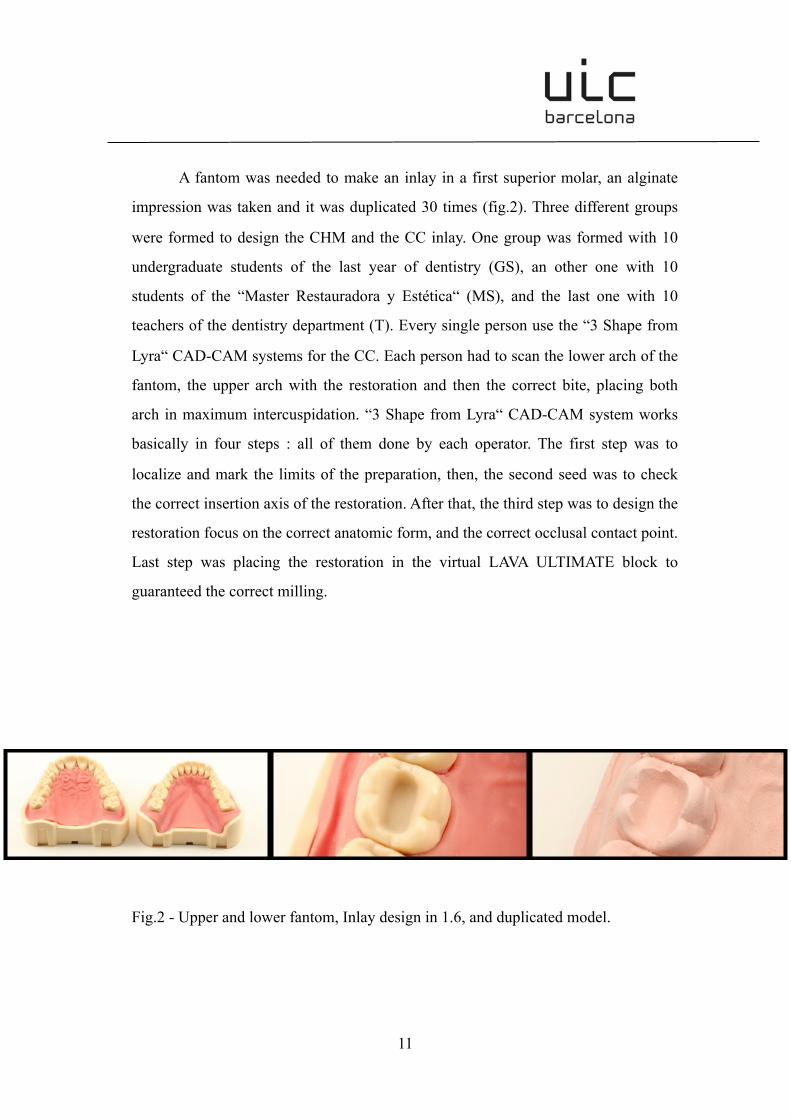
A fantom was needed to make an inlay in a first superior molar, an alginate
impression was taken and it was duplicated 30 times (fig.2). Three different groups
were formed to design the CHM and the CC inlay. One group was formed with 10
undergraduate students of the last year of dentistry (GS), an other one with 10
students of the “Master Restauradora y Estética“ (MS), and the last one with 10
teachers of the dentistry department (T). Every single person use the “3 Shape from
Lyra“ CAD-CAM systems for the CC. Each person had to scan the lower arch of the
fantom, the upper arch with the restoration and then the correct bite, placing both
arch in maximum intercuspidation. “3 Shape from Lyra“ CAD-CAM system works
basically in four steps : all of them done by each operator. The first step was to
localize and mark the limits of the preparation, then, the second seed was to check
the correct insertion axis of the restoration. After that, the third step was to design the
restoration focus on the correct anatomic form, and the correct occlusal contact point.
Last step was placing the restoration in the virtual LAVA ULTIMATE block to
guaranteed the correct milling.
Fig.2 - Upper and lower fantom, Inlay design in 1.6, and duplicated model.
! 11
!
From one part, to do the conventional design, this study asked every person to
make an inlay restoration, and they had to do the basic steps of a conventional inlay
prosthesis. This inlay was realized with composite FILTEK. On the other hand, this
study asked to the operators to design and mill the inlay restoration with the CAD-
CAM system “3 Shape from Lyra“ with an LAVA ULTIMATE block.
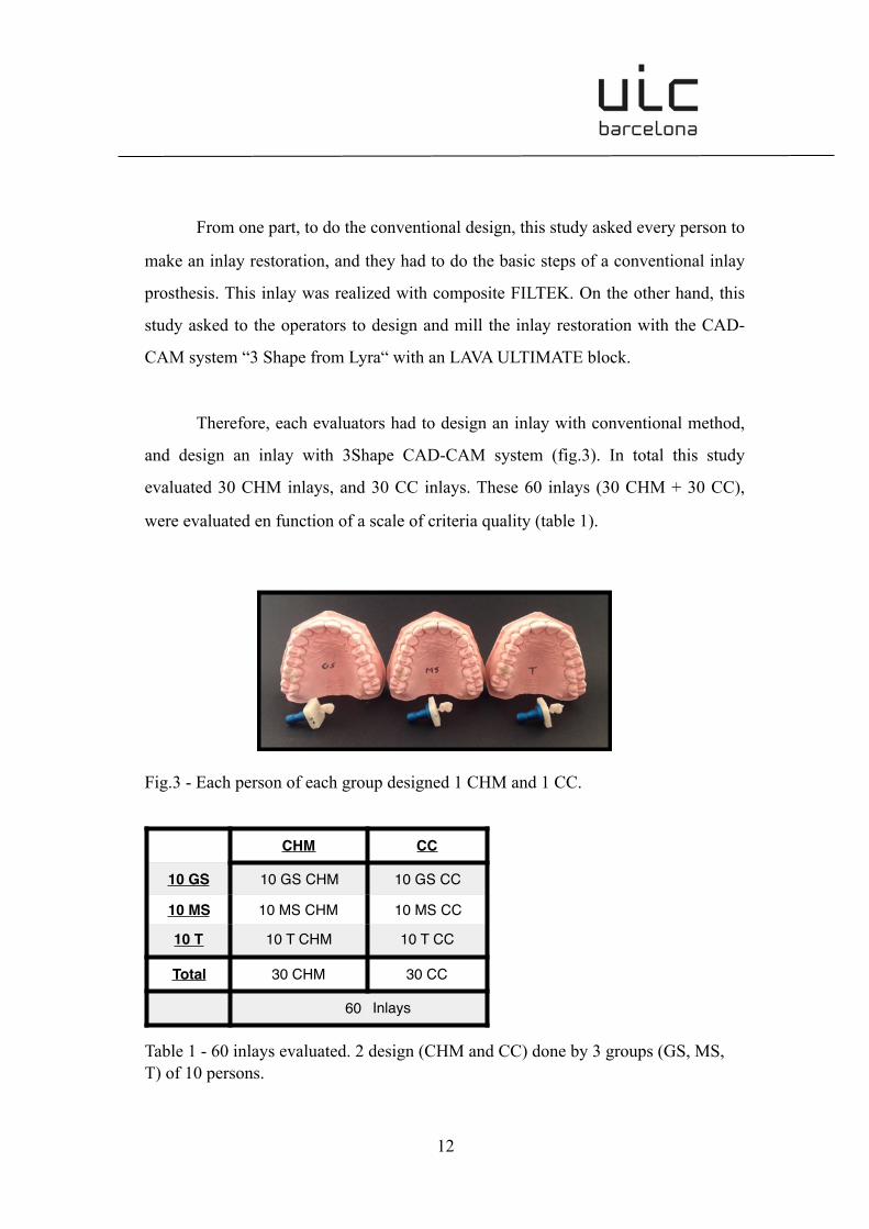
Therefore, each evaluators had to design an inlay with conventional method,
and design an inlay with 3Shape CAD-CAM system (fig.3). In total this study
evaluated 30 CHM inlays, and 30 CC inlays. These 60 inlays (30 CHM + 30 CC),
were evaluated en function of a scale of criteria quality (table 1).
Fig.3 - Each person of each group designed 1 CHM and 1 CC.
Table 1 - 60 inlays evaluated. 2 design (CHM and CC) done by 3 groups (GS, MS, T) of 10 persons.
CHM CC
10 GS 10 GS CHM 10 GS CC
10 MS 10 MS CHM 10 MS CC
10 T 10 T CHM 10 T CC
Total 30 CHM 30 CC
60 Inlays
! 12
!
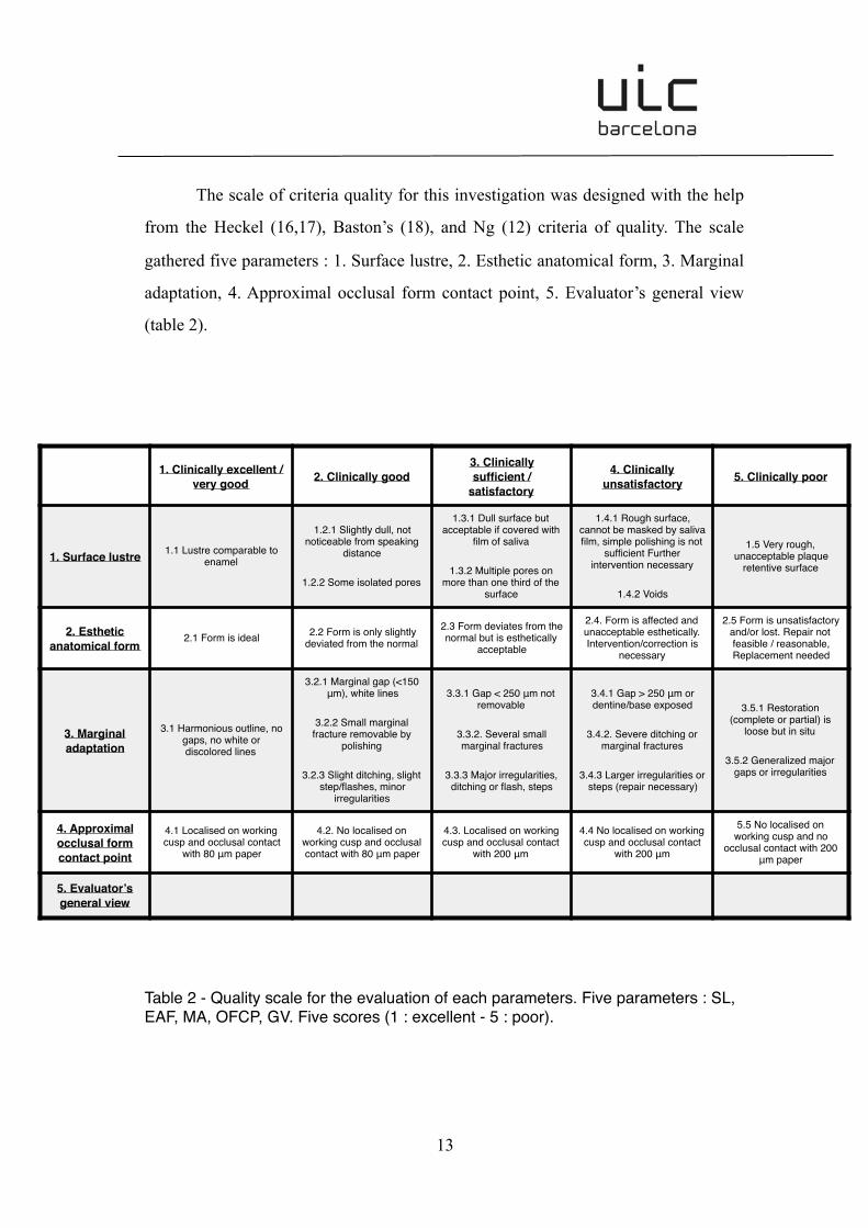
The scale of criteria quality for this investigation was designed with the help
from the Heckel (16,17), Baston’s (18), and Ng (12) criteria of quality. The scale
gathered five parameters : 1. Surface lustre, 2. Esthetic anatomical form, 3. Marginal
adaptation, 4. Approximal occlusal form contact point, 5. Evaluator’s general view
(table 2).
Table 2 - Quality scale for the evaluation of each parameters. Five parameters : SL, EAF, MA, OFCP, GV. Five scores (1 : excellent - 5 : poor).
! 13
1. Clinically excellent / very good 2. Clinically good
3. Clinically sufficient /
satisfactory
4. Clinically unsatisfactory 5. Clinically poor
1. Surface lustre 1.1 Lustre comparable to enamel
1.2.1 Slightly dull, not noticeable from speaking
distance
1.2.2 Some isolated pores
1.3.1 Dull surface but acceptable if covered with
film of saliva
1.3.2 Multiple pores on more than one third of the
surface
1.4.1 Rough surface, cannot be masked by saliva film, simple polishing is not
sufficient Further intervention necessary
1.4.2 Voids
1.5 Very rough, unacceptable plaque
retentive surface
2. Esthetic anatomical form
2.1 Form is ideal 2.2 Form is only slightly deviated from the normal
2.3 Form deviates from the normal but is esthetically
acceptable
2.4. Form is affected and unacceptable esthetically. Intervention/correction is
necessary
2.5 Form is unsatisfactory and/or lost. Repair not feasible / reasonable, Replacement needed
3. Marginal adaptation
3.1 Harmonious outline, no gaps, no white ordiscolored lines
3.2.1 Marginal gap (<150 μm), white lines
3.2.2 Small marginal fracture removable by
polishing
3.2.3 Slight ditching, slight step/flashes, minor
irregularities
3.3.1 Gap < 250 μm not removable
3.3.2. Several small marginal fractures
3.3.3 Major irregularities, ditching or flash, steps
3.4.1 Gap > 250 μm or dentine/base exposed
3.4.2. Severe ditching or marginal fractures
3.4.3 Larger irregularities or steps (repair necessary)
3.5.1 Restoration (complete or partial) is
loose but in situ
3.5.2 Generalized major gaps or irregularities
4. Approximal occlusal form contact point
4.1 Localised on working cusp and occlusal contact
with 80 μm paper
4.2. No localised on working cusp and occlusal contact with 80 μm paper
4.3. Localised on working cusp and occlusal contact
with 200 μm
4.4 No localised on working cusp and occlusal contact
with 200 μm
5.5 No localised on working cusp and no
occlusal contact with 200 μm paper
5. Evaluator’s general view
!
All of the 60 inlays were evaluated from 1 to 5. (1. Clinically excellent / very
good, 2. Clinically good, 3. Clinically sufficient / satisfactory, 4. Clinically
unsatisfactory, 5. Clinically poor).
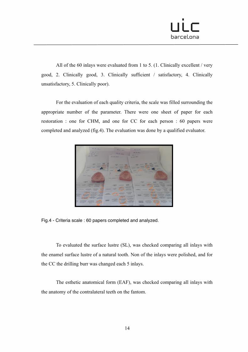
For the evaluation of each quality criteria, the scale was filled surrounding the
appropriate number of the parameter. There were one sheet of paper for each
restoration : one for CHM, and one for CC for each person : 60 papers were
completed and analyzed (fig.4). The evaluation was done by a qualified evaluator.
Fig.4 - Criteria scale : 60 papers completed and analyzed.
To evaluated the surface lustre (SL), was checked comparing all inlays with
the enamel surface lustre of a natural tooth. Non of the inlays were polished, and for
the CC the drilling burr was changed each 5 inlays.
The esthetic anatomical form (EAF), was checked comparing all inlays with
the anatomy of the contralateral teeth on the fantom.
! 14

!
The marginal adaptation (MA), was checked with endodontic files (from 15
to 30) to evaluate the gap between the inlay and the preparation (fig.5).
Fig.5 - Marginal adaptation (MA) measurment, checked with endodontic files.
The approximal occlusal form contact point (OFCP), was checked using two
types of articulating paper. One of 40 ym, and the other one of 200 ym (fig.6).
Fig.6 - Oclusal form contact point (OFCP) measurment, with two articulating papers.
! 15

!
All the data were collected and classified in an excel document, comparing all
parameters. The data were organized in 3 tables : one for each group (table 3, 4, 5).
After that, all data were analyzed comparing CHM and CC (table 6).
Table 3 - Data collected for GS group and comparison CHM/CC. Each 10 GS performed 1 CHM and 1 CC. Comparing all 5 parameters.
! 16
Restauration Group Person SL EAF MA OFCP GV Average
CHM GS 1 4 4 2 4 4 3,6
CC GS 1 3 1 2 1 2 1,8
CHM GS 2 4 2 2 3 3 2,8
CC GS 2 3 1 2 3 2 2,2
CHM GS 3 3 3 2 4 3 3
CC GS 3 2 1 1 1 1 3
CHM GS 4 4 3 2 3 3 3
CC GS 4 3 1 2 1 2 1,8
CHM GS 5 4 3 3 4 4 3,6
CC GS 5 2 1 2 3 2 2
CHM GS 6 3 3 2 4 3 3
CC GS 6 3 2 2 2 2 2,2
CHM GS 7 2 2 2 3 2 2,2
CC GS 7 3 2 2 2 2 2,2
CHM GS 8 3 3 2 2 3 2,6
CC GS 8 2 1 2 1 2 1,6
CHM GS 9 2 2 2 3 2 2,2
CC GS 9 2 2 1 3 2 2
CHM GS 10 3 3 2 3 3 2,8
CC GS 10 2 2 2 1 2 1,8
CHM 3,2 2,8 2,1 3,3 3,0 2,88
CC 2,5 1,4 1,8 1,8 1,9 2,06
Average comparison - GS
0
1
2
3
4
5
SL EAF MA OFCP GV Average
CHM CC

!
Table 4 - Data collected for MS group and comparison CHM/CC. Each 10 MS performed 1 CHM and 1 CC. Comparing all 5 parameters.
! 17
Restauration Group Person SL EAF MA OFCP GV Average
CHM MS 1 2 1 2 2 2 1,8
CC MS 1 3 1 1 2 2 1,8
CHM MS 2 2 2 2 1 2 1,8
CC MS 2 2 2 2 3 2 2,2
CHM MS 3 2 2 2 1 2 1,8
CC MS 3 2 2 1 1 2 1,6
CHM MS 4 1 1 1 1 1 1
CC MS 4 2 1 3 3 2 2,2
CHM MS 5 2 2 2 1 2 1,8
CC MS 5 3 1 2 1 2 1,8
CHM MS 6 2 1 2 2 2 1,8
CC MS 6 2 2 3 2 2 2,2
CHM MS 7 3 2 2 2 2 2,2
CC MS 7 3 2 2 2 2 2,2
CHM MS 8 1 1 1 1 1 1
CC MS 8 3 2 2 3 2 2,4
CHM MS 9 2 1 1 2 1 1,4
CC MS 9 2 1 1 1 1 1,2
CHM MS 10 2 2 2 1 2 1,8
CC MS 10 2 1 2 1 2 1,6
CHM 1,9 1,5 1,7 1,4 1,7 1,64
CC 2,4 1,5 1,9 1,9 1,9 1,92
Average comparison - MS
0
1
2
3
4
5
SL EAF MA OFCP GV Average
CHM CC
!
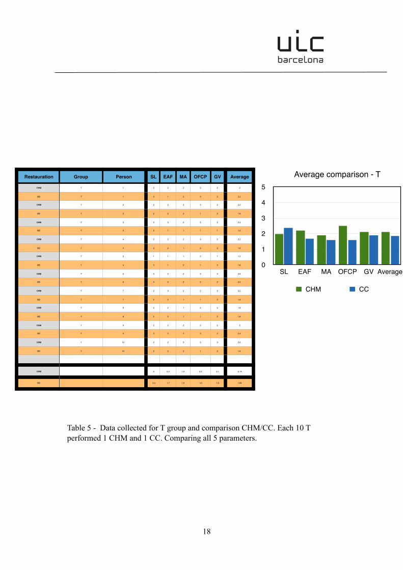
Table 5 - Data collected for T group and comparison CHM/CC. Each 10 T performed 1 CHM and 1 CC. Comparing all 5 parameters.
! 18
Restauration Group Person SL EAF MA OFCP GV Average
CHM T 1 2 2 2 2 2 2
CC T 1 3 1 2 3 2 2,2
CHM T 2 2 2 2 3 2 2,2
CC T 2 2 2 2 1 2 1,8
CHM T 3 2 3 2 3 2 2,4
CC T 3 2 1 1 1 1 1,2
CHM T 4 2 2 2 3 2 2,2
CC T 4 2 2 1 2 2 1,8
CHM T 5 1 1 1 2 1 1,2
CC T 5 3 1 2 1 2 1,8
CHM T 6 3 3 2 3 3 2,8
CC T 6 3 2 2 2 2 2,2
CHM T 7 2 3 2 2 2 2,2
CC T 7 2 2 1 1 2 1,6
CHM T 8 2 2 1 2 2 1,8
CC T 8 2 2 1 1 2 1,6
CHM T 9 2 2 2 2 2 2
CC T 9 3 2 2 3 2 2,4
CHM T 10 2 2 3 3 3 2,6
CC T 10 2 2 2 1 2 1,8
CHM 2 2,2 1,9 2,5 2,1 2,14
CC 2,4 1,7 1,6 1,6 1,9 1,84
Average comparison - T
0
1
2
3
4
5
SL EAF MA OFCP GV Average
CHM CC

!
Table 6 - Comparison CHM/CC. The 5 parameters compared depending on the operator. One for CHM and one for CC.
To test the normality of the data Shapiro-Wilk Test was performed. The
results was tested by using a one way ANOVA statistical analysis. All the data
analysis, were carried out using the STATGRAPHICS software (Statpoint
technologies, Warrenton, Virginia, USA) with a significance level set at p = 0,05.
! 19
Average comparison - CC
012345
SL EAF MA OFCP GV AVERAGE
GS MS T
Average comparison - CHM
012345
SL EAF MA OFCP GV AVERAGE
GS MS T

!
5. Results :
The principal objective of this study was to compare the quality of an inlay
done by two technique : composite hand-maid (CHM) versus CAD-CAM (CC).
Secondary objectives were to compare the quality of this same restoration, focusing
on each of the three groups individually. The results will first be described
considering all group together, then each criteria regarding to each group
individually.
In terms of quality, the results of the study showed statistically significant
difference between CHM and CC (p-valor < 0,05). The null hypothesis was rejected.
For all group mingled, the average score for the CC was 1,94 versus 2,22 for the
CHM (p-valor = 0,0102). More specifically, GS group showed worst results when
designing the inlay regardless of the type of method used (CHM and CC) comparing
to MS and T groups. However no statistically significant difference was observed
between MS and T groups. Analyzing with more details for each group, the study
reveled better results for CC than CHM for GS and T groups, but better results for
CHM than CC for MS group (table 7).
Table 7 - Analysis of Variance for Average.
! 20
!
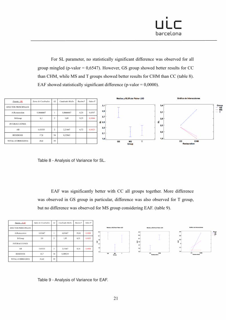
For SL parameter, no statistically significant difference was observed for all
group mingled (p-valor = 0,6547). However, GS group showed better results for CC
than CHM, while MS and T groups showed better results for CHM than CC (table 8).
EAF showed statistically significant difference (p-valor = 0,0000).
Table 8 - Analysis of Variance for SL.
EAF was significantly better with CC all groups together. More difference
was observed in GS group in particular, difference was also observed for T group,
but no difference was observed for MS group considering EAF. (table 9).
Table 9 - Analysis of Variance for EAF.
! 21

!
No statistically significant difference was observed for MA (p-valor =
0,3283) (table 10).
Table 10 - Analysis of Variance for MA.
OFCP was statistically significantly better for CC rather than CHM (p-valor =
0,017). In particular, OFCP was more accurate with CC rather than CHM for GS and
T group, but more accurate with CHM rather tan CC for MS group (table 11).
Table 11 - Analysis of Variance for OFCP.
! 22
!
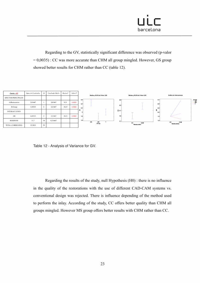
Regarding to the GV, statistically significant difference was observed (p-valor
= 0,0035) : CC was more accurate than CHM all group mingled. However, GS group
showed better results for CHM rather than CC (table 12).
Table 12 - Analysis of Variance for GV.
Regarding the results of the study, null Hypothesis (H0) : there is no influence
in the quality of the restorations with the use of different CAD-CAM systems vs.
conventional design was rejected. There is influence depending of the method used
to perform the inlay. According of the study, CC offers better quality than CHM all
groups mingled. However MS group offers better results with CHM rather than CC.
! 23
!
6. Discusion :
As Pascal Magne (19) described in a study on 2006, indirect techniques with
composite resins or ceramics are recommended for serial restorations when esthetics
and dynamic occlusion issues are of primary concern. Indirect composite inlays are
recommended for serial restorations without cusp coverage or with limited cuspal
coverage leaving at least one functional cusp. The past decades, CAD-CAM
techniques have been considerately growing, and today seems to bee an adequate
alternative to indirect hand-maid restoration (20). The implementation of this digital
method has decreased manufacturing costs by reducing technician time and material
costs while increasing productivity (21), but this technique must guaranty some
quality criteria : a correct esthetic anatomic form, an ideal contact and occlusal point,
a correct margin fitting, resistance, durability… Some studies have demonstrated the
advantage of using CAD-CAM system. In 2014 Ng et Al (12) compared the marginal
fitting of crowns fabricated with digital and conventional methods, and the study
concluded that the fully digital fabrication method provided better margin fit than the
conventional method. A lot of study have been describing the marginal fit of a
restoration after using a conventional impression and a digital impression. In a
systematic review of 2016, Tsirogiannis et al (22), reveled that no significant
difference was found regarding the marginal discrepancy of single unit ceramic
restorations fabricated after digital and conventional impressions. Both the digital
workflow and the conventional method ensure the clinically fully acceptable
fabrication of single-unit ceramic restorations. In 2014, Anadioti et Al (23) analyzed
the 3D and 2D marginal fit of pressed and CAD/CAM Lithium Disilicate crowns
made from digital and conventional impressions. The results of the study were that
the combination of PVS impression method and press fabrication technique produced
the most accurate 3D and 2D marginal fits rather than digital impression.
! 24

!
Correct occlusal contact point has been described in several studies,
comparing conventional and digital methods. In 2013, Litzenburger et Al (24)
analyzed the fully automatic CAD design of the occlusal morphology of partial
crowns compared to dental technicians' design. The results of the study show that in
the design of naturally shaped occlusal inlay/onlay surfaces, a fully automatic CAD
system can be at least as good as conventional wax-ups by dental technicians. In an
other study talking about occlusion, in 2010 : Reich et Al described (25) the occlusal
precision of laboratory versus CAD/CAM processed all-ceramic crowns. The results
show the time necessary to adjust the occlusion with both design, and time did not
differ significantly. Finally in 2015, a study of Kollmuss et Al (26) described the
comparison of chairside and laboratory CAD/CAM to conventional produced all-
ceramic crowns regarding morphology, occlusion, and aesthetics. The conclusions
were that all methods had pros and cons regarding different parameters. Further
improvements of CAD/CAM software shall lead to restorations comparable to
conventional restorations in all aspects, especially in aesthetics. All tested methods of
production for all- ceramic crowns produced clinically acceptable results. Thus, in an
individual case, the method chosen can be determined by the dentist’s preference.
Analyzing this study, the results show statistically significant difference
between the conventional and the digital methods, with better results for the digital
methods. This result is true considering all parameters together (esthetic, lustre,
occlusal point, marginal adaptation) and all three groups mingled (graduate student,
master student and teachers). Looking with more details each parameters and
comparing between each groups, some differences can be find. Results show better
quality of the inlay for master student when using conventional method rather tan
digital method.
! 25
!
7. Conclusion :
The principal objective of this study was to evaluate the restoration quality
between CAD-CAM systems and conventional prosthesis design on inlays. With the
limits of the study, we can say that CAD-CAM design showed better results than
composite hand-maid design for an inlay, considering all groups and all parameters
together. The secondary objective was to evaluate and compare the quality between
CAD-CAM systems and conventional prosthesis design on inlays depending on the
operator. Analyzing with more details, master student group had better results with
composite hand-maid design rather than composite hand-maid design. We can
conclude that CAD-CAM design is a good alternative to composite hand-maid
design, but depending on the operator.
8. Future expectations :
In a future, we can expect an evolution of the digital technique. In the last
decades the CAD-CAM design get closer to the conventional technique, but one
question is still without answer : one day, will the best digital technique better than
the best conventional technique ?
! 26

!
8. References :
1. Birnbaum NS, Aaronson HB. Dental impressions using 3D digital scanners:
virtual becomes reality. Compend Contin Educ Dent. 2008 Oct;29(8):494, 496,
498-505
2. Liu PR, Essig ME. Panorama of dental CAD/CAM restorative systems.
Compend Contin Educ Dent. 2008 Oct;29(8):482, 484, 486-8
3. Lee SJ, Gallucci GO. Digital vs. conventional implant impressions: efficiency
outcomes. Clin Oral Implants Res. 2013 Jan;24(1):111-5.
4. Bahillo J, Jané L, Bortolotto T, Krejci I, Roig M. Full-mouth composite
rehabilitation of a mixed erosion and attrition patient: a case report with v-
shaped veneers and ultra-thin CAD/CAM composite overlays. Quintessence Int.
2014 Oct;45(9):749-56.
5. Davidowitz G, Kotick PG. The use of CAD/CAM in dentistry. Dent Clin North
Am. 2011 Jul;55(3):559-70
6. Duret F, Preston JD. CAD/CAM imaging in dentistry. Curr Opin Dent. 1991 Apr;
1(2):150-4
7. Sneha S. Mantri. CAD-CAM in dental restorations: An overview. July 2010. Vol
I I . D i s p o n i b l e e n h t t p : / / w w w. r e s e a r c h g a t e . n e t / p u b l i c a t i o n /
228931572_CADCAM_in_Dental_Restorations_An_Overview
8. Nandini VV, Venkatesh KV, Nair KC. Alginate impressions: A practical
perspective. J Conserv Dent. 2008 Jan;11(1):37-41
9. Lee SJ, Gallucci GO. Digital vs. conventional implant impressions: efficiency
outcomes. Clin Oral Implants Res. 2013 Jan;24(1):111-5
10. Fuster-Torres MA, Albalat-Estela S, Alcañiz-Raya M, Peñarrocha-Diago M.
CAD/CAM dental systems in implant dentistry: update. Med Oral Patol Oral Cir
Bucal. 2009 Mar 1;14(3):E141-5
! 27

!
11. Hamza TA, Ezzat HA, El-Hossary MM, Katamish HA, Shokry TE, Rosenstiel
SF. Accuracy of ceramic restorations made with two CAD/CAM systems. J
Prosthet Dent. 2013 Feb;109(2):83-7
12. Ng J, Ruse D, Wyatt C. A comparison of the marginal fit of crowns fabricated
with digital and conventional methods. J Prosthet Dent. 2014 Sep;112(3):555-60.
13. Lemaire ED, Upton D, Paialunga J, Martel G, Boucher J. Clinical analysis of a
CAD/CAM system for custom seating: a comparison with hand-sculpting
methods. J Rehabil Res Dev. 1996 Jul;33(3):311-20.
14. Renne W, McGill ST, Forshee KV, DeFee MR, Mennito AS. Predicting marginal
fit of CAD/CAM crowns based on the presence or absence of common
preparation errors. J Prosthet Dent. 2012 Nov;108(5):310-5.
15. Mously HA, Finkelman M, Zandparsa R, Hirayama H. Marginal and internal
adaptation of ceramic crown restorations fabricated with CAD/CAM technology
and the heat-press technique. J Prosthet Dent. 2014 Aug;112(2):249-56.
16. Hickel R, Peschke A, Tyas M, Mjör I, Bayne S, Peters M, Hiller KA, Randall R,
Vanherle G, Heintze SD. FDI World Dental Federation: clinical criteria for the
evaluation of direct and indirect restorations-update and clinical examples. Clin
Oral Investig. 2010 Aug;14(4):349-66
17. Hickel R, Roulet JF, Bayne S, Heintze SD, Mjör IA, Peters M, Rousson V,
Randall R, Schmalz G, Tyas M, Vanherle G. Recommendations for conducting
controlled clinical studies of dental restorative materials. Science Committee
Project 2/98--FDI World Dental Federation study design (Part I) and criteria for
evaluation (Part II) of direct and indirect restorations including onlays and partial
crowns. J Adhes Dent. 2007;9 Suppl 1:121-47
18. Batson ER, Cooper LF, Duqum I, Mendonça G. Clinical outcomes of three
different crown systems with CAD/CAM technology. J Prosthet Dent. 2014 Oct;
112(4):770-7
19. Magne P. Composite resins and bonded porcelain: the postamalgam era? J Calif
Dent Assoc. 2006 Feb;34(2):135-47
! 28
!
20. Van Noort R. The future of dental devices is digital. Dent Mater 2012;28:3-12.
21. Beuer F, Schweiger J, Edelhoff D. Digital dentistry: an overview of recent
developments for CAD/CAM generated restorations. Br Dent J
2008;204:505-11.
22. Tsirogiannis P, Reissmann DR, Heydecke G. Evaluation of the marginal fit of
single-unit, complete-coverage ceramic restorations fabricated after digital and
conventional impressions: A systematic review and meta-analysis. J Prosthet
Dent. 2016 Apr 6. pii: S0022-3913(16)00139-6.
23. Anadioti E, Aquilino SA, Gratton DG, Holloway JA, Denry I, Thomas GW, Qian
F. 3D and 2D marginal fit of pressed and CAD/CAM lithium disilicate crowns
made from digital and conventional impressions. J Prosthodont. 2014 Dec;23(8):
610-7
24. Litzenburger AP, Hickel R, Richter MJ, Mehl AC, Probst FA. Fully automatic
CAD design of the occlusal morphology of partial crowns compared to dental
technicians' design. Clin Oral Investig. 2013 Mar;17(2):491-6.
25. Reich S, Brungsberg B, Teschner H, Frankenberger R. The occlusal precision of
laboratory versus CAD/CAM processed all-ceramic crowns. Am J Dent. 2010
Feb;23(1):53-6.
26. Kollmuss M, Kist S, Goeke JE, Hickel R, Huth KC. Comparison of chairside and
laboratory CAD/CAM to conventional produced all-ceramic crowns regarding
morphology, occlusion, and aesthetics. Clin Oral Investig. 2016 May;20(4):
791-7
! 29

!
9. Annexes
! 30

!
! 31
!
IP or tutor signature Supporting researcher:
�
! 32