cardiac electrophysiology qiang xia (夏强), phd department of physiology room c518, block c,...
TRANSCRIPT

CardiacElectrophysiology
Qiang XIA (夏强 ), PhDDepartment of Physiology
Room C518, Block C, Research Building, School of MedicineTel: 88208252
Email: [email protected]
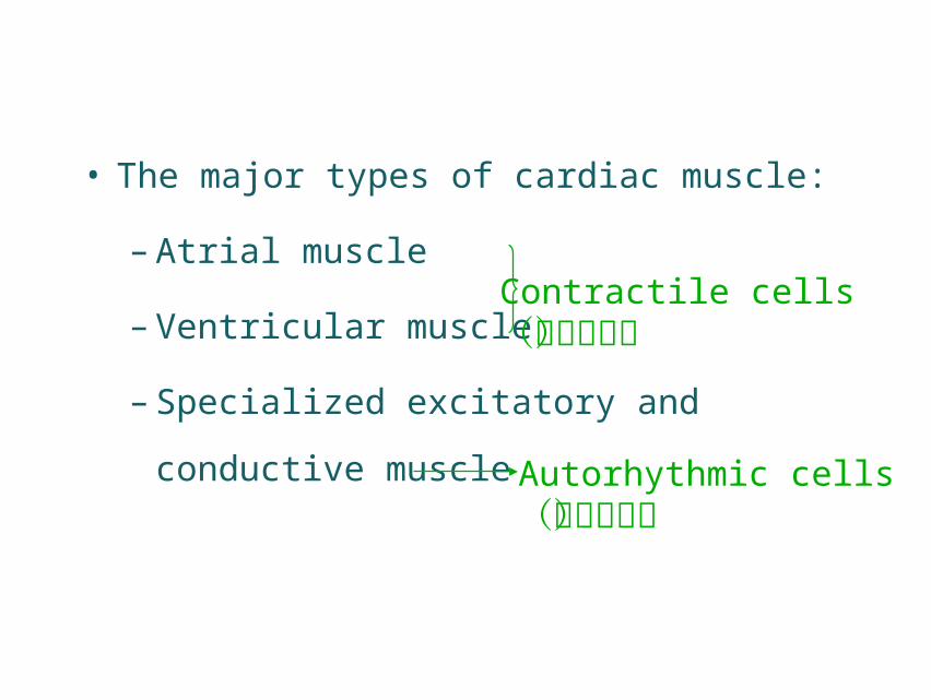
• The major types of cardiac muscle:
– Atrial muscle
– Ventricular muscle
– Specialized excitatory and conductive
muscle
Contractile cells(收缩细胞)
Autorhythmic cells(自律细胞)
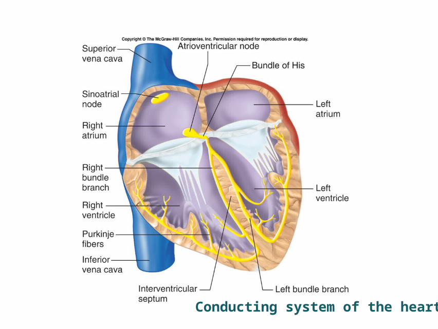
Conducting system of the heart
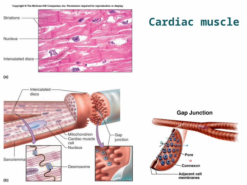
Cardiac muscle
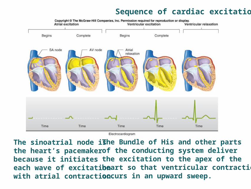
The sinoatrial node is the heart’s pacemaker because it initiates each wave of excitation with atrial contraction.
The Bundle of His and other parts of the conducting system deliver the excitation to the apex of the heart so that ventricular contraction occurs in an upward sweep.
Sequence of cardiac excitation
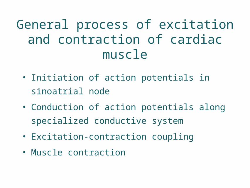
General process of excitation and contraction of cardiac muscle
• Initiation of action potentials in sinoatrial node
• Conduction of action potentials along
specialized conductive system
• Excitation-contraction coupling
• Muscle contraction
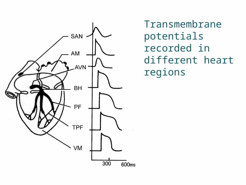
Transmembrane potentials recorded in different heart regions
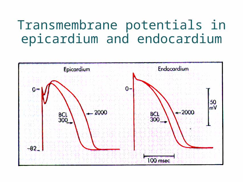
Transmembrane potentials in epicardium and endocardium
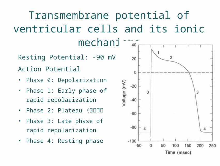
Transmembrane potential of ventricular cells and its ionic mechanisms
Resting Potential: -90 mV
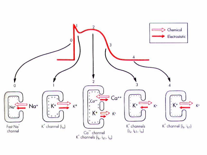
Action Potential
• Phase 0: Depolarization
• Phase 1: Early phase of rapid
repolarization
• Phase 2: Plateau(平台期)• Phase 3: Late phase of rapid
repolarization
• Phase 4: Resting phase
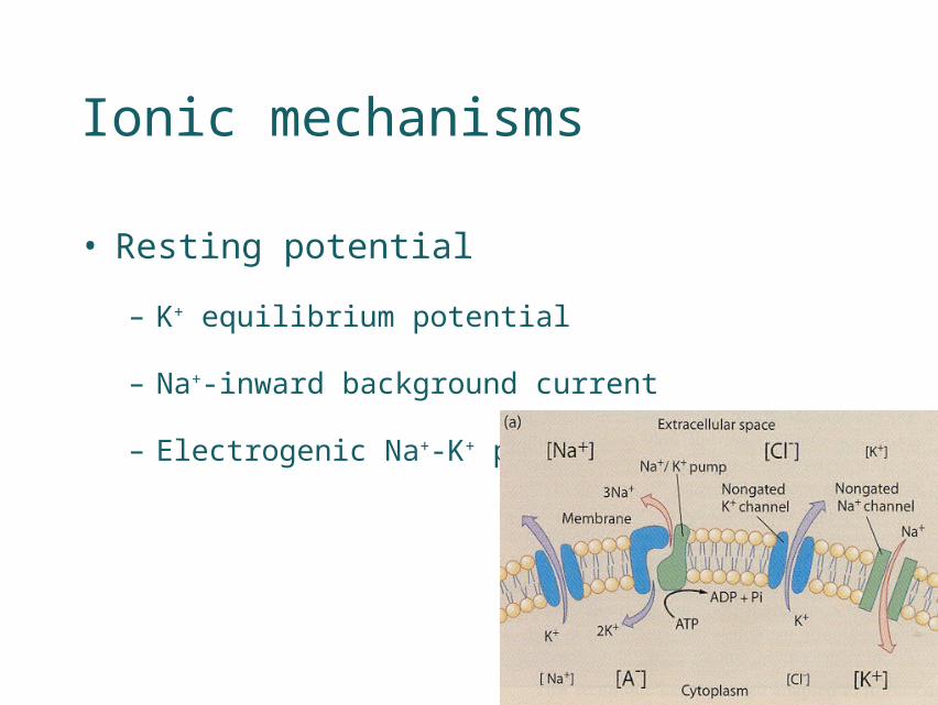
• Resting potential
– K+ equilibrium potential
– Na+-inward background current
– Electrogenic Na+-K+ pump
Ionic mechanisms
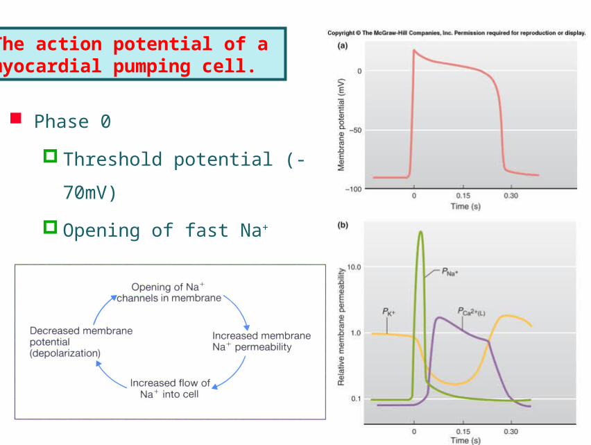
Phase 0
Threshold potential (-70mV)
Opening of fast Na+ channel
Regenerative cycle(再生性循环)
The action potential of a myocardial pumping cell.

Phase 1
Transient outward current, Ito
K+ current
activated at –20 mV
opening for 5~10 ms
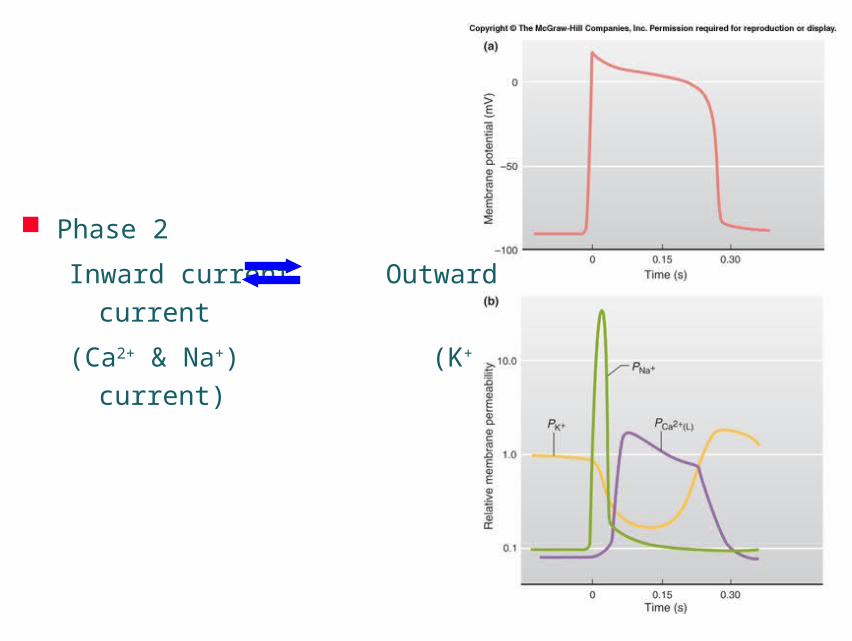
Phase 2
Inward current Outward current
(Ca2+ & Na+) (K+ current)

Types of Ca2+ channels in cardiac cells:
(1) L-type (long-lasting) (Nowycky, 1985)
(2) T-type (transient) (Nowycky, 1985)

Ca2+ channels
Duration of current long-lasting transient
Activation kinetics slower faster
Inactivation kinetics slower faster
Threshold high (-35mV) Low (-60mV)
cAMP/cGMP-regulated Yes No
Phosphorylation-regulated Yes No
Openers Bay-K-8644 -
Blockers varapamil Tetramethrin
nifedipine, diltiazem Ni2+
Inactivation by [Ca2+]i Yes slight
Patch-clamp recording run-down relatively stable
L-type T-
type
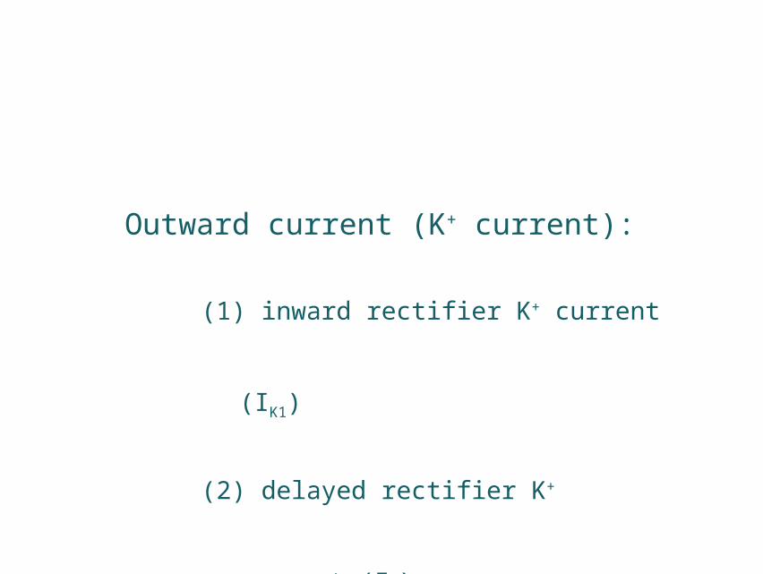
Outward current (K+ current):
(1) inward rectifier K+ current (IK1)
(2) delayed rectifier K+ current (IK)
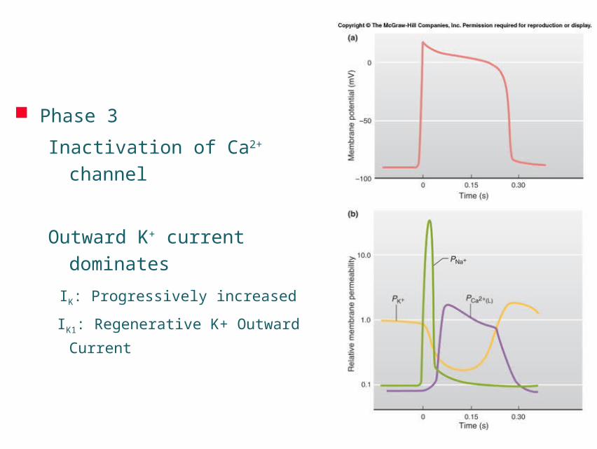
Phase 3
Inactivation of Ca2+ channel
Outward K+ current dominates
IK: Progressively increased
IK1: Regenerative K+ Outward Current
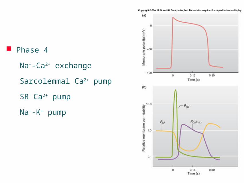
Phase 4
Na+-Ca2+ exchange
Sarcolemmal Ca2+ pump
SR Ca2+ pump
Na+-K+ pump

a, The key ion channels (and an electrogenic transporter) in cardiac cells. K+ channels (green) mediate K+ efflux from the cell; Na+ channels (purple) and Ca2+ channels (yellow) mediate Na+ and Ca2+ influx, respectively. The Na+/Ca2+ exchanger (red) is electrogenic, as it transports three Na+ ions for each Ca2+ ion across the surface membrane. b, Ionic currents and genes underlying the cardiac action potential. Top, depolarizing currents as functions of time, and their corresponding genes; centre, a ventricular action potential; bottom, repolarizing currents and their corresponding genes.
From the following article:Cardiac channelopathiesEduardo MarbánNature 415, 213-218(10 January 2002)doi:10.1038/415213a

Transmembrane potentials recorded in different heart regions
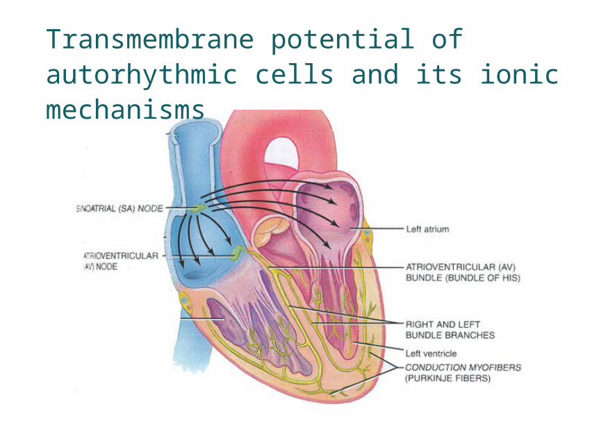
Transmembrane potential of autorhythmic cells and its ionic mechanisms
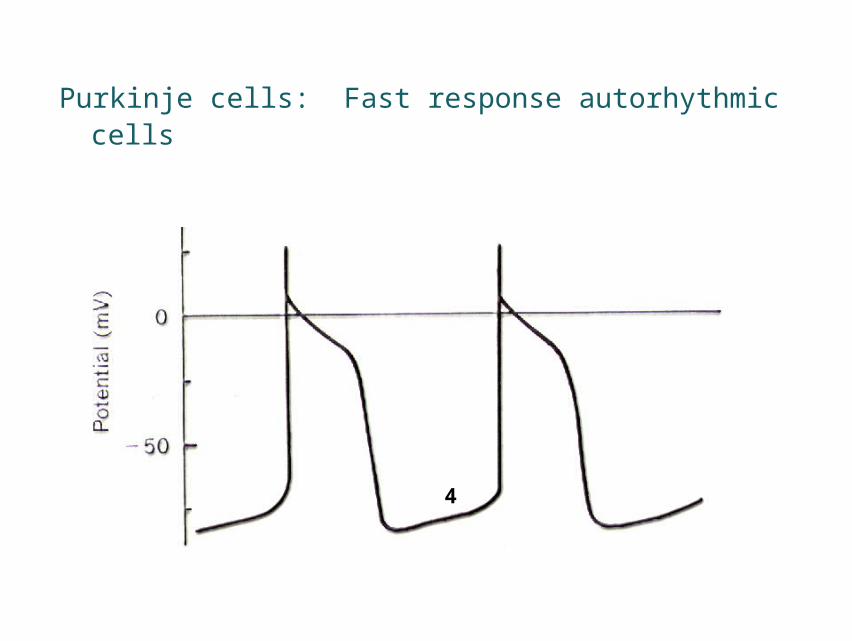
Purkinje cells: Fast response autorhythmic cells
4

Contractile cells Autorhythmic cells Phase 4 stable potential Phase 4 spontaneous depolarization
( 4期自动去极化)
Resting potential Maximal repolarization potential
(最大复极电位)
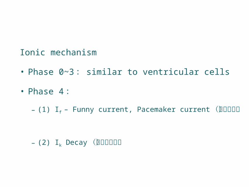
Ionic mechanism
• Phase 0~3 : similar to ventricular cells
• Phase 4:
– (1) If – Funny current, Pacemaker current(起搏电流)
– (2) Ik Decay(钾电流衰减)

Characteristics of If channel
• Na+, K+
• Voltage- & time-dependent
Activation── Repolarized to -60mV
Full activation── Hyperpolarized to -100mV
Inactivation── Depolarized to -50mV
• Blocked by Cs, not by TTX
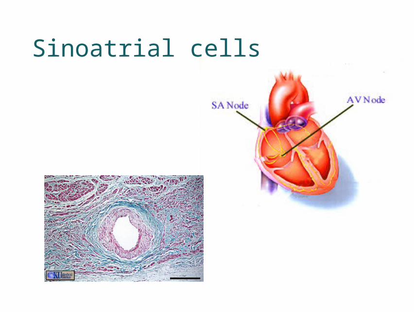
Sinoatrial cells
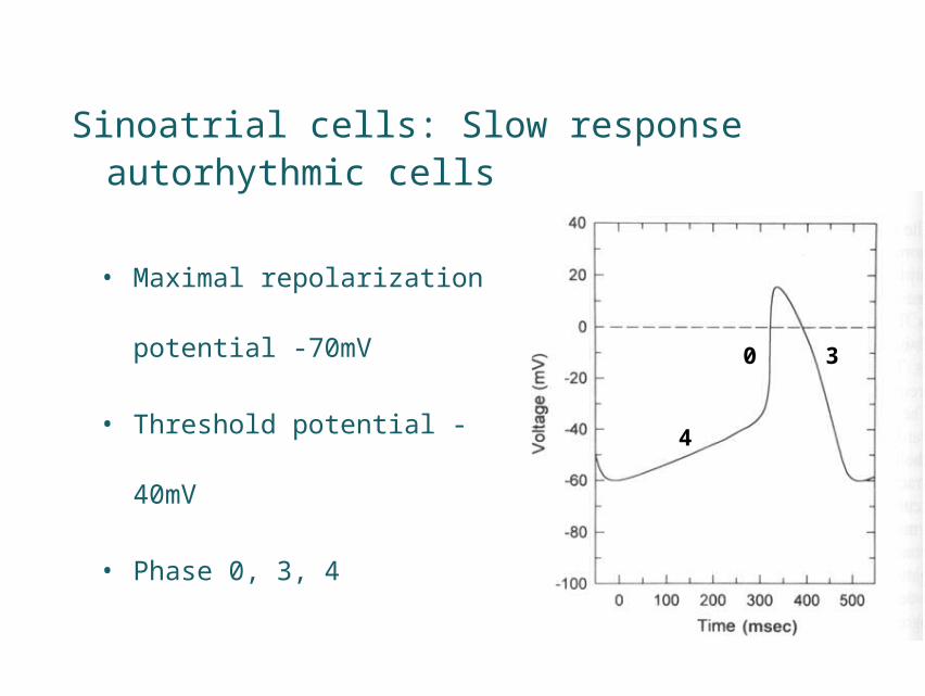
• Maximal repolarization
potential -70mV
• Threshold potential -40mV
• Phase 0, 3, 4
Sinoatrial cells: Slow response autorhythmic cells
4
0 3
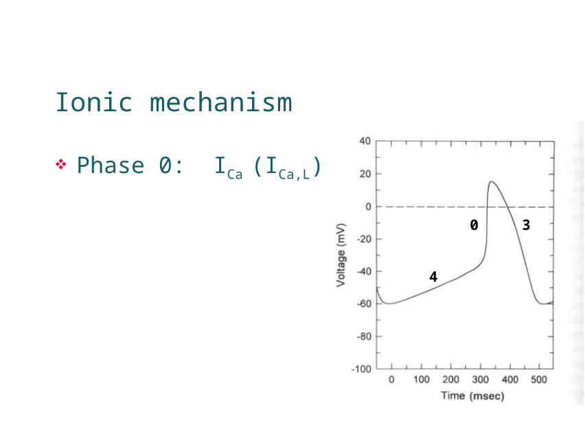
Ionic mechanism
Phase 0: ICa (ICa,L)
4
0 3
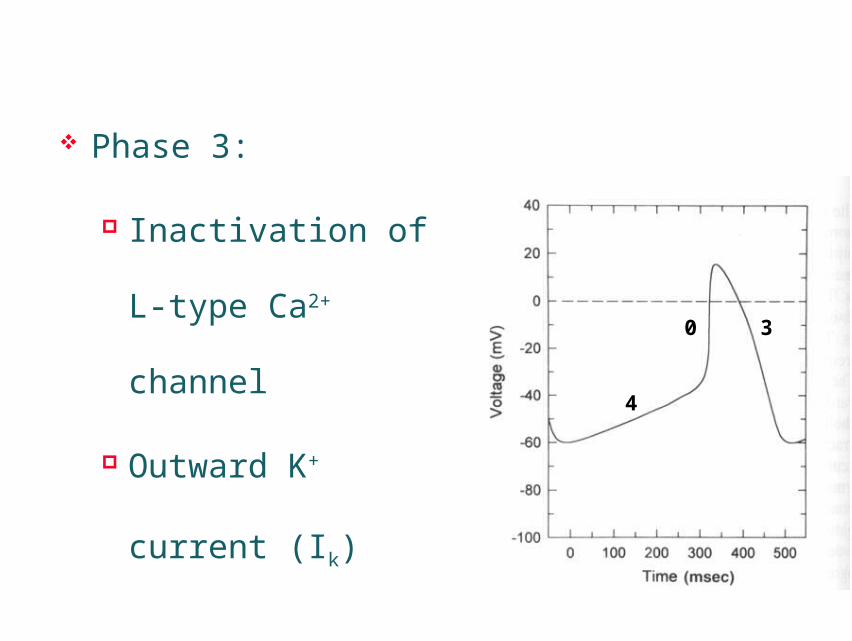
Phase 3:
Inactivation of L-type
Ca2+ channel
Outward K+ current
(Ik)
4
0 3

• Phase 4: Ik decay
Inactivated when repolarized to -60mV
ICa,T
Activated when depolarized to -50mV
If
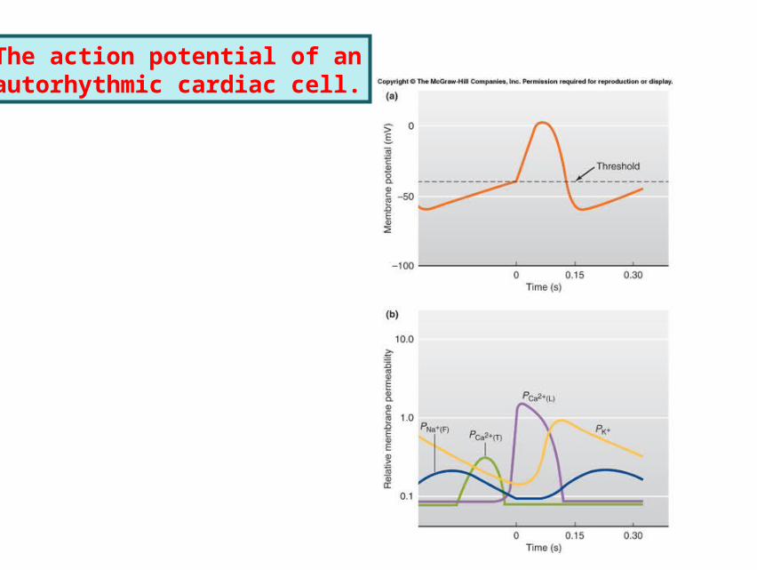
The action potential of anautorhythmic cardiac cell.
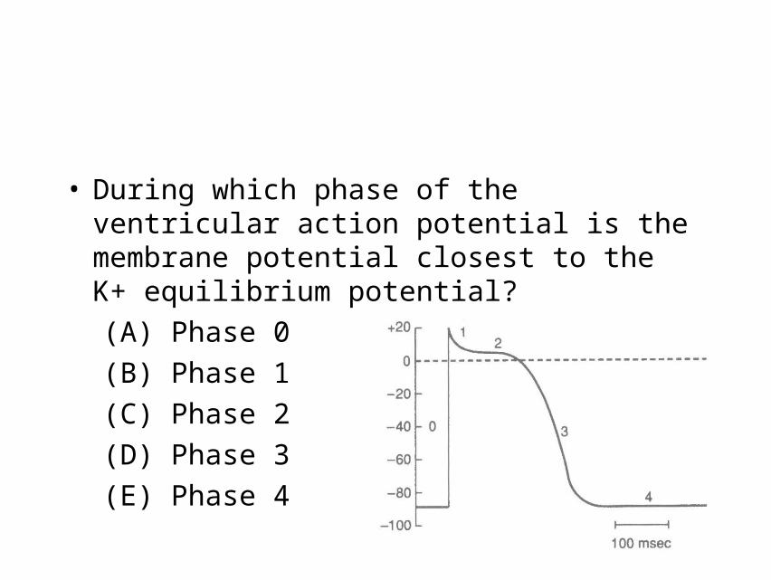
• During which phase of the ventricular action potential is the membrane potential closest to the K+ equilibrium potential?
(A) Phase 0
(B) Phase 1
(C) Phase 2
(D) Phase 3
(E) Phase 4
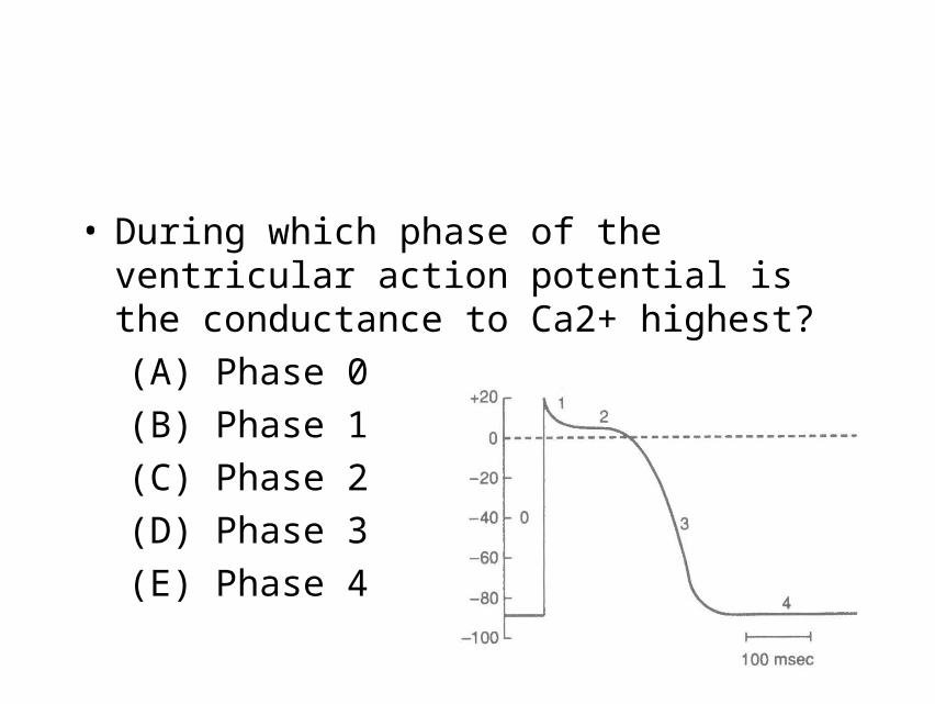
• During which phase of the ventricular action potential is the conductance to Ca2+ highest?
(A) Phase 0
(B) Phase 1
(C) Phase 2
(D) Phase 3
(E) Phase 4
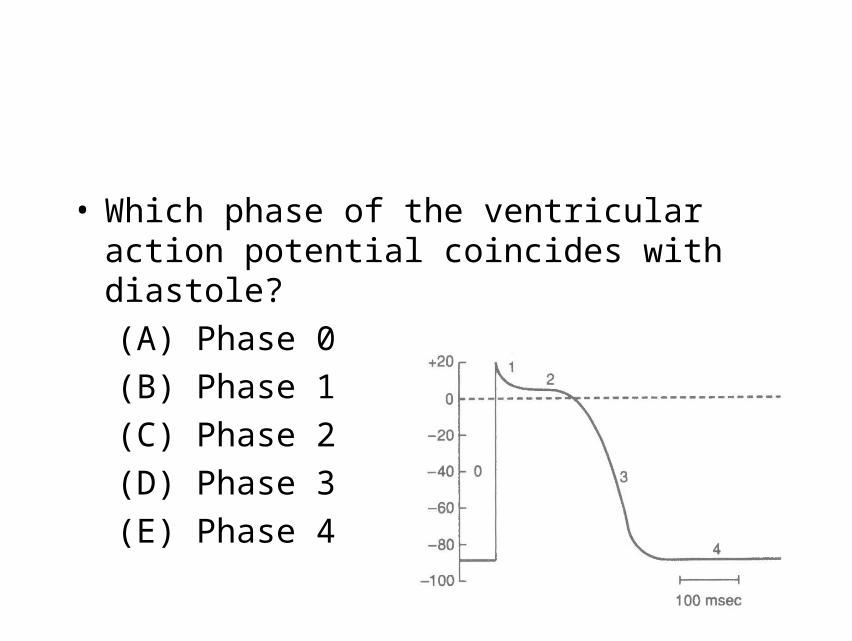
• Which phase of the ventricular action potential coincides with diastole?
(A) Phase 0
(B) Phase 1
(C) Phase 2
(D) Phase 3
(E) Phase 4

• The low-resistance pathways between myocardial cells that allow for the spread of action potentials are the
(A) gap junctions
(B) T tubules
(C) sarcoplasmic reticulum (SR)
(D) intercalated disks
(E) mitochondria
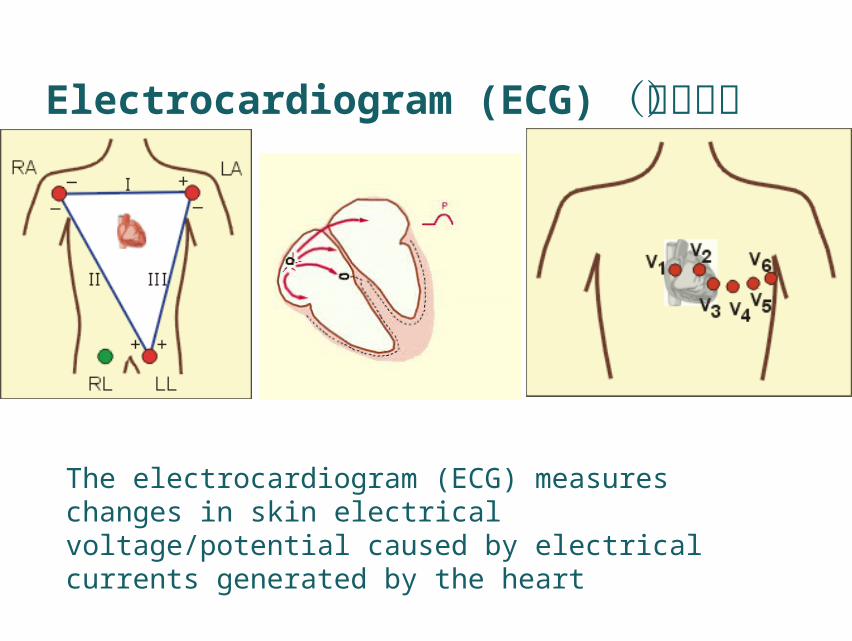
Electrocardiogram (ECG)(心电图)
The electrocardiogram (ECG) measures changes in skin electrical voltage/potential caused by electrical currents generated by the heart

The relationship between the electrocardiogram (ECG), recorded as the difference between currents at the left and right wrists,
and
an action potential typical of ventricular myocardial cells.
Electrocardiogram (ECG)
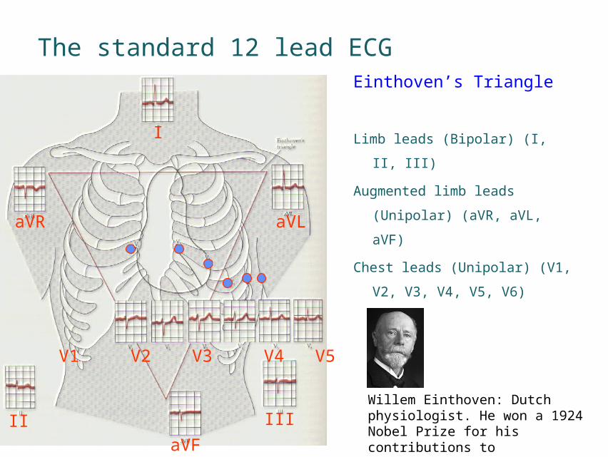
The standard 12 lead ECG Einthoven’s Triangle
Limb leads (Bipolar) (I, II, III)
Augmented limb leads
(Unipolar) (aVR, aVL, aVF)
Chest leads (Unipolar) (V1, V2,
V3, V4, V5, V6)
I
II III
aVR aVL
aVF
V1 V2 V3 V4 V5 V6
Willem Einthoven: Dutch physiologist. He won a 1924 Nobel Prize for his contributions to electrocardiography.

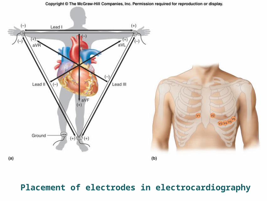
Placement of electrodes in electrocardiography

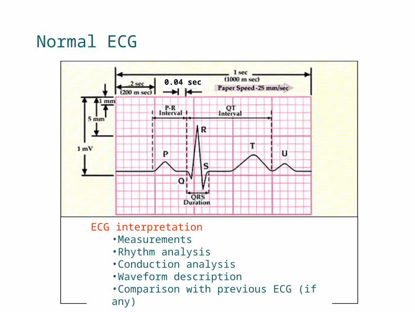
Normal ECG
0.04 sec
ECG interpretation•Measurements •Rhythm analysis•Conduction analysis•Waveform description •Comparison with previous ECG (if any)
Animation of a normal ECG wave
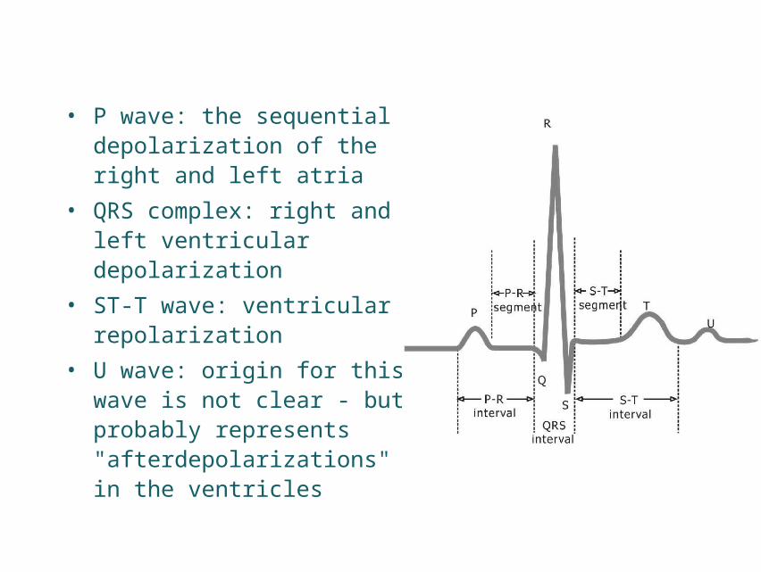
• P wave: the sequential depolarization of the right and left atria
• QRS complex: right and left ventricular depolarization
• ST-T wave: ventricular repolarization
• U wave: origin for this wave is not clear - but probably represents "afterdepolarizations" in the ventricles
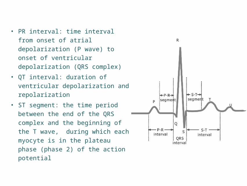
• PR interval: time interval from
onset of atrial depolarization (P
wave) to onset of ventricular
depolarization (QRS complex)
• QT interval: duration of
ventricular depolarization and
repolarization
• ST segment: the time period
between the end of the QRS
complex and the beginning of the
T wave, during which each
myocyte is in the plateau phase
(phase 2) of the action potential

Normal
Partial block
Complete block

• Excitability
• Autorhythmicity
• Conductivity
• Contractility
Electrophysiological properties(电生理特性)
Mechanical property(机械特性)
Physiological properties of cardiac cells
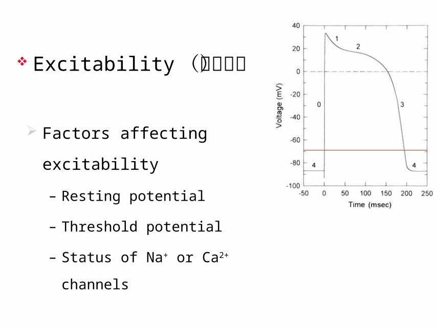
Factors affecting excitability
– Resting potential
– Threshold potential
– Status of Na+ or Ca2+ channels
Excitability(兴奋性)
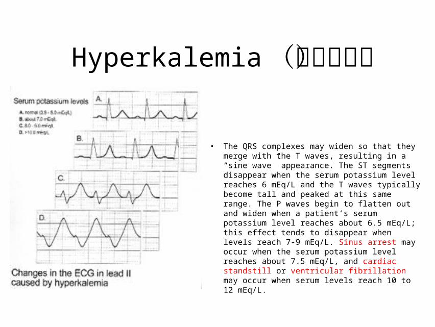
Hyperkalemia(高钾血症)
• The QRS complexes may widen so that they merge with the T waves, resulting in a “sine wave” appearance. The ST segments disappear when the serum potassium level reaches 6 mEq/L and the T waves typically become tall and peaked at this same range. The P waves begin to flatten out and widen when a patient‘s serum potassium level reaches about 6.5 mEq/L; this effect tends to disappear when levels reach 7-9 mEq/L. Sinus arrest may occur when the serum potassium level reaches about 7.5 mEq/L, and cardiac standstill or ventricular fibrillation may occur when serum levels reach 10 to 12 mEq/L.
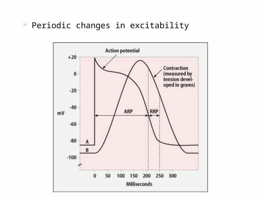
Periodic changes in excitability
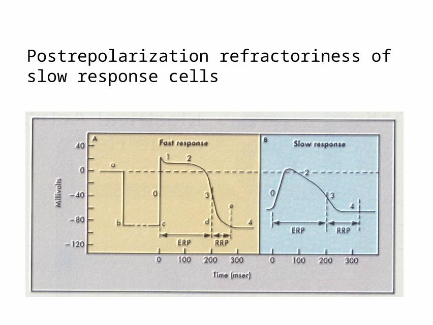
Postrepolarization refractoriness of slow response cells
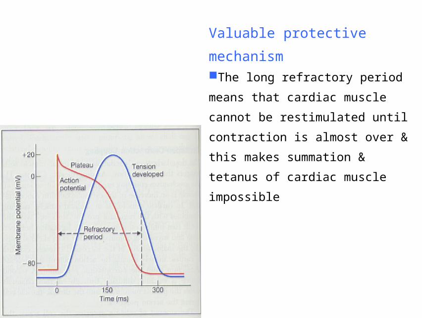
Valuable protective mechanismThe long refractory period means
that cardiac muscle cannot be
restimulated until contraction is almost
over & this makes summation &
tetanus of cardiac muscle impossible
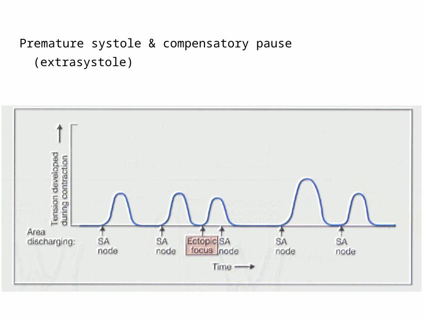
Premature systole & compensatory pause
(extrasystole)
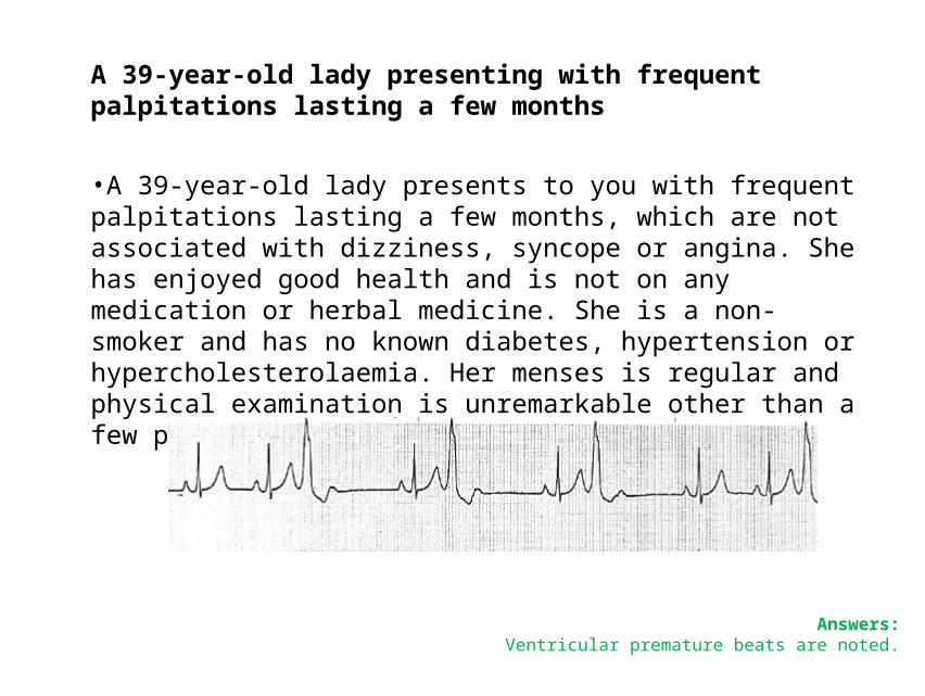
A 39-year-old lady presenting with frequent palpitations lasting a few months
•A 39-year-old lady presents to you with frequent palpitations lasting a few months, which are not associated with dizziness, syncope or angina. She has enjoyed good health and is not on any medication or herbal medicine. She is a non-smoker and has no known diabetes, hypertension or hypercholesterolaemia. Her menses is regular and physical examination is unremarkable other than a few premature beats. This is her ECG.
Answers:Ventricular premature beats are noted.
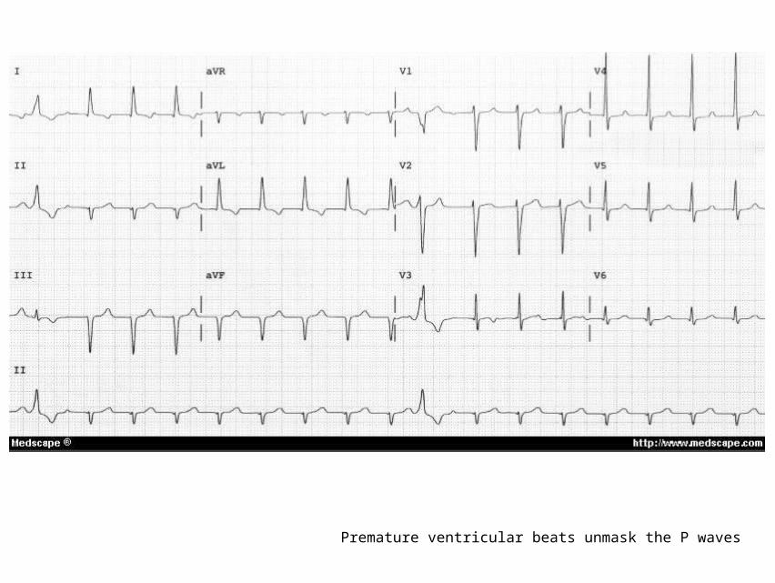
Premature ventricular beats unmask the P waves

Hemodynamic tracings to demonstrate the increased variability of systolic BP (SBP), diastolic BP (DBP), and heart period (HP) in MI rat with frequent VPB
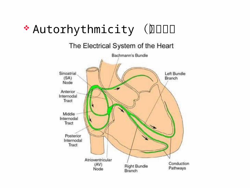
Autorhythmicity(自律性)
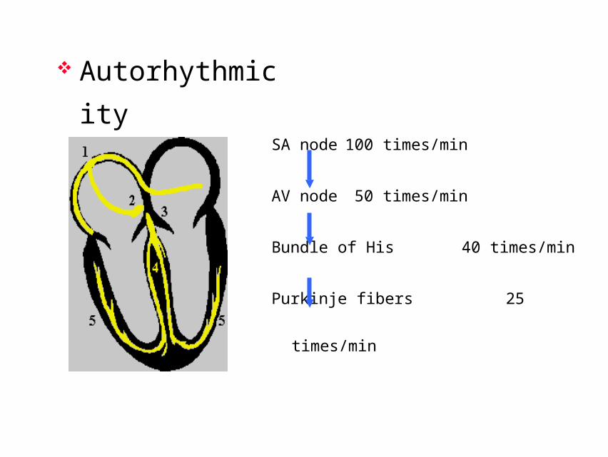
Autorhythmicity
SA node 100 times/min
AV node 50 times/min
Bundle of His 40 times/min
Purkinje fibers 25 times/min
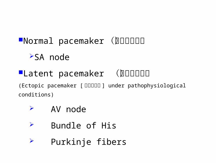
Normal pacemaker(正常起搏点)SA node
Latent pacemaker (潜在起搏点)(Ectopic pacemaker [异位起搏点 ] under pathophysiological conditions)
AV node
Bundle of His
Purkinje fibers

The mechanisms of SA node to control latent pacemakers
– Capture(夺获)
– Overdrive suppression(超速抑制)
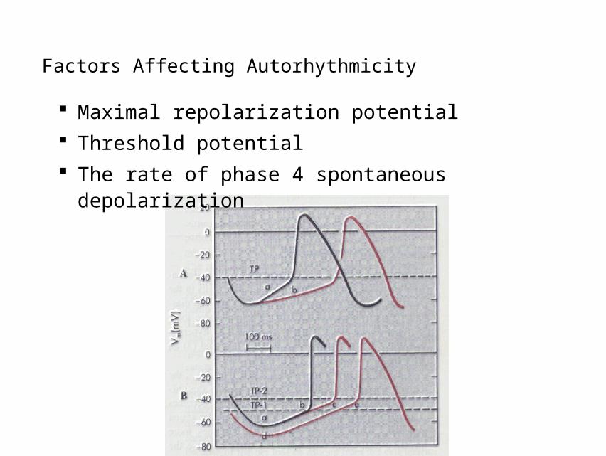
Factors Affecting Autorhythmicity
Maximal repolarization potential
Threshold potential
The rate of phase 4 spontaneous depolarization
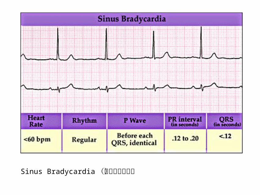
Sinus Bradycardia(窦性心动过缓)

Pacemaker

Conductivity(传导性)

Gap junction
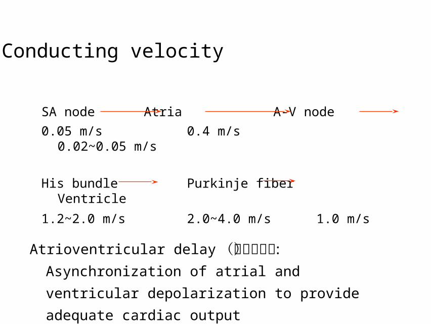
SA node Atria A-V node 0.05 m/s 0.4 m/s 0.02~0.05 m/s
His bundle Purkinje fiber Ventricle
1.2~2.0 m/s 2.0~4.0 m/s 1.0 m/s
Conducting velocity
Atrioventricular delay(房室延搁) : Asynchronization of
atrial and ventricular depolarization to provide
adequate cardiac output
Factors Affecting Conductivity
Structural factors
• Diameter of cardiac cells
• Gap junctions at Intercalated disk
Physiological factors
• The velocity and amplitude of phase 0
depolarization
• Excitability of adjacent region
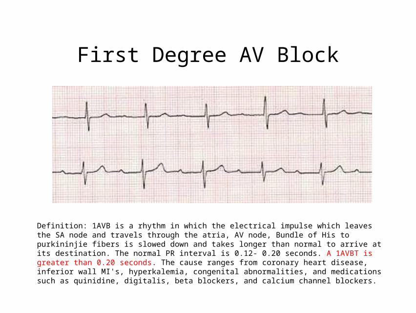
First Degree AV Block
Definition: 1AVB is a rhythm in which the electrical impulse which leaves the SA node and travels through the atria, AV node, Bundle of His to purkininjie fibers is slowed down and takes longer than normal to arrive at its destination. The normal PR interval is 0.12- 0.20 seconds. A 1AVBT is greater than 0.20 seconds. The cause ranges from coronary heart disease, inferior wall MI's, hyperkalemia, congenital abnormalities, and medications such as quinidine, digitalis, beta blockers, and calcium channel blockers.
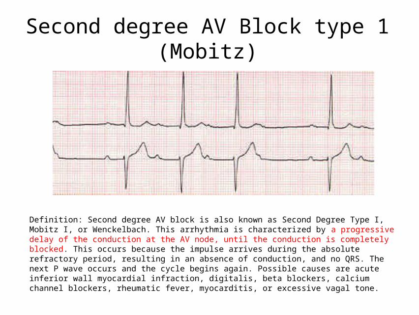
Second degree AV Block type 1 (Mobitz)
Definition: Second degree AV block is also known as Second Degree Type I, Mobitz I, or Wenckelbach. This arrhythmia is characterized by a progressive delay of the conduction at the AV node, until the conduction is completely blocked. This occurs because the impulse arrives during the absolute refractory period, resulting in an absence of conduction, and no QRS. The next P wave occurs and the cycle begins again. Possible causes are acute inferior wall myocardial infraction, digitalis, beta blockers, calcium channel blockers, rheumatic fever, myocarditis, or excessive vagal tone.

Mobitz II is characterized by 2-4 P waves before each QRS. The PR of the conducted P wave will be constant for each QRS. It is usually associated with acute anterior or anteroseptal myocardial infarction. Other causes are cardiomyopathy, rheumatic heart disease, coronary artery disease, digitalis, beta blockers, and calcium channel blockers. Mobitz II has the potential of progressing into a third degree heart block or ventricular standstill.
Second degree AV Block Type II
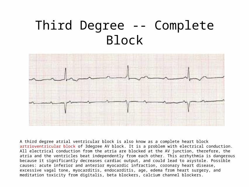
A third degree atrial ventricular block is also know as a complete heart block artrioventricular block of 3degree AV block. It is a problem with electrical conduction. All electrical conduction from the atria are blocked at the AV junction, therefore, the atria and the ventricles beat independently from each other. This arrhythmia is dangerous because it significantly decreases cardiac output, and could lead to asystole. Possible causes: acute inferior and anterior myocardic infraction, coronary heart disease, excessive vagal tone, myocarditis, endocarditis, age, edema from heart surgery, and meditation toxicity from digitalis, beta blockers, calcium channel blockers.
Third Degree -- Complete Block
------------------------++++++++++++++++++++++++++++++++++++++++++------------------------------------------------
------------------------++++++++++++++++++++++++++++++++++++++++++------------------------------------------------
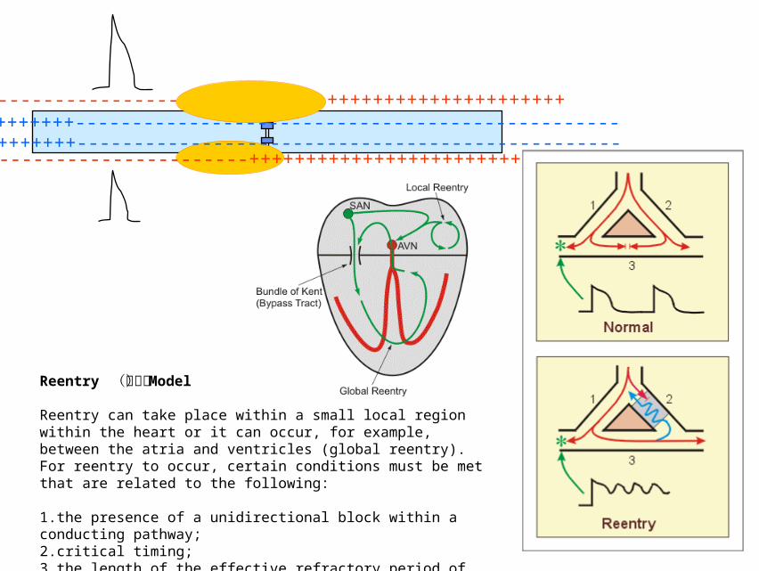
Reentry (折返)Model
Reentry can take place within a small local region within the heart or it can occur, for example, between the atria and ventricles (global reentry). For reentry to occur, certain conditions must be met that are related to the following:
1.the presence of a unidirectional block within a conducting pathway;2.critical timing;3.the length of the effective refractory period of the normal tissue.
• Q-T interval recorded on an ECG primarily corresponds to:
A Ventricular repolarization
B Ventricular depolarization plus ventricular repolarization
C Ventricular depolarization and atrial repolarization
D Atrial depolarization and conduction through AV node
E Purkinje fibers repolarization

• The resting membrane potential of a sinus nodal fiber is
A -124 mV
B -91 mV
C -85 mV
D -55 mV
E -25 mV

• You see a 55-year-old, white female for a routine check-up. On the ECG you see a prolonged PQ interval suggesting a first-degree atrioventricular block. What is the primary pacemaker of the heart?
A Sinoatrial node
B Atrioventricular node
C Atrioventricular bundle
D Right and left bundle branches
E Purkinje fibers

The End.