cardiology dr.ahmed mowafy
TRANSCRIPT
In Capsule Series
Internal Medicine
Cardiology
Second edition
By :
Dr. Ahmad M.Mowafy
ال يكمف المه نفسا إال وسعها لها ما كسبت وعميها ما اكتسبت ربنا ال تؤاخذنا إن نسينا أو أخطأنا ربنا وال تحمل عمينا إصرا كما حممته عمى الذين من قبمنا ربنا وال منا ما ال طاقة لنا به واعف عنا واغفر لنا وارحمنا أنت موالنا فانصرنا عمى تحم
ال و الكافرين
صد ا العظي
( 286آية)البقرة سورة
Preface
- First and foremost, thanks are due to ALLAH, to whom I relate
any success in achieving any work in my life.
- My words stand short of my supreme gratitude and thanks to
Prof. Dr. Hossam Mowafy ; Head of critical care unit, Kasr El Ainy.
- My thanks are extended to all my medical students whom I produce this
series:“In Capsule Series” to obtain a higher degree in internal medicine
by a simple effort.
- Finally, I wish to thank all members of My Family, my colleagues , my
Friends and even all my patients for their continuous help,
encouragement and support.
Ahmad Mowafy
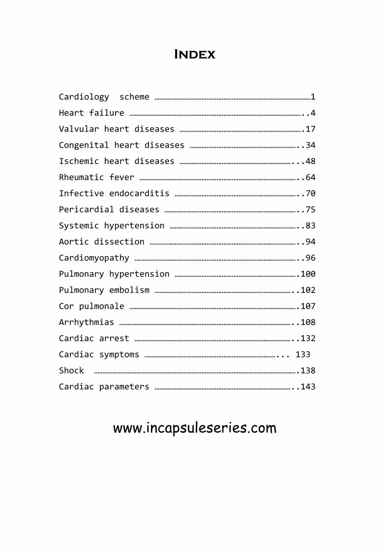
Index
Cardiology scheme …………………………………………………………………………………1
Heart failure …………………………………………………………………………………………..4
Valvular heart diseases ……………………………………………………………….17
Congenital heart diseases ………………………………………………………..34
Ischemic heart diseases …………………………………………………………...48
Rheumatic fever …………………………………………………………………………………..64
Infective endocarditis ………………………………………………………………..70
Pericardial diseases ……………………………………………………………………..75
Systemic hypertension …………………………………………………………………..83
Aortic dissection ……………………………………………………………………………..94
Cardiomyopathy ……………………………………………………………………………………..96
Pulmonary hypertension ……………………………………………………………….100
Pulmonary embolism ………………………………………………………………………..102
Cor pulmonale ……………………………………………………………………………………….107
Arrhythmias …………………………………………………………………………………………..108
Cardiac arrest …………………………………………………………………………………..132
Cardiac symptoms ……………………………………………………………………... 133
Shock ………………………………………………………………………………………………………….138
Cardiac parameters ………………………………………………………………………..143
www.incapsuleseries.com
In Capsule Series Cardiology
- 1 -
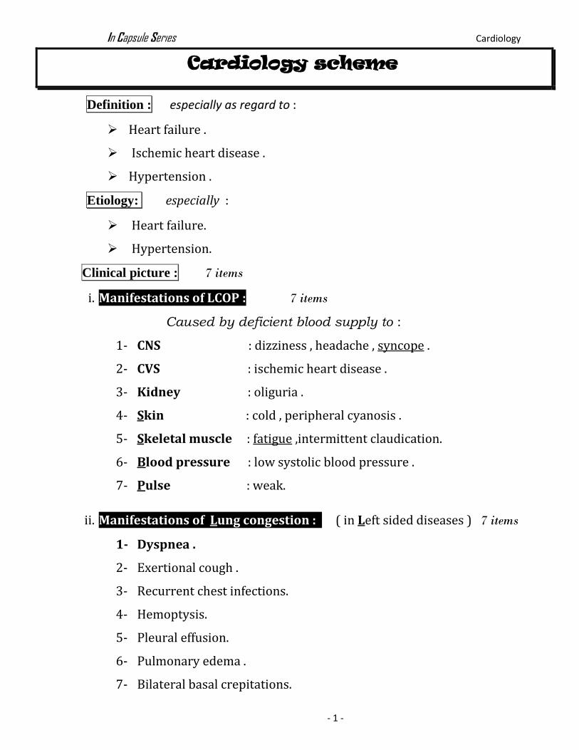
Cardiology scheme
Definition : especially as regard to :
Heart failure .
Ischemic heart disease .
Hypertension .
Etiology: especially :
Heart failure.
Hypertension.
Clinical picture : 7 items
i. Manifestations of LCOP : 7 items
Caused by deficient blood supply to :
1- CNS : dizziness , headache , syncope .
2- CVS : ischemic heart disease .
3- Kidney : oliguria .
4- Skin : cold , peripheral cyanosis .
5- Skeletal muscle : fatigue ,intermittent claudication.
6- Blood pressure : low systolic blood pressure .
7- Pulse : weak.
ii. Manifestations of Lung congestion : ( in Left sided diseases ) 7 items
1- Dyspnea .
2- Exertional cough .
3- Recurrent chest infections.
4- Hemoptysis.
5- Pleural effusion.
6- Pulmonary edema .
7- Bilateral basal crepitations.
In Capsule Series Cardiology
- 2 -
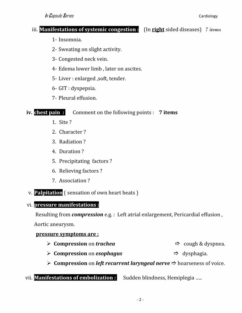
iii. Manifestations of systemic congestion : (In right sided diseases) 7 items
1- Insomnia.
2- Sweating on slight activity.
3- Congested neck vein.
4- Edema lower limb , later on ascites.
5- Liver : enlarged ,soft, tender.
6- GIT : dyspepsia.
7- Pleural effusion.
iv. chest pain : Comment on the following points : 7 items
1. Site ?
2. Character ?
3. Radiation ?
4. Duration ?
5. Precipitating factors ?
6. Relieving factors ?
7. Association ?
v. Palpitation ( sensation of own heart beats )
vi. pressure manifestations :
Resulting from compression e.g. : Left atrial enlargement, Pericardial effusion ,
Aortic aneurysm.
pressure symptoms are :
Compression on trachea cough & dyspnea.
Compression on esophagus dysphagia.
Compression on left recurrent laryngeal nerve hoarseness of voice.
vii. Manifestations of embolization : Sudden blindness, Hemiplegia …..
In Capsule Series Cardiology
- 3 -
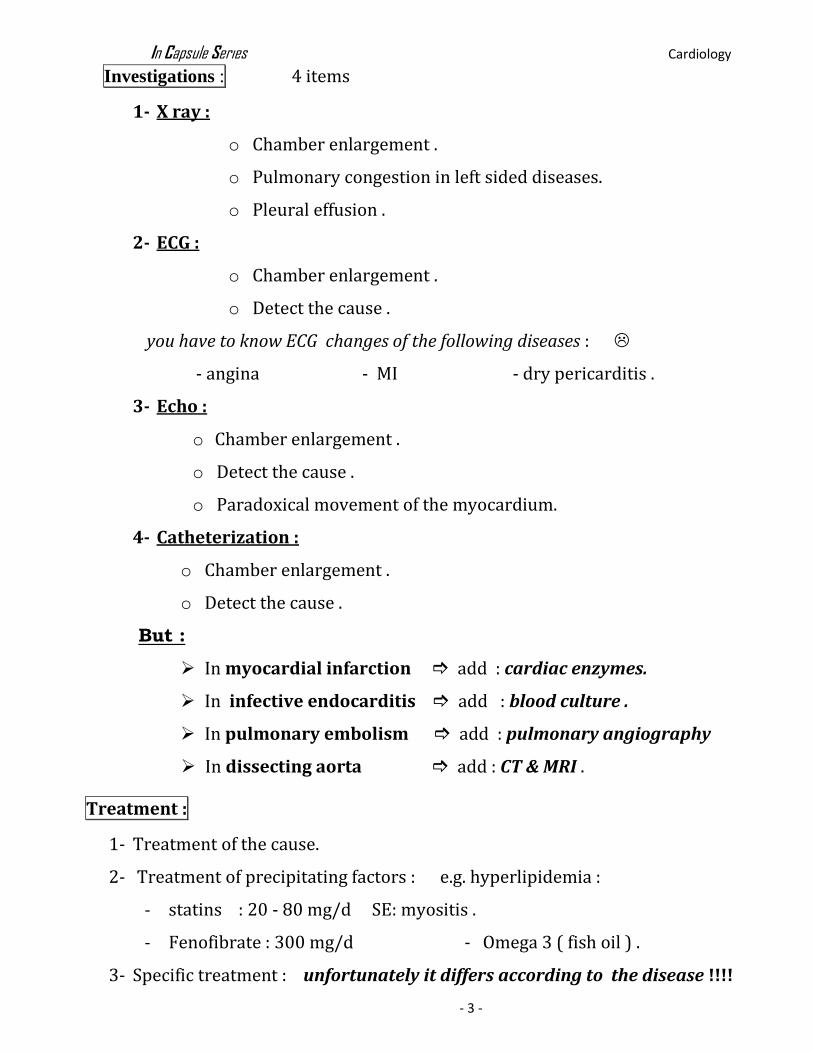
Investigations : 4 items
1- X ray :
o Chamber enlargement .
o Pulmonary congestion in left sided diseases.
o Pleural effusion .
2- ECG :
o Chamber enlargement .
o Detect the cause .
you have to know ECG changes of the following diseases :
- angina - MI - dry pericarditis .
3- Echo :
o Chamber enlargement .
o Detect the cause .
o Paradoxical movement of the myocardium.
4- Catheterization :
o Chamber enlargement .
o Detect the cause .
But :
In myocardial infarction add : cardiac enzymes.
In infective endocarditis add : blood culture .
In pulmonary embolism add : pulmonary angiography
In dissecting aorta add : CT & MRI .
Treatment :
1- Treatment of the cause.
2- Treatment of precipitating factors : e.g. hyperlipidemia :
- statins : 20 - 80 mg/d SE: myositis .
- Fenofibrate : 300 mg/d - Omega 3 ( fish oil ) .
3- Specific treatment : unfortunately it differs according to the disease !!!!
In Capsule Series Cardiology
- 4 -
Heart Failure
Definition:
It is a clinical syndrome in which the heart can’t maintain adequate cardiac
output to meet the metabolic needs of the body despite a normal ventricular
filling.
Classification:
1- Left sided Right sided Both (congestive HF)
2- Systolic Diastolic Both 3- Acute Chronic Acute on top of chronic
4- Low COP High COP
Etiology:
I- Left – sided heart failure :
A) Left atrial failure: MS , Myxoma .
B) Left ventricular failure: 3 x 3
1- Muscle disease :
Myocardial infarction
Myocarditis
CardioMyopathy
2- Volume (diastolic) overload: ( preload )
Hyperdynamic circulation .
Valvular disease: MR, AR
Congenital disease: VSD, PDA
3-pressure ( systolic ) overload: ( afterload )
Systemic hypertension .
AS .
Coarctation of aorta.
In Capsule Series Cardiology
- 5 -
II- Right sided heart failure:
A) Right atrial failure: TS , Myxoma
B) Right ventricular failure: 3×3
1- Muscle disease: The same as left .
2- Volume ( diastolic ) overload:
Hyperdynamic circulation
Valvular disease : TR, PR
Congenital disease: VSD, ASD
3- Pressure ( systolic ) over load:
pulmonary hypertension .
pulmonary stenosis .
pulmonary embolism .
The most common causes of LSHF are:
- Ischemic heart disease
- Systemic hypertension
The most common cause of RSHF is: LSHF
Diastolic heart failure: In this type of HF, the decrease in COP is due
to inadequate ventricular filling, not impaired systolic contraction.
High cardiac output HF: HF with hyperdynamic circulation e.g.
Thyrotoxicosis, anemia.
Precipitating factors: 2I, 2P, 2A
Infections: chest infections, infective endocarditis .
Iatrogenic: Calcium channel blocker (- ve inotropic ) .
Cortisone ( salt & water retention ).
Discontinuation of antifailure therapy.
Physical & emotional stress.
Pregnancy & delivery.
Anemia. Arrhythmias (tachy & brady arrhythmias) .
In Capsule Series Cardiology
- 6 -
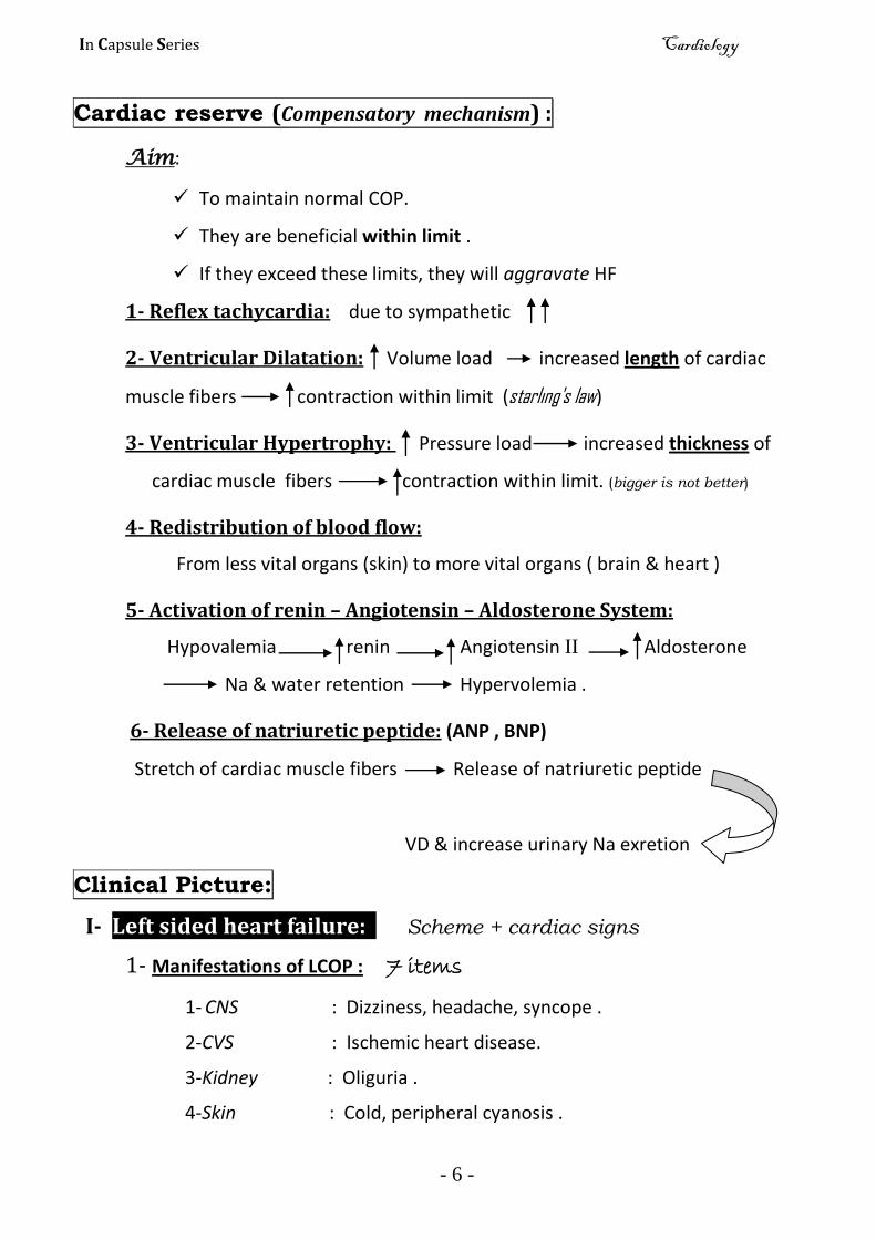
Cardiac reserve (Compensatory mechanism) :
Aim:
To maintain normal COP.
They are beneficial within limit .
If they exceed these limits, they will aggravate HF
1- Reflex tachycardia: due to sympathetic
2- Ventricular Dilatation: Volume load increased length of cardiac
muscle fibers contraction within limit (starling's law)
3- Ventricular Hypertrophy: Pressure load increased thickness of
cardiac muscle fibers contraction within limit. (bigger is not better)
4- Redistribution of blood flow:
From less vital organs (skin) to more vital organs ( brain & heart )
5- Activation of renin – Angiotensin – Aldosterone System:
Hypovalemia renin Angiotensin II Aldosterone
Na & water retention Hypervolemia .
6- Release of natriuretic peptide: (ANP , BNP)
Stretch of cardiac muscle fibers Release of natriuretic peptide
VD & increase urinary Na exretion
Clinical Picture:
І- Left sided heart failure: Scheme + cardiac signs
1- Manifestations of LCOP : 7 items
1- CNS : Dizziness, headache, syncope .
2-CVS : Ischemic heart disease.
3-Kidney : Oliguria .
4-Skin : Cold, peripheral cyanosis .
In Capsule Series Cardiology
- 7 -
5-Skletal muscle : fatigue , intermittent claudication .
6-Blood pressure: low systolic blood pressure.
7-Pulse : Weak.
2- Manifestations of pulmonary congestion: 7 items
1-Dyspnea: Exertional , orthopnea, paroxysmal nocturnal dyspnea or
dyspnea at rest .
2- Exertional Cough .
3-Recurrent chest infections.
4-Hemoptysis.
5-Pleural effusion.
6-Pulmonary edema .
7-Bilatera basal crepitation.
3-Features of the cause: Ischemic heart diseases , Systemic hypertension.
4-Cardiac signs:
a- Left ventricular enlargement .
b- Tachycardia.
c- Pulsus alternans: alternating strong & weak beats ( In advanced stage )
d- Gallop on the apex: due to flabby ventricle.
e- murmure of functional MR: pansystolic murmur due to LV dilatation .
П Right sided heart failure:
1-manifestations of LCOP: see before
2-manifestations of systemic congestion:
1-Insomnia.
2-Sweating on slight activity: due to sympathetic activation.
3-Congested neck vein.
4-Edema lower limb, later on ascites.
NB : ventricular gallop = S3 + tachycardia
In Capsule Series Cardiology
- 8 -
5-Liver: enlarged , soft, tender.
6-GIT: dyspepsia, malabsorption may lead to cardiac cachexia.
7-Pleural effusion.
3-Features of the cause: e.g. - LSHF .
- Pulmonary hypertension .
4-Cardiav Signs: ( the same as left – pulsus alternans )
1-Right ventricular enlargement .
2-Tachycardia
3-gallop (over tricuspid area).
4-murmure of functional TR .
N.B: LSHF Lung Congestion .
RSHF Systemic Congestion .
Differential Diagnosis :
LSHF RSHF
-Causes of dyspnea &orthopnea.
-Pericardial effusion
-COPD
-Obesity
-Liver cirrhosis
Investigations:
1- X ray:
o Chamber enlargement .
o Pulmonary congestion in LSHF.
o Pleural effusion
2-ECG:
o Detect the cause e.g. MI
o Chamber enlargement.
In Capsule Series Cardiology
- 10 -
3-Echocardiography: (key investigation)
o Chamber enlargement.
o Detect the cause.
o Paradoxical movement of the myocardium.
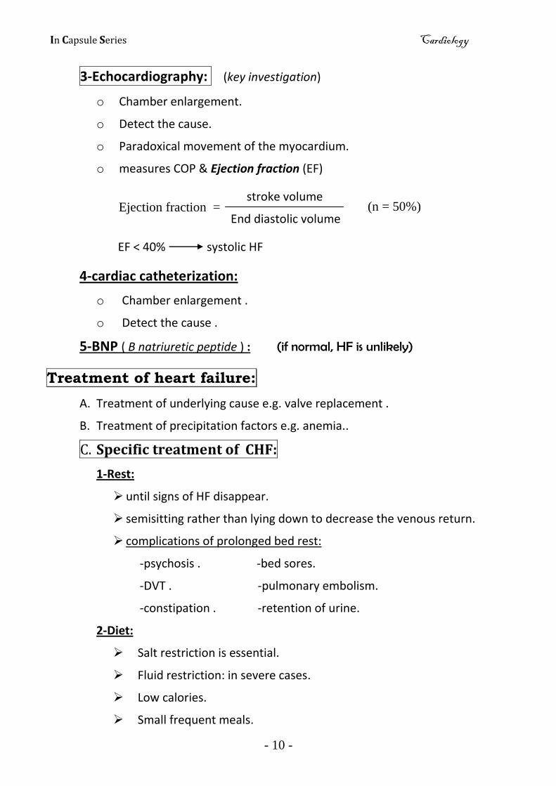
o measures COP & Ejection fraction (EF)
stroke volume End diastolic volume
EF < 40% systolic HF
4-cardiac catheterization:
o Chamber enlargement .
o Detect the cause .
5-BNP ( B natriuretic peptide ) : (if normal, HF is unlikely)
Treatment of heart failure:
A. Treatment of underlying cause e.g. valve replacement .
B. Treatment of precipitation factors e.g. anemia..
C. Specific treatment of CHF:
1-Rest:
until signs of HF disappear.
semisitting rather than lying down to decrease the venous return.
complications of prolonged bed rest:
-psychosis . -bed sores.
-DVT . -pulmonary embolism.
-constipation . -retention of urine.
2-Diet:
Salt restriction is essential.
Fluid restriction: in severe cases.
Low calories.
Small frequent meals.
Ejection fraction = (n = 50%)
In Capsule Series Cardiology
- 11 -
3-Sedation: as diazepam.
4- Diuretics:
Aim:
a. They increase salt & water excretion blood Volume So,
decrease the work of the heart .
b. Edema & visceral congestion .
Types:
ị- Loop diuretics:
-act on Loop of henle ( reabsorption of Na, H2O, K, Cl)
-e.g.
- Furosemide ( Lasix ) : 40-160 mg/d (oral, IV, IM).
- Bumetanide ( Burinex )
ị ị- Thiazides:
-act on distal tubules ( reabsorption of Na, H2O, K, Cl)
-e.g.
- Hydrochlorothiazide: 25-100 mg/d
- Chlorothalidone.
ị ị ị- potassium sparing diuretics:
- e.g. Spirnolactone (aldosterone antagonist)
-can be combined with lasix or thiazide to avoid hypokalemia.
Side effects of lasix & thiazides:
4 hypo:
-hypokalemia
-hypovolemia
-hyponatremia
- hypochloremic alkalosis
-4 hyper ( glucose )
-hyperglycemia
-hyperlipidemia
-hyperurecemia
-hypercalcemia (Thiazide only)
Lasix Ototoxicity & nephrotoxicity .
Spironolactone Hyperkalemia & gynecomastia.
In Capsule Series Cardiology
- 12 -
- In HF: lasix is more better than thiazide .
- Better given in the morning .
- It’s better to combine diuretics with ACEIs .
- Diuretics are the most effective treatment for symptoms of CHF .
5-Vasodilators: They are classified into:
Arteriolar Venous Both
- Reduce afterload Reduce preload Reduce both .
- Hydralazine
- Diazoxide
Nitrates - ACEIs.
- Na nitroprusside.
- Pharmacological details: see systemic hypertension
N.B: ACE inhibitors are the best vasodilator in the cases of CHF especially in LV failure .
6- Inotropic agents:
- Digitalis .
- Dopamine .
- Dobutamine .
- Milrinone: phospho diastrase inhibitors, used in emergency .
Digitalis
Action:
o Contractility of the ventricles .
o Excitability .
o Conductivity .
o HR : by direct action & vagal stimulation .
o On ECG: sagging depression of ST segment .
Mechanism of action : ( contractility )
Inhibition of Na - K ATPase (Na pump) intracellular Na intracellular
Ca increase muscle contraction by sliding of actin & myosin .
In Capsule Series Cardiology
- 13 -
Indications : long term control of : -
1- Heart failure : contractility .
2- Atrial fibrillation : conductivity of AV node .
Contraindications:
o Absolute contraindications :
- Digitals toxicity .
- Ventricular tachycardia (VT).
o Relative contraindications :
- Partial heart block .
- Peptic ulcer .
Administration :
o Digitalization : (to reach optimum therapeutic level )
2 tablets daily for 5 days (oral).
o Maintenance dose : (compensates for daily urinary excretion )
0.5 – 1 tablets daily (oral)
Preparations :
o Digoxin (Lanoxin):excreted mainly by the kidney (tab=0.25mg , amp=0.5mg )
o Digitoxin : metabolized mainly in the liver (digitoxine hepatic).
o Ouabain (IV) .
DIGITALIS TOXICITY :
o Precipitating factors:
Renal failure. Old age.
Hypokalemia. Hypercalcemia.
Thyroid disorders. Drugs : quinidine.
o Clinical picture:
Non cardiac :
GIT : Anorexia , nausea, vomiting (1STsymptom)
Neurological: Psychosis , yellow vision .
Gynecomasteia .
In Capsule Series Cardiology
- 13 -
Cardiac : (most life threatening )
excitability Arrhythmias .
AVN conduction heart block.
Almost any arrhythmia can be a manifestation of digitalis toxicity except
type 2 second degree heart block . MCQ
o Treatment :
Stop digitalis.
Stop diuretics.
Give K.
Digitalis antibodies (Digibind).
Anti-arrhythmic drugs (e.g. phenytoin , lidocaine ) .
o To avoid toxicity :
Decrease the dose.
Drug holiday.
Routine estimation of serum level of digitalis(N=0.5-2ng/ml).
7-β-blockers :
Historically , β blockers were contraindicated in HF due to their -ve
inotropic effect.
Recently : β blockers are indicated in HF because they were found to :
Reduce mortality & improve the prognosis.
Prevent arrhythmia.
Decrease blood pressure.
e.g.
Metoprolol ( 2nd generation β1 blocker )
Carvedilol (3rd generation β blocker).
Start with low doses with gradual increase .
In Capsule Series Cardiology
- 14 -
8- Aminophylline :
Action :
Bronchodilator . Vasodilator .
Diuretic effect . +ve inotropic .
Administration : Oral , suppositories , IV.
IV injection must be very slowly to avoid arrhythmia .
9- Oxygen therapy :
Especially in acute pulmonary edema , MI & hypoxic cor pulmonale
ACUTE HEART FALIURE
(Acute Cardiogenic Pulmonary Edema)
Etiology : (sudden in pulmonary venous pressure)
Acute left sided heart failure e.g. myocardial infarction.
On top of chronic LSHF : MS with aggravating factor as AF.
Clinical picture :
Severe dyspnea at rest & orthopnea.
Sense of impending death.
Sweating & irritability.
Cyanosis.
Crepitation .
Cough with frothy pink sputum.
Features of the Cause :MI.
Differential Diagnosis :
- Non Cardiogenic pulmonary edema ( ARDS ).
- DD of acute dyspnea : see page 104
In Capsule Series Cardiology
- 15 -
Treatment :
1) Hospitalization in ICU : bed rest in sitting position .
2) High dose oxygen correct hypoxia .
3) Morphine (IV) Reduce anxiety.
Reduce preload (venodilator).
4) Furosemide (IV) Decreases pulmonary congestion (venodilator)
Diuresis.
5) Vasodilators (IV):
Na nitroprusside IV infusion (0.5 – 5 mg / kg/ min)
Nitroglaycrin IV infusion (S/E: tolerance).
6) Inotropics :
- Dobutamine (β receptor agonist): +ve inotropic & vasodilatation (inodilator)
- Milrinone (phosphodiesterase inhibitor): inodilator.
NB :Milrinone is preferred to dobutamine in patients receiving β blocker because its
mechanism of action does not involve β receptors. Dobutamine also may precipitate
ischemic heart disease.
7) Aminophylline : 250 – 500 mg / IV infusion very slowly .
8) Treatment of the cause & the precipitating factors .
9) Advanced management : in refractory conditions
Mechanical ventilation .
Mechanical assist devices : Intra-aortic balloon counter pulsation.
In Capsule Series Cardiology
- 16 -
Refractory ( Intractable ) Heart Failure
ETIOLOGY :
Diagnostic error : the case may be pericardial effusion rather
heart failure .
Improper management : Inadequate salt restriction .
Discontinuation of treatment .
presence of a precipitating factor :e.g. infection .
presence of the cause : uncontrolled hypertension , AS .
Terminal cases of heart failure .
TREATMENT :
Reassess the cause.
Removal of mechanical factor : valve replacement .
Removal of precipitating factor.
Proper management :
Strict bed rest.
Salt & even fluid restriction.
Proper doses.
For terminal cases :
IV Lasix , morphine , dobutamine , nitrate may be used .
Mechanical ventilation .
Cardiac transplantation .
I know not with what weapons World War III will be fought, but World War IV will be
fought with sticks and stones.
Albert Einstein
In Capsule Series Cardiology
- 17 -
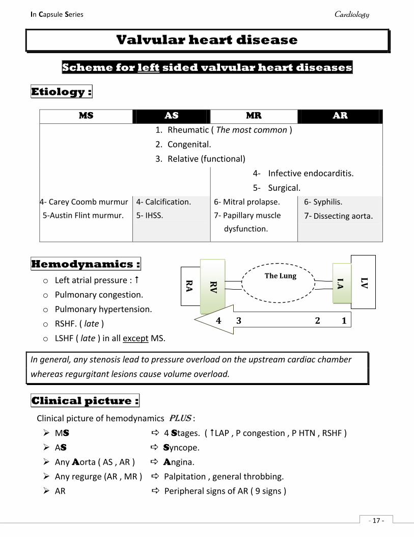
Valvular heart disease
Scheme for left sided valvular heart diseases
Etiology :
MS AS MR AR
1. Rheumatic ( The most common )
2. Congenital.
3. Relative (functional)
4- Infective endocarditis.
5- Surgical.
4- Carey Coomb murmur
5-Austin Flint murmur.
4- Calcification.
5- IHSS.
6- Mitral prolapse.
7- Papillary muscle
dysfunction.
6- Syphilis.
7- Dissecting aorta.
Hemodynamics :
o Left atrial pressure :
o Pulmonary congestion.
o Pulmonary hypertension.
o RSHF. ( late )
o LSHF ( late ) in all except MS.
In general, any stenosis lead to pressure overload on the upstream cardiac chamber
whereas regurgitant lesions cause volume overload.
Clinical picture :
Clinical picture of hemodynamics plus :
MS 4 Stages. ( LAP , P congestion , P HTN , RSHF )
AS Syncope.
Any Aorta ( AS , AR ) Angina.
Any regurge (AR , MR ) Palpitation , general throbbing.
AR Peripheral signs of AR ( 9 signs )
LV
LA
The Lung
RA
RV
4 3 2 1
In Capsule Series Cardiology
- 18 -
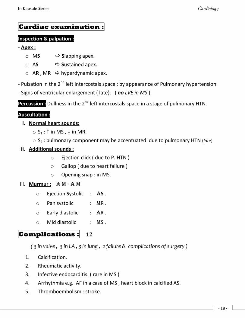
Cardiac examination :
Inspection & palpation :
- Apex :
o MS Slapping apex.
o AS Sustained apex.
o AR , MR hyperdynamic apex.
- Pulsation in the 2nd left intercostals space : by appearance of Pulmonary hypertension.
- Signs of ventricular enlargement ( late). ( no LVE in MS ).
Percussion : Dullness in the 2nd left intercostals space in a stage of pulmonary HTN.
Auscultation :
i. Normal heart sounds:
o S1 : in MS , in MR.
o S2 : pulmonary component may be accentuated due to pulmonary HTN (late)
ii. Additional sounds :
o Ejection click ( due to P. HTN )
o Gallop ( due to heart failure )
o Opening snap : in MS.
iii. Murmur : A M - A M
o Ejection Systolic : AS .
o Pan systolic : MR .
o Early diastolic : AR .
o Mid diastolic : MS .
Complications : 12
( 3 in valve , 3 in LA , 3 in lung , 2 failure & complications of surgery )
1. Calcification.
2. Rheumatic activity.
3. Infective endocarditis. ( rare in MS )
4. Arrhythmia e.g. AF in a case of MS , heart block in calcified AS.
5. Thromboembolism : stroke.
In Capsule Series Cardiology
- 19 -
6. LA enlargement compression on :
Lung dyspnea & cough.
Esophagus dysphagia.
Left recurrent laryngeal nerve hoarseness of voice.
7. Pulmonary congestion hemoptysis & recurrent chest infections.
8. Pulmonary infection.
9. Pulmonary embolism ( secondary to DVT )
10. RSHF.
11. LSHF except in MS.
12. Complications of surgery ( artificial valves ) :
Mechanical dysfunction.
Infective endocarditis.
Thromboembolism.
Hemolytic anemia.
Investigations :
X ray :
Chamber enlargement. Pulmonary congestion.
ECG :
Chamber enlargement e.g. LA P mitrale ( m shaped P wave )
Pulmonary hypertension P pulmonale ( Peaked P wave )
Echo & Doppler echo : ( The most important )
Chamber enlargement .
Detect the severity of the valve lesion.
Catheterization & angiography :
Detect the severity.
Chamber enlargement.
Treatment :
Medical :
1- Prophylaxis against IE & rheumatic activity.
2- Treatment of complications e.g. HF , AF , infections …
Surgical :
1- Balloon dilatation ( Percutaneous balloon valvuloplasty) for stenosis especially pure MS.
2- Valvotomy ( commissurotomy) : for stenosis.
3- Valve replacement : Tissue or synthetic valves .
In Capsule Series Cardiology
20
Mitral stenosis
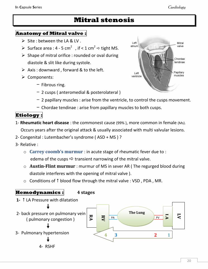
Anatomy of Mitral valve :
Site : between the LA & LV .
Surface area : 4 - 5 cm2 , if < 1 cm2 tight MS.
Shape of mitral orifice : rounded or oval during
diastole & slit like during systole.
Axis : downward , forward & to the left.
Components:
– Fibrous ring.
– 2 cusps ( anteromedial & posterolateral )
– 2 papillary muscles : arise from the ventricle, to control the cusps movement.
– Chordae tendinae : arise from papillary muscles to both cusps.
Etiology :
1- Rheumatic heart disease : the commonest cause (99% ), more common in female (Ms).
Occurs years after the original attack & usually associated with multi valvular lesions.
2- Congenital : Lutembacher’s syndrome ( ASD + MS ) ?
3- Relative :
o Carrey coomb’s murmur : in acute stage of rheumatic fever due to :
edema of the cusps transient narrowing of the mitral valve.
o Austin-Flint murmur : murmur of MS in sever AR ( The regurged blood during
diastole interferes with the opening of mitral valve ).
o Conditions of blood flow through the mitral valve : VSD , PDA , MR.
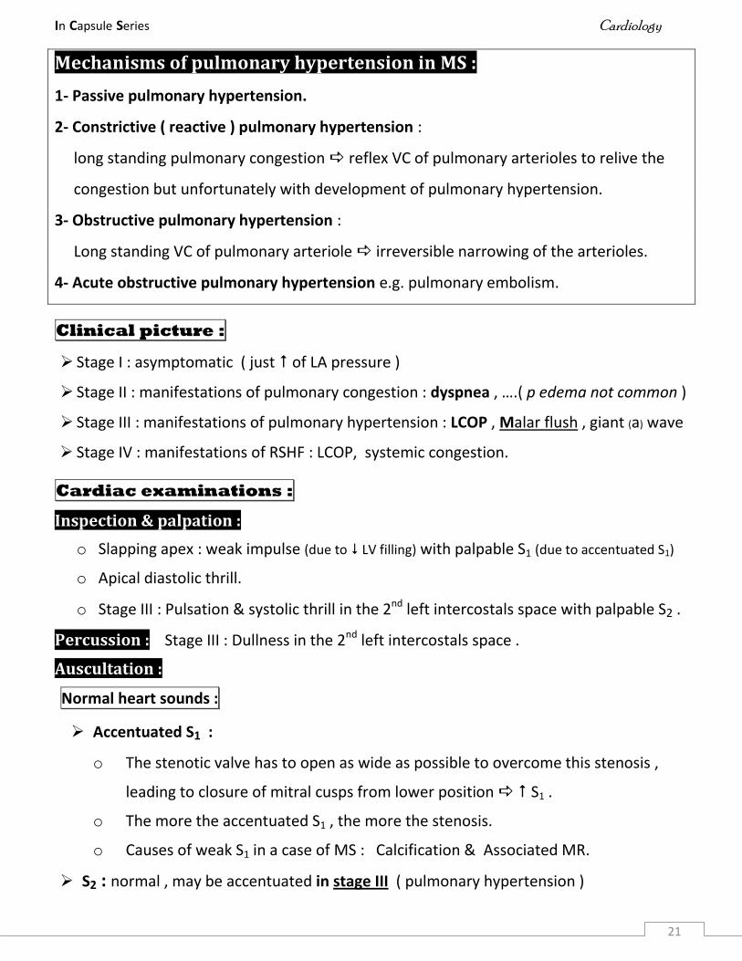
Hemodynamics : 4 stages
1- LA Pressure with dilatation
2- back pressure on pulmonary vein
( pulmonary congestion ) PA PV
3- Pulmonary hypertension
4- RSHF
LV
LA
The Lung
RA
RV
4 3 2 1
In Capsule Series Cardiology
21
Mechanisms of pulmonary hypertension in MS :
1- Passive pulmonary hypertension.
2- Constrictive ( reactive ) pulmonary hypertension :
long standing pulmonary congestion reflex VC of pulmonary arterioles to relive the
congestion but unfortunately with development of pulmonary hypertension.
3- Obstructive pulmonary hypertension :
Long standing VC of pulmonary arteriole irreversible narrowing of the arterioles.
4- Acute obstructive pulmonary hypertension e.g. pulmonary embolism.
Clinical picture :
Stage I : asymptomatic ( just of LA pressure )
Stage II : manifestations of pulmonary congestion : dyspnea , ….( p edema not common )
Stage III : manifestations of pulmonary hypertension : LCOP , Malar flush , giant (a) wave
Stage IV : manifestations of RSHF : LCOP, systemic congestion.
Cardiac examinations :
Inspection & palpation :
o Slapping apex : weak impulse (due to LV filling) with palpable S1 (due to accentuated S1)
o Apical diastolic thrill.
o Stage III : Pulsation & systolic thrill in the 2nd left intercostals space with palpable S2 .
Percussion : Stage III : Dullness in the 2nd left intercostals space .
Auscultation :
Normal heart sounds :
Accentuated S1 :
o The stenotic valve has to open as wide as possible to overcome this stenosis ,
leading to closure of mitral cusps from lower position S1 .
o The more the accentuated S1 , the more the stenosis.
o Causes of weak S1 in a case of MS : Calcification & Associated MR.
S2 : normal , may be accentuated in stage III ( pulmonary hypertension )
In Capsule Series Cardiology
22
Additional sounds :
1. Opening snap :
o Sharp snapping sound following S2 due to sudden opening of rigid cusps.
o The closer the snap to S2 , the more the stenosis.
o Calcification causes disappearance of opening snap.
2. Ejection click : in stage III ( Pulmonary hypertension )
3. Gallop on tricuspid area : in stage IV ( RSHF )
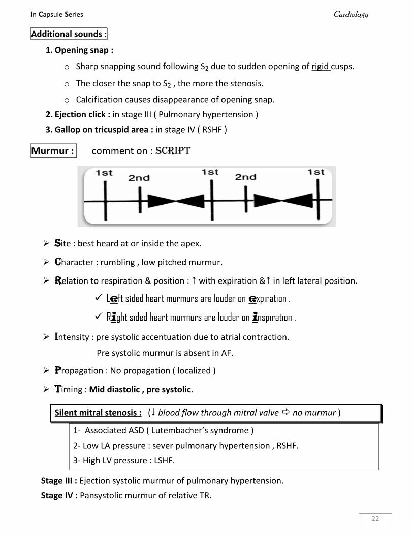
Murmur : comment on : SCRIPT
Site : best heard at or inside the apex.
Character : rumbling , low pitched murmur.
Relation to respiration & position : with expiration & in left lateral position.
Left sided heart murmurs are louder on expiration .
Right sided heart murmurs are louder on inspiration .
Intensity : pre systolic accentuation due to atrial contraction.
Pre systolic murmur is absent in AF.
Propagation : No propagation ( localized )
Timing : Mid diastolic , pre systolic.
Silent mitral stenosis : ( blood flow through mitral valve no murmur )
1- Associated ASD ( Lutembacher’s syndrome )
2- Low LA pressure : sever pulmonary hypertension , RSHF.
3- High LV pressure : LSHF.
Stage III : Ejection systolic murmur of pulmonary hypertension.
Stage IV : Pansystolic murmur of relative TR.
In Capsule Series Cardiology
23
Investigation :
X ray :
o LA enlargement ( lateral view with barium )
o Pulmonary congestion.
o Dilated pulmonary artery.
o RVE.
o Calcification of mitral valve.
ECG :
o LA enlargement ( P mitrale : m shaped P wave )
o Pulmonary hypertension ( P pulmonale : peaked P wave )
o RVE.
Echo & echo Doppler :
o Chamber enlargement . o Valve lesion. o Calcification.
Catheterization & angiography:
o Chamber enlargement.
o Mitral stenosis index = COP/LAP x 100 = 5/5 x 100 = 100% ( < 25 % is tight MS )
Complications : see scheme.
Treatment :
Medical :
1- Prophylaxis against IE & rheumatic activity.
2- Treatment of complications e.g. HF , AF , infections …
Surgical : Not very early , not very late
1- Balloon dilatation: In pure MS ( no calcification , not combined with MR , not severe )
2- Valvotomy ( commissurotomy) .
3- Valve replacement : Tissue or synthetic valves ,
Indications of valve replacement :
o Calcification o Associated MR
o Tight MS ( MS Index < 25 % , surface area < 1cm2 or severe manifestations)
o recurrent stenosis after balloon dilatation or valvotomy .
In Capsule Series Cardiology
24
Mitral Regurge
Etiology :
o Rheumatic ( the commonest )
o Congenital.
o Infective endocarditis.
o Surgical.
o Mitral valve prolapse.
o Papillary muscle dysfunction.
o Functional ( relative ) : dilatation of mitral ring due to dilatation of LV e.g. LSHF , AR.
Hemodynamics :
During systole : A part of blood regurgitates from LV to LA leading to LA dilatation .
During diastole : blood flow through the mitral valve volume load on LV LV
enlargement then failure.
In acute MR : as in myocardial infarction & IE , the LA has no time to dilate and
accommodate the regurgitant blood great of LAP rapid pulmonary
congestion then pulmonary edema & can lead to cardiogenic shock.
Clinical picture :
1- Asymptomatic for many years in mild cases.
2- Palpitation & general throbbing due to LV volume overload.
3- Manifestations of LSHF ( late )
4- Manifestations of pulmonary congestion then pulmonary hypertension RSHF later.
5- Acute pulmonary edema in a case of acute MR.
Cardiac examination :
Inspection & palpation :
o LVE .
o Hyperdynamic apex ( forcible , non sustained apex ).
o Systolic thrill over the apex.
Auscultation :
Normal heart sounds :
o S1 : weak (due to weak closure of mitral valve & masking by pansystolic murmur of MR)
o S2 : may be accentuated with development of pulmonary hypertension.
In Capsule Series Cardiology
25
Additional sounds:
o With HF Gallop.
o With pulmonary hypertension Ejection click.
Murmur :
- Murmur of MR :
Site : best heard at the apex.
Character : blowing.
Relation to respiration & position : with expiration & in left lateral position.
Propagation : to axilla ( except in posterior leaflet regurge radiate to the base of heart)
Timing : Pansystolic murmur. ( plateau )
- Relative MS : mid diastolic murmur due to excess blood flow across the mitral valve.
Complications : see scheme.
Investigations :
– X ray & ECG : Chamber enlargement e.g. LA , LV .
– Echo : - Chamber enlargement - Valve lesion.
– Catheterization : - Chamber enlargement - Valve lesion .
Treatment :
o Medical : see scheme .
o Surgical : valve replacement or mitral valve repair.
Mitral valve prolapse
Prolapse of one or both cusps of mitral valve into LA during systole.
Etiology : Unknown in most cases , more common in young female .
C/P : Asymptomatic in most cases.
– The same as MR but the most important symptoms are :
o Atypical chest pain . o Palpitation.
Investigations : Echo is diagnostic
Treatment :
o Reassurance. o blocker
o valve replacement in severe cases.
In Capsule Series Cardiology
26
Aortic Stenosis
Anatomy :
o 3 semilunar cusps attached to a fibrous valve ring .
o In about 1 % of individuals , only 2 cusps are present ( Bicuspid aortic valve ).
o Surface area is about 3 cm2 .
Etiology :
1- Rheumatic fever.
2- Congenital : it may be valvular , subvalvular or supravalvular.
3- Calcifications.
4- Hypertrophic cardiomyopathy : ( Idiopathic Hypertrophic Subaortic Stenosis - IHSS )
5- Relative :
o blood flow across the aortic valve : AR.
o Dilatation of aorta : Hypertension , atherosclerosis .
Hemodynamics :
During systole , there is obstruction of LV outflow results in :
o LCOP. o Pressure overload on LV leading to LV hypertrophy then failure.
Clinical picture :
Asymptomatic in mild cases. Manifestations of LCOP.
Syncope : especially Exertional due to low fixed COP.
Angina : Due to :
o LCOP coronary blood flow.
o LV hypertrophy O2 demand.
o Associated atherosclerosis or AR.
Manifestations of LSHF.
Cardiac examinations :
LVE.
Sustained apex : ( forcible ,sustained apex )
Systolic thrill over 2nd right intercostal space ( A1 ) & propagated to apex & neck.
In Capsule Series Cardiology
27
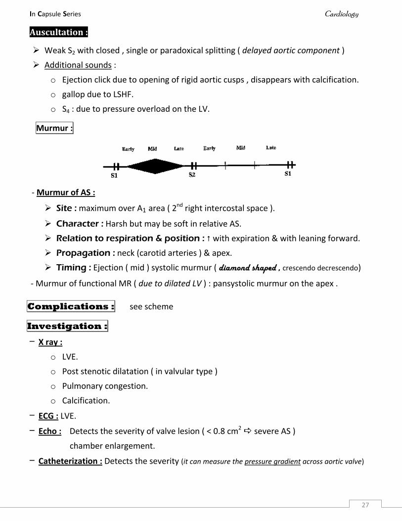
Auscultation :
Weak S2 with closed , single or paradoxical splitting ( delayed aortic component )
Additional sounds :
o Ejection click due to opening of rigid aortic cusps , disappears with calcification.
o gallop due to LSHF.
o S4 : due to pressure overload on the LV.
Murmur :
- Murmur of AS :
Site : maximum over A1 area ( 2nd right intercostal space ).
Character : Harsh but may be soft in relative AS.
Relation to respiration & position : with expiration & with leaning forward.
Propagation : neck (carotid arteries ) & apex.
Timing : Ejection ( mid ) systolic murmur ( diamond shaped , crescendo decrescendo)
- Murmur of functional MR ( due to dilated LV ) : pansystolic murmur on the apex .
Complications : see scheme
Investigation :
– X ray :
o LVE.
o Post stenotic dilatation ( in valvular type )
o Pulmonary congestion.
o Calcification.
– ECG : LVE.
– Echo : Detects the severity of valve lesion ( < 0.8 cm2 severe AS )
chamber enlargement.
– Catheterization : Detects the severity (it can measure the pressure gradient across aortic valve)
In Capsule Series Cardiology
28
Treatment :
Medical : same as scheme plus , blocker for angina.
Surgical :
Aortic valve replacement :
Indication : - Valve area < 0.8 cm2
- Systolic pressure gradient across the aortic valve > 50 mmHg.
- Severe symptoms.
Balloon dilatation & aortic Valvotomy ( associated with a high early restenosis rate )
Aortic Regurge
Etiology :
1- Rheumatic fever.
2- Congenital.
3- Infective endocarditis.
4- Surgical.
5- Dilatation of the ascending aorta :
Hemodynamics :
During diastole : regurgitation of blood from the aorta to the LV leading to :
Volume overload on the LV LVE the failure.
coronary blood flow Angina.
blood in LV LV contraction stroke volume systolic pressure .
This high systolic BP is compensated by peripheral VD which ( together with
regurgitation ) will decrease the diastolic BP. So in AR :
o Syphilis.
o Marfan syndrome.
o Severe hypertension.
o Aortic dissection.
o Ankylosing spondylitis.
Systolic BP : due to high stroke volume ( high COP ).
Diastolic BP : due to peripheral VD & regurgitation of blood during diastole.
In Capsule Series Cardiology
29
Clinical picture :
1- Asymptomatic in mild cases.
2- General throbbing : due to arterial pulsation.
3- Palpitation : due to forcible LV contraction.
4- Angina : there are 2 types
Classic angina : due to :
o Diastolic BP coronary blood flow.
o LVE O2 demand.
Angina of Lewis : Nocturnal , prolonged angina & associated with autonomic
disturbances (sweating , tachycardia)
5- Manifestations of LSHF : Pulmonary congestion & LCOP.
Peripheral signs of AR : ( due to big pulse volume ) 3 in neck , 3 in UL , 3 in LL
Head & neck :
o De Musset sign : nodding of the head.
o Corrigan’s sign : Marked visible carotid pulsation.
o Systolic thrill over the carotid artery.
Upper limb :
o BP : systolic & diastolic BP.
o Pulse : Water hammer pulse.
o Capillary pulsations : pressing on the nail tip moving red line.
Lower limb :
o Pistol shots : systolic femoral sound due to sudden distension of collapsed artery.
o Duroziez’s sign : systolic & diastolic murmur over the femoral artery if slight pressure
is applied to it by the stethoscope.
o Hill’s sign : The difference between systolic BP in LL & UL > 50 mmHg.
(Normally SBP in LL > UL by 10 - 20 mmHg )
NB : AR with minimal peripheral signs :
o Mild AR.
o systolic BP : MS , AS.
o Diastolic BP : Systemic hypertension.
In Capsule Series Cardiology
30
Cardiac examination :
LVE . Hyperdynamic apex.
No thrill over the aortic area in isolated AR.
Auscultation :
S2 : usually normal in isolated AR.
Additional sounds : gallop on the mitral area.
Murmur :
- Murmur of AR :
Site : maximum over the 3rd left intercostals space ( A2 area )
in syphilitic AR : maximum over A1 ( 2nd right intercostals space.
Character : Soft blowing , decrescendo .
Relation to respiration & position : with expiration & with leaning forward.
Propagation : To apex & left sterna border.
Timing : Early diastolic.
- Murmur of relative AS : ejection systolic murmur ( soft )
Murmurs over the mitral area ( apex ) in case of AR :
1- Mid diastolic murmur of relative MS ( Austin - Flint murmur )
2- Pansystolic murmur of relative MR.
3- Propagation of ejection systolic murmur of relative or combined AS.
4- Propagation of early diastolic murmur of AR itself.
Complications : see scheme .
Investigations :
– X ray : LVE & dilated aorta ( Boot - shaped heart )
– ECG : LVE.
– Echo : LVE , detects the severity of the valve lesion.
– Catheterization : LVE , detects the severity of the valve lesion.
In Capsule Series Cardiology
31
Treatment :
Medical : As scheme.
Surgical : Valve replacement in severe organic cases with LV dysfunction.
Rheumatic AR Syphilitic AR
Age 20 - 40 years > 40 years
History Of rheumatic fever Of syphilis
Valvular lesion yes no
Angina Less common. More common.
S2 Usually normal
Murmur (maximum intensity) Over A2 Over A1
X ray Calcification Aortic aneurysm.
Tricuspid Stenosis
Etiology :
It’s usually rheumatic in origin & usually associated with mitral or aortic valve diseases.
Hemodynamics : obstruction of tricuspid valve leading to :
- RA pressure RA enlargement & systemic congestion.
- RV filling COP.
Clinical picture:
- Symptoms of LCOP. - Symptoms of systemic congestion.
- Symptoms of associated lesions e.g. MS
NB : TS symptoms of MS due to restriction of pulmonary flow.
General signs :
- LCOP. - Systemic congestion.
- Neck vein : Giant (a) wave.
Cardiac sign :
- RA & RV enlargement.
- mid diastolic presystolic murmur at lower left sternal border, increases by inspiration.
Investigation :
- X ray , ECG : RA & RV enlargement.
In Capsule Series Cardiology
32
- Echo & Catheterization : diagnostic.
Treatment : Valve replacement.
Tricuspid Regurge
Etiology : TR is usually functional resulting from RVE dilatation of tricuspid ring.
Hemodynamics : During systole, part of blood regurgitates from RV to RA causing:
- RA pressure RA enlargement & systemic congestion.
- LCOP. - RVE then failure.
Clinical picture :
Symptoms : - of the cause. - Systemic congestion. - LCOP.
General signs :
Signs of systemic congestion :
- Congested pulsating neck vein with systolic expansion.
- Enlarged tender pulsating liver with mild jaundice.
- Ascites before edema LL.
Signs of LCOP : cold hand , weak pulse , SBP , peripheral cyanosis.
Mild jaundice ( liver congestion ) with peripheral cyanosis ( LCOP ) Cyano - ictric face.
Cardiac signs :
RA & RV enlargement
Systolic thrill over tricuspid area.
Murmur :
- Pansystolic murmur. - Increased by inspiration.
- Maximum over tricuspid area & propagated to the apex but not to the axilla.
Complications : - RSHF - IE - Cardiac cirrhosis.
Investigations :
- X ray & ECG : RA & RV enlargement.
- Echo & catheterization : Diagnostic.
Treatment :
- Treatment of RSHF . - Valve replacement.
In Capsule Series Cardiology
33
DD of systolic murmurs :
1. AS
2. PS
3. MR
4. TR.
5. VSD
6. PDA
7. Coarctation of aorta
Over the apex :
o MR
o Propagated from other area: all of the above 7 lesions may propagate to the apex.
Over the base :
o All of the above except MR & TR.
o Posterior leaflet MR may propagate to the base.
DD of diastolic murmurs :
1. AR
2. PR
3. MS
4. TS
5. PDA
6. Coarctation of aorta
Over the apex :
o MS : either organic or relative.
o Propagated from other area : AR , PR
Over the base :
o AR , PR , PDA
o coarctation of aorta ( due to collaterals )
In Capsule Series Cardiology
35
Congenital Heart Disease
Criteria to suspect Congenital HD:
o Cyanosis since birth.
o Murmur since birth.
o No history of rheumatic fever.
o Recurrent chest infection.
o Hypertensive child.
o +ve family history.
o Associated congenital anomalies.
Classification:
Cyanotic :
Fallot’s tetralogy ( F4 )
Fallot’s triology ( F3 )
Eisenmenger’s syndrome.
Tricuspid atresia.
Transposition of the great arteries.
Acyanotic :
With RV enlargement : ASD , PS .
With LV enlargement : AS , PDA , Coarctation of aorta.
With biventricular enlargement : VSD .
With NO ventricular enlargement : : Any mild lesion , Dextrocardia .
Atrial Septal Defect ( ASD )
Anatomy :
- There is an abnormal opening between the two atria, producing left to right shunt.
High ASD ( ostium secondum ) : the most common.
Low ASD ( ostium premium ) : may be associated with Mitral valve disease
( Lutembacher’s syndrome ) . MCQ
In Capsule Series Cardiology
35
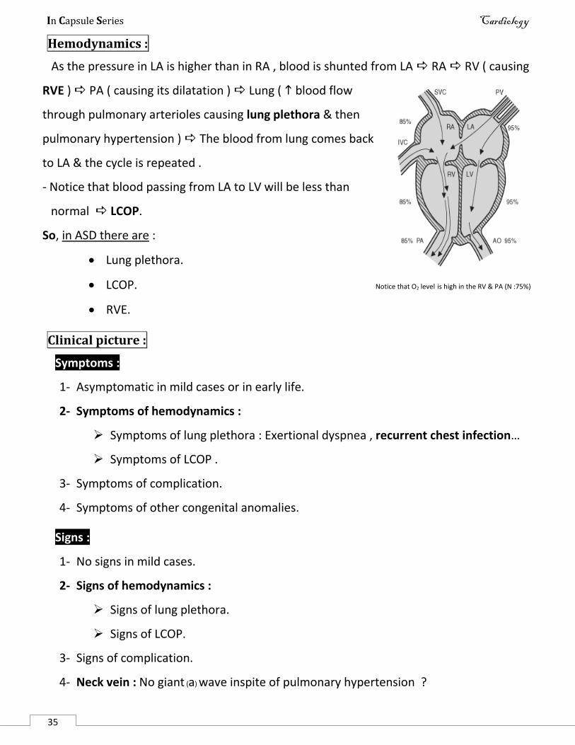
Hemodynamics :
As the pressure in LA is higher than in RA , blood is shunted from LA RA RV ( causing
RVE ) PA ( causing its dilatation ) Lung ( blood flow
through pulmonary arterioles causing lung plethora & then
pulmonary hypertension ) The blood from lung comes back
to LA & the cycle is repeated .
- Notice that blood passing from LA to LV will be less than
normal LCOP.
So, in ASD there are :
Lung plethora.
LCOP. Notice that O2 level is high in the RV & PA (N :75%)
RVE.
Clinical picture :
Symptoms :
1- Asymptomatic in mild cases or in early life.
2- Symptoms of hemodynamics :
Symptoms of lung plethora : Exertional dyspnea , recurrent chest infection…
Symptoms of LCOP .
3- Symptoms of complication.
4- Symptoms of other congenital anomalies.
Signs :
1- No signs in mild cases.
2- Signs of hemodynamics :
Signs of lung plethora.
Signs of LCOP.
3- Signs of complication.
4- Neck vein : No giant (a) wave inspite of pulmonary hypertension ?
In Capsule Series Cardiology
35
Cardiac examination :
1- RVE.
2- Auscultation :
i. S2 : Accentuated , wide fixed splitting S2
- Accentuated & Wide splitting : due to pulmonary hypertension.
- Fixed : because the VR to the RA during inspiration is compensated by blood
shunted from LA to RA .
ii. Murmur :
No murmur of ASD itself because of low pressure gradients between the 2 atria.
Murmur of relative TS & PS may be heard.
iii. Additional sounds : Ventricular Gallop due to RSHF.
Complication :
1- RSHF.
2- Paradoxical embolism e.g. stroke.
3- Eisenmenger’s syndrome: cyanosis with shunt reversal.
4- Infective endocarditis : rare due to low pressure gradient.
5- Arrhythmia : AF
Investigations :
X ray : RVE , dilated pulmonary artery , lung plethora.
ECG : RBBB in most cases , RVE.
Echo : RVE , detect the defect.
Catheterization :
Detect the defect : the catheter may pass through ASD.
pressure in the right side of the heart.
O2 level in RA in comparison to superior & inferior vena cava.
Treatment :
Closure of the defect : must be done before reversal of the shunt.
Treatment of complications.
In Capsule Series Cardiology
35
Pulmonary Stenosis ( PS )
Anatomy :
Valvular : the most common type (80 % ) .
Subvalvular ( Infundibular ). Supravalvular : rare.
Hemodynamics :
PS the resistance ( afterload ) against the RV leading to :
LCOP. RVE then failure.
Clinical picture :
Symptoms :
Asymptomatic in mild cases.
Symptoms of hemodynamics : LCOP , RSHF.
Signs :
General :
Signs of LCOP. Signs of RSHF. Giant (a) wave.
Cardiac :
RVE.
Systolic thrill on pulmonary area.
Auscultation :
- S2 : weak pulmonary component of S2 with wide splitting.
- Additional sounds : Ejection click in valvular type , S4 on tricuspid area.
- Murmur : ejection systolic murmur on pulmonary area.
Complications :
RSHF. Infective endocarditis.
TB due to lung oligemia.
Investigations :
X ray : RVE , Lung oligemia , Post stenotic dilatation in valvular type.
ECG : P pulmonale , RVE. Echo : Diagnostic.
Catheterization: Diagnostic .
detects the pressure gradient across the pulmonary valve : if >50 severe PS.
In Capsule Series Cardiology
35
Treatment :
Prophylaxis against infective endocardits Treatment of RSHF.
Surgical : in severe PS
- Valvular type : valvotomy or replacement.
- Subvalvular type : resection of infundibulum.
Patent Ductus Arteriosus( PDA )
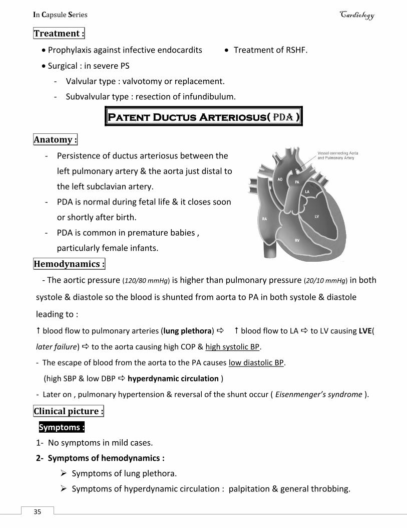
Anatomy :
- Persistence of ductus arteriosus between the
left pulmonary artery & the aorta just distal to
the left subclavian artery.
- PDA is normal during fetal life & it closes soon
or shortly after birth.
- PDA is common in premature babies ,
particularly female infants.
Hemodynamics :
- The aortic pressure (120/80 mmHg) is higher than pulmonary pressure (20/10 mmHg) in both
systole & diastole so the blood is shunted from aorta to PA in both systole & diastole
leading to :
blood flow to pulmonary arteries (lung plethora) blood flow to LA to LV causing LVE(
later failure) to the aorta causing high COP & high systolic BP.
- The escape of blood from the aorta to the PA causes low diastolic BP.
(high SBP & low DBP hyperdynamic circulation )
- Later on , pulmonary hypertension & reversal of the shunt occur ( Eisenmenger’s syndrome ).
Clinical picture :
Symptoms :
1- No symptoms in mild cases.
2- Symptoms of hemodynamics :
Symptoms of lung plethora.
Symptoms of hyperdynamic circulation : palpitation & general throbbing.
In Capsule Series Cardiology
35
3- Symptoms of complications.
4- Symptoms of other congenital anomalies.
Signs :
1- No signs in mild cases .
2- Signs of hemodynamics :
Signs of lung plethora.
Signs of hyperdynamic circulation : the same as peripheral signs of AR. (9)
3- Signs of complications.
4- Neck vein : Giant (a) wave due to pulmonary hypertension.
Cardiac examination :
1. LVE.
2. Continuous thrill over left infraclavicular area ( site of DA).
3. Auscultation :
S2 : Accentuated, reversed splitting S2 ( due to delayed evacuation of LV ).
Murmur : Continuous "machinery " murmur over left infraclavicular area.
DD of continuous murmur :
1- Arterio-venous fistula.
2- PDA.
3- Coarctation of aorta.
4- Double mitral ( combined MS & MR ).
Complications :
1- LSHF.
2- Paradoxical embolism e.g. stroke.
3- Infective endocarditis.
4- Eisenmenger’s syndrome differential cyanosis ( cyanosis only in LL ) because
the reversed cyanotic blood enter aorta distal to subclavian artery.
5- Arrhythmia.
In Capsule Series Cardiology
35
Investigation :
X ray : - Dilatation of aorta , PA , LA & LV. - Lung plethora.
ECG : LVE.
Echo : show chamber dilatation.
Catheterization :
Detect the defect : the catheter may pass through PDA.
Pulmonary pressure.
O2 level in PA in comparison to RV.
Treatment :
Medical :
Prophylaxis against infective endocarditis.
Treatment of complications
Medical closure of the duct : Indomethacin.
Surgical : Closure of the duct.
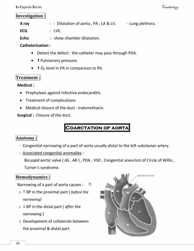
Coarctation of aorta
Anatomy :
- Congenital narrowing of a part of aorta usually distal to the left subclavian artery.
- Associated congenital anomalies :
Bicuspid aortic valve ( AS , AR ) , PDA , VSD , Congenital aneurism of Circle of Willis ,
Turner’s syndrome.
Hemodynamics :
Narrowing of a part of aorta causes :
o BP in the proximal part ( before the
narrowing)
o BP in the distal part ( after the
narrowing )
o Development of collaterals between
the proximal & distal part.
In Capsule Series Cardiology
35
Clinical picture :
Symptoms :
1- Asymptomatic in mild cases.
2- Symptoms of hemodynamics :
BP in the upper half Symptoms of hypertension e.g. headache , epistaxis ..
BP in the lower half Fatigue & Intermittent claudication of the LL.
Collaterals Pain around left shoulder.
3- Symptoms of complications.
4- Symptoms of other congenital anomalies e.g. AS , AR , PDA ….
Signs :
1- No signs in mild cases.
2- Signs of hemodynamics :
BP in arms, prominent carotid pulsation.
BP in legs , weak pulsations of LL e.g. dorsalis pedis.
Collaterals may be seen in interscapular area ( Suzman’s sign ).
3- Signs of complications.
4- Signs of other congenital anomalies.
Cardiac examination :
1. LV hypertrophy.
2. Auscultation :
Accentuated S2
Murmurs:
o Ejection systolic murmur due to :
Coarctation itself ( below left infraclavicular area ) , Associated AS ,Hypertension.
o Early diastolic murmur due to associated AR.
o Continuous murmur over the collaterals.
Complications :
1- Complications of hypertension e.g. cerebral hemorrhage …..
2- Heart failure.
3- Infective endocarditis.
In Capsule Series Cardiology
35
Investigations :
1- X ray :
LVE.
Rosler’s sign : Rib notching due to erosion by collaterals.
2- ECG : LVE .
3- Echo : LVE , can detect the coarctation .
4- Catheterization & aortography : can detect the site & severity of the coarctation.
Treatment :
Medical : prophylaxis against IE & treatment of the complications.
Surgical repair : in early childhood to avoid persistent hypertension.
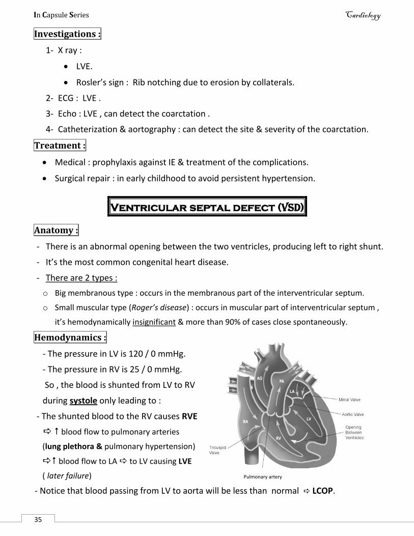
Ventricular septal defect (VSD)
Anatomy :
- There is an abnormal opening between the two ventricles, producing left to right shunt.
- It’s the most common congenital heart disease.
- There are 2 types :
o Big membranous type : occurs in the membranous part of the interventricular septum.
o Small muscular type (Roger’s disease) : occurs in muscular part of interventricular septum ,
it’s hemodynamically insignificant & more than 90% of cases close spontaneously.
Hemodynamics :
- The pressure in LV is 120 / 0 mmHg.
- The pressure in RV is 25 / 0 mmHg.
So , the blood is shunted from LV to RV
during systole only leading to :
- The shunted blood to the RV causes RVE
blood flow to pulmonary arteries
(lung plethora & pulmonary hypertension)
blood flow to LA to LV causing LVE
( later failure) Pulmonary artery
- Notice that blood passing from LV to aorta will be less than normal LCOP.
In Capsule Series Cardiology
35
So, in VSD there are :
Lung plethora.
Biventricular enlargement.
LCOP.
Clinical picture :
Symptoms :
1. Asymptomatic in mild cases & in Roger’s disease.
2. Symptoms of hemodynamics :
Symptoms of lung plethora : Exertional dyspnea ,recurrent chest infection…
Symptoms of LCOP.
3. Symptoms of the complications.
4. Symptoms of other congenital anomalies.
Signs :
1. No signs in mild cases.
2. Signs of hemodynamics : Lung plethora & LCOP.
3. Signs of complications.
4. Neck vein : Giant (a) wave.
Cardiac examination :
1. Biventricular enlargement with hyperdynamic apex.
2. Auscultation :
S2 : Accentuated pulmonary component , wide splitting.
Murmur :
- Harsh pansystolic murmur with thrill over the 3rd,4th intercostals spaces.
- Ejection systolic murmur of pulmonary hypertension.
- Mid diastolic murmur of relative MS ( blood flow across the mitral valve )
Complication :
1- HF.
2- Infective endocarditis.
3- Paradoxical embolism.
4- Eisenmenger’s syndrome : usually at 2nd - 3rd decade.
In Capsule Series Cardiology
35
Investigations :
1. X ray : Biventricular enlargement , lung plethora.
2. ECG : Biventricular enlargement.
3. Echo : Biventricular enlargement , diagnosis of anomaly.
4. Catheterization :
Detect the defect : the catheter may pass through VSD.
pressure in the RV & PA.
O2 level in RV in comparison to RA.
Treatment :
Prophylaxis against IE & treatment of complications.
Surgical closure of large defect.
Tetralogy of Fallot ( F4 )
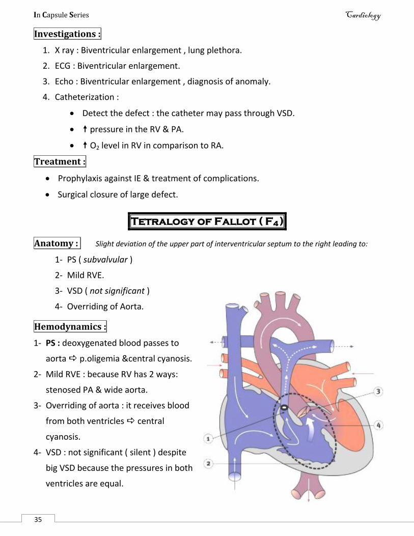
Anatomy : Slight deviation of the upper part of interventricular septum to the right leading to:
1- PS ( subvalvular )
2- Mild RVE.
3- VSD ( not significant )
4- Overriding of Aorta.
Hemodynamics :
1- PS : deoxygenated blood passes to
aorta p.oligemia ¢ral cyanosis.
2- Mild RVE : because RV has 2 ways:
stenosed PA & wide aorta.
3- Overriding of aorta : it receives blood
from both ventricles central
cyanosis.
4- VSD : not significant ( silent ) despite
big VSD because the pressures in both
ventricles are equal.
In Capsule Series Cardiology
35
Clinical picture :
Symptoms :
1- Central cyanosis since birth or shortly after.
2- Central dyspnea. ( hypoxia respiratory center )
3- Clubbing.
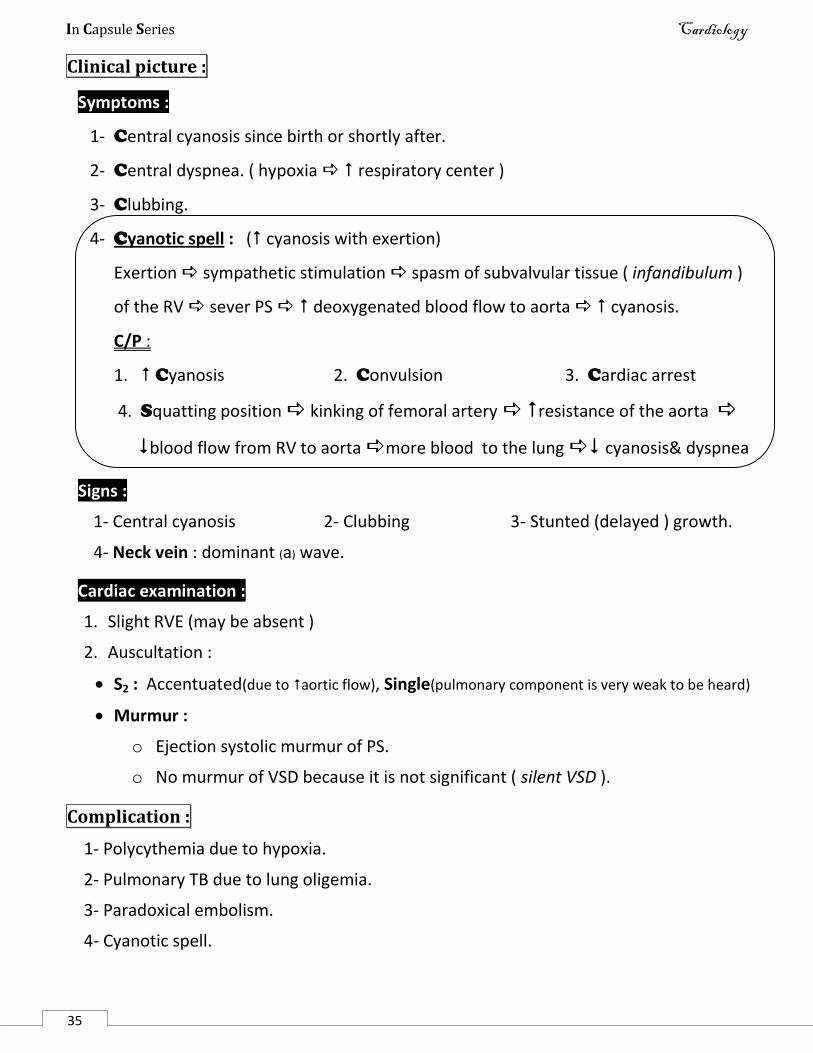
4- Cyanotic spell : ( cyanosis with exertion)
Exertion sympathetic stimulation spasm of subvalvular tissue ( infandibulum )
of the RV sever PS deoxygenated blood flow to aorta cyanosis.
C/P :
1. Cyanosis 2. Convulsion 3. Cardiac arrest
4. Squatting position kinking of femoral artery resistance of the aorta
blood flow from RV to aorta more blood to the lung cyanosis& dyspnea
Signs :
1- Central cyanosis 2- Clubbing 3- Stunted (delayed ) growth.
4- Neck vein : dominant (a) wave.
Cardiac examination :
1. Slight RVE (may be absent )
2. Auscultation :
S2 : Accentuated(due to aortic flow), Single(pulmonary component is very weak to be heard)
Murmur :
o Ejection systolic murmur of PS.
o No murmur of VSD because it is not significant ( silent VSD ).
Complication :
1- Polycythemia due to hypoxia.
2- Pulmonary TB due to lung oligemia.
3- Paradoxical embolism.
4- Cyanotic spell.
In Capsule Series Cardiology
35
Investigations :
1- X ray : Boot shaped heart : narrow base with elevated apex.
Pulmonary oligemia.
2- ECG : RVE.
3- Echo :diagnostic.
4- Catheterization : diagnostic.
Treatment :
1- Treatment of cyanotic spell : Squatting position , O2 , blocker.
2- Prophylaxis against infective endocarditis.
3- Blalock Taussig operation : acquired PDA.
4- Surgical total correction.
Triology of Fallot ( F3 )
1. PS ( valvular )
2. Marked RVE.
3. ASD.
Eisenmenger’s syndrome
Definition :
It is a condition in which a left-to-right shunt in the heart causes pulmonary hypertension,
which in turn ,causes increased pressure in the right side of the heart and reversal of the
shunt into a right-to-left shunt.
Etiology :
VSD.
PDA.
ASD.
Eisenmenger complex was applied to patients with reversal of shunt in a case of VSD
by Dr. Victor Eisenmenger in 1897 but the definition was extended by Dr. Paul Wood
to include shunts at any level VSD, ASD, PDA ,..
In Capsule Series Cardiology
35
Clinical picture :
1- History of congenital heart disease : VSD , PDA , ASD.
2- Pulmonary infection & hemoptysis.
3- C/P of pulmonary hypertension .
4- C/P of RSHF.
5- Decrease of the original murmur of the shunt due to low pressure gradient.
Treatment :
Prevention is best.
Closure of the defect is contraindicated as it increases the pressure in the right
side of the heart.
Symptomatic treatment e.g. HF. Heart lung transplantation.
Transposition of the great vessels (TGA)
The position of the aorta & the PA are reversed, this leads to separate 2 circuits :
The aorta arises from the RV ,so most of the blood returning to the heart from the
body is pumped back out through the aorta without going to the lung central
cyanosis.
The PA arises from the LV ,so the blood returning from the lungs goes back to the
lungs again.
To maintain life , an associated ASD , VSD , PDA ,PS should exist.
Treatment : Keep the PDA by Prostaglandin E1 , surgical correction.
Dextrocardia
Deviation of the heart to the right , it may be congenital or acquired.
Situs inversus totalis : mirror like transposition of the heart & all other viscera.
Isolated dextrocardia : mirror like transposition of the heart only.
Dextroversion: the heart is displaced to the right (RV remains to the right &LV to the left)
Acquired dextrocardia: acquired displacement of the heart to the right e.g. fibrosis.
In Capsule Series Cardiology
48
Ischemic Heart Disease (IHD)
Anatomy of coronary arteries:
There are two coronary arteries – left & right – originate from the root of
ascending aorta .
1- Left coronary artery : Passes forward & to the left in the left atrioventricular
groove for a short distance & then divides into :
a) Anterior descending artery : passes downward in anterior
interventricular groove to the apex & then turns backward to meet
posterior descending artery .
b) Circumflex artery : runs posteriorly in the left atrioventricular groove
to meet the right coronary .
2- Right coronary artery : runs in right atrioventricular groove to the
posterior surface of the heart to meet the circumflex artery.
posteriorly , it gives the posterior descending artery which runs in the
posterior interventricular groove to meet the anterior descending artery.
Patterns of coronary supply :
Balanced circulation:
- The left coronary artery supplies :
LA , LV , anterior wall of interventricular septum .
- The right coronary artery supplies :
RA , RV , posterior wall of interventricular septum .
Left coronary predominance :
-The left coronary supplies also: posterior wall of right ventricle.
Right coronary predominance :
- The right coronary supplies also: posterior wall of left ventricle .
Presentation of ischemic heart disease :
Asymptomatic ( silent ).
Angina.
Myocardial infarction.
Heart failure.
Arrhythmia.
Sudden death
In Capsule Series Cardiology
49
Angina Pectoris Definition :
It is a clinical syndrome of chest pain due to imbalance between oxygen supply & demands of the myocardium.
Etiology :
I. Decreased myocardial oxygen supply :
1) Decrease in quantity :
a) Coronary artery disease : Atherosclerosis. ( most common cause ) Arteritis : polyarteritis nodosa , SLE . Coronary spasm . Coronary embolism . Coronary osteial stenosis of syphilis . Congenital anomalies .
b) As a part of LCOP : AS , LSHF .
2) Decrease in quality : Anemia. Hypoxia.
II. Increased myocardial oxygen demand : Ventricular hypertrophy . Tachycardia .
Risk factors for atherosclerosis:
Non modifiable :
Age .
Sex : male > female .
+ve family history . Modifiable :
Hypertension : cause endothelial damage.
Hyperglycemia
Hyperlipidemia especially LDL .
Hyperuricemia .
Sedentary life style .
Smoking.
Stress & type A personality .
In Capsule Series Cardiology
- 50 -
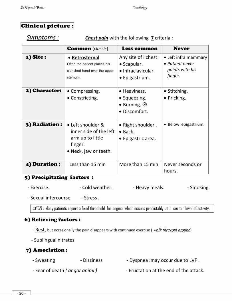
Clinical picture :
Symptoms : Chest pain with the following 7 criteria :
Common (classic) Less common Never
1) Site : Retrosternal Often the patient places his clenched hand over the upper sternum.
Any site of i chest:
Scapular.
Infraclavicular.
Epigastrium.
Left infra mammary
Patient never points with his finger.
2) Character: Compressing.
Constricting.
Heaviness.
Squeezing.
Burning.
Discomfort.
Stitching.
Pricking.
3) Radiation : Left shoulder & inner side of the left arm up to little finger.
Neck, jaw or teeth.
Right shoulder .
Back.
Epigastric area.
Below epigastrium.
4) Duration : Less than 15 min More than 15 min Never seconds or hours.
5) Precipitating factors :
- Exercise. - Cold weather. - Heavy meals. - Smoking.
- Sexual intercourse - Stress .
N.B : Many patients report a fixed threshold for angina, which occurs predictably at a certain level of activity.
6) Relieving factors :
- Rest, but occasionally the pain disappears with continued exercise ( walk through angina)
- Sublingual nitrates.
7) Association :
- Sweating - Dizziness - Dyspnea :may occur due to LVF .
- Fear of death ( angor animi ) - Eructation at the end of the attack.
In Capsule Series Cardiology
- 51 -
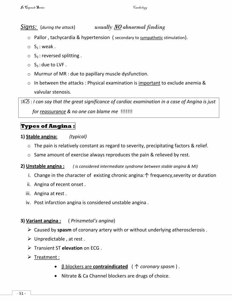
Signs: (during the attack) usually no abnormal finding
o Pallor , tachycardia & hypertension ( secondary to sympathetic stimulation).
o S1 : weak .
o S2 : reversed splitting .
o S3 : due to LVF .
o Murmur of MR : due to papillary muscle dysfunction.
o In between the attacks : Physical examination is important to exclude anemia &
valvular stenosis.
NB : I can say that the great significance of cardiac examination in a case of Angina is just
for reassurance & no one can blame me !!!!!!!
Types of Angina :
1) Stable angina: (typical)
o The pain is relatively constant as regard to severity, precipitating factors & relief.
o Same amount of exercise always reproduces the pain & relieved by rest.
2) Unstable angina : ( is considered intermediate syndrome between stable angina & MI)
i. Change in the character of existing chronic angina:↑ frequency,severity or duration
ii. Angina of recent onset .
iii. Angina at rest .
iv. Post infarction angina is considered unstable angina .
3) Variant angina : ( Prinzmetal’s angina)
Caused by spasm of coronary artery with or without underlying atherosclerosis .
Unpredictable , at rest .
Transient ST elevation on ECG .
Treatment :
β blockers are contraindicated ( ↑ coronary spasm ) .
Nitrate & Ca Channel blockers are drugs of choice.
In Capsule Series Cardiology
- 52 -
Decubitus Angina : usually on lying down (occurs in HF).
Nocturnal Angina : It awakens the patient from sleep , associated with dreaming .
Angina of Lewis : in cases of AR , it is nocturnal & prolonged .
Acute coronary syndrome :MI & unstable angina .
Investigation :
1- ECG :
A) Resting ECG :
In between the attacks :
usually normal.
ECG of old MI .
During the attack:
ST segment : depressed. ( more than 1mm )
T wave : Inverted .
B) Exercise ECG : ( in between the attacks only )
- The patient is exercises on a treadmill & ECG changes & vital signs are recorded.
- Stress test can be done with dobutamine in patients unable to do exertion.
- Stress test is considered +ve when : one or more of these changes are present :
Symptom : Typical anginal pain during the test.
Sign : Fall in blood pressure (10 mmHg or more) suggests ischemia
ECG : Depressed ST segment > 1mm .
NB : Exercise test can be misleading as there are :
False negative test : So normal test doesn’t exclude IHD .
False positive test :especially in patients with left ventricular hypertrophy.
Stress test is contraindicated in :
- Acute attacks. - Severe AS.
- Severe hypertension. - Congestive heart failure.
- Orthopedic problems.
In Capsule Series Cardiology
- 53 -
2- Echo & dobutamine Echo : may show abnormal motion of the myocardium .
3- Cardiac scan : ( Radioactive Thallium 201 )
Thallium 201: is taken up by healthy myocardium & not by ischemic myocardium (cold spot)
4- Coronary angiography : ( coronary catheter )
To detect the site & severity of coronary occlusion.
It’s generally used to determine whether mechanical revascularization (CABG
or PTCA) is possible & to guide this therapy.
5- Laboratory investigations:
For risk factors :Blood glucose level , Plasma lipid ( cholesterol ).
Cardiac enzymes : normal .
Treatment : 4
1- Control of risk factors :
Reassurance & sedation. No smoking.
Treatment of hyperlipidemia. Control of hypertension.
Control of diabetes. Weight loss.
Change of life style ( regular exercise program ).
2- Medical treatment of angina : in between the attacks
i. Nitrates :
Action :
Venodilator preload (venous return) myocardial oxygen demand.
Coronary dilatation increase coronary blood flow. ( mild effect )
Preparation :
Nitroglycerine ( nitromack ) : 2.5 mg twice daily orally or transdermal patches.
Isosorbid dinitrate ( dinitra ) : 10-20 mg twice daily.
Isosorbid mononitrate ( effox ) : 20-40 mg twice daily.
In Capsule Series Cardiology
- 54 -
Side effects :
Headache.
Hypotension.
Tolerance : so start with minimal effective dose with nitrate free interval periods.
ii. β blockers :
Action :
Reduce oxygen demand since they reduce heart rate, blood pressure & contractility.
Preparation :
Propranolol ( indral ) : non selective β blocker .
Atenolol (ateno), Metoprolol (betaloc) , Bisoprolol (concor) : Selective β blockers.
Carvedilol ( cardilol ) : β blocker with an arteriolar vasodilating action.
Side effects :
Lung : Bronchospasm.
Heart : Bradycardia , Heart block.
Depression , Impotence.
iii. Calcium channel blockers :
Action :
Reduce oxygen demand by : -ve inotropic action.
afterload ( arteriolar dilators ).
Coronary dilator : increase coronary blood flow ( effective in variant angina )
Preparation :
Verapamil ( Isopten ) : great -ve inotropic & weak vasodilator: 80 mg t.d.s.
Diltiazem : 60 mg twice daily.
Nifedipine ( adalat ) : mainly vasodilator & weak -ve inotropic: 10 – 20 mg t.d.s.
Recently : Amlodipine ( norvasc ) : mainly vasodilator .
Side effects :
Headache.
Hypotension.
In Capsule Series Cardiology
- 55 -
Precipitation of Heart failure.
Peripheral edema.
Verapamil & Diltiazem : bradycardia & heart block.
iv. Antiplatelet :
Aspirin : 75 mg single dose : it improves the prognosis.
Clopidogrel ( plavix ) :expensive.
3- Coronary revascularization :
Indications :
Angina not responding to medical treatment.
Post infarction angina to improve the prognosis.
Techniques :
1- PTCA ( Percutaneous Transluminal Coronary Angioplasty ):
Introduction of balloon or stent to dilate the stenotic artery( balloon-tipped catheter)
Indication of PTCA :
Stenosis of one or two vessels only ( except left main coronary artery )
2- CABG ( Coronary Artery Bypass Graft ) :
Grafting a piece of saphenous vein or internal mammary artery between the aorta &
the coronary artery distal to any obstruction.
Indication of CABG :
Stenosis of 3 or more vessels.
Stenosis of left main coronary artery.
4- Treatment of anginal attack :
Complete rest.
Nitroglycerine (0.5 mg) or isosorbide dinitrate (5mg) sublingually & repeated up to 3
times successively with interval of 3 minutes.
NB : If the patient is not relieved after the use of 2-3 tablets ,the patient should be immediately
transferred to hospital & evaluated for the possibility of myocardial infarction.
In Capsule Series Cardiology
- 56 -
Myocardial infarction
Definition :
Ischemic necrosis of part of the cardiac muscle due to sudden , persistent & complete
cessation of its blood supply.
Etiology :
Thrombosis on top of atherosclerosis. ♫♫
Coronary embolism ( rare ).
Severe coronary spasm.
Pathology :
Site:
1- Occlusion of the left anterior descending artery anterior infarction.
2- Occlusion of the circumflex artery lateral infarction.
3- Occlusion of the right coronary artery inferior infarction.
Types :
Transmural infarction ( ST elevation myocardial infarction - STEMI ) : infarction of full
thickness of the ventricular wall.
Subendocardial infarction ( Non ST elevation myocardial infarction -NSTEMI ) :
Transient or incomplete vessel occlusion.
Clinical picture : Pain and/or complications
I. Chest pain: Similar to angina but :
More severe, it may be severe enough to be described as the worst pain the patient has ever felt.
Radiates more : may below epigastric area but never below umbilicus.
More prolonged : up to several hours.
Unrelated to precipitating factors : may at rest.
Not relieved by rest or sublingual nitrate.
Associations: like angina & may also associated with complications.
In Capsule Series Cardiology
- 57 -
NB: Painless infarction:
o Elderly.
o Diabetic neuropathy.
o Patient under anesthesia.
o Transplanted heart ( denervated ).
II. Complications : 6 early & 6 late
Early complications : 6 items
1- Shock : see p133
Cardiogenic shock Neurogenic shock
Caused by massive infarction (> 40% of the
cardiac muscle) leading to severe pump failure
& high jugular venous pressure.
C/P : Hypotension , tachycardia ,pulmonary
edema.
ttt: The same as acute pulmonary edema &
mechanical assist devices: intraaortic
balloon counterpulsation.
Prognosis : very bad.
Caused by severe pain ( vagal stimulation ).
C/P : Hypotension, bradycardia .
ttt : morphine .
Prognosis : good .
2- Acute heart failure : with normal heart size.
3- Arrhythmia :
- All types may occur.
- The most serious are : VT , CHB .
4- Myocardial rupture :
Rupture of the septum acquired VSD .
Rupture of papillary muscles acute MR acute heart failure.
Rupture of the ventricular free wall blood fills the pericardium cardiac tamponade.
In Capsule Series Cardiology
- 58 -
5- Dry pericarditis : Hemorrhagic pericardial effusion may develop especially with
thrombolytic therapy.
6- Sudden death:
Arrhythmia (VT , VF ) : most deaths occur during few hours after MI .
Acute heart failure.
Cardiogenic shock.
Cardiac rupture.
Late complications : 6 items
1- Post infarction syndrome : ( Dressler’s syndrome ) within 4 weeks or more
Autoimmune phenomenon in response to necrotic cardiac tissue characterized by :
- Pericarditis - Pleurisy - Pneumonitis -fever.
2- Post infarction angina :
Due to affection of other diseased coronaries.
3- Myocardial aneurysm : ( dilatation of the scar tissue of MI )
On examination : double apex .
ECG : persistent ST segment elevation .
Fate : - Refractory heart failure.
- Rupture aneurysm.
- Recurrent embolism.
- Recurrent arrhythmia.
4- Thrombo-embolism :
Mural thrombosis :( infarction rough surface thrombosis systemic emboli )
DVT : due to prolonged recumbency pulmonary embolism .
5- Frozen shoulder : stiffness with limitation of movement due to :
Pain reflex arteriolar spasm & ischemia.
may be psychic.
6- Complications of treatment: anticoagulant , prolonged bed rest,….
In Capsule Series Cardiology
- 59 -
Signs : (not specific) nothing or anything
The physical examination may be entirely normal.
Pallor , sweating , nausea , vomiting & fever.
Pulse :
o Tachycardia : sympathetic stimulation , cardiogenic shock .
o Bradycardia : neurogenic shock , HB , inferior MI.
o Irregular : arrhythmias.
o weak : LVF .
NB : Bradycardia is often seen with inferior MI because the right coronary artery
supplies the SA node.
Blood pressure :
o Hypertension : sympathetic stimulation .
o Hypotension : LVF , shock .
Cardiac auscultation :
o S1 : weak.
o S2 : reversed splitting.
o S3 : due to LVF.
o S4 : due to decreased myocardial compliance.
o Murmur : of MR , VSD .
o Pericardial rub : Dry pericarditis.
Congested neck vein : in right ventricular infarction.
Differential Diagnosis :
Causes of acute chest pain :
o Ischemic heart diseases : Angina , MI.
o Pulmonary embolism.
o Aortic dissection.
o Pneumothorax.
o Acute dry pericarditis.
o Cardiac neurosis.
o Esophageal spasm , Perforating peptic ulcer , Cholecystitis.
In Capsule Series Cardiology
60
Investigations:
1- Cardiac enzymes :
Cardiac enzymes are released into blood from necrotic heart muscle after an acute MI.
Marker Initial rise Return to normal Notes
Creatine phosphokinase
( CPK )
4-8 h 2-4 days Non specific because it
may rise in damaged
skeletal muscles or brain.
CPK-MB 4-8 h 2-4 days It’s isoenzyme of CPK ,
specific to cardiac muscle
Lactic dehydrogenase
( LDH )
10 h 1-2 weeks Not specific .
Troponin ( cTnT , cTnI ) 2-6 h 1 week Most sensitive & specific
markers of myocardial
damage .
2- ECG :
In transmural infarction ( ST Elevation MI ):
1. Convex elevation of ST segment .
2. T wave :
Tall (hyperacute) in the first few minutes after vessel occlusion (the earliest change)
later on : Inverted T wave ( representing sever ischemia )
3. Finally, pathological Q waves occur, representing significant myocardial necrosis
& replacement by scar tissue.
In subendocardial infarction ( Non ST Elevation MI ) :
1. ST segment : normal or depressed.
2. No pathological Q waves ( non Q wave MI )
3. T wave : inverted.
NB: The ECG may be normal during the first few hours of infarction .
In old MI : The only residual change is the pathological Q wave.
In Capsule Series Cardiology
61
3- Echocardiography :
Ventricular wall motion abnormalities.
Complications : MR , myocardial aneurysm.
4- Cardiac scan : Like angina .
5- Coronary angiography :
reveals which vessels have been affected and the extent of damage.
6- Leukocytosis , ↑ ESR : as there is tissue damage.
Treatment : 3
I. Pre hospital :
1- Rapid transfer to hospital is a must ( Time lost is lives lost ) .
2- Oxygen inhalation.
3- Analgesics for pain Morphine 5 - 10 mg IV
4- For ventricular arrhythmias Lidocaine 50 – 100 mg IV ??
5- For heart block Atropine 0.5 – 1 mg IV .
II. Hospital care :
1- General :
a. Admission to CCU ( coronary care unit ) with hemodynamic monitoring & continuous ECG
b. Oxygen inhalation .
c. Complete rest .
d. Diet : Light frequent meals & avoid constipation .
e. Sedative : Diazepam .
f. Aspirin : is now considered an essential element ( 325 mg initial dose then 75 mg daily-oral)
g. ACE Inhibitor: Oral therapy e.g. Lisinopril 5mg on day1 & 2 ,then 10 mg daily.
NB : ACE Inhibitors are vasodilator that reduce cardiac work & decrease myocardial energy requirement .
ACE Inhibitors also have inhibitory effect on the cardiac remodeling.
In Capsule Series Cardiology
62
2- Relieving of chest pain :
a. Morphine ( 4 mg IV every 5 to 10 minutes as needed )
b. Nitroglycerine .
c. β blockers .
3- Thrombolytic therapy : ( time is muscle )
- The earlier that thrombolytic therapy is given after the onset of chest pain, the greater the
benefit (thrombolytic therapy is beneficial up to 6 hours but may be given for up to 12 hours)
Drugs :
o Streptokinase : 1.5 million units IV over 60 min. may cause allergy .
o Urokinase .
o Alteplase ( tissue plasminogen activator – tPA )
The important issue in thrombolytic therapy is not which drug to use, but how quickly to use it .
- Anticoagulant ( heparin ) & antiplatetelet ( aspirin ) are given with & after thrombolytic
therapy to reduce the risk of reocclusion.
Contrindication : the major risk is Bleeding
Bleeding disorders.
Major surgery within past 2 weeks .
Recent cerebral hemorrhage within past 12 months.
Active internal bleeding e.g. peptic ulcer.
Sever hypertension.
Diabetic retinopathy with recent bleed.
Aortic dissection.
Pericarditis.
4- Angioplasty : Percutaneous Transluminal Coronary Angioplasty ( PTCA ) :
- Introduction of balloon or stent to dilate the stenotic artery ( balloon-tipped catheter)
- More effective than thrombolytic therapy ( fewer complication , shorter hospitalization ).
to relieve pain of post infarction angina.
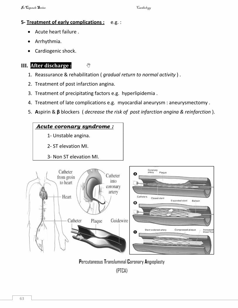
In Capsule Series Cardiology
63
5- Treatment of early complications : e.g. :
Acute heart failure .
Arrhythmia.
Cardiogenic shock.
III. After discharge :
1. Reassurance & rehabilitation ( gradual return to normal activity ) .
2. Treatment of post infarction angina.
3. Treatment of precipitating factors e.g. hyperlipidemia .
4. Treatment of late complications e.g. myocardial aneurysm : aneurysmectomy .
5. Aspirin & β blockers ( decrease the risk of post infarction angina & reinfarction ).
Acute coronary syndrome :
1- Unstable angina.
2- ST elevation MI.
3- Non ST elevation MI.
Percutaneous Transluminal Coronary Angioplasty
(PTCA)
In Capsule Series Cardiology
- 64 -
Rheumatic fever
Definition:
Diffuse inflammatory disease typically caused by an abnormal immune response to
upper respiratory tract infection with group A - hemolytic streptococci .
Etiology : There are 2 theories
1- Antigenic similarity :
The antigen of streptococci is similar to the antigens of heart & connective tissue so,
antibodies produced against streptococci can react with cardiac muscle & other CT .
2- Altered antigenicity :
Streptococci or its toxins may change the character of the connective tissue making
it abnormal to the body so acting as an antigen of auto antibodies against it.
Pathology :
I- Site : ( Rheumatic fever bites the heart & licks the joints )
1- Joints : synovial membrane .
2- Heart : Pancarditis .
3- CNS .
4- Skin & subcutaneous tissue .
5- Small blood vessels .
6- Serous membrane . ( Pleura , Peritoneum , Pericardium )
II- Types : There are 2 types
1- Exudative lesion :
Affecting mainly the serous membrane .
Heals mostly without scaring or deformity .
2- Proliferative lesion : formation of Aschoff nodules
Affects mainly the heart & the skin .
Heals by fibrosis .
In Capsule Series Cardiology
- 65 -
Consists of : ( from in to out )
Fibrinoid degeneration .
Lymphocytes & plasma cells .
Aschoff giant cells .
Fibroblasts .
Layer of fibrosis .
Predisposing factors :
1- Recurrent streptococcal infections of the pharynx & tonsils .
2- Age : most common between 5-15 years(rare below 5 & above 25 ).
3- Sex : equal in both , but chorea is more common in females .
4- Familial : due to similar environment . ( overcrowding )
Clinical picture :
Latent period : 1- 3 weeks
I- Major criteria : ( mnemonic : C.A.N.C.ER )
1- Carditis : found in 50 % of patients
Rheumatic fever affects the 3 layers of the heart ( pancarditis )
Pericarditis :
o Dry pericarditis : causing stitching pain & pericardial rub .
o Pericardial effusion : less common .
o Never constrictive pericarditis .
Myocarditis :
o Tachycardia : not proportional to the degree of fever.
o Tic - Tac rhythm : due to loss of muscular component of S1 .
o Heart failure .
o Arrhythmia & may be heart block .
Endocarditis : (valvulitis )
o Order of frequency : Mitral > Aorta > Tricuspid > Pulmonary (MAT P)
In Capsule Series Cardiology
- 66 -
o Carey Coomb’s murmur: Transient mid diastolic murmur of mitral
stenosis in acute stage due to swelling of the mitral cusps causing
narrowing of the mitral valve .
o Later on (over years): fibrosis may lead to stenosis , regurge or both .
2- Arthritis : found in 75% of patients
o Polyarthritis .
o Affects big joints : Knees , ankles , elbows .
o Asymmetrical .
o Migratory ( flitting )
o The affected joints show hotness , redness , tenderness , swelling with
limitation of movement .
o Dramatic response to salicylates .
o Duration of arthritis is about 2-6 weeks .
o No deformity : leaving the joints with complete resolution .
3- Subcutaneous Nodule : rare
o Description: Small ,painless ,tenderless ,firm nodule ¬ adherent to skin.
o Etiology : accumulation of Aschoff nodule .
o Site : over bony prominences & tendons e.g. dorsum of the hands & feet
o Importance : associated with severe carditis .
4- Chorea : ( Sydenham’s chorea ) : 10 % of cases
o Involuntary , sudden movements with emotional instability… See neurology
o More common in female.
o It appears very late after streptococcal infection ( 6 months ) , so it’s not
associated with arthritis & ESR is not raised.
o It is self limiting.
In Children : Carditis > arthritis .
In Adult : Arthritis > carditis .
In Capsule Series Cardiology
- 67 -
5- ERythema marginatum :
o Occurs in the form of areas of erythema with central pallor.
o Site : trunk & proximal part of limbs.
o Non pruritic ,non painful.
II- Minor criteria : ( mnemonic : P.E.A.C.E )
1. Prolonged PR interval in the ECG : > 0.22 sec , due to myocarditis .
2. ESR : elevated .
3. Arthralagia : painful joints without swelling.
4. CRP ( C reactive protein ) : elevated .
5. Elevated temperature ( fever ).
III - Other manifestations of rheumatic fever :
Pallor .
Pleurisy .
Pneumonia .
Peritonitis Abdominal pain.
Diagnosis of rheumatic fever :
2 major criteria or 1 major & 2 minor criteria . plus ,
Evidence of recent streptococcal infection (+ve throat culture , ASO , Scarlet fever)
- No need to say that if arthritis is taken as a major criteria , don’t consider arthralagia as a minor criteria.
- Also if carditis is taken as a major criteria , don’t consider prolonged PR interval as a minor criteria.
Complications :
Acute complications : i. Heart failure ii. Arrhythmia & heart block .
Chronic complications = Complications of valvular heart diseases .
Differential diagnosis :
Fever of unknown origin.
Infective endocarditis.
Juvenile rheumatoid arthritis.
Acute leukemia.
In Capsule Series Cardiology
- 65 -
Investigations : There is NO specific confirmatory test .
a) Laboratory : ( evidence of recent streptococcal infection )
Blood picture : Leucocytosis .
ESR :
CRP ( C reactive protein ) :
ASO titre ( Anti Streptolysin O titre ) : Normally up to 150 Todd units
Higher than 250 Todd units in adult & 333 Todd units in children indicates
recent streptococcal infection .
Throat culture : may reveal group A streptococci .
b) Cardiac : ( evidence of carditis )
X ray : Cardiac enlargement.
ECG : Prolonged PR interval , arrhythmia & heart block.
Echo : Cardiac enlargement & valve lesions.
Signs of rheumatic activity :
Major or minor criteria.
Laboratory tests: ESR , CRP , ASO & Leucocytosis .
Treatment :
Prophylactic :
i. Primary prevention : ( to prevent rheumatic fever )
Proper management of URT infection .
Tonsillectomy of chronically infected tonsils .
ii. Secondary prevention : ( to prevent recurrence of rheumatic activity )
Long acting penicillin: Benzathine penicillin (Retarpen):1.2 million U/2-4 weeks IM or
Penicillin V ( Ospen ) 250 mg /12 hr Orally . or
Erythromycin 250 mg /12 hr Orally . (in a case of allergy to penicillin)
For 5 years after the last attack or till age of 25 ( which is longer) & may for ever.
iii. Tertiary prevention : prevention of infective endocarditis.
In Capsule Series Cardiology
- 66 -
Curative :
1- Complete bed rest : till improvement of all symptoms & signs.
2- Diet : Light meals & salt restriction.
3- Antibiotics :
Benzyl penicillin G ( Crystalline penicillin ) : 1 million units / 12 hr IM for about 10 days.
Penicillin V ( Ospen ) : 250 mg / 6 hr orally for about 10 days.
Erythromycin 250 mg / 6 hr orally for about 10 days. (in a case of allergy to penicillin)
4- Anti-inflammatory drugs :
Aspirin : for Arthritis
- 100 mg / kg / day in 6 divided doses up to satisfactory laboratory findings .
- Gradual withdrawal is important to avoid rebound arthritis.
Corticosteroids : for Carditis ?
- Predinsone (Hostacortin) : 1 mg / kg / day in 4 divided doses for 4 weeks then gradual
withdrawal in 4 weeks .
- Aspirin should be added during steroid withdrawal to avoid rebound activity .
5- Treatment of complications :
Congestive heart failure : Digitalis , Dilator , Diuretic. Unlike normal heart failure,
rheumatic heart failure responds well to cortisone.
Chorea : Chlorpromazine , Haloperidol.
Try to learn something about everything and everything about something.
Thomas Henry Huxley
In Capsule Series Cardiology
70
Infective endocarditis
Definition : Infection of the endocardium.
Etiology :
The development of infective endocarditis requires combination of 2 factors :
I. Infection.
II. Underlying cardiac disease.
I- Infection : ( any organism but commonly bacteria )
a) Gram +ve cocci :
o Strept. viridans (the most common organism): e.g. tooth extraction.
o Strept. foecalis : e.g. GIT procedures.
o Staph. aureus :e.g. cardiac catheterization.
b) Gram –ve bacilli : Haemophilus influenza , Klebsiella, Pseudomonas.
c) Others : Fungi , Rickettsia , Chlamydia ……
II- Underlying cardiac disease :
(In fact, all situations inducing a turbulent blood flow are a favorable to an infective endocarditis)
a) Valvular disease : ( left > right , regurge > stenosis )
o All valvular heart diseases except MS (due to marked fibrosis) .
o Prosthetic ( artificial ) valve endocarditis.
b) Congenital heart disease :
o Especially : VSD , PDA, coarctation of aorta.
o Uncommon in ASD ( low pressure gradient between the 2 atria ).
c) Myocardial infarction.
Types:
Sub acute IE : most common & requires combinations of the 2 factors (infection & cardiac lesion).
Acute IE : affecting the healthy endocardium ,usually occurs in the right side of the heart in addicts.
In Capsule Series Cardiology
71
Pathology :
Vegetations are formed over the valvular & mural endocardium causing valvular
damage.(The organism in the center surrounded by fibrin & platelets) Vegetation may
detach leading to embolization.
Notice that this vegetation is big , friable & septic.
Also the myocardium may contain foci of inflammation.
Clinical picture :
Manifestations of the disease are due to :
Toxemia.
Embolization.
Immune complexes.
A) Extra cardiac manifestations :
Face :
i. Fever ( usually low grade & prolonged ).
Any prolonged unexplained fever in cardiac patient is considered & treated as
infective endocarditis until proved otherwise.
ii. Pallor & toxic facies.
iii. Eye :
Subcojunctival hemorrhage . ( Toxemia )
Roth spots (area of retinal hemorrhage with pale center)
Sudden blindness : due to embolism of the central retinal artery.
Upper & lower limbs :
i. Clubbing : It occurs with long standing cases .
ii. Splinter hemorrhage : longitudinal hemorrhage under the nails due to rupture
capillaries (toxemia)
In Capsule Series Cardiology
72
iii. Osler’s nodule : Small , painful , intracutaneous in the pulps of fingers & toes.( due
to toxic endothelial hyperplasia of the capillary .
iv. Janeway’s nodule : Small painless patches on the palms .
v. Abnormalities of the redial pulse :
Tachycardia : due to fever .
Absent pulse : due to embolization of brachial artery.
Spleen :
Infection : mild enlargement .
Infarction : Stitching pain & splenic rub.
Kidney :
Infection ( Immune complexes ) : AGN.
Infarction : loin pain & hematuria.
CNS :
Infection : Meningitis & encephalitis .
Infarction : Embolic hemiplegia , Subarachnoid hemorrhage .
Lung :
Infection : pneumonia.
Infarction : due to pulmonary embolism in right sided endocarditis .(rare)
B) Cardiac manifestations :
1- Features of the underlying cardiac disease which already existed before IE.
2- Precipitation of heart failure.
3- New murmurs due to perforated cusps or rupture of chordae tendineae.
In Capsule Series Cardiology
73
Investigations :
1- Blood culture : for organism ( the most important investigation )
It is +ve in most cases ( 90 % )
At least 3 samples are taken during fever & cultured under aerobic & anaerobic
conditions.
The result are observed from 3 days up to 3 weeks.
Negative culture may be seen in :
o Antibiotic treatment before taking the blood sample .
o Some organisms e.g. fungi .
2- Echo : for vegitations
Small vegitations can detected by trans esophageal echo.
3- Blood picture :
o Anemia .
o Leucocytosis.
o ↑ ESR .
4- Urine analysis : for proteinuria & hematuria .
5- Immune complexes : are presented in the circulation of most patients .
Treatment :
I- Prophylactic :
a) Correction of the underlying cardiac lesion e.g. closure of VSD.
b) Prevention of infection : Antibiotics
For tooth extraction & upper respiratory tract surgery :
Amoxicillin 3 g orally 1 hour before & 1g orally 6 hours after .
For GIT & genitourinary procedures :
Ampicillin : 2 g IV plus Gentamycin : 80 mg IV half an hour before & 6 hours after.
For cardiac surgery & catheterization :
Cefotaxime : 2g IV , 2 hours before & 1 gm / 6h for 1 day after.
In Capsule Series Cardiology
74
II- Curative :
a) Medical treatment :
1- Antibiotics :
o Once infective endocarditis is suspected , the patient must rest in bed & blood
samples are taken for culture & sensitivity test .
o Treatment with antibiotics must start immediately without waiting for the result of
blood culture.
o Strong antibiotics in large doses are given parenterally for at least 4-6 weeks.
o Start with : ( even before the result of blood culture )
Penicillin G : 24 million units/day IV. plus
Gentamycin 80 mg /8h IM.
o Resistant cases should be treated according to the result of blood culture e.g. :
For Strept. viridans : penicillin G + gentamycin as before
For Staphyloccoci : ox , clox , diclox acillin . or vancomycin 1gm/12h IV.
For pseudomonas : ceftazidime 2g /8h or carbinicillin .
For fungal infections : Amphotericin B .
2- Treatment of complications : e.g. heart failure.
NB : Heparin & other anticoagulants are contraindicated in IE for fear of rupture of
mycotic aneurysm .
b) Surgical treatment :
o Replacement of the severely damaged valve.
o Replacement of infected prosthetic valve.
o Removal of myocardial abscess .
In Capsule Series Cardiology
- 75 -
Acute dry pericarditis
Etiology :
1- Idiopathic : most probably viral.
2- Infection : ◊ Viral : Coxackie B or Echo virus ( commonest ) ◊ Bacterial : TB.
3- Infarction : may occur as early or late complication.
4- Irradiation .
5- Immunological : SLE , RA .
6- Iatrogenic : hydralazine , procainamide .
7- Infiltration : leukemia , lymphoma , breast cancer .
8- Rheumatic fever .
9- Renal failure .
10- Trauma : post cardiotomy syndrome .
Clinical picture :
1- General symptoms : FHMA ( fever, headache , malaise , anorexia )
2- Local symptoms : Pain .
3- Signs : Pericardial rub .
4- Features of the cause .
The characteristics of the pain :
Site : pericardial.
Character : sever stitching .
Radiation : may radiate to the shoulders .
Duration : continuous .
Increased by : movement & respiration .
Decreased by : sitting & leaning forward .
Associated with : pleurisy .
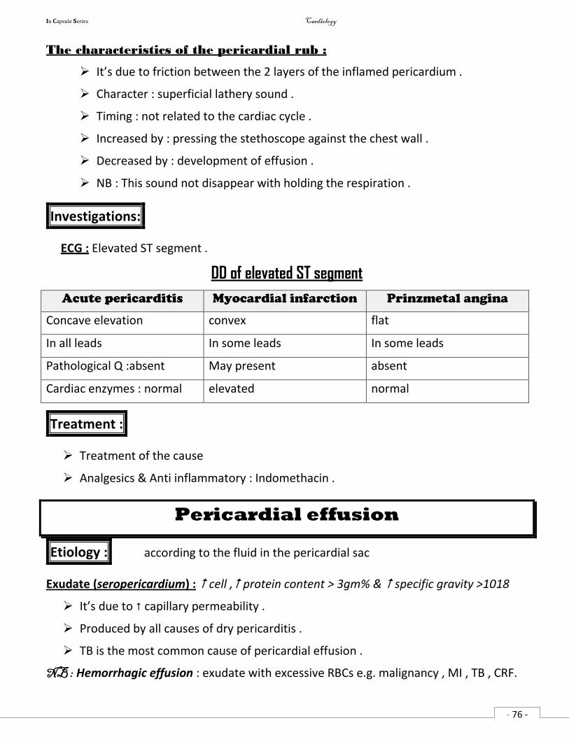
In Capsule Series Cardiology
- 76 -
The characteristics of the pericardial rub :
It’s due to friction between the 2 layers of the inflamed pericardium .
Character : superficial lathery sound .
Timing : not related to the cardiac cycle .
Increased by : pressing the stethoscope against the chest wall .
Decreased by : development of effusion .
NB : This sound not disappear with holding the respiration .
Investigations:
ECG : Elevated ST segment .
DD of elevated ST segment
Acute pericarditis Myocardial infarction Prinzmetal angina
Concave elevation convex flat
In all leads In some leads In some leads
Pathological Q :absent May present absent
Cardiac enzymes : normal elevated normal
Treatment :
Treatment of the cause
Analgesics & Anti inflammatory : Indomethacin .
Pericardial effusion
Etiology : according to the fluid in the pericardial sac
Exudate (seropericardium) : cell , protein content > 3gm% & specific gravity >1018
It’s due to capillary permeability .
Produced by all causes of dry pericarditis .
TB is the most common cause of pericardial effusion .
NB : Hemorrhagic effusion : exudate with excessive RBCs e.g. malignancy , MI , TB , CRF.
In Capsule Series Cardiology
- 77 -
Transudate ( hydropericardium ): low cell & protein content < 3gm% & specific gravity<1018
The same causes of generalized edema :
o Heart failure hydrostatic pressure .
o Liver cirrhosis, Nephrotic syndrome , Malnutrition hypoproteinemia.
Hemopericardium : Accumulation of blood
◊ Trauma . ◊ Rupture aortic aneurysm .
Chylous effusion : Accumulation of lymph e.g. Obstruction or injury of thoracic duct .
Hemodynamics :
- It depends on the amount & rate of accumulation .
e.g. 200 ml with rapid rate of accumulation may lead to cardiac tamponade & in contrast a slowly developing
effusion of 2 litres can be accommodated by pericardial stretching .
- The effusion results in compression of the heart so interferes with cardiac relaxation &
limit ventricular filling .
- It affects right side of the heart more than left side due to high pressure in the left side.
- This results in : ◊ Systemic congestion .
◊ LCOP .
Clinical picture :
Symptoms : 2 hemodynamic + 2 P
1- Symptoms of systemic congestion .
2- Symptoms of LCOP .
3- Pain : dull aching pain due to stretch of parietal pericardium .
4- Pressure manifestations :
o On lung : dyspnea , improved by sitting up & leaning forward .
o On esophagus : dysphagia .
o On left recurrent laryngeal nerve : hoarseness of voice .
In Capsule Series Cardiology
- 78 -
Signs : 2 hemodynamic + 2 P
1- Signs of systemic congestion :
i - Neck vein :
o Congested neck vein .
o Kussmaul’s sign : inspiratory filling of neck vein due to failure of the heart to
accept the increased VR during inspiration .
o Friedreich’s sign : Rapid deep Y wave . ( Y wave = atrial emptying )
ii – Enlarged tender liver .
iii – Ascites before edema LL ( ascites precox ) due to :
o Kinking of hepatic vein .
o Obstruction of lymphatics passing through central tendon of
diaphragm causes accumulation of lymph in peritoneum .
2- Signs of LCOP : cold hand , pallor , los systolic blood pressure , weak pulse .
3- Prayer’s position .
4- Pulsus pardoxus : exaggerated inspiratory fall in systolic BP ( >10 mmHg ).
Normally , there is slight fall in systolic BP during inspiration ( < 10 mmHg )
Notice that Pulsus paradoxus is an exaggerated of the normal & not a paradox .
Explanation :
Normally : During inspiration :
i - VR to the right side of the heart COP .
ii – Expansion of the lung taking part of COP of the right ventricle.
So, the net result is : normal or Minimal of COP .
In pericardial effusion : During inspiration :
i - VR but the compression of the right side of the heart prevents the proper
filling of ventricle no COP.
ii – Expansion of the lung taking part of COP of the right ventricle .
So, the net result is massive COP .
In Capsule Series Cardiology
- 79 -
Cardiac examination :
Inspection & palpation :
Apex is not visible or palpable .
Pericardial pulge in children .
Percussion :
Dullness outside the apex .
Dullness to the right border of the sternum .
Dullness over the second space ( disappears on sitting )
Wide bare area.
Dullness below the left scapula due to compression of the left lung ( Ewart’s sign )
Auscultation : weak distant heart sounds.
Complications :
1- Cardiac tamponade .
2- Constrictive pericarditis .
Investigations :
X ray : Flask shaped heart ( narrow base ) with sharp border .
ECG : low voltage .
Causes of pulsus pardoxus :
1- Pericardial effusion .
2- Constrictive pericarditis .
3- Restrictive cardiomyopathy .
4- COPD .
5- Acute severe asthma .
In Capsule Series Cardiology
- 80 -
Echo : The most important for diagnosis of pericardial effusion & for detection of
severity
Catheterization : Not needed any more after echo .
Aspiration ( pericardiocentesis ) : may needed for diagnosis of the cause by analysis
of the pericardial fluid .
DD of pericardial effusion :
Right sided heart failure .
Constrictive pericarditis .
Restrictive cardiomyopathy .
TS & TR .
Generalized edema : liver cirrhosis , nephrotic syndrome .
Treatment :
1- Treatment of the cause e.g. TB
2- Pericardial aspiration ( pericardiocentesis ) :
For of cardiac tamponade .
For drainage of pus & injection of antibiotics or cytotoxic drugs.
Site of aspiration : at the angle between xiphoid process & left costal margine.
3- Surgical : Pericadiectomy or pericardio – pleural window .
Cardiac tamponade
It’s a severest form of pericardial effusion.
Etiology :
The same as pericardial effusion .
But , the most common causes are :
Idiopathic.
CRF.
Neoplastic.
In Capsule Series Cardiology
- 81 -
Clinical picture :
- The same as pericardial effusion Plus
- Beck’s triad : 3D
Decrease of systolic BP .
Distended Jugular vein .
Diminished heart sounds ( quite heart ) .
Investigations & treatment : The same as pericardial effusion .
Constrictive pericarditis
Fibrosis & marked adhesion between the 2 layer of the pericardium.
Etiology :
o The same as dry pericarditis with exception of rheumatic fever.
o The most common cause is TB & irradiation.
Hemodynamics :
The same as pericardial effusion with the following exception :
o The ventricular filling is reduced in late diastole when the elastic limit of the
pericardium is reached.
o Calcification of pericardium is common.
Clinical picture :
Symptoms :
Similar to pericardial effusion but :
o Without or with minimal pressure manifestations.
o AF in 30 % of cases.
Signs :
Similar to pericardial effusion but with no special decubitus ( no prayer’s position)
In Capsule Series Cardiology
- 82 -
Cardiac signs :
o Weak or absent apical pulsation.
o Pericardial knock : High pitched early diastolic sound due to sudden halting of the
relaxing ventricles by rigid pericardium.
Investigations :
o X ray : Calcification.
o ECG : Low voltage , Flat or inverted T wave .
o Echo : Pericardial thickening , calcification , exclude the effusion.
o CT & MRI : Highly diagnostic.
Treatment :
o Treatment of the cause e.g. TB.
o Pericardiectomy.
Adhesive pericarditis
o Adhesions between the pericardium & the surrounding mediastinal strictures &
chest wall.
o It occurs as a late complication of rheumatic fever or may be due to an extension of
inflammation from neighbor strictures.
A doctor must work eighteen hours a day and seven days a week. If you cannot console
yourself to this, get out of the profession.
Martin H. Fischer
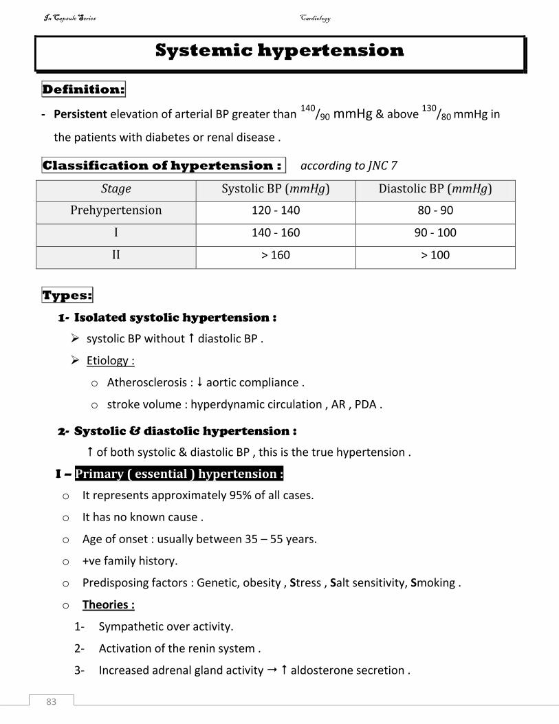
In Capsule Series Cardiology
83
Systemic hypertension
Definition:
- Persistent elevation of arterial BP greater than 140/90 mmHg & above
130/80 mmHg in
the patients with diabetes or renal disease .
Classification of hypertension : according to JNC 7
Stage Systolic BP (mmHg) Diastolic BP (mmHg)
Prehypertension 120 - 140 80 - 90
I 140 - 160 90 - 100
II > 160 > 100
Types:
1- Isolated systolic hypertension :
systolic BP without diastolic BP .
Etiology :
o Atherosclerosis : aortic compliance .
o stroke volume : hyperdynamic circulation , AR , PDA .
2- Systolic & diastolic hypertension :
of both systolic & diastolic BP , this is the true hypertension .
I – Primary ( essential ) hypertension :
o It represents approximately 95% of all cases.
o It has no known cause .
o Age of onset : usually between 35 – 55 years.
o +ve family history.
o Predisposing factors : Genetic, obesity , Stress , Salt sensitivity, Smoking .
o Theories :
1- Sympathetic over activity.
2- Activation of the renin system .
3- Increased adrenal gland activity aldosterone secretion .
In Capsule Series Cardiology
84
4- Multifactorial theory :
Stress sympathetic renal ischemia rennin aldosterone BP .
5- Hyperinsulinemia due to peripheral insulin resistance .
6- Decreased atrial natriuretic peptide ( ANP )
7- Barroreceptors resetting .
II – Secondary hypertension : ( curable hypertension )
o Hypertension with a known underlying cause .
o It represents approximately 5% of all cases .
o Secondary hypertension is suspected when the patient has any of the following :
a. Age of onset : before 25 or after 55 years .
b. -ve family history .
c. Rapidly progressive hypertension with early complications .
1- Renal :
i – Parenchymal : ( volume dependent hypertension )
GN , diabetic nephropathy , pyelonephritis , polycystic kidney , ………………..
Mechanism :
- Ineffective in disposing Na .
- Fail to produce necessary VD substances (PG ).
ii – Renovascular : renal artery stenosis which by turn activate the renin system.
C/P : Generalized atherosclerosi & flank bruits.
2- Endocrinal :
o Pituitary : Acromegaly ( endothelial hyperplasia , Na & water retention )
o Thyroid : - Hypothyroidism.
- Hyperthyroidism isolated systolic hypertension .
o Parathyroid : Hyperparathyroidism .
o DM .
o SRG : - Conn’s syndrome : never sever HTN ,muscle weakness &hypokalemia
- Cushing syndrome . - Pheochromocytoma : paroxysmal HTN.
In Capsule Series Cardiology
85
3- CNS :
o ICT .
o Lesions of the medulla .
4- Vascular :
o Polyarteritis nodosa .
o Polycythemia .
o Coarctation of the aorta .
5- Iatrogenic :
o Contraceptive pills .
o Cortisone .
o Catecholamine .
o Calcium .
Clinical picture:
Symptoms :
1- Asymptomatic in most cases .
2- May discovered accidentally .
3- Headache after information .
4- Headache is usually occipital .
5- Blurring of vision , tinnitus , epistaxis , nausea & vomiting .
6- Complications of HTN may be the first presentation .
Signs :
Blood pressure : persistent elevation > 140/90 mmHg .
Signs of left ventricular hypertrophy .
Auscultation :
o Accentuated S2 .
o S4 .
o Closed splitting S2 .
o Ejection click .
o Ejection systolic murmur : due to relative AS .
o Early diastolic murmur : due to relative AR .
In Capsule Series Cardiology
86
Hypertensive urgency:
Rapid rise of BP > 220/120 mmHg & not associated with target organ damage e.g.
renal failure , heart failure .
Hypertensive emergency:
Rapid rise of BP > 220/120 mmHg & associated with target organ damage .
Malignant HTN : Hypertensive emergency with development of papilloedema .
Accelerated HTN : Similar to malignant HTN without papilloedema .
Complications :
1- Cardiac :
o LSHF : due to pressure overload .
o Ischemic heart disease : due to atherosclerosis & hypertrophy .
o Bernheim effect : ( signs of RSHF )
Hypertrophy oh LV may cause bulging of the septum in th RV leading to
slight impairment of the filling of RV signs of RSHF .
2- Cerebral :
o Cerebral atherosclerosis .
o Cerebral ischemia & thrombosis ( infarction )
o Cerebral hemorrhage ( stroke )
o Hypertensive encephalopathy :
As a result of acute rise of BP , the cerebral blood vessels are no longer able
to maintain the necessary degree of constriction ( failure of auto regulation )
& they begin to dilate cerebral blood flow ICT , brain edema , coma &
convulsion may occur .
NB : How to differentiate between stroke & hypertensive encephalopathy ?
Stroke : Signs of lateralization ( unilateral )
Hypertensive encephalopathy : No signs of lateralization ( bilateral )
In Capsule Series Cardiology
87
3- Renal :
o Renal failure .
o Hematuria & proteinuria .
4- Retinal : 4 grades
o Grade I : Thickening of retinal arterioles ( silver wire appearance ).
o Grade II : Kinking of retinal veins .
o Grade III : Hemorrhage & exudates .
o Grade IV : Papilloedema .
5- Vascular :
o Atherosclerosis .
o Aortic dissection .
Investigations :
1- Investigations for complications :
o Cardiac : X ray , ECG , Echo , ….
o Cerebral : CT, MRI brain .
o Renal : urine analysis , renal function , renal imaging .
2- Investigations for the cause : when secondary HTN is suspected or in a case of
refractory hypertension .
Treatment :
The target BP is lower than 140
/90 mmHg , unless the patient has diabetes or renal
disease, in which case the target would be lower than 130
/80 mmHg.
A) Lines of treatment :
I – Non pharmacological ( lifestyle modification ) .
II – Pharmacological :
Treatment of associated risk factors e.g. hyperlipidemia
Treatment of the cause : in a case of secondary hypertension .
Antihypertensive drugs
B) Choice of treatment.
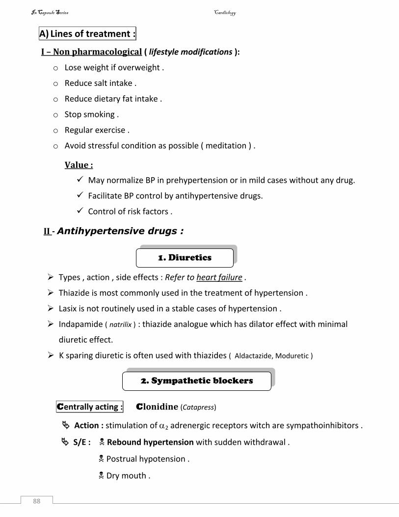
In Capsule Series Cardiology
88
A) Lines of treatment :
I – Non pharmacological ( lifestyle modifications ):
o Lose weight if overweight .
o Reduce salt intake .
o Reduce dietary fat intake .
o Stop smoking .
o Regular exercise .
o Avoid stressful condition as possible ( meditation ) .
Value :
May normalize BP in prehypertension or in mild cases without any drug.
Facilitate BP control by antihypertensive drugs.
Control of risk factors .
II - Antihypertensive drugs :
Types , action , side effects : Refer to heart failure .
Thiazide is most commonly used in the treatment of hypertension .
Lasix is not routinely used in a stable cases of hypertension .
Indapamide ( natrilix ) : thiazide analogue which has dilator effect with minimal
diuretic effect.
K sparing diuretic is often used with thiazides ( Aldactazide, Moduretic )
Centrally acting : Clonidine (Catapress)
Action : stimulation of 2 adrenergic receptors witch are sympathoinhibitors .
S/E : Rebound hypertension with sudden withdrawal .
Postrual hypotension .
Dry mouth .
1. Diuretics
2. Sympathetic blockers
In Capsule Series Cardiology
89
Ganglion blockers : Trimethaphan .
Nerve ending blockers :
i. methyl dopa ( Aldomet ) :
Action : synthesis of catecholamines ,also has central inhibiting action .
S/E : Postural hypotension , Hepatitis , Hemolysis . ( 3 H )
ii. Reserpine ( Brinerdin ) : Reuptake of catecholamines ( stores )
S/E : Depression , Nasal congestion , hypertonia .
blockers : Prazosin ( minipress )
Action : vasodilatation .
S/E : First dose syncope , tachycardia .
blockers :
Mechanism of action :
Is still questionable .
contractility , HR COP .
renin release .
Preparation :
Propranolol ( indral ) : non selective β blocker .
Atenolol (ateno) , Metoprolol (betaloc) , Bisoprolol (concor) : Selective β1 blockers.
Carvedilol (cardilol) ,Labetalol : Combined β & blockers ( blockers with vasodilation)
Side effects :
Lung : Bronchospasm.
Heart : Bradycardia , Heart block.
Depression , Impotence.
CVS uses of blockers : Hypertension Angina Heart failure
Arrhythmia F4 Mitral valve prolapse.
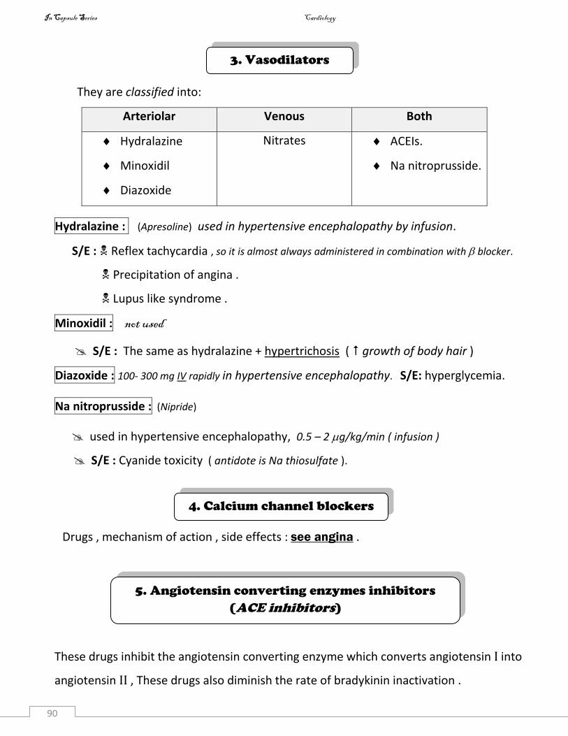
In Capsule Series Cardiology
90
They are classified into:
Arteriolar Venous Both
Hydralazine
Minoxidil
Diazoxide
Nitrates ACEIs.
Na nitroprusside.
Hydralazine : (Apresoline) used in hypertensive encephalopathy by infusion.
S/E : Reflex tachycardia , so it is almost always administered in combination with blocker.
Precipitation of angina .
Lupus like syndrome .
Minoxidil : not used
S/E : The same as hydralazine + hypertrichosis ( growth of body hair )
Diazoxide : 100- 300 mg IV rapidly in hypertensive encephalopathy. S/E: hyperglycemia.
Na nitroprusside : (Nipride)
used in hypertensive encephalopathy, 0.5 – 2 g/kg/min ( infusion )
S/E : Cyanide toxicity ( antidote is Na thiosulfate ).
Drugs , mechanism of action , side effects : see angina .
These drugs inhibit the angiotensin converting enzyme which converts angiotensin I into
angiotensin II , These drugs also diminish the rate of bradykinin inactivation .
3. Vasodilators
4. Calcium channel blockers
5. Angiotensin converting enzymes inhibitors
(ACE inhibitors)
In Capsule Series Cardiology
91
Short acting : Captopril ( capoten ) : ½ - 2 tablet t.d.s. ( tab = 25 mg )
Long acting : 1 tab / day Enalapril ( Ezapril ) , Lisinopril ( Zestril ) , Ramipril ( Tritace ).
S/E : Dry cough . Hyperkalemia . Skin rash first dose phenomenon.
Losartan (CozAAr )
Valsartan ( Tareg )
S/E : Similar to ACE inhibitors but no cough .
B) Choice of treatment :
Non pharmacological measures ( lifestyle modification) should be initiated
in all hypertensive patients & those with prehypertension .
The selection of a specific antihypertensive drug should take into
consideration comorbid conditions associated with hypertension as well as
the patient’s personal, response & financial .
1- Uncomplicated hypertension : Stepped antihypertensive therapy
The treatment passes in steps & if there is not an adequate response go to the
next step .
VD .
Decreased angiotensin II
secretion of aldosterone retension of Na.
Decreased bradykinin inactivation bradykinin which is vasodilator .
6. Angiotensin II receptor blockers
(ARBs)
In Capsule Series Cardiology
92
Step 1 : Initiate therapy with one of the following :
ACE inhibitors or blockers or Ca channel blockers or Diuretics .
Step 2 : Combination of 2 drugs of step 1 ( include a diuretic ) .
Step 3 : Combination of 3 drugs of step 1 ( include a diuretic ) .
Step 4 : Add blocker or hydralazine …..to step 3 .
NB : The use of lower doses of 2 or more drugs may lower BP with fewer adverse effects
than the use of higher dose of a single agent .
2- Hypertensive crisis:
The aim of treatment is to lower the BP rapidly to terminate ongoing target
organ damage (TOD ).
It is unwise to lower the BP too quickly as it may lead to organ hypoperfusion .
Avoid initial reduction in BP more than 25 % & remember that the patients
with chronic hypertension may not tolerate a normal BP so , be judicious
when lowering the BP .
i. Rapid acting antihypertensive drugs :
Na nitroprusside ( Nipride ) : 0.5 – 2 g/kg/min ( infusion ) .
Nitroglycerine : 10 – 100 g/kg/min.
Diazoxide : 100 – 300 mg rapidly IV .
Hydralazine : 20 mg IV .
Labetalol : 20 mg IV every 10 minutes until control of BP ( maximum 200 mg)
Fenoldopam : is a new dopamine receptor agonist .
Lasix may be used with one of the above agents .
ii. Specific treatment :
a) Hypertensive encephalopathy : add
Anticonvulsant : Diazepam IV .
Cerebral dehydrating measures : 25 % Mannitol infusion with lasix .
b) Treatment of the target organ damage ( TOD ) :
Cerebral stroke , acute LSHF & renal failure .
In Capsule Series Cardiology
93
3- Tretment of hypertension with certain concomitant diseases :
Hypertension with heart failure
Use : ACE inhibitors , Diuretics . Avoid : Ca channel blockers
Hypertension with ischemic heart disease
Use: blocker or Ca channel blocker . Avoid : Hydralazine .
Hypertension with DM
Use: ACE inhibitors , Ca channel blockers .
Avoid : blockers ( masking of warning signs of hypoglycemic coma ).
Hypertension with renal impairment
Use : blockers , Ca channel blockers , Diuretics (Lasix) , methyl dopa & ACE
inhibitors but with monitoring of cretinin level .
Avoid : ACE inhibitors in bilateral renal stenosis .
Hypertension with Asthma or COPD
Avoid blockers .
Hypertension with pregnancy
Use : methyl dopa , Ca channel blocker , Hydralazine or Labetalol .
Avoid : ACE inhibitors , blockers .Diuretics .
Hypertension with hyperthyroidism
Use : blocker .
Hypertension with peripheral vascular disease
Use : Ca channel blockers , blocker . Avoid : blockers .
NB : Hypokalemic hypertension :
1- Conn’s syndrome . 2- Cushing syndrome .
3- Renal artery stenosis . 4- Iatrogenic : Diuretics .
Acute complications of hypertension :
- CNS : H encephalopathy , SAH , stroke.
- CVS : Acute pulmonary edema, Dissecting aorta, IHD
- ARF. - Epistaxis.
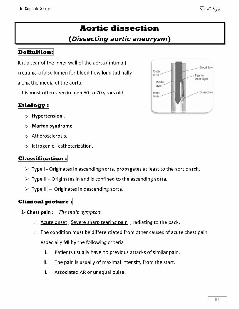
In Capsule Series Cardiology
94
Aortic dissection
(Dissecting aortic aneurysm)
Definition:
It is a tear of the inner wall of the aorta ( intima ) ,
creating a false lumen for blood flow longitudinally
along the media of the aorta.
- It is most often seen in men 50 to 70 years old.
Etiology :
o Hypertension .
o Marfan syndrome.
o Atherosclerosis.
o Iatrogenic : catheterization.
Classification :
Type I - Originates in ascending aorta, propagates at least to the aortic arch.
Type II – Originates in and is confined to the ascending aorta.
Type III – Originates in descending aorta.
Clinical picture :
1- Chest pain : The main symptom
o Acute onset , Severe sharp tearing pain , radiating to the back.
o The condition must be differentiated from other causes of acute chest pain
especially MI by the following criteria :
i. Patients usually have no previous attacks of similar pain.
ii. The pain is usually of maximal intensity from the start.
iii. Associated AR or unequal pulse.
In Capsule Series Cardiology
95
The use of anticoagulants or thrombolytic therapy in a patient with aortic dissection
in one word is fatal.
2- Hypertension in most cases. Hypotension may suggests aortic rupture .
3- AR in more than 50 % of cases.
4- Obstruction of the opening of aortic branches: depends on which arteries are blocked.
o Coronary artery Coronary insufficiency .
o Subclavian artery Unequal pulse.
o Ischemic symptoms of the brain Stroke , Syncope.
o Ischemic symptoms of the spinal cord paraplegia.
o Mesenteric arteries abdominal pain.
o Renal artery renal failure.
5- Rupture aneurysm into pericardium Cardiac tamponade.
6- Sudden death : 40 % of cases.
Investigations :
o X ray :Mediastinal widening.
o ECG : to exclude other causes of acute chest pain e.g. MI.
o Trans Esophageal Echo (TEE) , Aortography , CT : diagnostic.
o MRI : gold standard for final diagnosis.
Treatment :
1- Control of hypertension : blocker ( Esmolol ) with Nitroprusside.
2- Surgical correction :
o Acute dissection in ascending aorta is a surgical emergency.
o Prognosis without surgery is very poor.
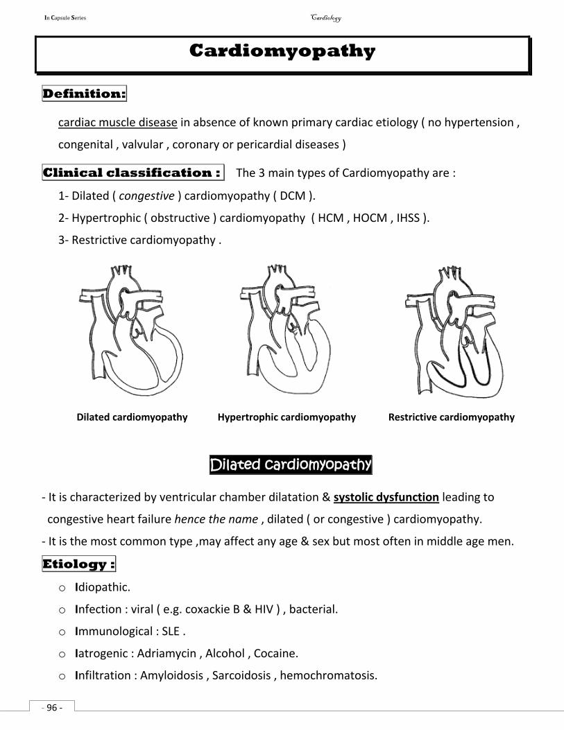
In Capsule Series Cardiology
- 96 -
Cardiomyopathy
Definition:
cardiac muscle disease in absence of known primary cardiac etiology ( no hypertension ,
congenital , valvular , coronary or pericardial diseases )
Clinical classification : The 3 main types of Cardiomyopathy are :
1- Dilated ( congestive ) cardiomyopathy ( DCM ).
2- Hypertrophic ( obstructive ) cardiomyopathy ( HCM , HOCM , IHSS ).
3- Restrictive cardiomyopathy .
Dilated cardiomyopathy Hypertrophic cardiomyopathy Restrictive cardiomyopathy
Dilated cardiomyopathy
- It is characterized by ventricular chamber dilatation & systolic dysfunction leading to
congestive heart failure hence the name , dilated ( or congestive ) cardiomyopathy.
- It is the most common type ,may affect any age & sex but most often in middle age men.
Etiology :
o Idiopathic.
o Infection : viral ( e.g. coxackie B & HIV ) , bacterial.
o Immunological : SLE .
o Iatrogenic : Adriamycin , Alcohol , Cocaine.
o Infiltration : Amyloidosis , Sarcoidosis , hemochromatosis.
In Capsule Series Cardiology
- 97 -
o Endocinal : DM , Acromegaly .
o Nurological : Freidreich’s ataxia , Duchenne myopathy , myotonia atrophica . MCQ
Clinical picture :
o Congestive heart failure.( LSHF , RSHF )
o Arrhythmia.
o Thromboembolism.
Investigations :
o X ray , ECG , Echo : show dilated heart.
o Biopsy : for amyloidosis or sarcoidosis.
Treatment :
o Treatment of HF : ACEI , blocker , Diuretic & heart transplant in refractory cases.
o Treatment of arrhythmia .
o Treatment of thromboembolism : anticoagulant.
Hypertrophic cardiomyopathy
- Asymmetrical ventricular hypertrophy especially in the interventricular septum & LV.
- This thickening of myocardium results in diastolic dysfunction due to myocardial stiffness
- Also , it may result in an obstruction in the LV outflow tract subvalvular AS .
- Hypertrophic cardiomyopathy is considered to be the most common cause of sudden
death in young athletes. MCQ
Etiology : Autosomal dominant disease
o Idiopathic Hypertrophic Sub-aortic Stenosis ( IHSS )
o Associated with Freidreich’s ataxia.
Clinical picture :
o Diastolic dysfunction : Pulmonary congestion & LCOP.
o Manifestations of AS : Angina , Syncope.
o Sudden death : usually associated with sports most probably due to ventricular
arrhythmias.
In Capsule Series Cardiology
- 98 -
General signs :
o Giant (a) wave if there is right ventricular outflow tract obstruction.
o Jerky ( bifid ) carotid pulse.
Cardiac examination :
o Double apical impulse : due to palpable atrial systole ( S4 ).
o Paradoxical splitting of S2
o No click as obstruction is subvalvular and not valvular.
o Left parasternal late ejection systolic murmur.
o Associated MR is common : apical pansystolic murmur.
o The murmur of HCM , contrary to valvular AS , decreases by leg raising or squatting
because of venous return which fills the ventricle and so decrease obstruction.
Investigations :
o X ray : No cardiomegaly , pulmonary congestion.
o ECG : Left ventricular hypertrophy , deep wide Q wave , arrhythmias.
o Echo : Ventricular hypertrophy , mitral systolic anterior motion ( SAM )
Treatment :
o Amiodarone for arrhythmias.
o blockers : contractility & outflow obstruction.
o Ca Channel blockers : contractility & outflow obstruction.
o Avoid digitalis & other inotropics outflow obstruction .
o Surgical resection of hypertrophied septum.
Restrictive cardiomyopathy
- It is characterized by restrictive diastolic ventricular filling with normal systolic function.
- It is the least common form of cardiomyopathy.
Etiology :
o Idiopathic endomyocardial fibrosis.
o Infiltration : Amyloidosis , Sarcoidosis , Hemochromatosis. ♫♫
In Capsule Series Cardiology
- 99 -
Clinical picture :
- Similar to constrictive pericarditis ( IF RV is involved )
o Systemic congestion.
o LCOP.
o AF.
- Pulmonary congestion if LV is involved.
Cardiac signs :
o Signs of RVE : Late & mild.
o Murmur of mitral & tricuspid regurge : due to involvement of the papillary muscles
by fibrosis.
o No pericardial knock.
Investigations :
o X ray : No or mild RVE , no calcification ( to differentiate it from constrictive
pericarditis )
o ECG : Low voltage.
o Echo : Diastolic dysfunction.
o Endomyocardial biopsy : Diagnostic.
Treatment :
o Treatment of the cause e.g. Hemochromatosis.
o Symptomatic treatment : HF , Arrhythmias , Thromboembolism.
o Cardiac transplant in refractory cases.
Medicine cannot, except over a short period, increase the population of the world.
Bertrand Russell
In Capsule Series Cardiology
- 100 -
Pulmonary hypertension
Definition:
Elevation of the pulmonary arterial pressure above 35
/17 mmHg.
Etiology :
Primary pulmonary hypertension (PPH) :
Very rare ( represents less than 1 % of all cases of pulmonary hypertension ).
Etiology is unknown , occurs more commonly in middle aged female with repeated
pregnancies.
May be due to repeated thromboembolism , pulmonary vasculitis.
Secondary pulmonary hypertension :
1- Hyperdynamic pulmonary hypertension :
Due to increased pulmonary arterial blood flow :
Hyperdynamic circulation.
Congenital heart diseases : ASD , VSD , PDA .
2- Passive pulmonary hypertension :
Due to pulmonary congestion : Mitral valve disease . LSHF .
3- Vasoconstrictive ( reactive ) Pulmonary hypertension :
Due to reflex VC of pulmonary arteriole in response to :
Long standing pulmonary congestion.
Long standing hyperdynamic pulmonary hypertension .
Hypoxia : COPD .
4- Obstructive pulmonary hypertension :
Long standing vasoconstrictive pulmonary hypertension Irreversible VC of
pulmonary arterioles.
5- Acute obstructive pulmonary hypertension :
Massive pulmonary embolism .
Clinical picture :
1- C/P of the cause .
2- Right ventricular enlargement .
3- RSHF .
4- Signs of pulmonary hypertension :
General : Giant “a” wave . ( no giant “a” wave in ASD ?? )
In Capsule Series Cardiology
- 101 -
Cardiac :
Inspection : Pulsation in the 2nd left intercostals space .
Palpation : Diastolic shock ( palpable accentuated pulmonary component of S2 )
Percussion : Dullness in the 2nd left intercostals space .
Auscultation :
o Accentuated S2 .
o Wide splitting S2 .
o S4 .
o Ejection click .
o Ejection systolic murmur : due to relative PS .
o Early diastolic murmur : due to relative PR .
Investigations :
X ray :
o Dilated pulmonary artery .
o RVE .
ECG :
o Peaked P wave ( P pulmonale ) .
Echo :
o Detection of the cause .
o Estimation of the pulmonary artery pressure .
o RVE .
Catheterization :
o Detection of the cause .
o Estimation of the pulmonary artery pressure .
o RVE .
Treatment :
Treatment of the cause .
Treatment of RSHF .
Drugs : ( limited benefit )
o ACE inhibitors , Anticoagulants .
o PGI2 . ( potent systemic & pulmonary vasodilator )
o Ca channel blockers .
o Diuretics .
Heart lung transplantation : is the chief therapeutic option .
In Capsule Series Cardiology
102
Pulmonary embolism
Etiology : ( source of emboli )
1- Deep venous thrombosis ( DVT ) : Commonest cause ( 90 % )
Predisposing factors of DVT : ( Virchow triad )
i. Slow circulation. ii. Injury of endothelium. iii. Hypercoagulability.
Risk factors for DVT : 6 “ O "
Operative : especially pelvic & bone surgery .
Oncology : Cancer stomach , leukemia & lymphoma .
Old age .
Oral contraceptive pills Obesity.
Others : 2 “C " - Congestive heart failure
- Coagulation disorders : protein C & S deficiency .
Sites of DVT : Deep veins of the leg particularly the iliac, femoral & popliteal veins .
2- Detached thrombi from right side of the heart :
Infective endocarditis , Atrial fibrillation , Myocardial infarction.
3- Paradoxical embolism :from the left side of the heart if there is left to right shunt:VSD
4- Rare : Fat embolism , Air embolism , Amniotic fluid embolism .
Clinical picture :
Unfortunately, there are no clinical or laboratory findings that will confirm or
exclude the diagnosis of pulmonary embolism .
The clinical picture of pulmonary embolism is not specific , so successful
management of pulmonary embolism requires a combination of clinical
suspicion & appropriate use of diagnostic tools .
The clinical presentations are ranging from asymptomatic to sudden death
depending on the size of the embolus .
In Capsule Series Cardiology
103
1- Small sized emboli :
Recurrent small pulmonary emboli results in occlusion of large number of
pulmonary arterioles.
Usually asymptomatic , but non specific symptoms of tachypnea , dyspnea,
tachycardia or pleuritic chest pain may occur .
May result in the development of pulmonary hypertension & subacute
corpulmonale over years .
2- Medium sized emboli : ( pulmonary infarction )
If occlusion involves less than 60% of the vascular bed .
C/P :
Pleuritic chest pain.
Cough & hemoptysis.
Dyspnea .
3- Acute massive pulmonary embolism : occlusion > 60% of the vascular bed
i. Acute dyspnea .
ii. Acute chest pain : ( similar to angina pain ) due to :
o Distension of the pulmonary artery .
o Reflex coronary spasm .
iii. Sudden appearance of pulmonary hypertension .
iv. Acute right sided heart failure . ( see HF )
v. Shock : It is an obstructive shock in which there is marked of blood flow to
the lung blood flow to left atrium COP shock .
vi. Sudden death : may be the first manifestation if occlusion involves more
than 80% of the vascular bed .
vii. Finding suggestive for DVT : Tenderness , swelling , redness of the LL .
unfortunately, DVT of the leg is often clinically silent .
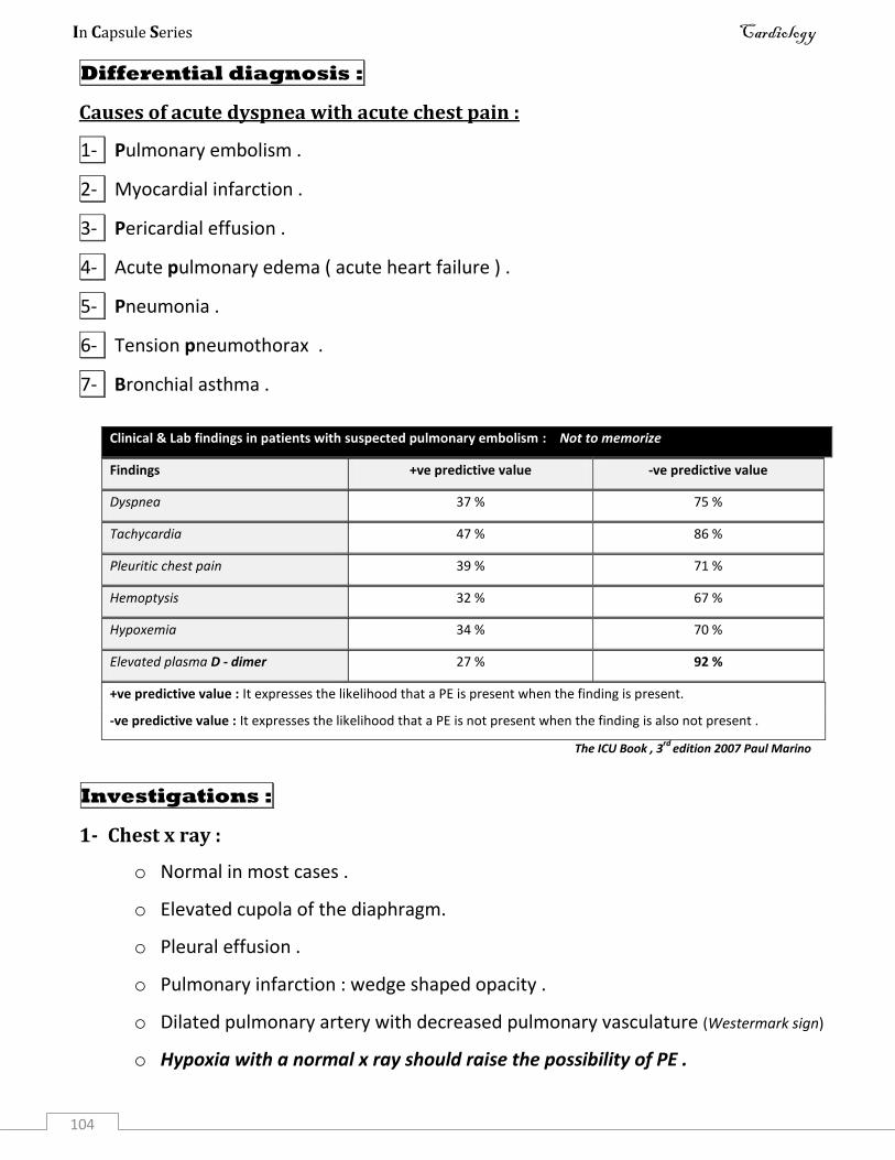
In Capsule Series Cardiology
104
Differential diagnosis :
Causes of acute dyspnea with acute chest pain :
1- Pulmonary embolism .
2- Myocardial infarction .
3- Pericardial effusion .
4- Acute pulmonary edema ( acute heart failure ) .
5- Pneumonia .
6- Tension pneumothorax .
7- Bronchial asthma .
Clinical & Lab findings in patients with suspected pulmonary embolism : Not to memorize
Findings +ve predictive value -ve predictive value
Dyspnea 37 % 75 %
Tachycardia 47 % 86 %
Pleuritic chest pain 39 % 71 %
Hemoptysis 32 % 67 %
Hypoxemia 34 % 70 %
Elevated plasma D - dimer 27 % 92 %
+ve predictive value : It expresses the likelihood that a PE is present when the finding is present.
-ve predictive value : It expresses the likelihood that a PE is not present when the finding is also not present .
The ICU Book , 3rd
edition 2007 Paul Marino
Investigations :
1- Chest x ray :
o Normal in most cases .
o Elevated cupola of the diaphragm.
o Pleural effusion .
o Pulmonary infarction : wedge shaped opacity .
o Dilated pulmonary artery with decreased pulmonary vasculature (Westermark sign)
o Hypoxia with a normal x ray should raise the possibility of PE .
In Capsule Series Cardiology
105
2- ECG :
o May be normal .
o Sinus tachycardia & atrial fibrillation .
o S1 Q3 T3 pattern ( S wave in lead I , Q wave in lead III & an inverted T in lead III )
o The most important value is to exclude MI as a cause of dyspnea & chest pain.
3- Echo : Enlargement of RA , RV & pulmonary artery .
4- Pulmonary angiography : is the gold standard for making a diagnosis .
5- Spiral CT angiography : less invasive than standard angiography .
6- Ventilation – perfusion scan : Areas of normal ventilation & hypoperfusion .
7- Laboratory :
o Arterial blood gases : hypoxia , hypocapnia .
o Elevated serum LDH .
o D – Dimer : (an end product of fibrin digestion )
It is a good –ve test : if D-dimer is not elevated , PE is unlikely .
8- Investigation for diagnosis of DVT : Duplex ultrasound .
Treatment of pulmonary embolism & DVT :
I- Prophylaxis :
1. Early post operative ambulation .
2. Elastic stocking .
3. Elevation of the leg .
4. Anticoagulants in high risk patients : for about one week
Low dose heparin 5000 units SC / 8-12 hr .
Low Molecular Weight heparin ( enoxaparine : Clexane) 20-40mg/d.
II- Curative :
1- Hospitalization in CCU .
In Capsule Series Cardiology
106
2- Anticoagulants :
Heparin :
Mechanism of action : activation of antithrombin III .
5000 – 10000 units IV as a loading dose then ,1000 units /h (infusion)
Duration : for about one week or till clinical improvement .
Follow up: by estimation of PTT, should be maintained twice the normal
Antidote : protamine sulphate IV .
LMW heparin : 1mg/kg SC/12 h. No need for routine anticoagulant monitoring.
Oral anticoagulant : warfarin ( Marevan )
Mechanism of action : Competes with vitamin K formation of
coagulation factors II , VII , IX & X .
Dose : 2-10 mg/d . It is determined according to the PT .
Because of the delayed onset of action , it is started 3 days before
discontinuation of heparin & is continued for at least 3 months & for life
in recurrent cases .
Warfarin should never be used in pregnancy .
3- Thrombolytic therapy : Streptokinase , tPA
Indicated in life threatening massive pulmonary embolism when the patient
is hemodynamically unstable ( RSHF or shock )
4- Pulmonary embolectomy :
Is used for those with massive PE when thrombolysis is contraindicated.
5- Symptomatic treatment :
Pain : Analgesics e.g. pethidine ( avoid morphine )
Dyspnea : Oxygen inhalation & mechanical ventilation in severe cases .
6- Treatment of complications : - Shock - RSHF .
In Capsule Series Cardiology
107
7- Inferior vena caval filters : e.g. Greenfield filter
Meshlike filter can be placed in the inferior vena cava to trap emboli &
prevent them from traveling to the lungs .
Should be considered when anticoagulants are contraindicated .
Cor pulmonale
Definition :
Cor pulmonale is right ventricular enlargement with or without failure resulting from
pulmonary disease after exclusion of LSHF & congenital heart diseases .
Pulmonary disease Pulmonary HTN RV enlargement RSHF
Etiology :
1- Acute cor pulmonale :
o Acute massive pulmonary embolism .
o Acute massive lung collapse .
o Tension pneumothorax .
2- Subacute cor pulmonale :
o Recurrent small pulmonary embolization.
o Lumphangitis carcinomatosis of the lung .
3- Chronic cor pulmonale :
o Parenchymal lung diseases : COPD , IPF .
o Pulmonary vascular diseases : Bilharziasis , primary pulmonary hypertension .
o Thoracic wall diseases : kyphoscoliosis , Pickwickian syndrome .
Clinical picture , investigations , treatment : 4
i. Of the cause : Bilhrziasis : see GIT book p 50 , COPD : see chest .
ii. Of pulmonary hypertension .
iii. Of RV enlargement : may be masked by emphysema in a case of COPD .
iv. Of RSHF .
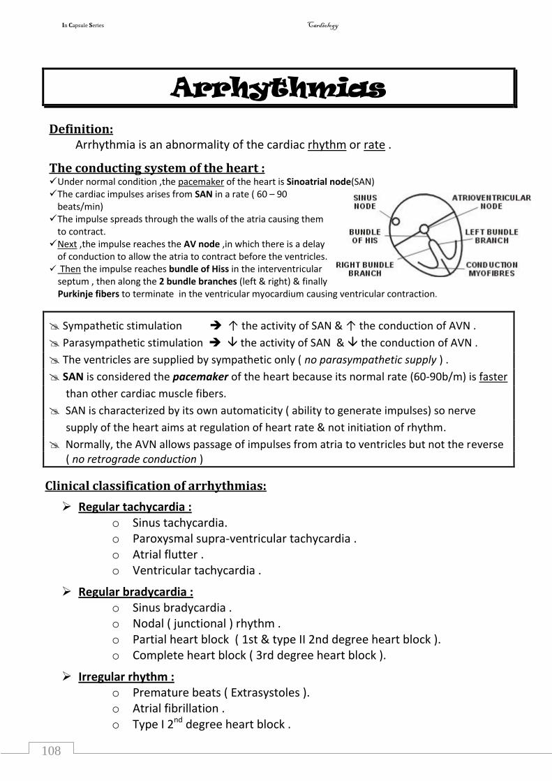
In Capsule Series Cardiology
108
Arrhythmias
Definition: Arrhythmia is an abnormality of the cardiac rhythm or rate .
The conducting system of the heart : Under normal condition ,the pacemaker of the heart is Sinoatrial node(SAN) The cardiac impulses arises from SAN in a rate ( 60 – 90
beats/min) The impulse spreads through the walls of the atria causing them
to contract. Next ,the impulse reaches the AV node ,in which there is a delay
of conduction to allow the atria to contract before the ventricles. Then the impulse reaches bundle of Hiss in the interventricular
septum , then along the 2 bundle branches (left & right) & finally Purkinje fibers to terminate in the ventricular myocardium causing ventricular contraction.
Sympathetic stimulation ↑ the activity of SAN & ↑ the conduction of AVN .
Parasympathetic stimulation the activity of SAN & the conduction of AVN .
The ventricles are supplied by sympathetic only ( no parasympathetic supply ) .
SAN is considered the pacemaker of the heart because its normal rate (60-90b/m) is faster
than other cardiac muscle fibers.
SAN is characterized by its own automaticity ( ability to generate impulses) so nerve
supply of the heart aims at regulation of heart rate & not initiation of rhythm.
Normally, the AVN allows passage of impulses from atria to ventricles but not the reverse ( no retrograde conduction )
Clinical classification of arrhythmias:
Regular tachycardia : o Sinus tachycardia. o Paroxysmal supra-ventricular tachycardia . o Atrial flutter . o Ventricular tachycardia .
Regular bradycardia : o Sinus bradycardia . o Nodal ( junctional ) rhythm . o Partial heart block ( 1st & type II 2nd degree heart block ). o Complete heart block ( 3rd degree heart block ).
Irregular rhythm : o Premature beats ( Extrasystoles ). o Atrial fibrillation . o Type I 2nd degree heart block .
In Capsule Series Cardiology
- 109
-
Arrhythmia scheme
I- Etiology of any arrhythmia : 7
Tachyarrhythmia Bradyarrhythmia
1- Myocarditis.
2- Ischemic heart disease ( Myocardial infarction ).
3- Rheumatic heart disease .
4- Congenital heart disease .
5- Digitalis.
6- Sympathomimetics .
7- Thyrotoxicosis .
6- Sympatholytics
7- Hypothyroidism .
Exceptions :
Sinus ( tachy or brady ) arrhythmias : Physiological & pathological causes .
Atrial flutter or Atrial fibrillation : begin the etiology by : MS & thyrotoxicosis.
II- Clinical picture :
Symptoms of tachyarrhythmias :
1- Asymptomatic .
2- palpitation :
o Onset :
o Offset :
o Duration of the disease : short in serious arrhythmias e.g. VT,CHB.
3- Manifestations of LCOP .
4- Precipitation of HF & angina.
5- Features of the cause e.g. : MI , Rheumatic heart disease, digitalis toxicity ……
Symptoms of bradyarrhythmias :
The same but no precipitation of angina .
In Capsule Series Cardiology
- 110
-
Exceptions :
Atrial fibrillation ( AF ) : add thromboembolism.
Ventricular tachycardia ( VT ) : add Sudden death .
Complete heart block : add Syncope , Sudden death .
Signs :
1- Radial pulse : ( test for ventricle ) 4
a) Rate : ↑ ( Uncountable pulse ) in tachy , in bradyarrhythmias .
b) Rhythm : all are regular except AF & extrasystoles.
c) Response to carotid sinus massage ( in tachy ): HR in any tachyarrhythmia
except arrhythmias that originate in the ventricle .
simply: any arrhythmia contain this word , ventricular , in its name no effect
( no parasympathetic supply )
NB : In bradyarrhythmias : Response to atropine instead .
d) Respiratory sinus arrhythmia: -ve in all arrhythmias except in both sinus tachy
& bradyarrhythmias .
Simply, it is –ve in any arrhythmia except when arrhythmia contains this word , sinus ,
it is +ve .
Respiratory sinus arrhythmia : ( HR is increased during inspiration )
- Inspiration ↑ VR ↑ of SAN ↑ HR .
- This is a physiological process indicating that the pacemaker is the SAN .
2- Neck vein : ( test for atrium )
Rapid A wave in atrial tachyarrhythmias.
Loss of A wave in atrial fibrillation.
Cannon A wave in any arrhythmia containing this word : nodal, either :
paroxysmal nodal tachycardia or nodal rhythm.
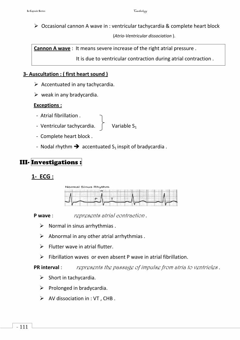
In Capsule Series Cardiology
- 111
-
Occasional cannon A wave in : ventricular tachycardia & complete heart block
(Atrio-Ventricular dissociation ).
Cannon A wave : It means severe increase of the right atrial pressure .
It is due to ventricular contraction during atrial contraction .
3- Auscultation : ( first heart sound )
Accentuated in any tachycardia.
weak in any bradycardia.
Exceptions :
- Atrial fibrillation .
- Ventricular tachycardia. Variable S1
- Complete heart block .
- Nodal rhythm accentuated S1 inspit of bradycardia .
III- Investigations :
1- ECG :
P wave : represents atrial contraction .
Normal in sinus arrhythmias .
Abnormal in any other atrial arrhythmias .
Flutter wave in atrial flutter.
Fibrillation waves or even absent P wave in atrial fibrillation.
PR interval : represents the passage of impulse from atria to ventricles .
Short in tachycardia.
Prolonged in bradycardia.
AV dissociation in : VT , CHB .
In Capsule Series Cardiology
- 112
-
QRS complex : represents the ventricular contraction .
Regular except in AF & extrasystole.
deformed ( bizarre ) : in VT & CHB .
2- Investigations for the cause :
Echo : Congenital or valvular heart diseases.
Thyroid function tests .
IV- Treatment : See later
NB : This scheme is more than enough for undergraduates
Imagination is more important than knowledge.
Albert Einstein
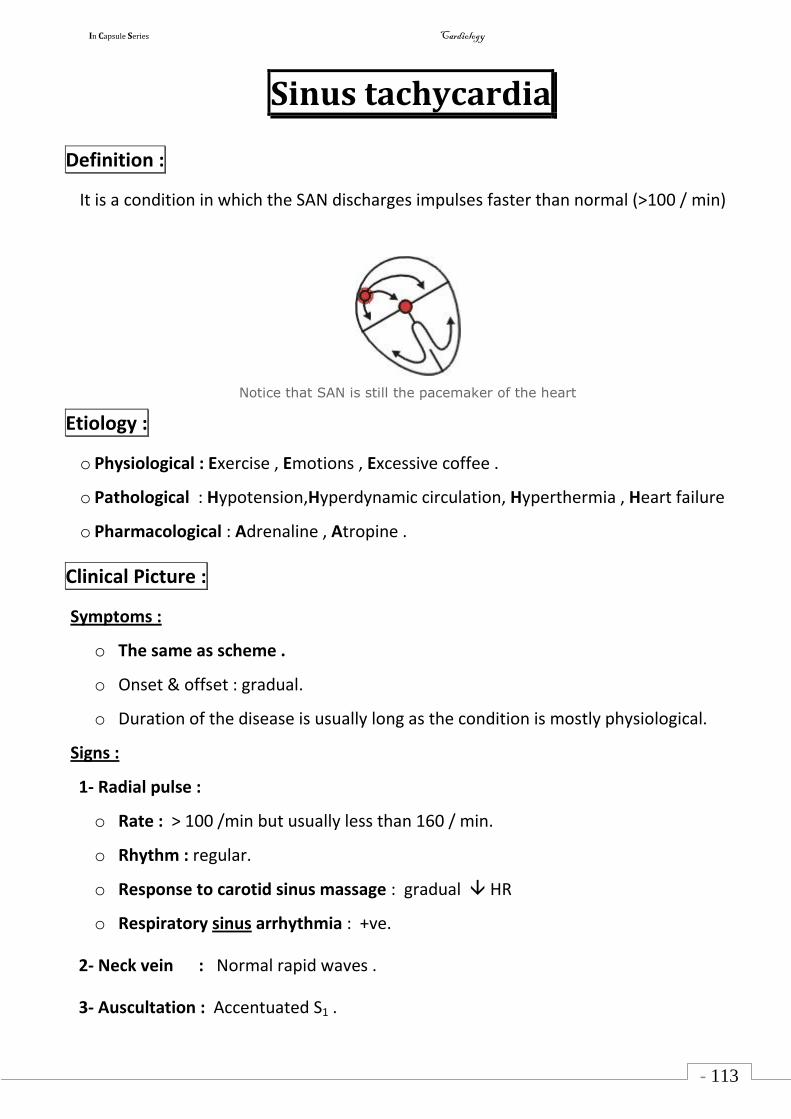
In Capsule Series Cardiology
- 113
-
Sinus tachycardia
Definition :
It is a condition in which the SAN discharges impulses faster than normal (>100 / min)
Notice that SAN is still the pacemaker of the heart
Etiology :
o Physiological : Exercise , Emotions , Excessive coffee .
o Pathological : Hypotension,Hyperdynamic circulation, Hyperthermia , Heart failure
o Pharmacological : Adrenaline , Atropine .
Clinical Picture :
Symptoms :
o The same as scheme .
o Onset & offset : gradual.
o Duration of the disease is usually long as the condition is mostly physiological.
Signs :
1- Radial pulse :
o Rate : > 100 /min but usually less than 160 / min.
o Rhythm : regular.
o Response to carotid sinus massage : gradual HR
o Respiratory sinus arrhythmia : +ve.
2- Neck vein : Normal rapid waves .
3- Auscultation : Accentuated S1 .
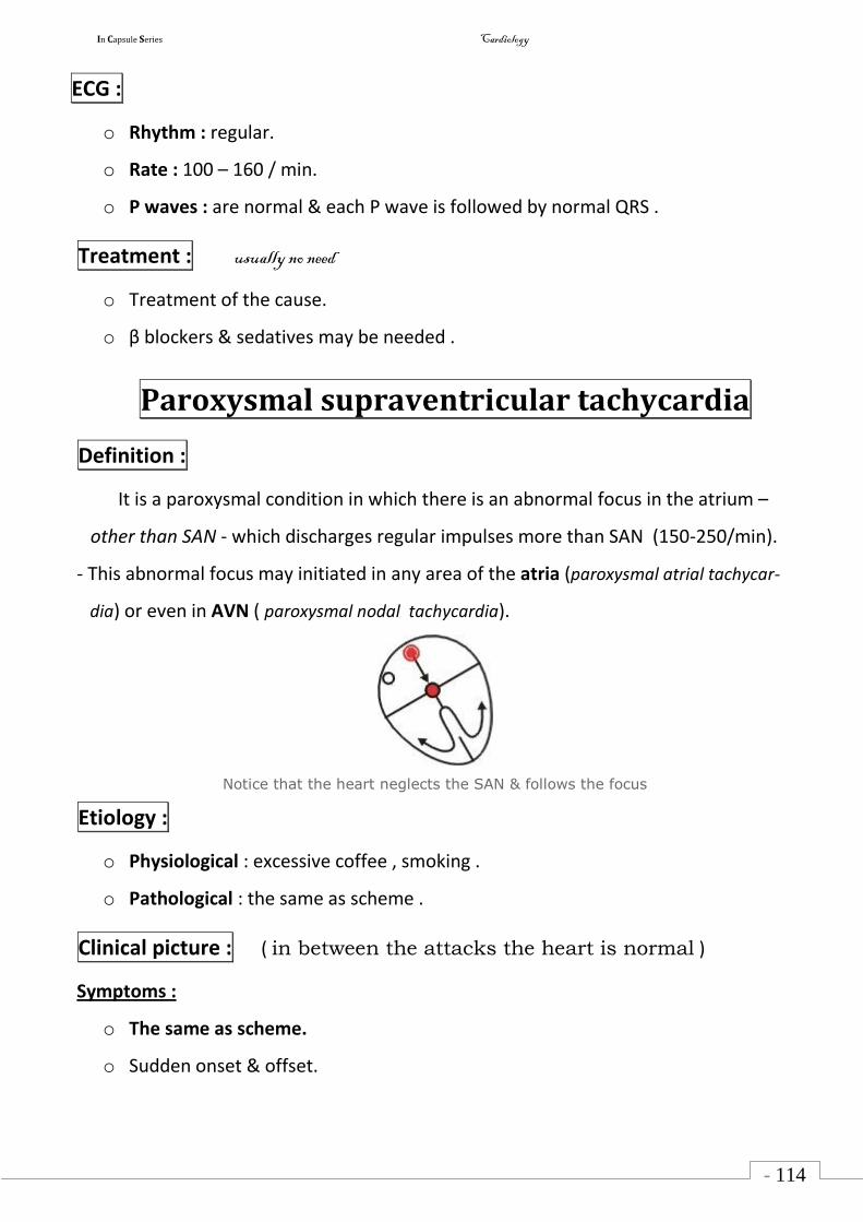
In Capsule Series Cardiology
- 114
-
ECG :
o Rhythm : regular.
o Rate : 100 – 160 / min.
o P waves : are normal & each P wave is followed by normal QRS .
Treatment : usually no need
o Treatment of the cause.
o β blockers & sedatives may be needed .
Paroxysmal supraventricular tachycardia
Definition :
It is a paroxysmal condition in which there is an abnormal focus in the atrium –
other than SAN - which discharges regular impulses more than SAN (150-250/min).
- This abnormal focus may initiated in any area of the atria (paroxysmal atrial tachycar-
dia) or even in AVN ( paroxysmal nodal tachycardia).
Notice that the heart neglects the SAN & follows the focus
Etiology :
o Physiological : excessive coffee , smoking .
o Pathological : the same as scheme .
Clinical picture : ( in between the attacks the heart is normal )
Symptoms :
o The same as scheme.
o Sudden onset & offset.
In Capsule Series Cardiology
- 115
-
o Duration of the disease:usually long history as the condition is mostly physiolog-
ical.
o Duration of the attack : Variable , usually few minutes but may lasts for hours.
NB : PSVT that lasts for more than 50 % of the day is considered a permanent PSVT.
Signs : during the attack
1- Redial pulse :
o Rate : 150 – 250 beats/min. (uncountable ).
o Rhythm : regular .
o Response to carotid massage : sudden HR .
o Respiratory sinus arrhythmia : -ve . ( SAN is not the pacemaker )
2- Neck vein :
Atrial tachycardia : Normal rapid waves .
Nodal tachycardia : Cannon A waves .
3- Auscultation : Accentuated S1 .
ECG :
o P wave : - In atrial tachycardia : deformed.
- In nodal tachycardia : absent or inverted.
o QRS : rapid , regular with normal shape.
Treatment : During the attack
1- Vagal stimulation : Carotid sinus massage or pressure on eye ball.
2- Drugs : A B C D
Adenosine , β blockers , Ca channel blockers (verapamil) , Digitalis. ( IV )
3- If there is no response or if the patient is hemodynamically unstable :
DC cardioversion.
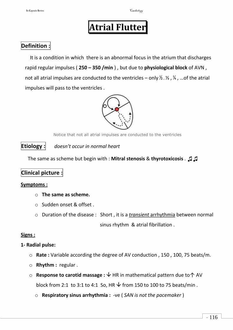
In Capsule Series Cardiology
- 116
-
Atrial Flutter
Definition :
It is a condition in which there is an abnormal focus in the atrium that discharges
rapid regular impulses ( 250 – 350 /min ) , but due to physiological block of AVN ,
not all atrial impulses are conducted to the ventricles – only ½ , ⅓ , ¼ , …of the atrial
impulses will pass to the ventricles .
Notice that not all atrial impulses are conducted to the ventricles
Etiology : doesn’t occur in normal heart
The same as scheme but begin with : Mitral stenosis & thyrotoxicosis . ♫♫
Clinical picture :
Symptoms :
o The same as scheme.
o Sudden onset & offset .
o Duration of the disease : Short , it is a transient arrhythmia between normal
sinus rhythm & atrial fibrillation .
Signs :
1- Radial pulse:
o Rate : Variable according the degree of AV conduction , 150 , 100, 75 beats/m.
o Rhythm : regular .
o Response to carotid massage : HR in mathematical pattern due to↑ AV
block from 2:1 to 3:1 to 4:1 So, HR from 150 to 100 to 75 beats/min .
o Respiratory sinus arrhythmia : -ve ( SAN is not the pacemaker )
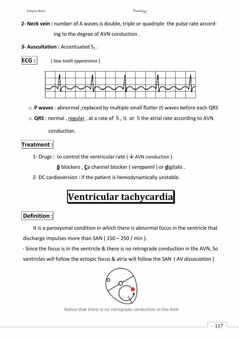
In Capsule Series Cardiology
- 117
-
2- Neck vein : number of A waves is double, triple or quadriple the pulse rate accord-
ing to the degree of AVN conduction .
3- Auscultation : Accentuated S1 .
ECG : ( Saw tooth appearance )
o P waves : abnormal ,replaced by multiple small flutter (f) waves before each QRS
o QRS : normal , regular , at a rate of ½ , ⅓ or ¼ the atrial rate according to AVN
conduction.
Treatment :
1- Drugs : to control the ventricular rate ( AVN conduction )
β blockers , Ca channel blocker ( verapamil ) or digitalis .
2- DC cardioversion : if the patient is hemodynamically unstable.
Ventricular tachycardia
Definition :
It is a paroxysmal condition in which there is abnormal focus in the ventricle that
discharge impulses more than SAN ( 150 – 250 / min ).
- Since the focus is in the ventricle & there is no retrograde conduction in the AVN, So
ventricles will follow the ectopic focus & atria will follow the SAN ( AV dissociation )
Notice that there is no retrograde conduction in the AVN
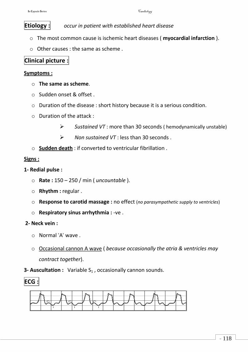
In Capsule Series Cardiology
- 118
-
Etiology : occur in patient with established heart disease
o The most common cause is ischemic heart diseases ( myocardial infarction ).
o Other causes : the same as scheme .
Clinical picture :
Symptoms :
o The same as scheme.
o Sudden onset & offset .
o Duration of the disease : short history because it is a serious condition.
o Duration of the attack :
Sustained VT : more than 30 seconds ( hemodynamically unstable)
Non sustained VT : less than 30 seconds .
o Sudden death : if converted to ventricular fibrillation .
Signs :
1- Redial pulse :
o Rate : 150 – 250 / min ( uncountable ).
o Rhythm : regular .
o Response to carotid massage : no effect (no parasympathetic supply to ventricles)
o Respiratory sinus arrhythmia : -ve .
2- Neck vein :
o Normal "A" wave .
o Occasional cannon A wave ( because occasionally the atria & ventricles may
contract together).
3- Auscultation : Variable S1 , occasionally cannon sounds.
ECG :
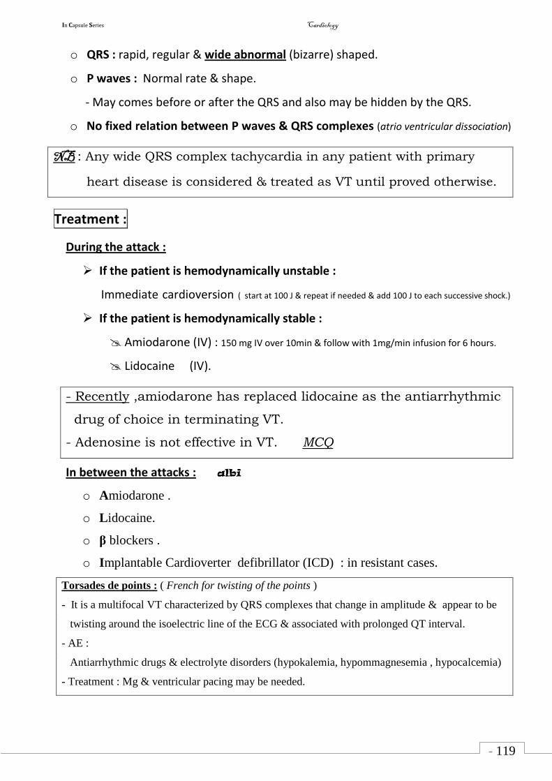
In Capsule Series Cardiology
- 119
-
o QRS : rapid, regular & wide abnormal (bizarre) shaped.
o P waves : Normal rate & shape.
- May comes before or after the QRS and also may be hidden by the QRS.
o No fixed relation between P waves & QRS complexes (atrio ventricular dissociation)
NB : Any wide QRS complex tachycardia in any patient with primary
heart disease is considered & treated as VT until proved otherwise.
Treatment :
During the attack :
If the patient is hemodynamically unstable :
Immediate cardioversion ( start at 100 J & repeat if needed & add 100 J to each successive shock.)
If the patient is hemodynamically stable :
Amiodarone (IV) : 150 mg IV over 10min & follow with 1mg/min infusion for 6 hours.
Lidocaine (IV).
- Recently ,amiodarone has replaced lidocaine as the antiarrhythmic
drug of choice in terminating VT.
- Adenosine is not effective in VT. MCQ
In between the attacks : albi
o Amiodarone .
o Lidocaine.
o β blockers .
o Implantable Cardioverter defibrillator (ICD) : in resistant cases.
Torsades de points : ( French for twisting of the points )
- It is a multifocal VT characterized by QRS complexes that change in amplitude & appear to be
twisting around the isoelectric line of the ECG & associated with prolonged QT interval.
- AE :
Antiarrhythmic drugs & electrolyte disorders (hypokalemia, hypommagnesemia , hypocalcemia)
- Treatment : Mg & ventricular pacing may be needed.
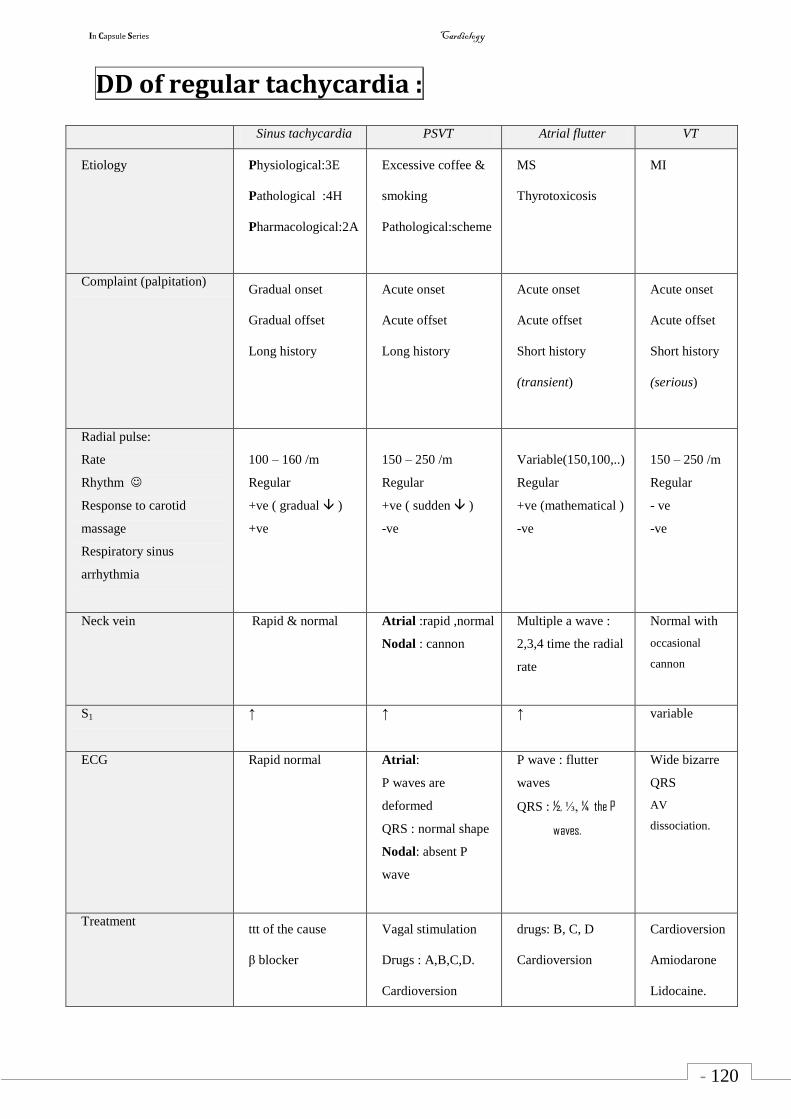
In Capsule Series Cardiology
- 120
-
DD of regular tachycardia :
Sinus tachycardia PSVT Atrial flutter VT
Etiology Physiological:3E
Pathological :4H
Pharmacological:2A
Excessive coffee &
smoking
Pathological:scheme
MS
Thyrotoxicosis
MI
Complaint (palpitation) Gradual onset
Gradual offset
Long history
Acute onset
Acute offset
Long history
Acute onset
Acute offset
Short history
(transient)
Acute onset
Acute offset
Short history
(serious)
Radial pulse:
Rate
Rhythm
Response to carotid
massage
Respiratory sinus
arrhythmia
100 – 160 /m
Regular
+ve ( gradual )
+ve
150 – 250 /m
Regular
+ve ( sudden )
-ve
Variable(150,100,..)
Regular
+ve (mathematical )
-ve
150 – 250 /m
Regular
- ve
-ve
Neck vein Rapid & normal Atrial :rapid ,normal
Nodal : cannon
Multiple a wave :
2,3,4 time the radial
rate
Normal with
occasional
cannon
S1 ↑ ↑ ↑
variable
ECG Rapid normal Atrial:
P waves are
deformed
QRS : normal shape
Nodal: absent P
wave
P wave : flutter
waves
QRS : ½, ⅓, ¼ the P
waves.
Wide bizarre
QRS
AV
dissociation.
Treatment ttt of the cause
β blocker
Vagal stimulation
Drugs : A,B,C,D.
Cardioversion
drugs: B, C, D
Cardioversion
Cardioversion
Amiodarone
Lidocaine.
In Capsule Series Cardiology
- 121
-
Sinus bradycardia
Definition :
It is a condition in which the SAN discharges impulses by a rate less than 60 / min
Etiology :
o Physiological : During sleep , Athletes .
o Pathological : Obstructive jaundice , Hypothyroidism.
o Pharmacological : β blockers , Ca channel blockers , Digitalis .
Clinical picture:
Symptoms : usually asymptomatic
o The same as scheme ( notice that there is no precipitation of angina )
o Onset & offset : gradual .
o Duration of the disease is usually long as the condition is mostly physiological.
Signs :
1- Radial pulse :
o Rate : < 60 /min.
o Rhythm : regular.
o Response to exercise or atropine : gradual ↑ HR
o Respiratory sinus arrhythmia : +ve .
2- Neck vein : Slow – normal shape .
3- Auscultation : Weak S1 .
ECG :
o Rhythm : regular.
o Rate : < 60/min.
o P waves : are normal & each P wave is followed by normal QRS .
In Capsule Series Cardiology
- 122
-
Treatment : usually no need
o Treatment of the cause.
o Atropine may be needed.
o Artificial pacemaker may be needed in sever chronic cases or when sinus
bradycardia is a part of Sick Sinus Syndrome .
Nodal ( Junctional ) rhythm
Definition :
- It is a condition in which the heart is controlled by the AVN .
- Here , the impulses reach the atria & ventricles in the same time .
Etiology : The same as scheme ( the most common causes are digitalis & MI )
Clinical picture :
Symptoms :
o The same as scheme.
o Sudden onset & offset .
o Duration of the disease : usually short history except if congenital .
Signs :
1- Radial pulse :
o Rate : slow (40 – 50 /min) .
o Rhythm : regular.
o Response to exercise or atropine : gradual ↑ HR .
o Respiratory sinus arrhythmia : -ve . ( SAN is not the pacemaker )
2- Neck vein : Cannon A waves .
3- Auscultation : accentuated S1 ( cannon sounds )
This is an exception in bradyarrhythmia.
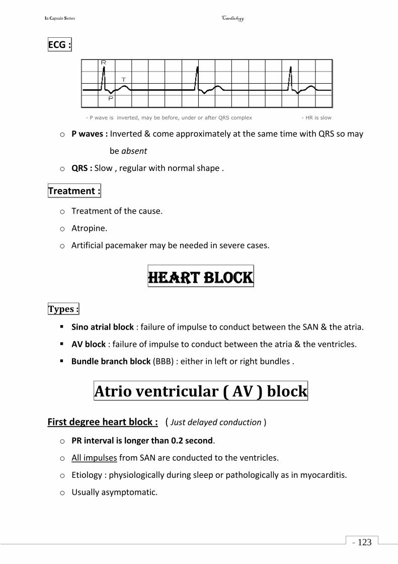
In Capsule Series Cardiology
- 123
-
ECG :
- P wave is inverted, may be before, under or after QRS complex - HR is slow
o P waves : Inverted & come approximately at the same time with QRS so may
be absent
o QRS : Slow , regular with normal shape .
Treatment :
o Treatment of the cause.
o Atropine.
o Artificial pacemaker may be needed in severe cases.
Heart block
Types :
Sino atrial block : failure of impulse to conduct between the SAN & the atria.
AV block : failure of impulse to conduct between the atria & the ventricles.
Bundle branch block (BBB) : either in left or right bundles .
Atrio ventricular ( AV ) block
First degree heart block : ( Just delayed conduction )
o PR interval is longer than 0.2 second.
o All impulses from SAN are conducted to the ventricles.
o Etiology : physiologically during sleep or pathologically as in myocarditis.
o Usually asymptomatic.
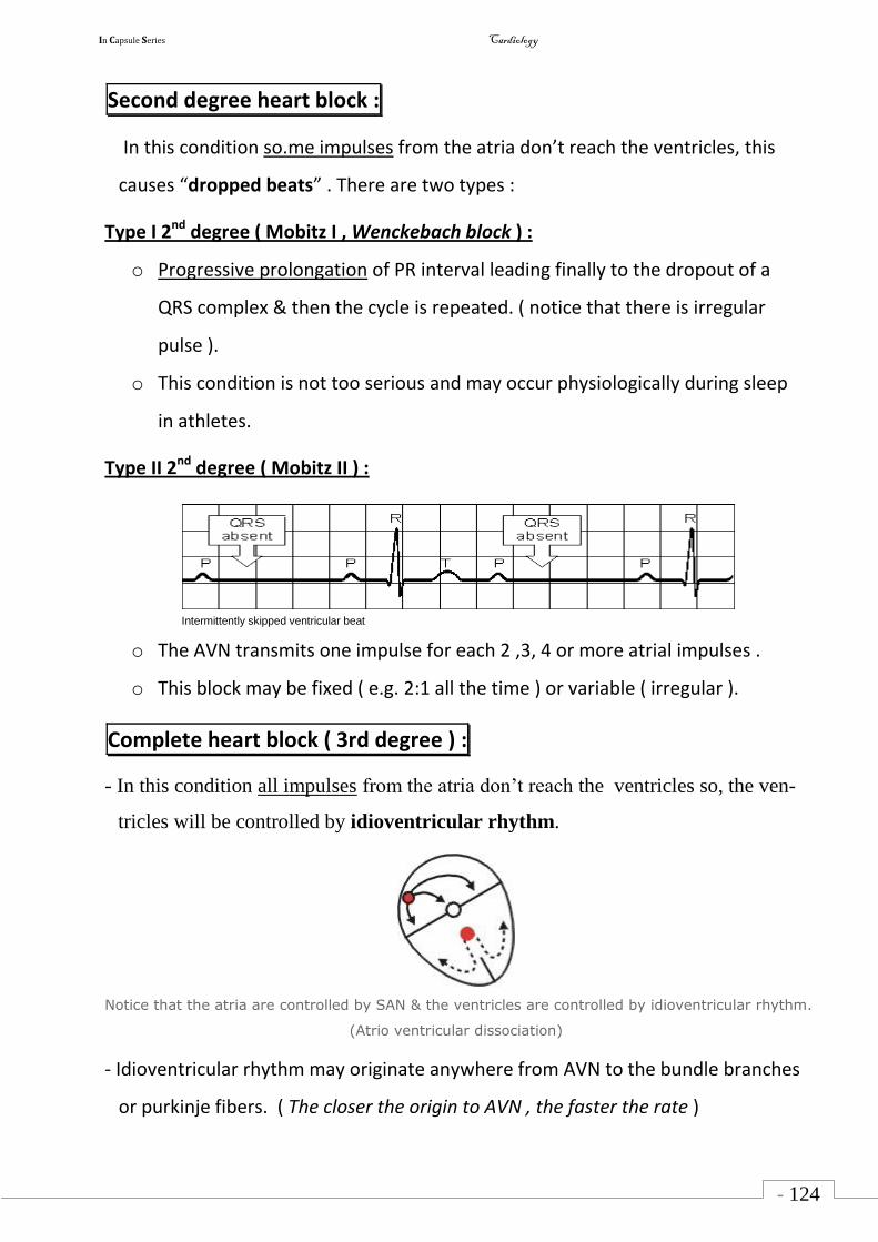
In Capsule Series Cardiology
- 124
-
Second degree heart block :
In this condition so.me impulses from the atria don’t reach the ventricles, this
causes “dropped beats” . There are two types :
Type I 2nd degree ( Mobitz I , Wenckebach block ) :
o Progressive prolongation of PR interval leading finally to the dropout of a
QRS complex & then the cycle is repeated. ( notice that there is irregular
pulse ).
o This condition is not too serious and may occur physiologically during sleep
in athletes.
Type II 2nd degree ( Mobitz II ) :
Intermittently skipped ventricular beat
o The AVN transmits one impulse for each 2 ,3, 4 or more atrial impulses .
o This block may be fixed ( e.g. 2:1 all the time ) or variable ( irregular ).
Complete heart block ( 3rd degree ) :
- In this condition all impulses from the atria don’t reach the ventricles so, the ven-
tricles will be controlled by idioventricular rhythm.
Notice that the atria are controlled by SAN & the ventricles are controlled by idioventricular rhythm.
(Atrio ventricular dissociation)
- Idioventricular rhythm may originate anywhere from AVN to the bundle branches
or purkinje fibers. ( The closer the origin to AVN , the faster the rate )
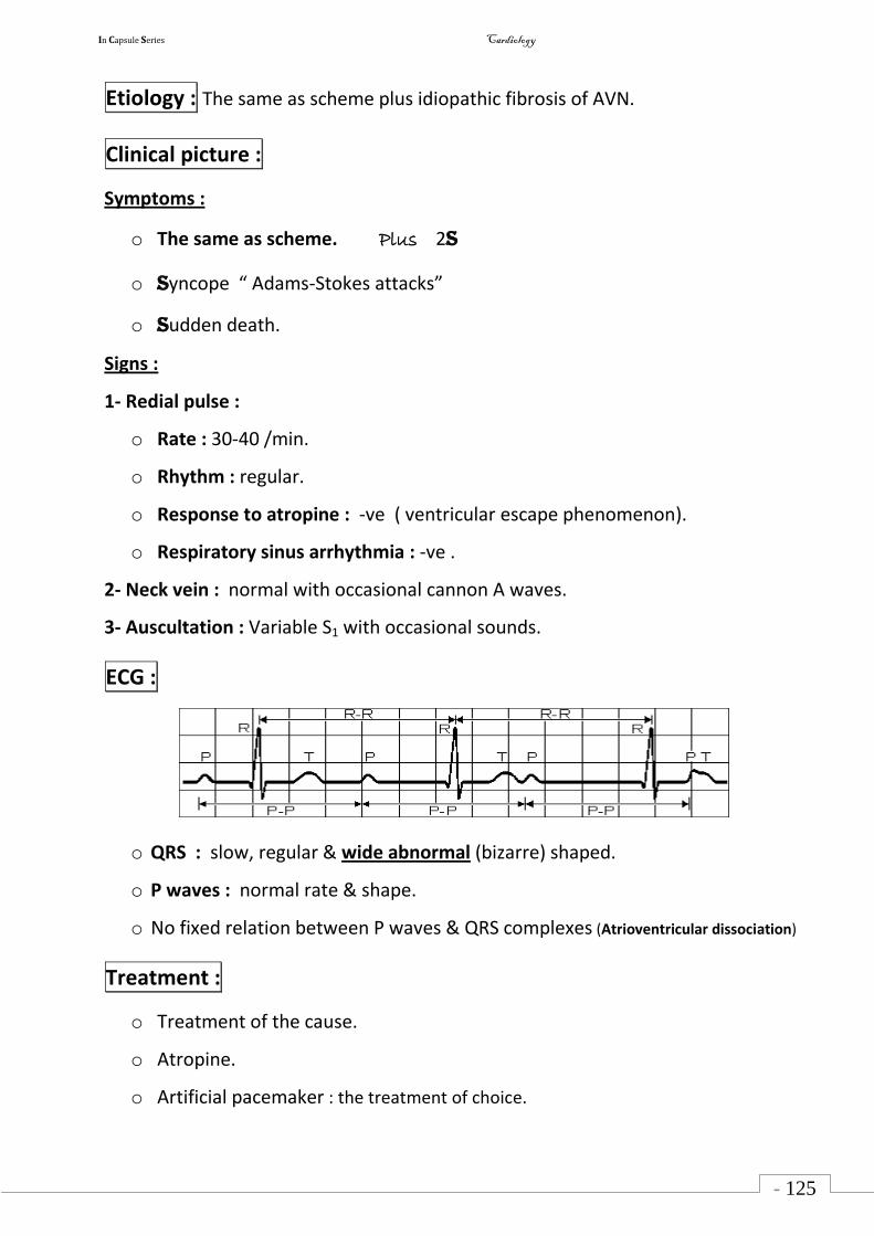
In Capsule Series Cardiology
- 125
-
Etiology : The same as scheme plus idiopathic fibrosis of AVN.
Clinical picture :
Symptoms :
o The same as scheme. Plus 2S
o Syncope “ Adams-Stokes attacks”
o Sudden death.
Signs :
1- Redial pulse :
o Rate : 30-40 /min.
o Rhythm : regular.
o Response to atropine : -ve ( ventricular escape phenomenon).
o Respiratory sinus arrhythmia : -ve .
2- Neck vein : normal with occasional cannon A waves.
3- Auscultation : Variable S1 with occasional sounds.
ECG :
o QRS : slow, regular & wide abnormal (bizarre) shaped.
o P waves : normal rate & shape.
o No fixed relation between P waves & QRS complexes (Atrioventricular dissociation)
Treatment :
o Treatment of the cause.
o Atropine.
o Artificial pacemaker : the treatment of choice.
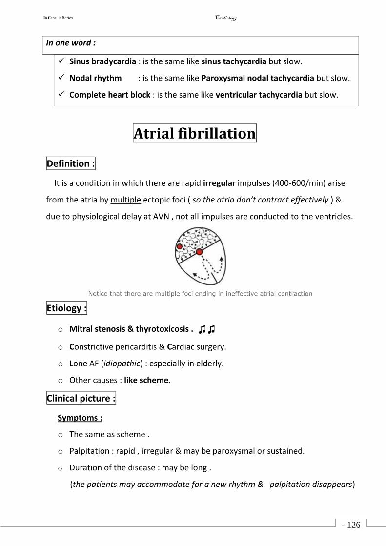
In Capsule Series Cardiology
- 126
-
In one word :
Sinus bradycardia : is the same like sinus tachycardia but slow.
Nodal rhythm : is the same like Paroxysmal nodal tachycardia but slow.
Complete heart block : is the same like ventricular tachycardia but slow.
Atrial fibrillation
Definition :
It is a condition in which there are rapid irregular impulses (400-600/min) arise
from the atria by multiple ectopic foci ( so the atria don’t contract effectively ) &
due to physiological delay at AVN , not all impulses are conducted to the ventricles.
Notice that there are multiple foci ending in ineffective atrial contraction
Etiology :
o Mitral stenosis & thyrotoxicosis . ♫♫
o Constrictive pericarditis & Cardiac surgery.
o Lone AF (idiopathic) : especially in elderly.
o Other causes : like scheme.
Clinical picture :
Symptoms :
o The same as scheme .
o Palpitation : rapid , irregular & may be paroxysmal or sustained.
o Duration of the disease : may be long .
(the patients may accommodate for a new rhythm & palpitation disappears)
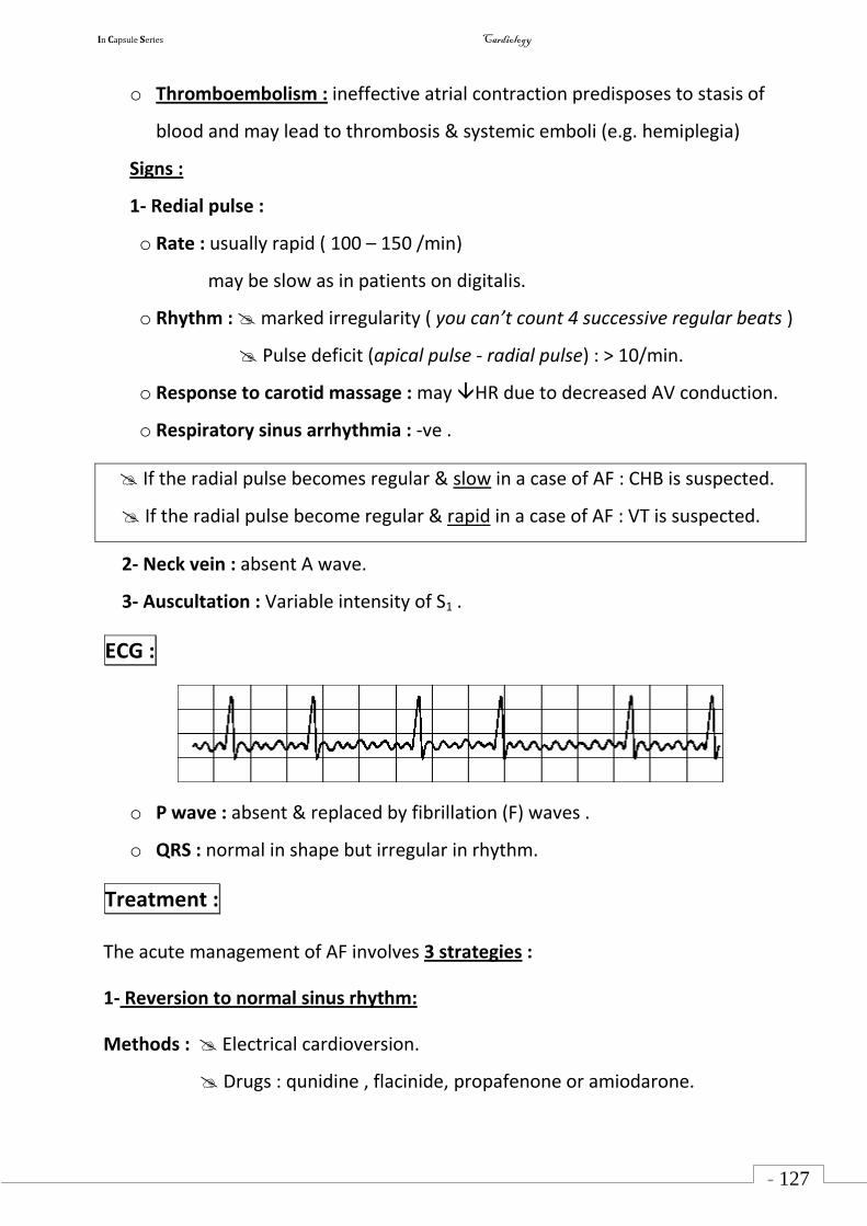
In Capsule Series Cardiology
- 127
-
o Thromboembolism : ineffective atrial contraction predisposes to stasis of
blood and may lead to thrombosis & systemic emboli (e.g. hemiplegia)
Signs :
1- Redial pulse :
o Rate : usually rapid ( 100 – 150 /min)
may be slow as in patients on digitalis.
o Rhythm : marked irregularity ( you can’t count 4 successive regular beats )
Pulse deficit (apical pulse - radial pulse) : > 10/min.
o Response to carotid massage : may HR due to decreased AV conduction.
o Respiratory sinus arrhythmia : -ve .
If the radial pulse becomes regular & slow in a case of AF : CHB is suspected.
If the radial pulse become regular & rapid in a case of AF : VT is suspected.
2- Neck vein : absent A wave.
3- Auscultation : Variable intensity of S1 .
ECG :
o P wave : absent & replaced by fibrillation (F) waves .
o QRS : normal in shape but irregular in rhythm.
Treatment :
The acute management of AF involves 3 strategies :
1- Reversion to normal sinus rhythm:
Methods : Electrical cardioversion.
Drugs : qunidine , flacinide, propafenone or amiodarone.
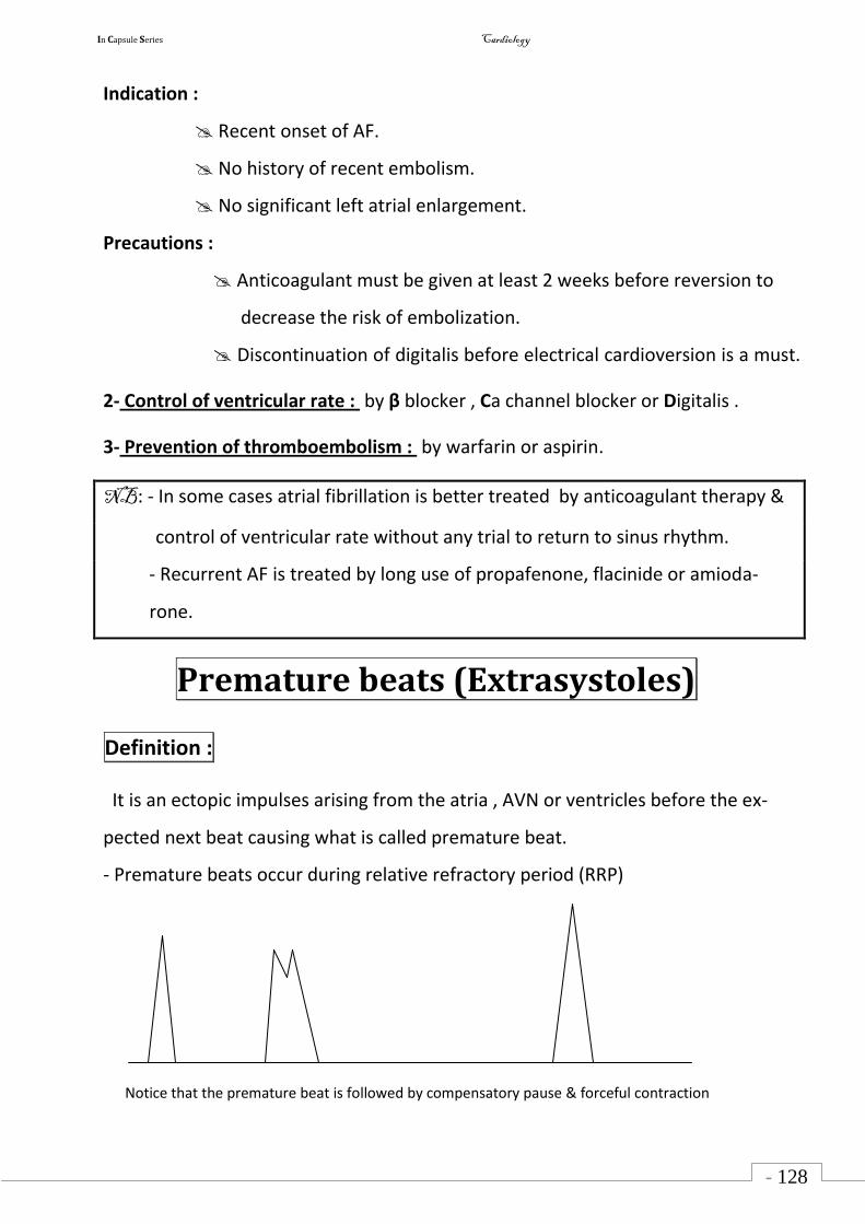
In Capsule Series Cardiology
- 128
-
Indication :
Recent onset of AF.
No history of recent embolism.
No significant left atrial enlargement.
Precautions :
Anticoagulant must be given at least 2 weeks before reversion to
decrease the risk of embolization.
Discontinuation of digitalis before electrical cardioversion is a must.
2- Control of ventricular rate : by β blocker , Ca channel blocker or Digitalis .
3- Prevention of thromboembolism : by warfarin or aspirin.
NB: - In some cases atrial fibrillation is better treated by anticoagulant therapy &
control of ventricular rate without any trial to return to sinus rhythm.
- Recurrent AF is treated by long use of propafenone, flacinide or amioda-
rone.
Premature beats (Extrasystoles)
Definition :
It is an ectopic impulses arising from the atria , AVN or ventricles before the ex-
pected next beat causing what is called premature beat.
- Premature beats occur during relative refractory period (RRP)
Notice that the premature beat is followed by compensatory pause & forceful contraction
In Capsule Series Cardiology
- 129
-
Etiology :
o Physiological : Emotions , smoking or excessive coffee.
o Pathological : The same as scheme.
Clinical picture :
Symptoms :
o Asymptomatic in most cases.
o Occasional irregular palpitation .
Signs :
1- Redial pulse :
o Rate : normal , tachy or bradycardia.
o Rhythm : Occasional irregularity.
Pulse deficit : < 10 / min.
o Response to exercise : The irregularity disappears due to in diastolic
period.
2- Neck vein : Normal wave with occasional irregularity.
3- Auscultation : Normal sounds with occasional irregularity.
ECG :
ventricular premature beats are wide bizarre QRS not preceded by P wave & fol-
lowed by compensatory pause.
Treatment :
1- Reassurance .
2- Treatment of the cause.
3- In chronic stable cases : Amiodarone, β blocker, Ca channel blocker or qunidine.
4- Lidocaine (IV) in emergency cases.
In Capsule Series Cardiology
- 130
-
Wolf-Parkinson-White (WPW) syndrome :
- It is accessory pathway that connects the atrium & ventricle & can bypass the AVN.
- So, AF is a very serious arrhythmia in these patients,it may lead to ventricular fibrillation.
- WPW is associated with thyrotoxicosis, mitral valve prolapse, HCM & more commen in
men.
- Treatment : Amiodarone , β blocker . Radiofrequency ablation is the treatment of choice.
- Digitalis & verapamil should be avoided ( ↑ conduction through the accessory pathway).
Treatment of arrhythmias
I- Pharmacological (Antiarrhythmic drugs) :
CLASS DRUGS MAIN USES
Class I : Na channel blockers
(slows the depolarization)
Class IA
Class IB
Class IC
Qunidine , Procainamide.
Lidocaine , Phenytoin.
Flacainide , Propafenone.
Broad spectrum.
Ventricular arrhythmias.
Broad spectrum.
Class II : β blockers Propranolol , Atenolol , Esmolol Tachyarrhythmias.
Premature beats.
Class III : K channel blockers Amiodarone , Bretylium.
Broad spectrum.
Class IV : Ca channel blockers Verapamil , Diltiazem.
Atrial tachyarrhyth-
mias.
Others : Adenosine(automaticity&conductivity)
Digitalis (automaticity&conductivity)
PSVT
Atrial tachyarrhythmias
Side effects of antiarrhythmic drugs :
Proarrhythmias : new arrhythmias induced by the drug .
In Capsule Series Cardiology
- 131
-
Qunidine :
Allergy & hypotension .
Cinchonism ( headache, vomiting , tinnitus & blurring of vision ).
Digitalis toxicity .
Lidocaine : 3m
Mental confusion.
Myocardial depression.
Muscle twitching.
Amiodarone : due to its tendency to accumulate in body tissue it may lead to:
CNS : Dizziness, depression , tremors.
Corneal deposits.
Thyroid dysfunctions ( hyper or hypo thyroidism )
Pulmonary fibrosis.
Elevation of hepatic enzymes.
Constipation.
Skin pigmentation.
II- Non pharmacological :
DC cardioversion .
Implantable cardioverter defibrillator ( ICD).
Radiofrequency catheter ablation .
Artificial pacemaker .( temporary , permanent ).
I have not failed. I've just found 10,000 ways that won't work.
Thomas Edison
In Capsule Series Cardiology
- 132
-
Causes of cardiac arrest : 5 H & 5 T
5 Hs :
o Hypovolemia
o Hypoxia.
o Hydrogen ions ( acidosis )
o Hyperkalemia & Hypokalemia.( Hyper more common )
o Hypothermia.
5 Ts :
o Toxins : CO , Cyanide .
o Tamponade. ( I mean cardiac tamponade )
o Tension pneumothorax.
o Thrombosis of coronary artery.
o Thrombosis of pulmonary artery.
In Capsule Series Cardiology
- 133 -
Pathogenesis of cardiac symptoms
Dyspnea
Definition :
Awareness or difficulty of breathing.
Etiology :
CVS : Any left sided heart disease lung congestion.
LSHF , MS , MR , AS , AR , myocardial infarction …….
Chest : All chest diseases
o Chest wall : obesity , Kyphoscoliosis …
o Bronchial : COPD , bronchial asthma ….
o Pleural : pleurisy , pleural effusion ….
o Pulmonary circulation diseases : pulmonary embolism , pulmonary hypertension.
CNS :
Increased ICT of respiratory center.
Abdominal : of intra abdominal pressure.
Pregnancy , Ascites , HSM ….
Metabolic : Any acidosis
DKA , Lactic acidosis , Uremic acidosis …
Pathogenesis of cardiac dyspnea : 7
1- Decrease in the vital capacity due to increase the amount of blood present in the lungs.
2- Exaggeration of Hering-Breuer reflex due to rigid alveoli.
3- Churchil-Cope reflex : It occurs in pulmonary congestion which leads to stimulation of
respiratory center.
4- Fatigue of respiratory muscle due to LCOP.
5- Pulmonary edema non functioning alveoli.
In Capsule Series Cardiology
- 134 -
6- Pleural & pericardial effusion lead to mechanical compression of the lungs.
7- Hypoxia stimulation of respiratory center.
Grades of dyspnea : New York Heart Association ( NYHA )
o Grade I : Dyspnea on extra ordinary effort.
o Grade II : Dyspnea on ordinary effort.
o Grade III : Dyspnea on less than ordinary effort .
o Grade IV : Dyspnea even at rest.
Orthopnea :
- Dyspnea on lying flat , partially relieved by sitting.
- This occurs due to :
o Increased venous return which increases the lung congestion.
o Elevation of diaphragm.
- No one can blame me when I say that orthopnea is not specific to cardiac diseases , It
also may occur due to chest or abdominal causes.
Paroxysmal Nocturnal Dyspnea ( PND ) :
- Attacks of dyspnea & cough with frothy expectoration occurring during night 1 – 2
hours after sleep & after a few minutes the patient feels better & goes back to sleep.
- If the condition is accompanied with chest wheezes , it is called cardiac asthma.
- Cardiac asthma is a state of impending pulmonary edema.
- PND is a specific symptom of left sided heart lesion especially LSHF.
- Pathogenesis of PND : During sleep , there are :
i- Vagal predominance causes further weakness of the contraction of left ventricle.
ii- Absorption of edema fluid into the circulation VR pulmonary congestion.
- The condition must be differentiated from bronchial asthma.
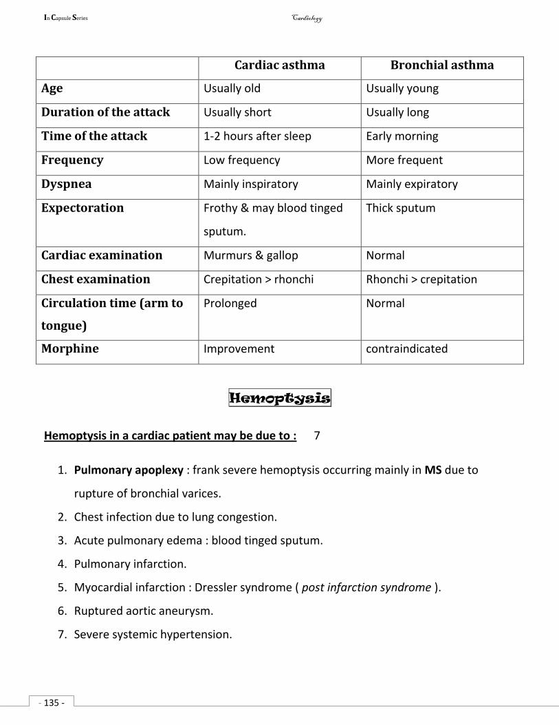
In Capsule Series Cardiology
- 135 -
Cardiac asthma Bronchial asthma
Age Usually old Usually young
Duration of the attack Usually short Usually long
Time of the attack 1-2 hours after sleep Early morning
Frequency Low frequency More frequent
Dyspnea Mainly inspiratory Mainly expiratory
Expectoration Frothy & may blood tinged
sputum.
Thick sputum
Cardiac examination Murmurs & gallop Normal
Chest examination Crepitation > rhonchi Rhonchi > crepitation
Circulation time (arm to
tongue)
Prolonged Normal
Morphine Improvement contraindicated
Hemoptysis
Hemoptysis in a cardiac patient may be due to : 7
1. Pulmonary apoplexy : frank severe hemoptysis occurring mainly in MS due to
rupture of bronchial varices.
2. Chest infection due to lung congestion.
3. Acute pulmonary edema : blood tinged sputum.
4. Pulmonary infarction.
5. Myocardial infarction : Dressler syndrome ( post infarction syndrome ).
6. Ruptured aortic aneurysm.
7. Severe systemic hypertension.
In Capsule Series Cardiology
- 136 -
Acute pulmonary edema
Etiology :
A) Cardiogenic pulmonary edema :
Acute left sided heart failure e.g. myocardial infarction.
On top of chronic LSHF : MS with aggravating factor as AF.
B) Non cardiogenic pulmonary edema : (Acute Respiratory Distress Syndrome – ARDS)
Diffuse alveolar damage (DAD) characterized by capillary permeability , pulmonary
edema & refractory hypoxia , caused by :
Sepsis .
Aspiration of gastric contents.
2 P : pneumonia , pancreatitis.
2 B : Burn , Blood transfusion.
Terminal renal & hepatic failure.
Clinical picture of acute pulmonary edema :
Severe dyspnea at rest & orthopnea.
Sense of impending death.
Sweating & irritability.
Cyanosis. Crepitation.
Cough with frothy pink sputum.
Features of the Cause :MI.
Differential Diagnosis : DD of acute dyspnea : MI & 6 P see p 104
Investigation :
Investigations of heart failure.
Investigations for the cause e.g. MI : ECG & cardiac enzymes.
Arterial blood gases : O2 , PCO2 ( PCO2 in late cases )
Investigations for ARDS : No LSHF ( PCWP < 18 mmHg , EF : normal)
Treatment : see p 15
In Capsule Series Cardiology
- 133 -
Syncope
Definition :
Sudden, transient loss of consciousness due to cerebral ischemia followed by spontaneous
recovery.
Causes :
1- Vasomotor syncope :
i- Vaso vagal attack : bad sight , bad smell , exposure to fear…..
ii- Carotid sinus syndrome : shaving , carotid sinus massage.
2- Orthostatic syncope : ( postural hypotension )
Autonomic neuropathy as in DM.
Adisson’s disease.
Antihypertensive drugs : blockers , methyl dopa .
Weakness of the muscles of the lower limbs.
3- Cardiac :
A) Exertional : AS , LSHF .
B) At rest : CHB , VT.
C) Positional ( syncope in certain position ) : Left atrial myxoma.
D) Acute decrease of VR e.g.hemorrhage.
E) Hypoxia : as in F4 ( Cyanotic spell )
4- Cerebral :
Transient ischemic attack (TIA).
Hypertensive encephalopathy. Cerebral embolism.
5- Situational :
Cough syncope :
Severe cough intrathoracic pressure VR cerebral blood flow syncope.
Micturation syncope : occurs in old men with BPH.
In Capsule Series Cardiology
138
Shock
Definition: Medically , shock is defined as inadequate tissue perfusion.
Types & Causes: there are 4 types :
1- Hypovolemic shock : ( the most common )
It is due to volume loss e.g.
o Blood loss : hemorrhage.
o Plasma loss : burn.
o Fluid loss : vomiting , diarrhea , dehydration ( DKA )
2- Cardiogenic shock : Pump failure ( contractility )
o Extensive MI (> 40 % of LV mass )
o Arrhythmias.
o Acute MR .
3- Obstructive shock. Mechanical obstruction to COP ( extrinsic cardiogenic )
o Cardiac tamponade
o Tension pneumothorax
o Massive pulmonary embolism.
4- Distributive shock : systemic vasodilatation peripheral vascular resistance
o Septic shock : sepsis with hypotension.
o Anaphylactic shock : caused by a hypersensitivity reaction to an allergen.
o Neurogenic shock : Failure of the nervous system to control diameter of blood vessels.
o Adrenal insufficiency.
Pathphysiology : ( all types of shock )
o Reduction of tissue perfusion.
o Damage of cellular membranes.
o Leakage of lysosomal enzymes.
o Reduction of cellular energy stores.
o Cell death.
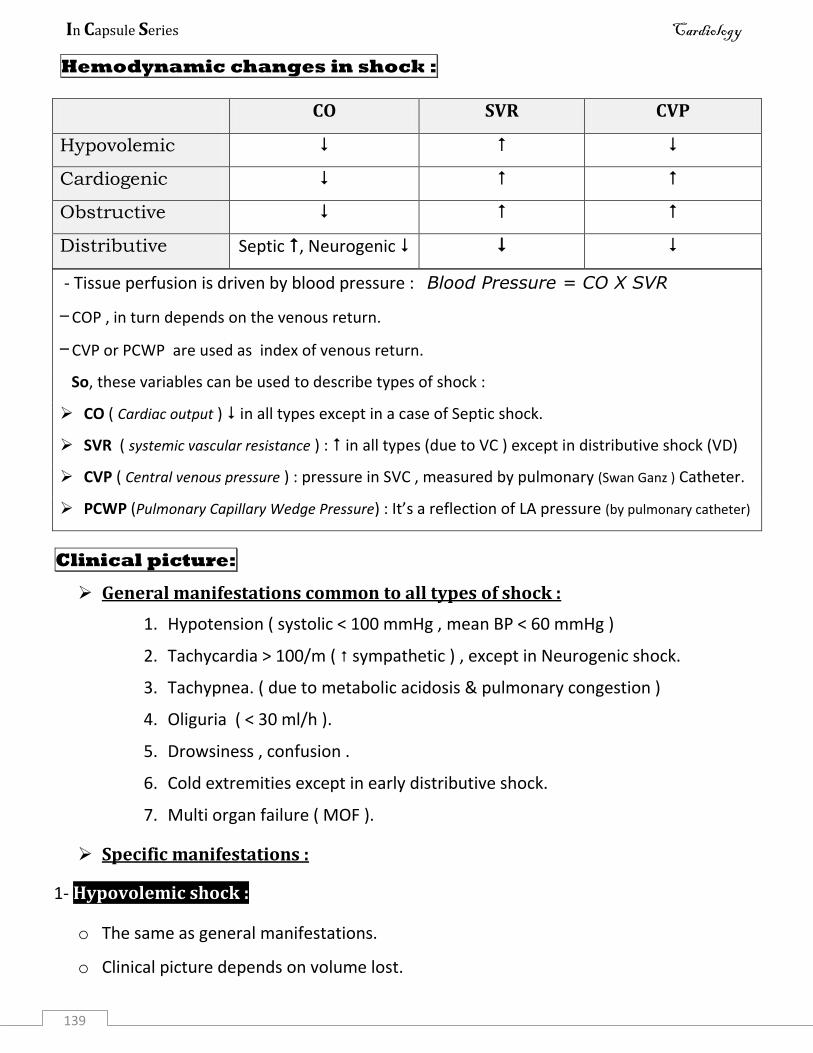
In Capsule Series Cardiology
139
Hemodynamic changes in shock :
CO SVR CVP
Hypovolemic
Cardiogenic
Obstructive
Distributive Septic , Neurogenic
- Tissue perfusion is driven by blood pressure : Blood Pressure = CO X SVR
– COP , in turn depends on the venous return.
– CVP or PCWP are used as index of venous return.
So, these variables can be used to describe types of shock :
CO ( Cardiac output ) in all types except in a case of Septic shock.
SVR ( systemic vascular resistance ) : in all types (due to VC ) except in distributive shock (VD)
CVP ( Central venous pressure ) : pressure in SVC , measured by pulmonary (Swan Ganz ) Catheter.
PCWP (Pulmonary Capillary Wedge Pressure) : It’s a reflection of LA pressure (by pulmonary catheter)
Clinical picture:
General manifestations common to all types of shock :
1. Hypotension ( systolic < 100 mmHg , mean BP < 60 mmHg )
2. Tachycardia > 100/m ( sympathetic ) , except in Neurogenic shock.
3. Tachypnea. ( due to metabolic acidosis & pulmonary congestion )
4. Oliguria ( < 30 ml/h ).
5. Drowsiness , confusion .
6. Cold extremities except in early distributive shock.
7. Multi organ failure ( MOF ).
Specific manifestations :
1- Hypovolemic shock :
o The same as general manifestations.
o Clinical picture depends on volume lost.
In Capsule Series Cardiology
140
o It is classified according to the percentage of blood loss into 4 classes :
( The same as the score in a tennis game : 15 - 30 - 40 - game over )
- Class I : up to 15 % - Class II : 15 - 30 %
- Class III : 30 - 40 % - Class IV : > 40 %
2- Cardiogenic shock :
o The same as general manifestations.
o Clinical picture of the cause e.g. MI : chest pain.
o Clinical manifestations of acute pulmonary edema : dyspnea , crepitations…
o Congested neck vein.
3- Obstructive shock :
o The same as general manifestations.
o C/P of the cause e.g. Cardiac tamponade (JVP, BP , distant heart sounds)
4- Distributive shock :
Septic shock :
o The same as general manifestations.
o Evidence of fever & localized infection.
o Warm flushed extremities ( warm shock )
o Strong pulse.
Anaphylactic shock :
o Warm flushed extremities.
o Erythema , Urticaria , angio edema , bronchospasm.
Neurogenic shock : Hypotension with bradycardia.
In Capsule Series Cardiology
141
Investigations :
Laboratory :
o Blood picture.
o Blood culture.
o Blood gases.
o Blood glucose.
o Liver & renal function tests. o Lactic acid level.
Imaging :
o X ray , ECG , Echo : for cardiac causes of shock.
o US & CT abdomen : for abdominal infections & intraperitoneal hemorrhage .
Treatment :
Goal : to restore normal tissue perfusion.
General measures :
o Monitoring of hemodynamics : CVP , CO , SVR.
o Trendelenburg position . ( legs up , head down )
o Patent airway.
o Oxygen therapy.
o IV fluid therapy in noncardiogenic shock patients.
Specific treatment : according to the type of shock.
Hypovolemic shock :
o Identify source of volume depletion.
o Blood transfusion in a cases of bleeding.
Cardiogenic shock :
o Treatment of the cause e.g. MI
o Dopamine , Dobutamine .
o Mechanical assist devices : Intra aortic balloon counterpulsation.
In Capsule Series Cardiology
142
Obstructive shock: relief of the obstruction is life saving
o If cardiac tamponade is present, urgent pericardiocentesis is essential.
o Tension pneumothorax must be treated with needle thoracostomy.
o Massive pulmonary embolism requires urgent thrombolysis or surgical removal.
Anaphylactic shock :
o Adrenaline .
o Hydrocortisone IV
o Antihistaminic.
Septic shock :
o Treatment of infection by antibiotics .
o Vasopressor as Adrenaline.
In Capsule Series Cardiology
- 143 -
Cardiac parameters
– Heart rate ( HR ) : 60 - 90 beats / min.
– Cardiac output ( CO ) : 4 - 6 L / min.
– Stroke volume ( SV ) : 60 - 100 ml / beat.
– Right atrial pressure ( RAP ) : 0 - 6 mmHg.
– Central venous pressure ( CVP ) : 0 - 6 mmHg.
– Left atrial pressure ( LAP ) : 6 - 12 mmHg.
– Pulmonary capillary wedge pressure ( PCWP ) : 6 - 12 mmHg.
– Right ventricular pressure ( RVP ) :
Systolic : 15 - 30 mmHg.
Diastolic : 0 - 8 mmHg.
– Right ventricular End Diastolic volume ( RVEDV ) : 100 - 160 ml.
– Right ventricular Ejection fraction ( RVEF ) : 40 - 60 %
– Pulmonary artery pressure :
Systolic : 15 - 30 mmHg.
Diastolic : 8 - 15 mmHg.
Mean : 10 - 20 mmHg.
– Left ventricular pressure ( LVP ) :
Systolic : 90 - 140 mmHg.
Diastolic : 4 - 12 mmHg.
– Arterial blood pressure :
Systolic ( SBP ) : 90 - 140 mmHg.
Diastolic ( DBP ) : 60 - 90 mmHg.
In Capsule Series Cardiology
- 144 -
Mean ( MAP ) : 70 - 105 mmHg.
Publisher
Dr Ramy Mohamed Saafan
010 52 92 500