case prsentation tmj ankylosis
TRANSCRIPT

Case prsentationBilateral TMJ Ankylosis

History takingเพศ หญิง อาย ุ8 ปีCC: อ้าปากได้น้อยมา 8ปีPI: ตอนอายุ 1 เดือน พลดัตกจากแม่ พ่ึงสงัเกตว่าลกูอ้าปากได้น้อยตอนฟันน ้านมซ่ีแรกขึน้
3 ปี 5 เดือน Known case Lt TMJ ankyloses type III, มีแผนผา่ตดัตอนอาย ุ6 ปี
5 ปี เร่ิมมีปัญหาหายใจล าบากตอนนอน นอนกรนเสียงดงั ไม่มีหยดุหายใจ Dx เป็น OSA ใช้ CPAPPMN: เป็น Asthma ตอน 1 ปี หลงัจากนัน้แขง็แรงดี, ปฏิเสธการแพ้ยา, ได้รบัวคัซีนครบ และมีพฒันาการสมวยั

Extra-oral examination
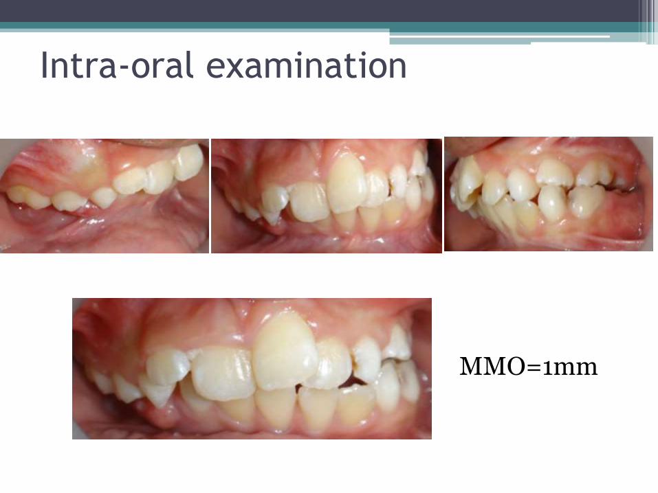
Intra-oral examination
MMO=1mm

OPG

Lat ceph

PA ceph

CXR

CT
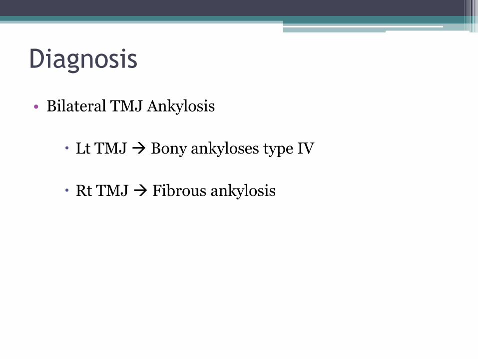
Diagnosis
• Bilateral TMJ Ankylosis
Lt TMJ Bony ankyloses type IV
Rt TMJ Fibrous ankylosis

Treatment plan
Surgery treatment
Gap arthroplasty at Rt TMJ
Reconstruction at Lt TMJ with costochondral graft
Postoperative Physiotherapy
Pateint should be encouraged to start active exercises of jaw as soon as it can be to lolerated(mouth gag, finger exerciser)
Follow-up

Ankylosis (joint stiffness) ▫ is the pathological fusion of parts of a joint resulting in
restricted movement across the joint
▫ Ankylosis of the Temporomandibular joint, an arthrogenic disorder of the TMJ, refers to restricted mandibular movements (hypomobility) with deviation to the affected side on opening of the mouth.

Anatomy
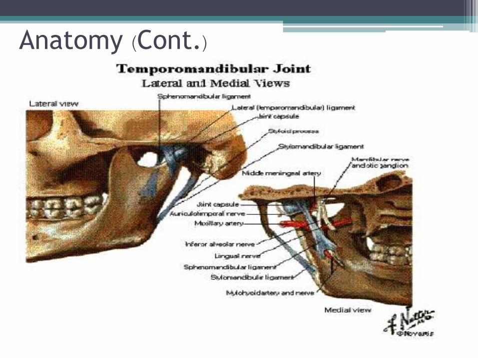
Anatomy (Cont.)

Classifications
• Bilateral or Unilateral ankylosis
• Fibrous ankylosis or Bony ankylosis
• Intra-articular or Extra-articular ankylosis
• Complete or Partial ankylosis
• True or false ankylosis

AetiologyTrauma
- At birth (with forceps)
- Blow to the chin (causing
haemarthrosis)
- Condylar fracture
Infections and Inflammatory
- Rheumatoid Arthritis
- Septic arthritis
- Otitis media
- Mastoditis
- Parotitis
- Osteoarthritis
Systemic disease
- Small pox
- Ankylosing spondylitis
- Syphilis
- Typhoid fever
- Scarlet fever
Others
- Malignancies
- Post radiology
- Post surgery
- Prolonged trismus

Pathophysiology
Truma
Extravasation of blood into the joint space
haemarthrosis
Calcificatiion and obliteration of the joint space
Intra-capsular ankylosis Extra-capsular ankylosis

Pathophysiology
Infection
Degenerative changesRoughness, limitation
Calcificatiion and obliteration of the joint space
Intra-capsular ankylosis Extra-capsular ankylosis

Unilateral clinical features • Mouth opening is very limited• Asymmetry of face with fullness of the affected side &
relative flattening of the unaffected side.• Face is deviated towards the affected side.• Chin is retracted on the affected side & slightly bypass the
midline.• Slight gliding movement towards the affected side.• Cross bite is present.• Well defined antegonial notch on affected side.

Bilateral clinical features• Bird face appearance/ micrognathia.
• No gliding movement neither protrusive nor lateral movement.
• Presence of scar on the chin (possibly due to trauma)
• Class II malocclusion, protrusive incisors & anterior open bite.
• In a long standing case there is atrophy or fibrosis of muscle.
• In congenital case-difficulty of introducing the nipple into the mouth of newborn infants.

Investigations
• For definitive diagnosis & to confirm the extent of bony growth imaging may be required.
1. Lateral oblique view
2. O. P. G. view
3. Cephalometric radiograph
4. Submentovertex view
5. PA view
6. C T Scan

Radiographic features
• Fusion of joint
• Loss of joint space
• Prominent antigonial notch
• Coronoid hyperplasia
Sequelae of TMJ ankylosis
• Facial growth distortion
• Nutritional impairment
• Respiratory disorders
• Malocclusion
• Poor oral hygiene
• Multiple carious and impacted teeth
Management
• Non surgical management
• Surgical treatment

SURGICAL MANAGEMENTAims and Objectives of surgery
To release ankylosed mass and creation of a gap
Creation of functional joint (improve patient’s oral hygiene, nutrition
and good speech)
To reconstruct the joint and restore the vertical height of the ramus
To prevent recurrence
To restore normal facial growth pattern
Procedures
1. Condylectomy
2. Gap arthroplasty
3. Interpositional arthroplasty

Condylectomy
• Fibrous ankylosis
• Pre-auricular incision is made
• Cut at the level of the condylar neck
• The head (condyle) should be separated
from the superior attachment carefully
• The wound is then sutured in layers
• The usual complication of this procedure is an ipsilateral deviation to
the affected side. And anterior open bite if the procedure was
bilaterally.
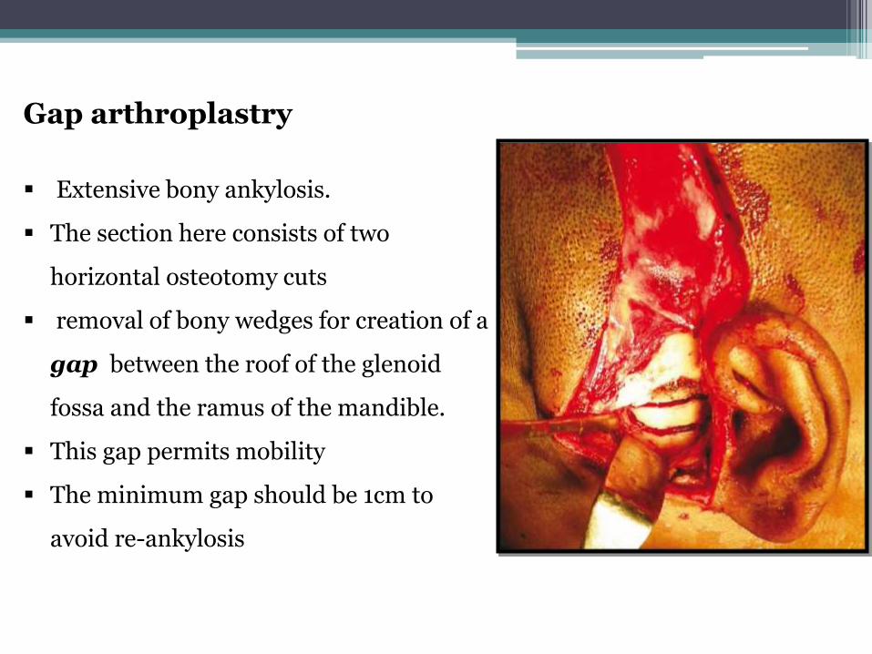
Gap arthroplastry
Extensive bony ankylosis.
The section here consists of two
horizontal osteotomy cuts
removal of bony wedges for creation of a
gap between the roof of the glenoid
fossa and the ramus of the mandible.
This gap permits mobility
The minimum gap should be 1cm to
avoid re-ankylosis

Interpositional arthroplasty
This is actually an improvement/modification on gap arthroplasty
Currently the surgical protocol of choice
Materials are used to interpose between the ramus of the mandible and
base of the skull to avoid re-ankylosis
The procedure involves the creation of gap, but in addition, a barrier is
inserted between the two surfaces to avoid reoccurrence and to
maintain the vertical height of the ramus

MATERIALS USED IN INTERPOSITIONAL ARTHROPLASTY
Autogenous Heterogenous Alloplastic
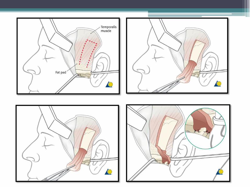
I. Temporalis muscles
II. Temporalis fascia
III. Fascia lata
IV. Cartiligenous grafts
CostochondralMetatartsalSternoclavicularAuricular graft
V. Dermis
I. chromatised submucosa of pig’s bladder
II. lyophilized bovine cartilage
Metallic: tantalum foil and plate, stainless steel, Titanium, Gold.
Nonmetallic: silastic, Teflon, acrylic, nylon, ceramic

Autografts, such as skin, temporalis muscle, or
fascia lata, are presently considered the material
of choice for interposition.
Advantages of these flaps in TMJ reconstruction include
close proximity to the TMJ without involving an additional surgical site.

Use of costochondral graft
• In children, after the release of the ankylosis. It is necessary to place a material that will allow groth
• A costochondral graft is harvested from the 5th
6th or 7th rib
• A costochondral junction about 1.5 cm is harvested and attached to lateral surface of ramus of the mandible to reconstruct the ramus
• Cosmetic surgery is carried out at the later date when the growth of the patient is completed.

Complicatipon of costochondral grafting procedure
1. Second surgical site is necessary.
2. Difficulty in suturing or stablizing the interpositional material on the medial aspect of joint.
3. Doner site complication such as pleuriticpain, pneumothorax.
4. Excessive growth of graft beyond what is required. This can be minimised by taking not more than 1.5 cm of costochondral graft.

Intra-Operative Haemorrhage (damage of any superficial temporal vessels, transverse facial
artery, etc) Damage to the external auditory meatus Damage to the Zygomatic and temp. branch of facial nerve Damage to the Auriculotemporal nerve Damage to the Parotid gland Damage to the teeth
Post Operative infection open bite
Complications of surgery
• Inadequate gap created between the fragments
• Fracture of the costochondral graft
• Inadequate coverage of the glenoid fossa surface
• Inadequate post-op physiotherapy
• Higher osteogenic potential and periostal osteogenic power may be
responsible for high rate of recurrence in children
Recurrence of TMJ ankylosis