case report kras mutation-positive bronchial surface ... mutation-positive bse-type adenocarcinoma...
TRANSCRIPT
Int J Clin Exp Pathol 2015;8(11):15338-15343www.ijcep.com /ISSN:1936-2625/IJCEP0015605
Case ReportKRAS mutation-positive bronchial surface epithelium (BSE)-type lung adenocarcinoma with strong expression of TTF-1: a case providing a further insight as for the role of TTF-1 in the oncogenesis
Yusuke Takanashi1, Shogo Tajima2, Takamitsu Hayakawa1, Hiroshi Neyatani1, Kazuhito Funai3
1Department of Thoracic Surgery, Fujieda Municipal General Hospital, Fujieda, Japan; 2Department of Pathology, University of Tokyo, Graduate School of Medicine, Tokyo, Japan; 3Department of Surgery 1, Hamamatsu University School of Medicine, Hamamatsu, Japan
Received September 5, 2015; Accepted October 20, 2015; Epub November 1, 2015; Published November 15, 2015
Abstract: Bronchial surface epithelium (BSE)-type lung adenocarcinoma is a subtype of non-terminal respiratory unit (TRU)-type lung adenocarcinoma originating in the bronchial surface epithelium. However, there are few known cases of BSE-type adenocarcinoma with marked expression of thyroid transcription factor-1 (TTF-1). This paper describes a very rare case of KRAS mutation-positive BSE-type adenocarcinoma that exhibited strong expression of TTF-1 that was putatively involved in oncogenesis. An 84-year-old woman, a never smoker, was referred to our hospital because of an abnormal chest radiograph. Chest computed tomography (CT) showed a solid mass lesion, 15 mm × 10 mm, with a relatively smooth margin in the left upper lobe. The patient underwent partial resection of the left upper lobe for strongly suspected lung cancer with a clinical stage of cT1aN0M0. Histopathological find-ings showed continuous migration of papillary, hyperplastic, atypical columnar tumor cells originating from normal bronchial surface epithelium, leading to a diagnosis of BSE-type adenocarcinoma. TTF-1 was strongly expressed in almost 100% of the tumor cells, which tested positive for the KRAS mutation. TTF-1 has recently attracted attention as an oncogene, and it is purportedly involved in the carcinogenesis and survival of lung adenocarcinoma cells. There is typically an inverse correlation between the respective expressions of KRAS and TTF-1, but in the present study, they appeared simultaneously and were both putatively involved as oncogenic driver alterations. This case is important in that it sheds some light on the largely unknown pathogenic mechanism of BSE-type adenocarcinoma.
Keywords: BSE-type adenocarcinoma, TTF-1, KRAS
Introduction
Among the lung adenocarcinomas that occur in the bronchial surface epithelium (BSE) or respiratory submucosal glands, non-terminal respiratory unit (TRU)-type adenocarcinoma is most often negative for expression of thyroid transcription factor-1 (TTF-1) and is closely associated with the KRAS mutation and smok-ing status [1]. As its name suggests, BSE-type adenocarcinoma is a subtype of non-TRU type adenocarcinoma that originates in the bronchi-al surface epithelium [1]. There are very few documented cases of BSE-type adenocarcino-ma with marked expression of TTF-1 [2], and few studies have focused on the pathogenic mechanism of this tumor type [1-4]. This paper
describes a very rare case of KRAS mutation-positive BSE-type adenocarcinoma that exhib-ited strong expression of TTF-1, which was putatively involved in oncogenesis.
Clinical summary
An 84-year-old woman, a never smoker, was referred to our hospital because of an abnormal chest radiograph. Chest computed tomography (CT) showed a solid mass lesion, 15 mm × 10 mm, with a relatively smooth margin in the left upper lobe (Figure 1). No significant lymph node swelling was seen. Although fluorodeoxy-glucose positron emission tomography (FDG-PET) detected significant accumulation at the mass lesion, suggesting malignancy, the mass

KRAS mutation-positive BSE-type adenocarcinoma with strong expression of TTF-1
15339 Int J Clin Exp Pathol 2015;8(11):15338-15343
lesion in the peripheral lung parenchyma was difficult to diagnose by bronchoscopy. The patient underwent surgery for strongly sus- pected lung cancer with a clinical stage of cT1aN0M0. The mass lesion was palpable with ease, and partial resection of the left upper lobe was performed due to the patient’s im- paired pulmonary function and extreme age. The patient’s postoperative course was un- eventful. Twelve months after the surgery, the patient is currently free of recurrent disease.
Pathological findings
Magnification of a 15 mm × 10 mm nodule with central necrosis in the lung parenchyma (Figure 2A) showed papillary, hyperplastic, atypical columnar epithelium (Figure 2B) that was diagnosed as a papillary-predominant in- vasive adenocarcinoma. The tumor cells con-tained eosinophilic cytoplasm, while the oval-shaped nuclei were arranged on the basolater-al side and were partially pseudostratified. Magnifying the edge of the papillary prolifera-tive structure revealed serial migration of tum- or cells from healthy bronchial surface epitheli-um (Figure 2C, arrow). The lumen structure of the tumor cells was smooth, and the cilia along the border of the tumor cell migration from the healthy bronchial surface epithelium had dis- appeared (Figure 2C). Based on the migrat- ion from healthy bronchial surface epithelium and cytomorphology, the tumor was deemed to be a BSE-type adenocarcinoma originating in the bronchial surface epithelium. The entire tumor consisted solely of BSE-type adenocarci-noma and did not include any other subtypes.
Immunohistochemistry findings indicated that the tumor cells were CK7-positive (Figure 3A)
Figure 1. Chest computed tomography (CT) shows a solid mass lesion, 15 mm × 10 mm, with a relatively smooth margin in the left upper lobe.
Figure 2. A. A 15 mm × 10 mm nodule with central necrosis in the lung parenchyma (hematoxylin and eosin [HE] staining; 12.5 × magnification) is seen; B. The nodule exhibits papillary, hyperplastic, atypi-cal columnar epithelium that was diagnosed as a papillary-predominant invasive adenocarcinoma (HE staining; 200 × magnification). C. Magnifying the edge of the papillary proliferative structure shows serial migration of tumor cells from healthy bron-chial surface epithelium and disappearance of the cilia along the border of the tumor cell migration (HE staining; 400 × magnification). Based on these findings, the tumor was diagnosed as a BSE-type adenocarcinoma originating in the bronchial surface epithelium.

KRAS mutation-positive BSE-type adenocarcinoma with strong expression of TTF-1
15340 Int J Clin Exp Pathol 2015;8(11):15338-15343
and CK20-negative, which was consistent with primary lung cancer. The main characteristic of this case was the consistently strong expres-sion of TTF-1, which is closely linked to TRU-type adenocarcinoma, throughout the entire tumor (Figure 3B, nucleus; brown). This feature contradicted the cytomorphological finding of
BSE-type adenocarcinoma. The expression of TTF-1 was considerably weaker in healthy bron-chial surface epithelium (Figure 3B, arrow) than in the tumor cells. The tumor cells were also diffusively positive for MUC4 (Figure 3C) and p53 (Figure 3D), which supported the diagnosis of BSE-type adenocarcinoma. Moreover, the tumor cells were negative for MUC5AC.
Genetic analysis
Direct-sequencing of KRAS gene codons 12 and 13 showed a GGT→GTT mutation at codon 12 (Figure 4), which supported the diagnosis of BSE-type adenocarcinoma. The never-smoker type p53 gene mutation was predicted based on the patient’s never-smoker status, so direct sequencing was performed on P53 gene exons 5 to 8, but no mutations were detected. Other genetic analyses were also negative for epider-mal growth factor receptor (EGFR) mutation on polymerase chain reaction (PCR) and anaplas-tic lymphoma kinase (ALK) fusion genes on flu-orescent in situ hybridization (FISH).
Figure 3. The tumor cells are: (A) CK7-positive; (B) strongly TTF-1-positive; (C) MUC4-positive; and (D) p53-positive. Specifically, TTF-1 is expressed in almost 100% of tumor cells and is only weakly expressed in healthy respiratory tract mucosa. TTF-1, Thyroid transcription factor-1.
Figure 4. Direct-sequencing of KRAS gene codons 12 and 13 shows a GGT→GTT mutation at codon 12.
KRAS mutation-positive BSE-type adenocarcinoma with strong expression of TTF-1
15341 Int J Clin Exp Pathol 2015;8(11):15338-15343
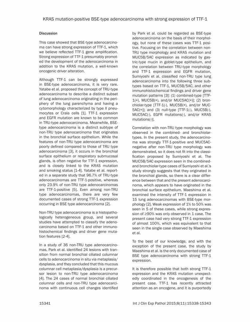
Discussion
This case showed that BSE-type adenocarcino-ma can have strong expression of TTF-1, which we believe reflected TTF-1 gene amplification. Strong expression of TTF-1 presumably promot-ed the development of the adenocarcinoma in addition to the KRAS mutation, a well-known oncogenic driver alteration.
Although TTF-1 can be strongly expressed in BSE-type adenocarcinoma, it is very rare. Yatabe et al. proposed the concept of TRU-type adenocarcinoma to describe a distinct subset of lung adenocarcinoma originating in the peri- phery of the lung parenchyma and having a cytomorphology characterized by type II pneu-mocytes or Crala cells [1]. TTF-1 expression and EGFR mutation are known to be common in TRU-type adenocarcinoma. Meanwhile, BSE-type adenocarcinoma is a distinct subtype of non-TRU type adenocarcinoma that originates in the bronchial surface epithelium. While the features of non-TRU type adenocarcinoma are poorly defined compared to those of TRU type adenocarcinoma [3], it occurs in the bronchial surface epithelium or respiratory submucosal glands, is often negative for TTF-1 expression, and is closely linked to the KRAS mutation and smoking status [1-4]. Yatabe et al. report- ed in a separate study that 96.7% of TRU-type adenocarcinomas are TTF-1-positive, whereas only 23.9% of non-TRU type adenocarcinomas are TTF-1-positive [5]. Even among non-TRU type adenocarcinomas, there are very few documented cases of strong TTF-1 expression occurring in BSE type adenocarcinoma [2].
Non-TRU type adenocarcinoma is a histopatho-logically heterogeneous group, and several studies have attempted to classify the adeno-carcinoma based on TTF-1 and other immuno-histochemical findings and driver gene muta-tion features [2-4].
In a study of 36 non-TRU type adenocarcino-mas, Park et al. identified 24 lesions with tran-sition from normal bronchial ciliated columnar cells to adenocarcinoma in situ via metaplasia/dysplasia, and they concluded that this mucous columnar cell metaplasia/dysplasia is a precur-sor lesion to non-TRU type adenocarcinoma [4]. The 24 cases of normal bronchial ciliated columnar cells and non-TRU type adenocarci-noma with continuous cell changes identified
by Park et al. could be regarded as BSE-type adenocarcinoma on the basis of their morphol-ogy, but none of these cases was TTF-1 posi-tive. Focusing on the correlation between non-TRU type morphology and KRAS mutation and MUC5B/5AC expression as indicated by gas-tric-type mucin in goblet-type epithelium, and the correlation between TRU-type morphology and TTF-1 expression and EGFR mutation, Sumiyoshi et al. classified non-TRU type lung adenocarcinoma into the following three sub-types based on TTF-1, MUC5B/5AC, and other immunohistochemical findings and driver gene mutation patterns [3]: (1) combined-type [TTF-1(+), MUC5B(+), and/or MUC5AC(+)]; (2) bron-chiolar-type [TTF-1(-), MUC5B(+), and/or MUC- 5AC(+)]; and (3) null-type [TTF-1(-), MUC5B(-), MUC5AC(-), EGFR mutations(-), and/or KRAS mutations(-)].
Correlation with non-TRU type morphology was observed in the combined- and bronchiolar-types. In the present study, the adenocarcino-ma was strongly TTF-1-positive and MUC5AC-negative after non-TRU type morphology was demonstrated, so it does not fit into the classi-fication proposed by Sumiyoshi et al. The MUC5B/5AC expression seen in the combined- and bronchiolar-type cases of Sumiyoshi et al.’s study strongly suggests that they originated in the bronchial glands, so there is a clear differ-ence between that and the present adenocarci-noma, which appears to have originated in the bronchial surface epithelium. Maeshima et al. examined the intensity of TTF-1 expression in 15 lung adenocarcinomas with BSE-type mor-phology [2]. Weak expression of 1% to 50% was seen in 5 of these cases, while strong expres-sion of ≥90% was only observed in 1 case. The present case had very strong TTF-1 expression of almost 100%, which was equivalent to that seen in the single case observed by Maeshima et al.
To the best of our knowledge, and with the exception of the present case, the study by Maeshima et al. is the only documented case of BSE type adenocarcinoma with strong TTF-1 expression.
It is therefore possible that both strong TTF-1 expression and the KRAS mutation unexpect-edly coordinated in the oncogenesis of the present case. TTF-1 has recently attracted attention as an oncogene, and it is purportedly
KRAS mutation-positive BSE-type adenocarcinoma with strong expression of TTF-1
15342 Int J Clin Exp Pathol 2015;8(11):15338-15343
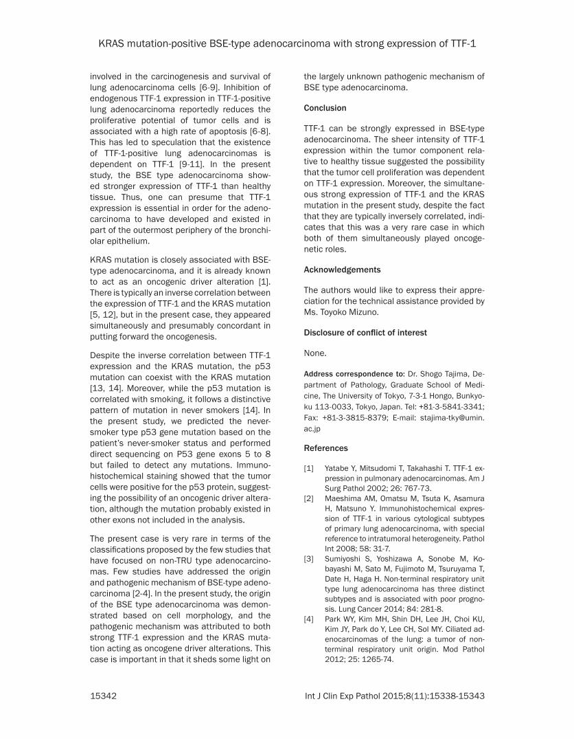
involved in the carcinogenesis and survival of lung adenocarcinoma cells [6-9]. Inhibition of endogenous TTF-1 expression in TTF-1-positive lung adenocarcinoma reportedly reduces the proliferative potential of tumor cells and is associated with a high rate of apoptosis [6-8]. This has led to speculation that the existence of TTF-1-positive lung adenocarcinomas is dependent on TTF-1 [9-11]. In the present study, the BSE type adenocarcinoma show- ed stronger expression of TTF-1 than healthy tissue. Thus, one can presume that TTF-1 expression is essential in order for the adeno-carcinoma to have developed and existed in part of the outermost periphery of the bronchi-olar epithelium.
KRAS mutation is closely associated with BSE-type adenocarcinoma, and it is already known to act as an oncogenic driver alteration [1]. There is typically an inverse correlation between the expression of TTF-1 and the KRAS mutation [5, 12], but in the present case, they appeared simultaneously and presumably concordant in putting forward the oncogenesis.
Despite the inverse correlation between TTF-1 expression and the KRAS mutation, the p53 mutation can coexist with the KRAS mutation [13, 14]. Moreover, while the p53 mutation is correlated with smoking, it follows a distinctive pattern of mutation in never smokers [14]. In the present study, we predicted the never-smoker type p53 gene mutation based on the patient’s never-smoker status and performed direct sequencing on P53 gene exons 5 to 8 but failed to detect any mutations. Immuno- histochemical staining showed that the tumor cells were positive for the p53 protein, suggest-ing the possibility of an oncogenic driver altera-tion, although the mutation probably existed in other exons not included in the analysis.
The present case is very rare in terms of the classifications proposed by the few studies that have focused on non-TRU type adenocarcino-mas. Few studies have addressed the origin and pathogenic mechanism of BSE-type adeno-carcinoma [2-4]. In the present study, the origin of the BSE type adenocarcinoma was demon-strated based on cell morphology, and the pathogenic mechanism was attributed to both strong TTF-1 expression and the KRAS muta-tion acting as oncogene driver alterations. This case is important in that it sheds some light on
the largely unknown pathogenic mechanism of BSE type adenocarcinoma.
Conclusion
TTF-1 can be strongly expressed in BSE-type adenocarcinoma. The sheer intensity of TTF-1 expression within the tumor component rela-tive to healthy tissue suggested the possibility that the tumor cell proliferation was dependent on TTF-1 expression. Moreover, the simultane-ous strong expression of TTF-1 and the KRAS mutation in the present study, despite the fact that they are typically inversely correlated, indi-cates that this was a very rare case in which both of them simultaneously played oncoge-netic roles.
Acknowledgements
The authors would like to express their appre-ciation for the technical assistance provided by Ms. Toyoko Mizuno.
Disclosure of conflict of interest
None.
Address correspondence to: Dr. Shogo Tajima, De- partment of Pathology, Graduate School of Medi- cine, The University of Tokyo, 7-3-1 Hongo, Bunkyo-ku 113-0033, Tokyo, Japan. Tel: +81-3-5841-3341; Fax: +81-3-3815-8379; E-mail: [email protected]
References
[1] Yatabe Y, Mitsudomi T, Takahashi T. TTF-1 ex-pression in pulmonary adenocarcinomas. Am J Surg Pathol 2002; 26: 767-73.
[2] Maeshima AM, Omatsu M, Tsuta K, Asamura H, Matsuno Y. Immunohistochemical expres-sion of TTF-1 in various cytological subtypes of primary lung adenocarcinoma, with special reference to intratumoral heterogeneity. Pathol Int 2008; 58: 31-7.
[3] Sumiyoshi S, Yoshizawa A, Sonobe M, Ko-bayashi M, Sato M, Fujimoto M, Tsuruyama T, Date H, Haga H. Non-terminal respiratory unit type lung adenocarcinoma has three distinct subtypes and is associated with poor progno-sis. Lung Cancer 2014; 84: 281-8.
[4] Park WY, Kim MH, Shin DH, Lee JH, Choi KU, Kim JY, Park do Y, Lee CH, Sol MY. Ciliated ad-enocarcinomas of the lung: a tumor of non-terminal respiratory unit origin. Mod Pathol 2012; 25: 1265-74.

KRAS mutation-positive BSE-type adenocarcinoma with strong expression of TTF-1
15343 Int J Clin Exp Pathol 2015;8(11):15338-15343
[5] Yatabe Y, Kosaka T, Takahashi T, Mitsudomi T. EGFR mutation is specific for terminal respira-tory unit type adenocarcinoma. Am J Surg Pathol 2005; 29: 633-9.
[6] Kwei KA, Kim YH, Girard L, Kao J, Pacyna-Gengelbach M, Salari K, Lee J, Choi YL, Sato M, Wang P, Hernandez-Boussard T, Gazdar AF, Pe-tersen I, Minna JD, Pollack JR. Genomic profil-ing identifies TITF1 as a lineage-specific onco-gene amplified in lung cancer. Oncogene 2008; 27: 3635-40.
[7] Tanaka H, Yanagisawa K, Shinjo K, Taguchi A, Maeno K, Tomida S, Shimada Y, Osada H, Ko-saka T, Matsubara H, Mitsudomi T, Sekido Y, Tanimoto M, Yatabe Y, Takahashi T. Lineage-specific dependency of lung adenocarcinomas on the lung development regulator TTF-1. Can-cer Res 2007; 67: 6007-11.
[8] Weir BA, Woo MS, Getz G, Perner S, Ding L, Beroukhim R, Lin WM, Province MA, Kraja A, Johnson LA, Shah K, Sato M, Thomas RK, Bar-letta JA, Borecki IB, Broderick S, Chang AC, Chi-ang DY, Chirieac LR, Cho J, Fujii Y, Gazdar AF, Giordano T, Greulich H, Hanna M, Johnson BE, Kris MG, Lash A, Lin L, Lindeman N, Mardis ER, McPherson JD, Minna JD, Morgan MB, Nadel M, Orringer MB, Osborne JR, Ozenberger B, Ra-mos AH, Robinson J, Roth JA, Rusch V, Sasaki H, Shepherd F, Sougnez C, Spitz MR, Tsao MS, Twomey D, Verhaak RG, Weinstock GM, Wheel-er DA, Winckler W, Yoshizawa A, Yu S, Zakowski MF, Zhang Q, Beer DG, Wistuba II, Watson MA, Garraway LA, Ladanyi M, Travis WD, Pao W, Ru-bin MA, Gabriel SB, Gibbs RA, Varmus HE, Wil-son RK, Lander ES, Meyerson M. Characteriz-ing the cancer genome in lung adenocarcino- ma. Nature 2007; 450: 893-8
[9] Yamaguchi T, Hosono Y, Yanagisawa K, Taka-hashi T. NKX2-1/TTF-1: an enigmatic onco-gene that functions as a double-edged sword for cancer cell survival and progression. Can-cer Cell 2013; 23: 718-23.
[10] Mu D. The complexity of thyroid transcription factor 1 with both pro- and anti-oncogenic ac-tivities. J Biol Chem 2013; 288: 24992-5000.
[11] Boggaram V. Thyroid transcription factor-1 (TTF-1/Nkx2.1/TITF1) gene regulation in the lung. Clin Sci (Lond) 2009; 116: 27-35.
[12] Takeuchi T, Tomida S, Yatabe Y, Kosaka T, Osa-da H, Yanagisawa K, Mitsudomi T, Takahashi T. Expression profile-defined classification of lung adenocarcinoma shows close relation-ship with underlying major genetic changes and clinicopathologic behaviors. J Clin Oncol 2006; 24: 1679-88.
[13] Kosaka T, Yatabe Y, Endoh H, Kuwano H, Taka-hashi T, Mitsudomi T. Mutations of the epider-mal growth factor receptor gene in lung can-cer: biological and clinical implications. Can- cer Res 2004; 64: 8919-23.
[14] Le Calvez F, Mukeria A, Hunt JD, Kelm O, Hung RJ, Tanière P, Brennan P, Boffetta P, Zaridze DG, Hainaut P. TP53 and KRAS mutation load and types in lung cancers in relation to tobac-co smoke: distinct patterns in never, former, and current smokers. Cancer Res 2005; 65: 5076-83.