chronic hypomagnesaemia on bovine ketosis of related clinical symptoms, such as lack of appetite,...
TRANSCRIPT
生物圏科学Biosphere Sci. 55:31-38 (2016)
総 説
Chronic hypomagnesaemia on bovine ketosis
Shigeru Yoshida
Professor emeritus
Graduate school of Biosphere Science, Hiroshima University,Higashi-Hiroshima 739-8528, Japan
Abstract Cows in the dairy farm of Hiroshima University were affected by abnormal milk secretion named “the Utrecht abnormality of milk”, and these cows were suffering from osteoporosis. Calcium was released from bone to milk by the increased serum calcium level. The increased serum calcium was caused by the decreased serum magnesium level by a low magnesium intake from roughage less than 0.2 % in dry matter. These cows were suffering from chronic hypomagnesaemia. Mass outbreak of bovine ketosis was occurred in this dairy farm. The levels of the blood serum Ca, Mg and inorganic phosphate (Pi) were 3.93 mEq, 1.84 mEq, and 4.95 mg/dl, respectively, in the average of milking 20 cows. These cows showed a low level of serum magnesium for many years. The 72 cows suffering from ketosis were also showed low serum magnesium level (1.77 mEq), a low serum calcium level (3.91 mEq) and low serum Pi level 5.06 mg/dl), compared with normal 93 cows (Ca 4.28 mEq, Mg 2.00 mEq, and Pi 7.81 mg/dl). They also showed low serum magnesium level as well as the cows of University dairy farm. In the relationship between ketosis and chronic hypomagnesaemia, ketosis was caused by the metabolic disturbance from α-ketoglutarate to succinyl-CoA, i.e., oxidative decarboxylation in TCA cycle. This reaction needs TDP, Lipoate, CoA, NAD, FAD, and Mg as cofactors. This disturbance must be act the accumulation of acetoacetate and ketone bodies, such as acetoacetate, acetone and β-OH-butyrate. The injection of magnesium, such as MgSO4 and MgCl2, have an effect on bovine ketosis. New hypothesis can be proposed that ketosis and downer cow, suffering from chronic hypomagnesaemia, must be come down with beriberi of human, because beriberi is a one of metabolic disturbance of oxidative decarboxylation reaction.Key words: Oxidative decarboxylation reaction, Low serum magnesium, Chronic hypomagnesaemia,
The Utrecht abnormality of milk, Osteoporosis.
A certain dairy farm reported frequent occurrences of “the Utrecht abnormality of milk”, of reproductive disturbances, and ketosis among lactating dairy cows. Through clinical, biochemical and pathological analysis, all the cows in this dairy herd were diagnosed as suffering from osteoporosis accompanied by a low serum Mg level. All cows were closely observed in order to follow this metabolic disorder. These dairy cows had been fed on rations with adequate in Ca and P but they low Mg for several years. The Mg content was below 0.2% of ration dry matter as shown in the former paper (Yoshida, 2015).
Accepted on September 1, 2016 *E-mail: [email protected], http://www.osteoporosis-cow.com
32 Shigeru YOSHIDA
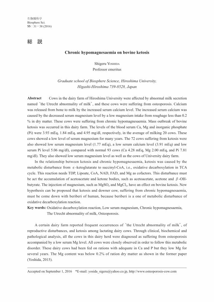
Most of the lactating cows spontaneously exhibited ketosis at the same time in the summer of 1971. Abnormal changes in blood serum values were noticed in Miyuki dairy farm as shown in Fig. 1. The observed changes included marked decrease of serum Ca from 4.17 mEq to 3.93 mEq and Pi from 6.91 mg/dl to 4.95 mg/dl, while, serum Mg was slightly increased from 1.76 mEq to 1.84 mEq. A veterinarian who regularly came to the dairy farm diagnosed the cows as ketotic after analysis of urinary acetone and observation of related clinical symptoms, such as lack of appetite, loss of body weight, decrease of milk yield, and lethargy. Fifteen of the twenty cows were ketotic at the same time as indicated in Table 1. As shown in Fig. 1, 7 ketotic cows of the Miyuki dairy farm were at low serum Ca and Mg. These lactating dairy cows were diagnosed with disturbances of Mg metabolism, and Mg-deficiency was suggested to be associated with a shortage of Mg intake. The therapeutic effects of Mg salts have been tried on these ketotic cows.
Fig.1. Changes of blood serum values (mean of 20 cows) during a mass outbreak of ketosis in Miyuki dairy farm.
Table 1. Blood serum values of ketotic cows of mass outbreak of ketosis of Miyuki dairy farm
Body weight(kg/day)
Milk yield(kg)
Ca(mEq)
Mg(mEq)
Pi(mg/dl)
Al(% )
Gl(% )
GOT(K-unit)
GPT(K-unit)
ALP(KA-unit)
Before outbreak (Jul. 7) 536 18.0 4.17 ↓1.76 6.82 3.32 4.79 58.2 18.2 5.9After outbreak (Aug. 7) 533 15.3 ↓3.93 1.84 ↓4.95 3.32 4.65 53.7 16.1 6.1
P<0.001 N.S. P<0.001 Urinary acetone levels after ketosis were +++ for 10 cows, ++ for 5cows, + for 2 cows, and - for 3 cows.P: calculated by t-test
Table 2. Blood serum values of ketotic cows of Miyuki dairy farm
Ca(mEq)
Mg(mEq)
Pi(mg/dl)
Al(% )
Gl(% )
GOT(K-unit)
ALP(KA-unit)
Glucose(mg/dl)
Acetone(mg/dl)
Urinaryacetone
Control (93 cows) 4.28 2.00 7.81 2.73 4.58 56.3 5.12 57.1 - -Ketosis (7 cows) ↓3.88 ↓1.69 ↓4.02 3.06 4.61 47.9 8.12 53.2 8.12 +++
Chronic hypomagnesemia on bovine ketosis 33
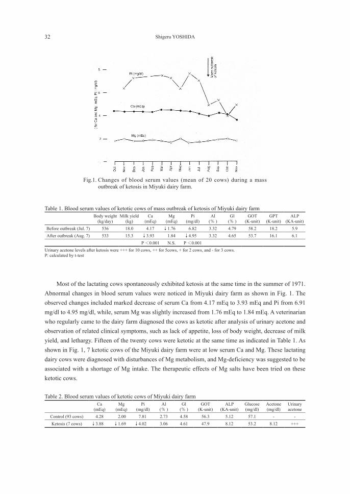
Case 1: Cow No.10, one of the 20 cows of the same dairy farm, was a lactating Holstein cow whose last calving was on July 15, 1971. This cow was 7 years old, and 504 kg in body weight, and produced 17 kg of milk per day in October, 1971. Cow No 10 had been kept under close observation and was declared ketotic on August 10, 1971. Ketone bodies had been evacuated in the cow’s urine for two months, from August to October. Magnesium salts were given to this ketotic cow after based on the hypothesis of a possible relation between ketosis or ketone body metabolism and magnesium metabolism. One gram of net Mg content in 10 gram of MgSO4・7H2O with 50 ml distilled water was given for 5 consecutive days by intravenous injection. Ketone bodies in blood serum and urine came down to normal values after this treatment. Blood serum samples were collected before and after treatment, and are shown in Table 3 and Fig. 2.
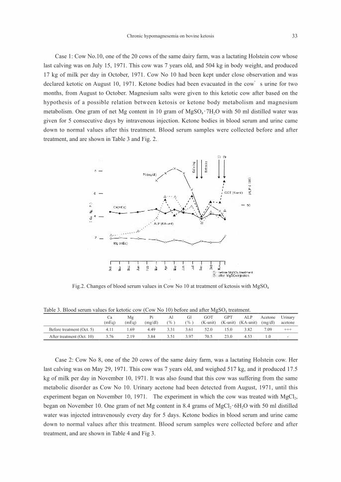
Case 2: Cow No 8, one of the 20 cows of the same dairy farm, was a lactating Holstein cow. Her last calving was on May 29, 1971. This cow was 7 years old, and weighed 517 kg, and it produced 17.5 kg of milk per day in November 10, 1971. It was also found that this cow was suffering from the same metabolic disorder as Cow No 10. Urinary acetone had been detected from August, 1971, until this experiment began on November 10, 1971. The experiment in which the cow was treated with MgCl2, began on November 10. One gram of net Mg content in 8.4 grams of MgCl2・6H2O with 50 ml distilled water was injected intravenously every day for 5 days. Ketone bodies in blood serum and urine came down to normal values after this treatment. Blood serum samples were collected before and after treatment, and are shown in Table 4 and Fig 3.
Fig.2. Changes of blood serum values in Cow No 10 at treatment of ketosis with MgSO4
Table 3. Blood serum values for ketotic cow (Cow No 10) before and after MgSO4 treatment.
Ca(mEq)
Mg(mEq)
Pi(mg/dl)
Al(% )
Gl(% )
GOT(K-unit)
GPT(K-unit)
ALP(KA-unit)
Acetone(mg/dl)
Urinaryacetone
Before treatment (Oct. 5) 4.11 1.69 4.49 3.31 3.61 52.0 15.0 3.82 7.09 +++After treatment (Oct. 10) 3.76 2.19 3.84 3.51 3.97 70.5 23.0 4.53 1.0 -
34 Shigeru YOSHIDA
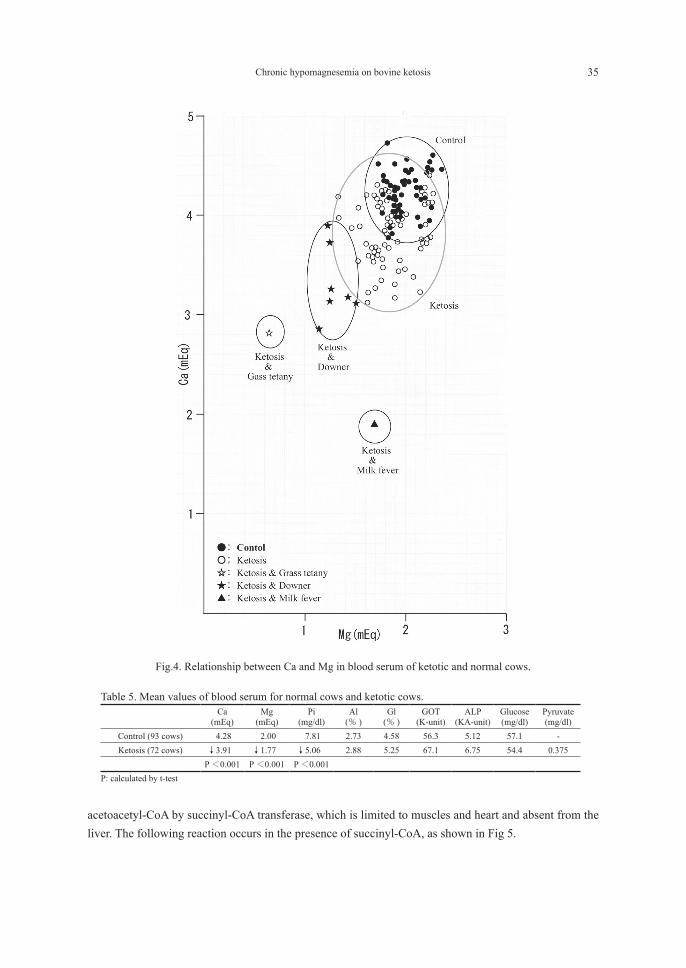
Abnormality in the levels of blood serum Ca, Mg, and Pi was observed in ketotic cows of the Miyuki dairy farm. Blood serum samples were then collected from ketotic cows as well as normal cows at other dairy farms in the immediate vicinity. A veterinary surgeon diagnosed the pathology cases as having ketosis on the basis of serum samples and clinical symptoms such as urinary acetone excretion, lack of appetite, nervous disturbance, decreased activity of stomach and intestine, deceased body weight, and decreased milk yield. It can be observed clearly that serum Ca, Mg and Pi were lower than the normal control samples at the time of ketotic condition as shown in Table 5 and Fig. 4. A decrease in the glucose level of serum was also observed, while the levels of serum GOT, ALP, and globulin were higher than those in normal controls. Ketosis is a metabolic disorder of the carbohydrate metabolism in which the level of ketone bodies in the fluids, including milk, and urine, is too high. However, it has not been revealed the mechanism that the ketone bodies accumulate in the living cow. It has been recognized that the accumulation of ketone bodies develops when the formation of acetoacetate is accelerated in the liver. But, ketone body accumulation is also supposed to be due to the reduction of the disintegration of acetoacetate in the muscles. From the results of the present investigations, a theory on ketosis and the ketone body metabolism can be proposed. The accumulation of ketone bodies, develops when the rate of formation of acetoacetate in the liver exceeds that at which acetoacetate can be oxidized in the muscle. The most important mechanism in ketone body oxidation depends on the conversion of free acetoacetate into
Fig.3. Changes of blood serum values in ketotic cow (Cow No 8) at treated with MgCl2.
Table 4. Blood serum values for ketotic cow (Cow No 8) before and after MgCl2 treatment.
Ca(mEq)
Mg(mEq)
Pi(mg/dl)
Al(% )
Gl(% )
GOT(K-unit)
ALP(KA-unit)
Glucose(mg/dl)
Acetone(mg/dl)
Urinaryacetone
Before treatment (Nov. 10) 4.01 1.63 3.85 3.28 4.51 39.5 8.8 60.8 5.98 +++After treatment (Nov. 14) 4.16 1.82 5.12 3.49 4.30 49.0 4.3 60.0 1.17 -
Chronic hypomagnesemia on bovine ketosis 35
acetoacetyl-CoA by succinyl-CoA transferase, which is limited to muscles and heart and absent from the liver. The following reaction occurs in the presence of succinyl-CoA, as shown in Fig 5.
Fig.4. Relationship between Ca and Mg in blood serum of ketotic and normal cows.
Table 5. Mean values of blood serum for normal cows and ketotic cows.
Ca(mEq)
Mg(mEq)
Pi(mg/dl)
Al(% )
Gl(% )
GOT(K-unit)
ALP(KA-unit)
Glucose(mg/dl)
Pyruvate(mg/dl)
Control (93 cows) 4.28 2.00 7.81 2.73 4.58 56.3 5.12 57.1 -Ketosis (72 cows) ↓3.91 ↓1.77 ↓5.06 2.88 5.25 67.1 6.75 54.4 0.375
P<0.001 P<0.001 P<0.001 P: calculated by t-test

36 Shigeru YOSHIDA
CONCLUSION
When succinyl-CoA is depressed, acetoacetate accumulates and converted into acetone and β-hydroxybutyrate. Reduced oxidative decarboxylation reaction from α-ketoglutarate results in a reduced yield of succinyl-CoA. Accumulation of α-ketoglutarate and reduced succinate have been reported. The complete reaction system is summarized in Fig 6. An inhibition or metabolic disturbance occurring in oxidative decarboxylation of carboxyhydrate from pyruvate to citrate and from α-ketoglutarate to succinate. However, further research with partially purified enzyme preparations associated with this reaction showed that the process as a whole requires the presence of a considerable group of small molecular materials, including TDP, Lipoate, CoA, NAD, FAD, and Mg. Thus, the oxidative decarboxylation reaction can be hampered by a deficiency in one of these cofactors leading to a deficit in succinyl-CoA.
Fig.5 Oxidative decarboxylation in muscle
Fig.6 Map of TCA cycle and ketone bodies production
Chronic hypomagnesemia on bovine ketosis 37
ACKNOWLEDGEMENT
The author wish to thanks veterinarians for collecting blood samples with this study.
REFFERENCES
Bach.S.J., and Hibitt. K.C., 1959. Biochemial Aspect of Bovine Ketosis. Biochem.J. 72:87.Ballard.F.J., Hanson. R.W., Kronfeld. D.S., Raggi. F., 1968. Metabolic Changes in Liver Associated with
Spontaneous Ketosis and Starvation in Cows., J.Nutrition 95:160.Yoshida,S., 2015. Osteoporosis in lactating dairy cows. Biosphere Sci. 54: 99.
38 Shigeru YOSHIDA
慢性Mg欠乏により発生した乳牛のケトージスについて
吉田 繁広島大学名誉教授
〒720-0076 福山市本庄町中1-4-9, [email protected], http://www.osteoporosis-cow.com
要 旨 前号で,大学付属牧場の全ての乳牛が低酸度二等乳を泌乳し,その乳牛群に骨粗鬆症が発生していることを論じた。即ち,その原因は粗飼料のMg不足により乳牛の血清Mgが低下し血清 Caが増加したために,骨から多量の Caが乳汁に移行した為に生じたものである。なお牛乳中に Caが多いとアルコール試験で不安定になることが知られている。 粗飼料中のMg含量は DM中として0.2%以下でグラステタニーが発生すると云われている。この乳牛群20頭を追跡調査する過程で血清 Pの急激な低下から代謝異常の発生を指摘し,獣医師が検診したところケトージスが集団発生していることが判明した。発病前は血清 Ca 4.17 mEq,Mg1.78 mEq,P 6.82mg/dlに対し,発病後は血清 Ca3.93 mEq,Mg1.84 mEq,P 4.95 mg/dlであった。この乳牛にMgSO4とMgCl2を投与したところケトージスは回復したので血清Mgがケトージス発病に深く関与していることが推定される。ケトージス発生時は血清中のα-Ketoglutarateが増加しているとの報告があり,TCAサイクルの酸化的脱炭酸反応の過程で補酵素としてMgが必須である。この乳牛群でのケトージスの発生はMg不足に要因があると考えられる。人の脚気はビタミン B1欠乏症であるが乳牛のケトージスは同一の部位の代謝障害であり,乳牛の起立不能症も深く関連していると推定される。 大学周辺の民間牧場でケトージスが発生した際,健康な乳牛93頭の平均値 Ca 4.28 mEq,Mg 2.00 mEq,P 7.81 mg/dlに比べて,罹患乳牛群72頭では Ca 3.76 mEq,Mg 1.77 mEq,P 5.06 mg/dlであり,付属牧場同様に Ca,Mg,Pともに低下する傾向を示した。 この牧場では乳牛の急性疾患である起立不能症のグラステタニーのほか,慢性疾患である低酸度二等乳と骨粗鬆症が発生しており,亜急性のケトージスが発生した。これらの疾病の原因は粗飼料に由来するMgの不足に起因すると考えられる。キーワード:ケトージス,酸化的脱炭酸反応,低Mg血症,Mg欠乏症,低酸度二等乳,骨粗鬆症