circulatory system class notes - san dieguito union high...
TRANSCRIPT

Circulatory System
Unit 6.8 (6th Edition)
Chapter 7.8 (7th Edition)1
Learning Objectives
• Label the layers, chambers, valves, and
major blood vessels of the heart.
• Differentiate between systole and diastole.
• Trace the path of blood, O2, & CO2 during cardiac cycle.
• Explain the conductive pathway of electrical impulses.
• Identify abnormal heart rhythms (arrhythmias)
• Distinguish between the 3 major types of blood vessels.
• Compare/contrast the three main types of blood cells by
describing the function of each.
• Describe the major diseases of the circulatory system.
2
Fun Facts About the Circulatory System
• The heart beats ~ 2.5 billion times in an average life span.
• ~ 8 million blood cells die in human body each second.
• ~ 8 million blood cells are born each second.
• A tiny droplet of blood has ~ 5 million red blood cells.
• It takes ~ 20 seconds for a red blood cell to circle the body.
• Red blood cells make ~ 250,000 round trips of the body
before returning to the bone marrow, where they were
born, to die.
• Red blood cells may live for ~ 4 months circulating
throughout the body, feeding the 60 trillion other cells.
• Though weighing only 11 ounces on average, a healthy
heart pumps 2,000 gallons of blood through 60,000
miles of blood vessels each day.
• Adult body has 4-6 quarts of blood in circulation. 3
Overview of Circulatory System
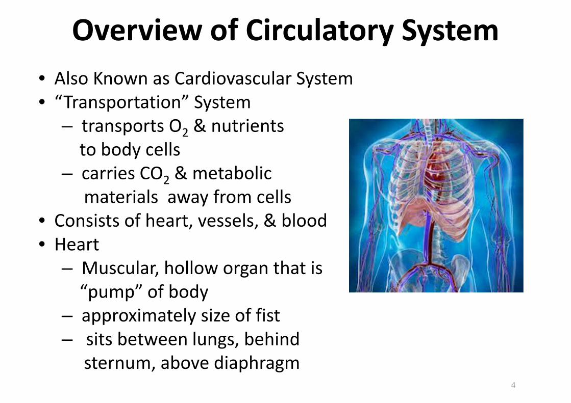
• Also Known as Cardiovascular System
• “Transportation” System
– transports O2 & nutrients
to body cells
– carries CO2 & metabolic
materials away from cells
• Consists of heart, vessels, & blood
• Heart
– Muscular, hollow organ that is
“pump” of body
– approximately size of fist
– sits between lungs, behind
sternum, above diaphragm 4
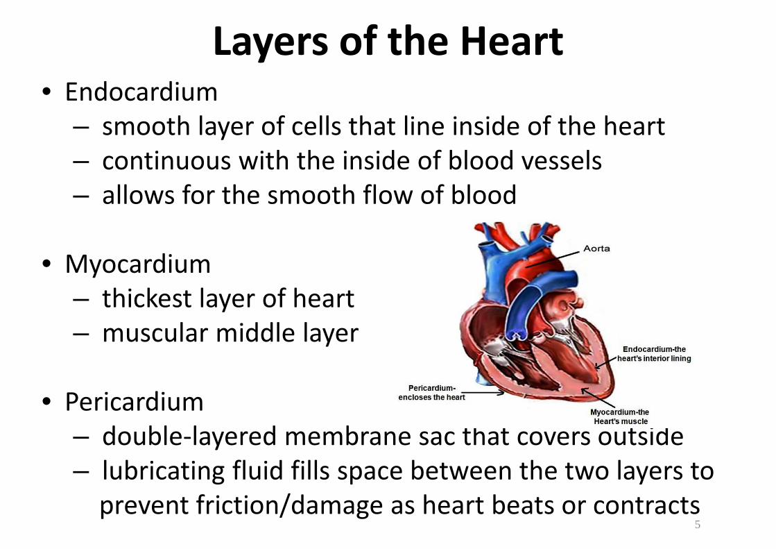
Layers of the Heart• Endocardium
– smooth layer of cells that line inside of the heart
– continuous with the inside of blood vessels
– allows for the smooth flow of blood
• Myocardium
– thickest layer of heart
– muscular middle layer
• Pericardium
– double-layered membrane sac that covers outside
– lubricating fluid fills space between the two layers to
prevent friction/damage as heart beats or contracts5

Four Chambers of the Heart
• Septum
– muscular wall
– divides heart into left & right
– prevents blood from moving
between left & right side
• Atrium (plural-atria)
– Latin word “entrance hall”
– two upper chambers
– Left & Right Atria
• Ventricles
– Latin word “little belly”
– two lower chambers
– Left and Right Ventricles6
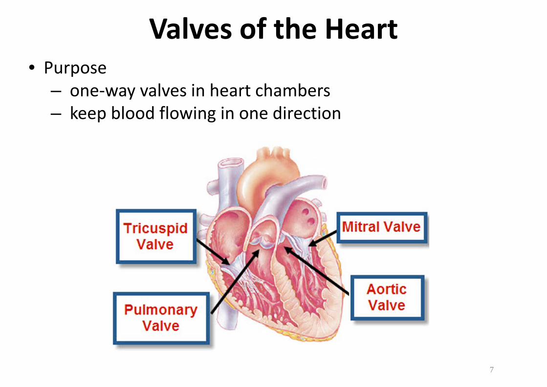
Valves of the Heart
• Purpose
– one-way valves in heart chambers
– keep blood flowing in one direction
7
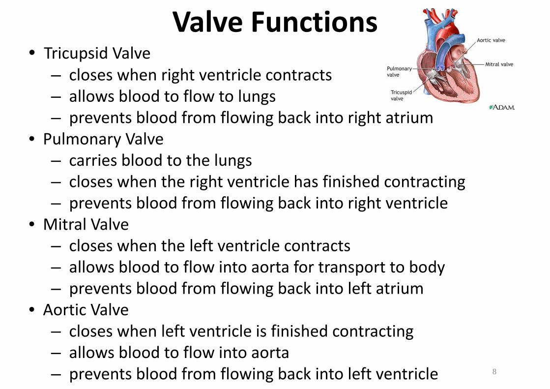
Valve Functions• Tricupsid Valve
– closes when right ventricle contracts
– allows blood to flow to lungs
– prevents blood from flowing back into right atrium
• Pulmonary Valve
– carries blood to the lungs
– closes when the right ventricle has finished contracting
– prevents blood from flowing back into right ventricle
• Mitral Valve
– closes when the left ventricle contracts
– allows blood to flow into aorta for transport to body
– prevents blood from flowing back into left atrium
• Aortic Valve
– closes when left ventricle is finished contracting
– allows blood to flow into aorta
– prevents blood from flowing back into left ventricle 8

Blood Circulation
• Cardiopulmonary Circulation
– from heart to lungs and
– from lungs to heart
• Systemic (General) Circulation
– carry blood through rest of
body
• Blood Vessels
– carry blood leaving heart
– arteries carry blood away
from heart
– veins carry blood to heart9

How the Heart Works
10
• You Tube Video– http://www.youtube.com/watch?v=84PrHxJri9Q
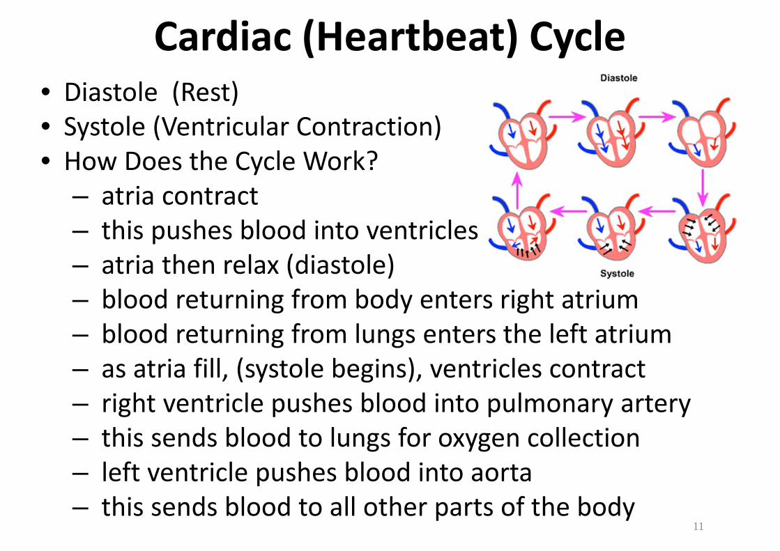
Cardiac (Heartbeat) Cycle
• Diastole (Rest)
• Systole (Ventricular Contraction)
• How Does the Cycle Work?
– atria contract
– this pushes blood into ventricles
– atria then relax (diastole)
– blood returning from body enters right atrium
– blood returning from lungs enters the left atrium
– as atria fill, (systole begins), ventricles contract
– right ventricle pushes blood into pulmonary artery
– this sends blood to lungs for oxygen collection
– left ventricle pushes blood into aorta
– this sends blood to all other parts of the body11
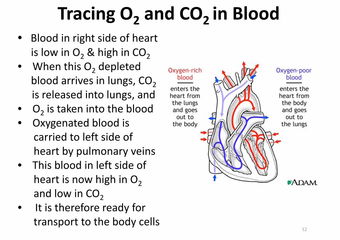
Tracing O2 and CO2 in Blood
• Blood in right side of heart
is low in O2 & high in CO2
• When this O2 depleted
blood arrives in lungs, CO2
is released into lungs, and
• O2 is taken into the blood
• Oxygenated blood is
carried to left side of
heart by pulmonary veins
• This blood in left side of
heart is now high in O2
and low in CO2
• It is therefore ready for
transport to the body cells12
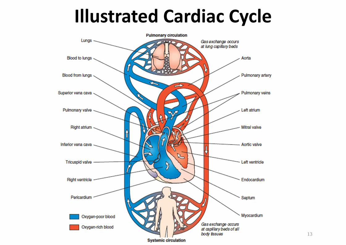
Illustrated Cardiac Cycle
13
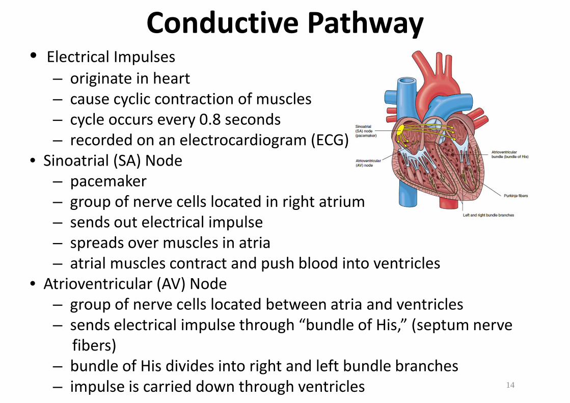
Conductive Pathway• Electrical Impulses
– originate in heart
– cause cyclic contraction of muscles
– cycle occurs every 0.8 seconds
– recorded on an electrocardiogram (ECG)
• Sinoatrial (SA) Node
– pacemaker
– group of nerve cells located in right atrium
– sends out electrical impulse
– spreads over muscles in atria
– atrial muscles contract and push blood into ventricles
• Atrioventricular (AV) Node
– group of nerve cells located between atria and ventricles
– sends electrical impulse through “bundle of His,” (septum nerve
fibers)
– bundle of His divides into right and left bundle branches
– impulse is carried down through ventricles 14

Arrhythmias• Definition
– abnormal heart rhythms
– interference to natural heart rhythms
– can be mild to life-threatening
• Premature Atrial Contraction (PAC)
– early contraction of atria
– usually goes unnoticed
• Ventricle Fibrillation
– ventricles contract at random without coordination
– decreases blood output and causes death if untreated
• Diagnosis
– cardiac monitors
– electrocardiograms
• Treatment
– defibrillator: device that shocks the heart with electrical current
– stops uncoordinated contraction & SA node regains control
– pacemakers: monitors and delivers electrical impulses 15
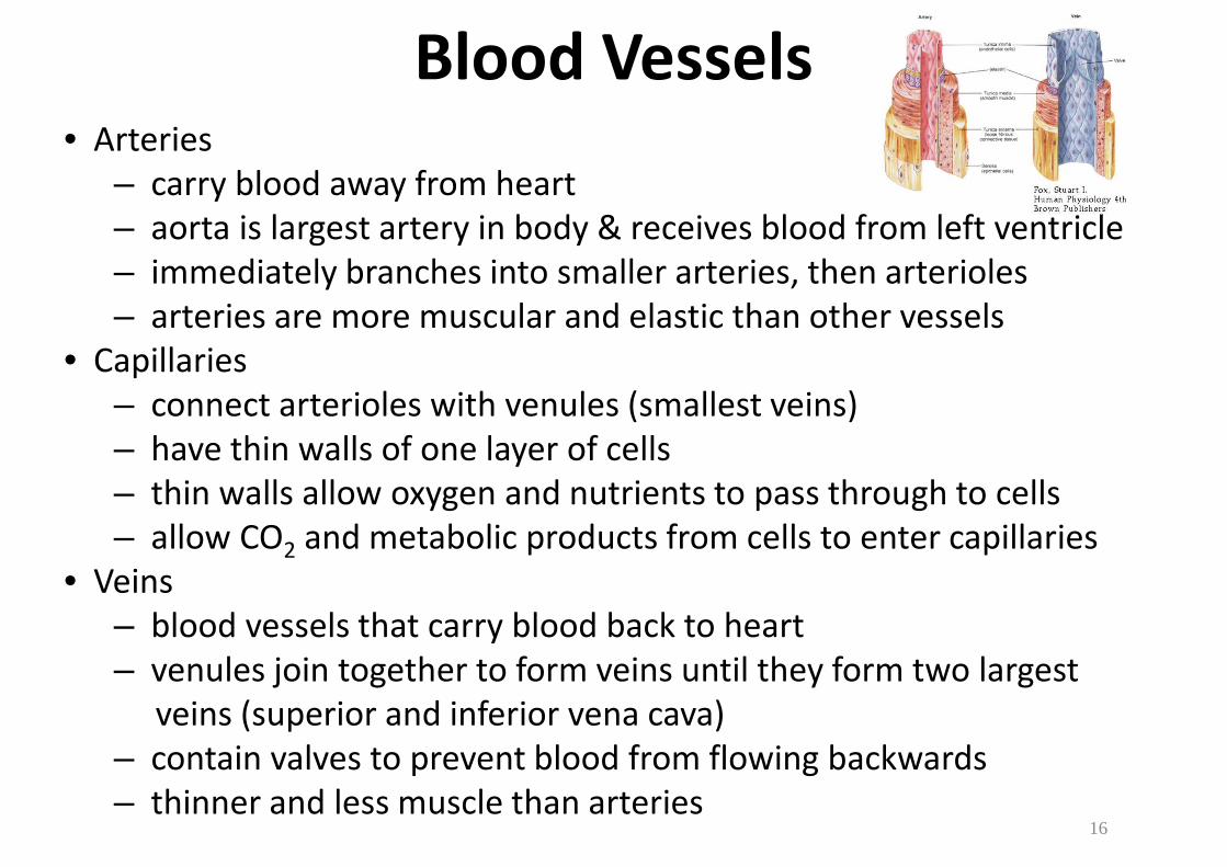
Blood Vessels• Arteries
– carry blood away from heart
– aorta is largest artery in body & receives blood from left ventricle
– immediately branches into smaller arteries, then arterioles
– arteries are more muscular and elastic than other vessels
• Capillaries
– connect arterioles with venules (smallest veins)
– have thin walls of one layer of cells
– thin walls allow oxygen and nutrients to pass through to cells
– allow CO2 and metabolic products from cells to enter capillaries
• Veins
– blood vessels that carry blood back to heart
– venules join together to form veins until they form two largest
veins (superior and inferior vena cava)
– contain valves to prevent blood from flowing backwards
– thinner and less muscle than arteries16
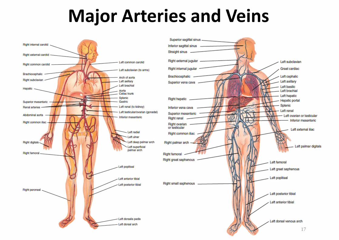
Major Arteries and Veins
17
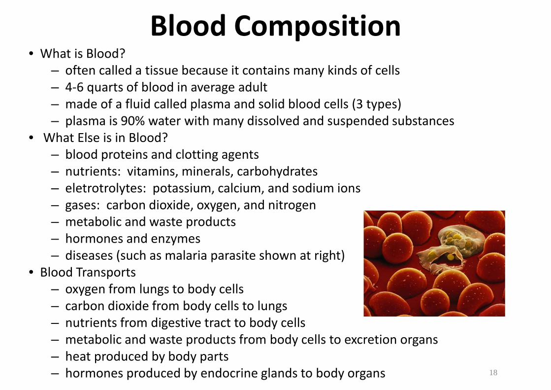
Blood Composition• What is Blood?
– often called a tissue because it contains many kinds of cells
– 4-6 quarts of blood in average adult
– made of a fluid called plasma and solid blood cells (3 types)
– plasma is 90% water with many dissolved and suspended substances
• What Else is in Blood?
– blood proteins and clotting agents
– nutrients: vitamins, minerals, carbohydrates
– eletrotrolytes: potassium, calcium, and sodium ions
– gases: carbon dioxide, oxygen, and nitrogen
– metabolic and waste products
– hormones and enzymes
– diseases (such as malaria parasite shown at right)
• Blood Transports
– oxygen from lungs to body cells
– carbon dioxide from body cells to lungs
– nutrients from digestive tract to body cells
– metabolic and waste products from body cells to excretion organs
– heat produced by body parts
– hormones produced by endocrine glands to body organs 18
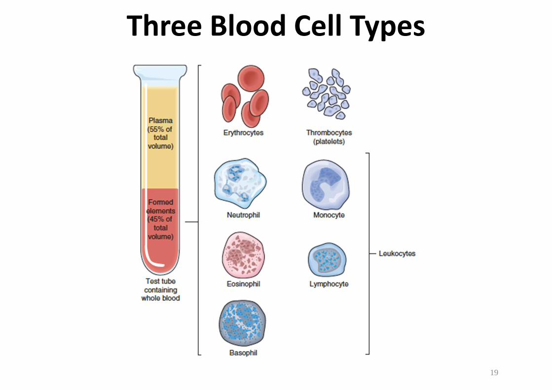
Three Blood Cell Types
19
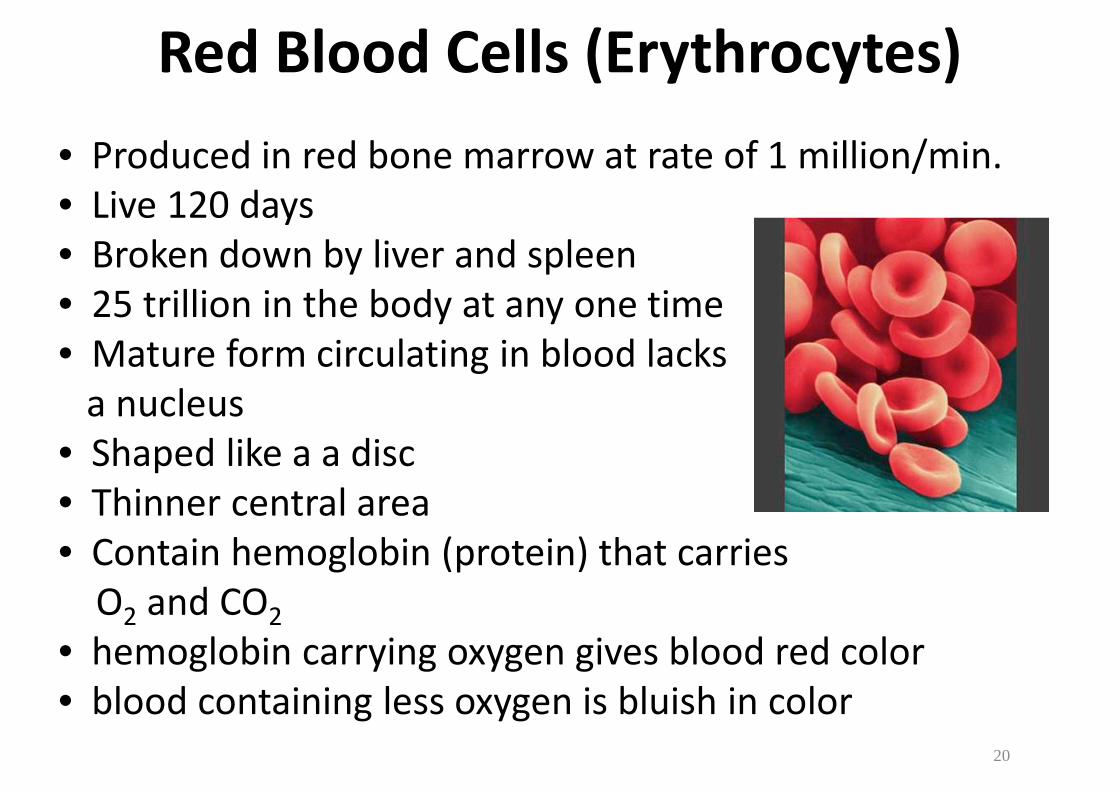
Red Blood Cells (Erythrocytes)
• Produced in red bone marrow at rate of 1 million/min.
• Live 120 days
• Broken down by liver and spleen
• 25 trillion in the body at any one time
• Mature form circulating in blood lacks
a nucleus
• Shaped like a a disc
• Thinner central area
• Contain hemoglobin (protein) that carries
O2 and CO2
• hemoglobin carrying oxygen gives blood red color
• blood containing less oxygen is bluish in color
20
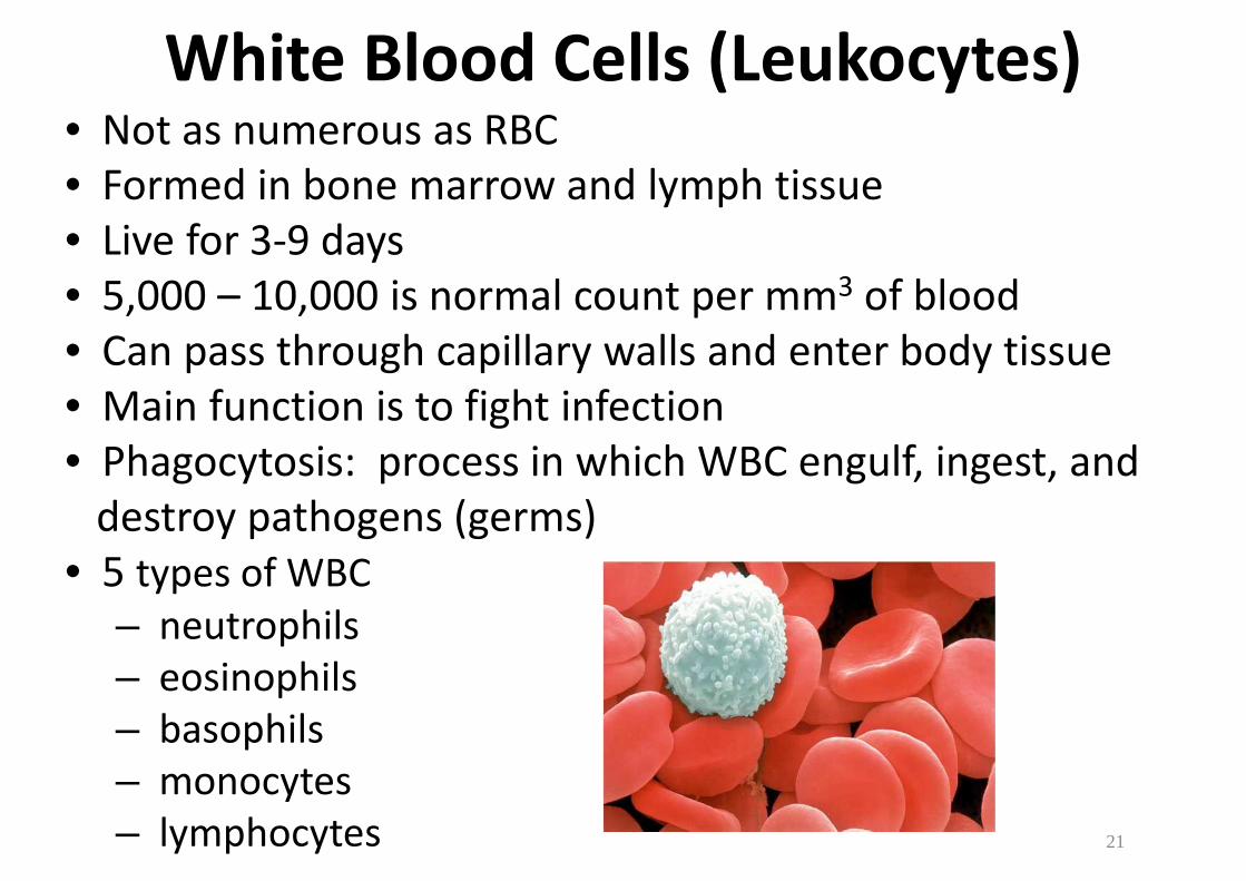
White Blood Cells (Leukocytes)• Not as numerous as RBC
• Formed in bone marrow and lymph tissue
• Live for 3-9 days
• 5,000 – 10,000 is normal count per mm3 of blood
• Can pass through capillary walls and enter body tissue
• Main function is to fight infection
• Phagocytosis: process in which WBC engulf, ingest, and
destroy pathogens (germs)
• 5 types of WBC
– neutrophils
– eosinophils
– basophils
– monocytes
– lymphocytes 21

Platelets (Thrombocytes)
• Pieces of cells
• Shown in middle at right
• Lack nuclei
• Vary in shape and size
• Formed in bone marrow
• Live for 5-9 days
• Normal count is 250,000 – 400,000 per mm3 of blood
• Important for clotting process (stop bleeding)
• Collect at sight of cut and form a sticky plug
• Secrete serotonin that causes blood vessel to spasm and
narrow (decreasing blood flow)
• Release enzyme that forms net-like fiber/clot22

Diseases & Abnormalities• Anemia
– inadequate number of RBC, hemoglobin, or both
– types: iron deficiency, aplastic, pernicious, and sickle cell
• Aneurysm
– ballooning out of, or saclike formation of artery wall
• Arteriosclerosis
– hardening or thickening of arterial walls (result of aging)
– causes high blood pressure (hypertension)
• Atherosclerosis
– fatty plaques (often cholesterol) deposit on
artery walls
• Congestive Heart Failure (CHF)
– heart muscles do not beat adequately to supply blood
• Embolus
– foreign substance in bloodstream 23

Diseases & Abnormalities Continued• Hemophilia
– inherited disease common in males
– blood is not able to clot (missing plasma protein)
• Hypertension
– high blood pressure (above 140 / 90 mm Hg)
• Leukemia
– malignant disease of bone marrow or lymph tissue
– results in high number of immature WBC
• Myocardial Infraction
– heart attack
– caused by blockage in coronary arteries
– cuts off supply of blood to heart
• Phlebitis
– inflammation of vein, frequently in legs
• Varicose Veins
– swollen, dilated veins that have lost elasticity24