common lesions and conditions of the oral...
TRANSCRIPT
Common Lesions and Conditions of the Oral Cavity
K. Mark Anderson DDS, MS
University of Tennessee College of Dentistry

Everyday Lumps and Bumps
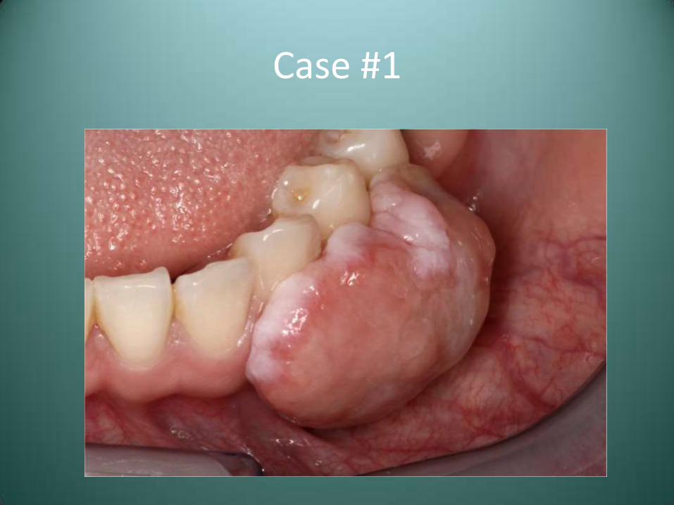
Case #1
• This patient presented with the gingival swelling seen here
Case #1
Case #2
• A 14 year old female presented with this lesion of the gingiva

Case #2

Cases 1 and 2
Differential Diagnosis – “The 3 P’s”
• Pyogenic Granuloma
• Peripheral Ossifying Fibroma
• Peripheral Giant Cell Granuloma
Pyogenic Granuloma (Pregnancy Tumor)
• Common non-neoplastic proliferation of granulation tissue
• Not a true granuloma
• Response to local irritation or trauma
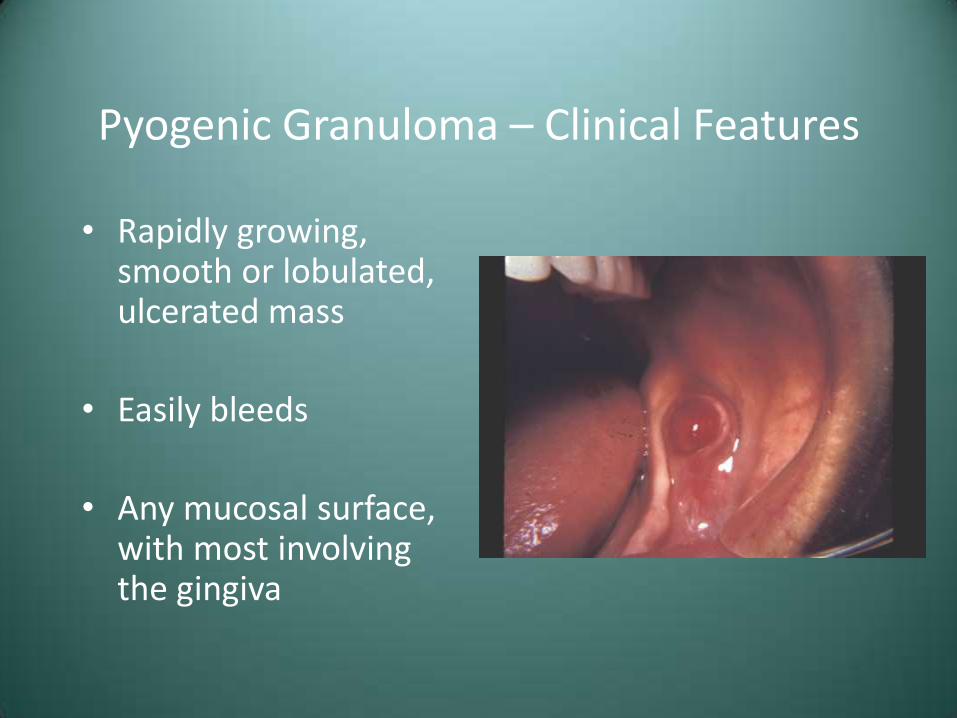
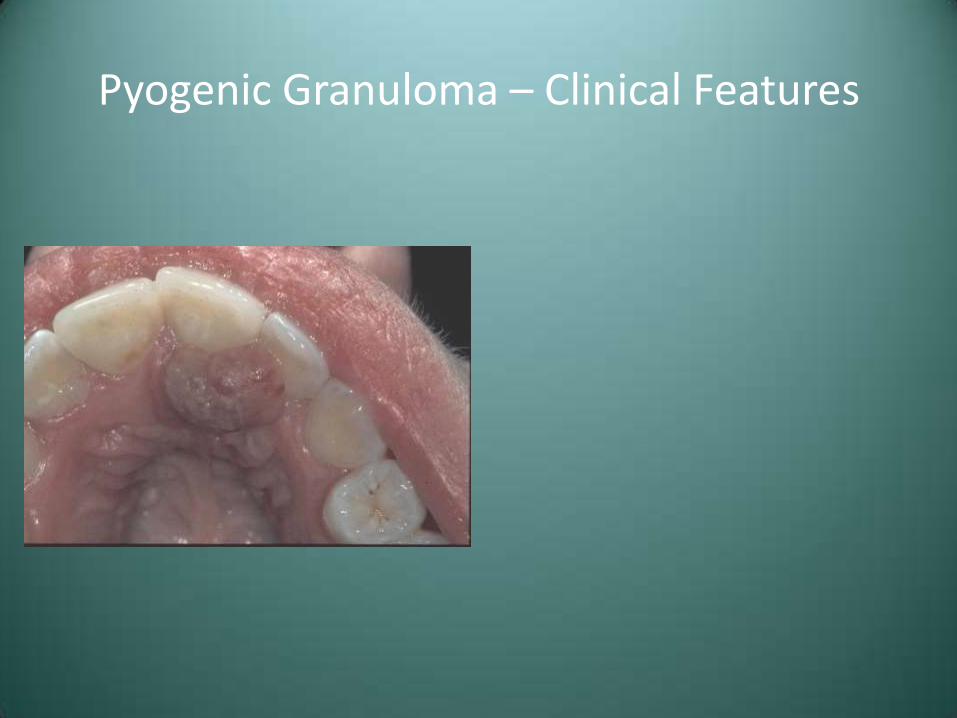
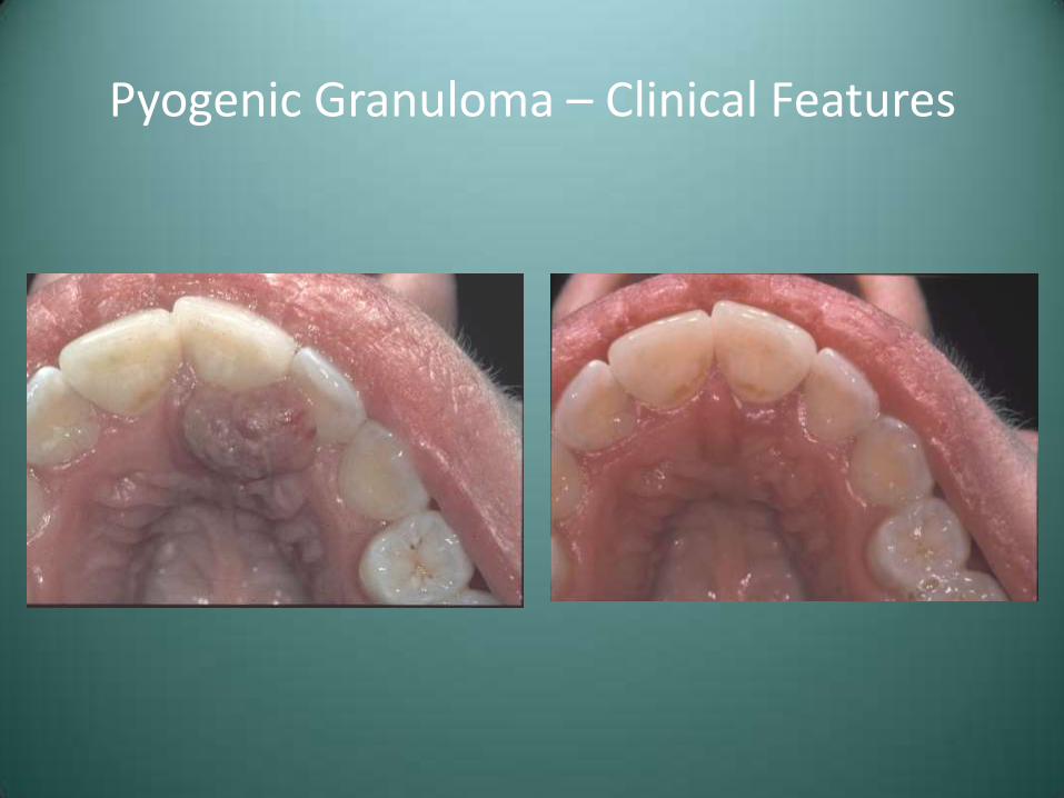
Pyogenic Granuloma – Clinical Features
• F>M, children and young adults
• Common during pregnancy
Pyogenic Granuloma – Clinical Features
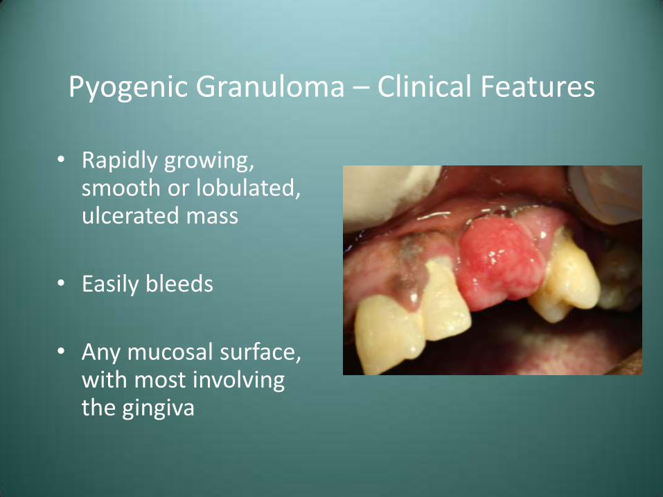
• Rapidly growing, smooth or lobulated, ulcerated mass
• Easily bleeds
• Any mucosal surface, with most involving the gingiva
Pyogenic Granuloma – Clinical Features
• Rapidly growing, smooth or lobulated, ulcerated mass
• Easily bleeds
• Any mucosal surface, with most involving the gingiva
Pyogenic Granuloma – Clinical Features
Pyogenic Granuloma – Clinical Features

Pyogenic Granuloma – Treatment and Prognosis
• Conservative surgical excision with removal of any local factors
• Lesions associated with pregnancy may spontaneously regress postpartum
• Recurrences occur due to remaining local factors (calculus)
Peripheral Ossifying Fibroma
• Relatively common reactive lesion, probably arising from periodontal ligament
• This lesion is unrelated to the central ossifying fibroma
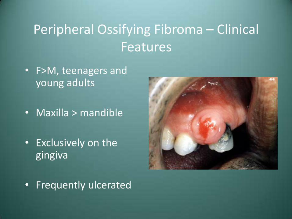
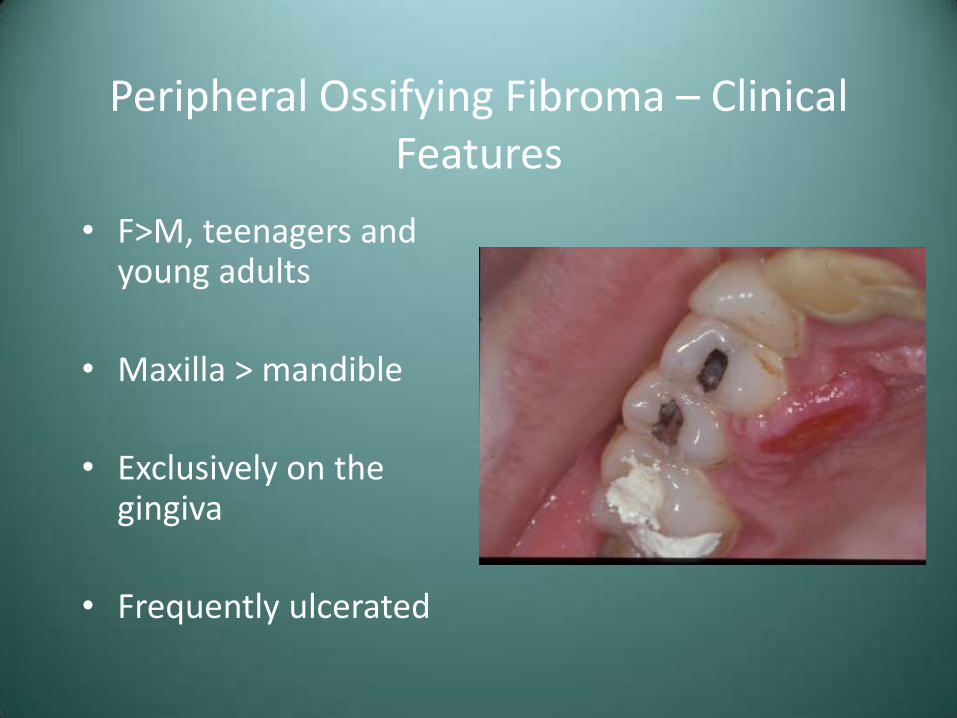
Peripheral Ossifying Fibroma – Clinical Features
• F>M, teenagers and young adults
• Maxilla > mandible
• Exclusively on the gingiva
• Frequently ulcerated
Peripheral Ossifying Fibroma – Clinical Features
• F>M, teenagers and young adults
• Maxilla > mandible
• Exclusively on the gingiva
• Frequently ulcerated
Peripheral Ossifying Fibroma – Clinical Features
• F>M, teenagers and young adults
• Maxilla > mandible
• Exclusively on the gingiva
• Frequently ulcerated
Peripheral Ossifying Fibroma – Treatment and Prognosis
• Local excision down to the periosteum
• Elimination of local factors or irritants
• Approximately 16% recurrence rate

Peripheral Giant Cell Granuloma
• Relatively common reactive lesion of the gingiva
• Histologically identical to the central giant cell granuloma
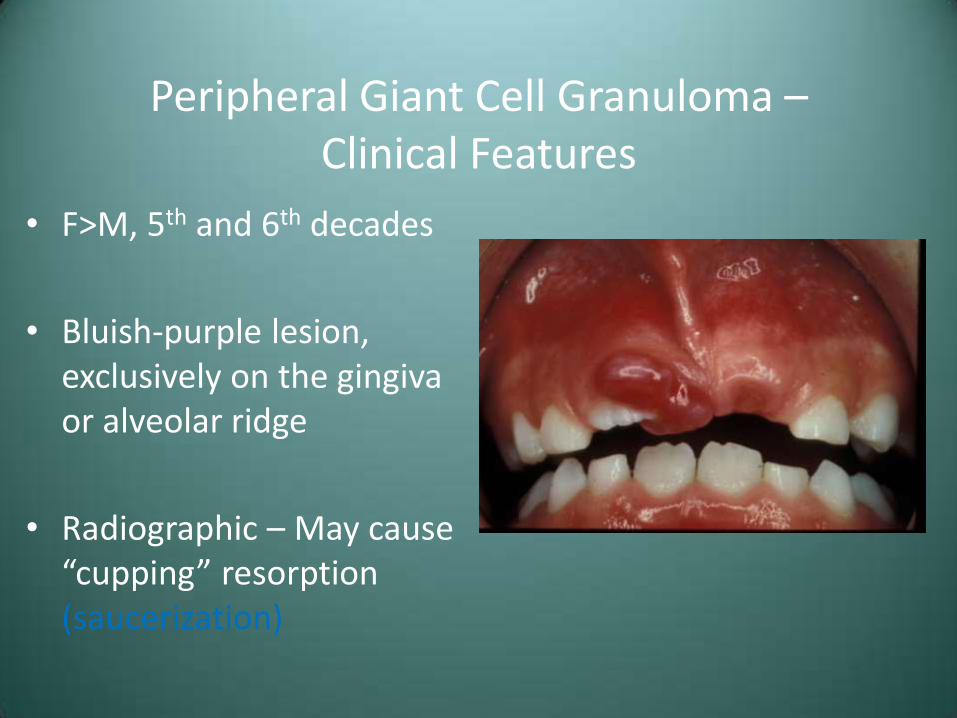
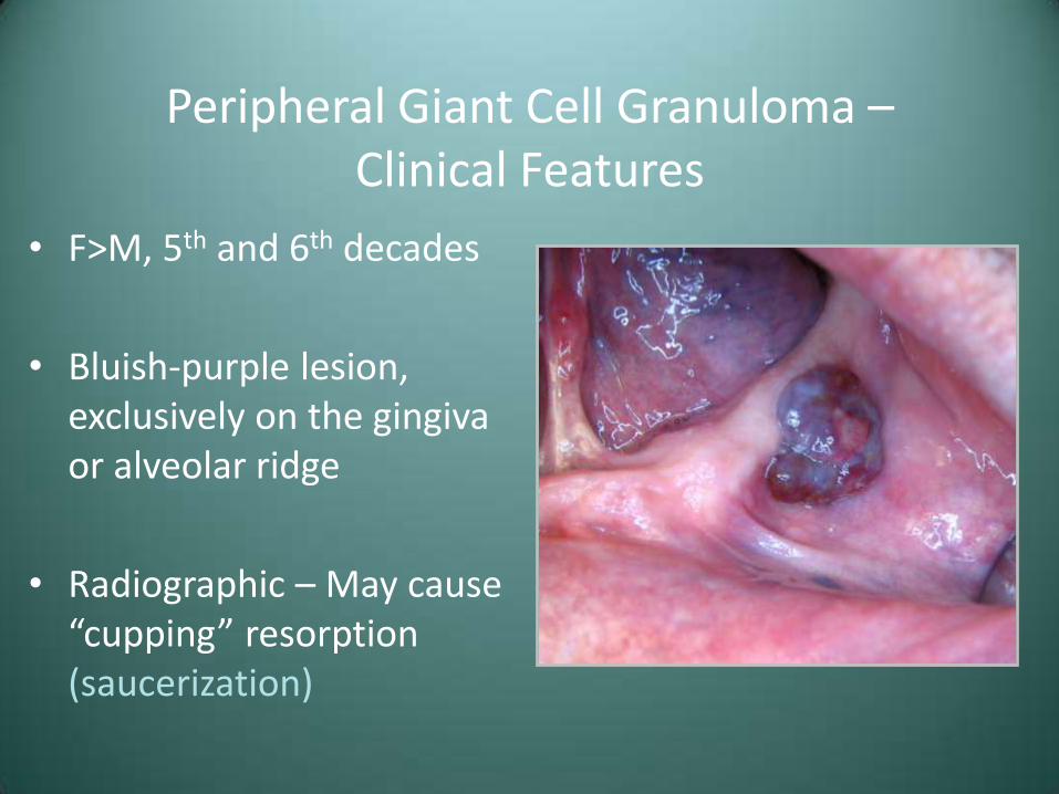
Peripheral Giant Cell Granuloma – Clinical Features
• F>M, 5th and 6th decades
• Bluish-purple lesion, exclusively on the gingiva or alveolar ridge
• Radiographic – May cause “cupping” resorption (saucerization)
Peripheral Giant Cell Granuloma – Clinical Features
• F>M, 5th and 6th decades
• Bluish-purple lesion, exclusively on the gingiva or alveolar ridge
• Radiographic – May cause “cupping” resorption (saucerization)

Peripheral Giant Cell Granuloma – Clinical Features
• F>M, 5th and 6th decades
• Bluish-purple lesion, exclusively on the gingiva or alveolar ridge
• Radiographic – May cause “cupping” resorption (saucerization)
Peripheral Giant Cell Granuloma – Clinical Features
• F>M, 5th and 6th decades
• Bluish-purple lesion, exclusively on the gingiva or alveolar ridge
• Radiographic – May cause “cupping” resorption (saucerization)
Peripheral Giant Cell Granuloma – Treatment and Prognosis
• Local excision down to underlying bone
• Removal of local factors
• Approximately 10% recurrence rate

Additional Considerations
Fibroma (Irritation Fibroma, Traumatic Fibroma)
• The most common tumor of the oral cavity
• Probably not a true neoplasm
• Reactive lesion, secondary to trauma or chronic irritation

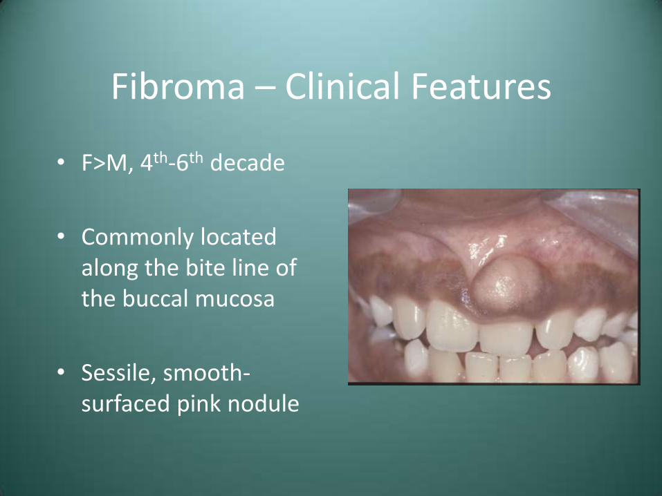
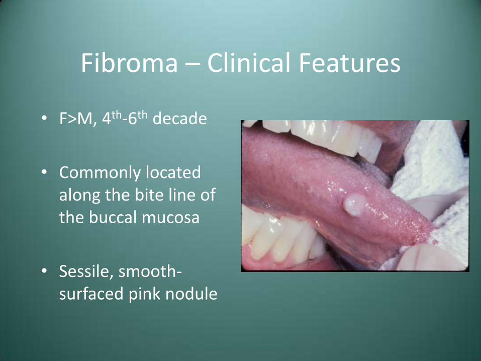
Fibroma – Clinical Features
• F>M, 4th-6th decade
• Commonly located along the bite line of the buccal mucosa
• Sessile, smooth-surfaced pink nodule
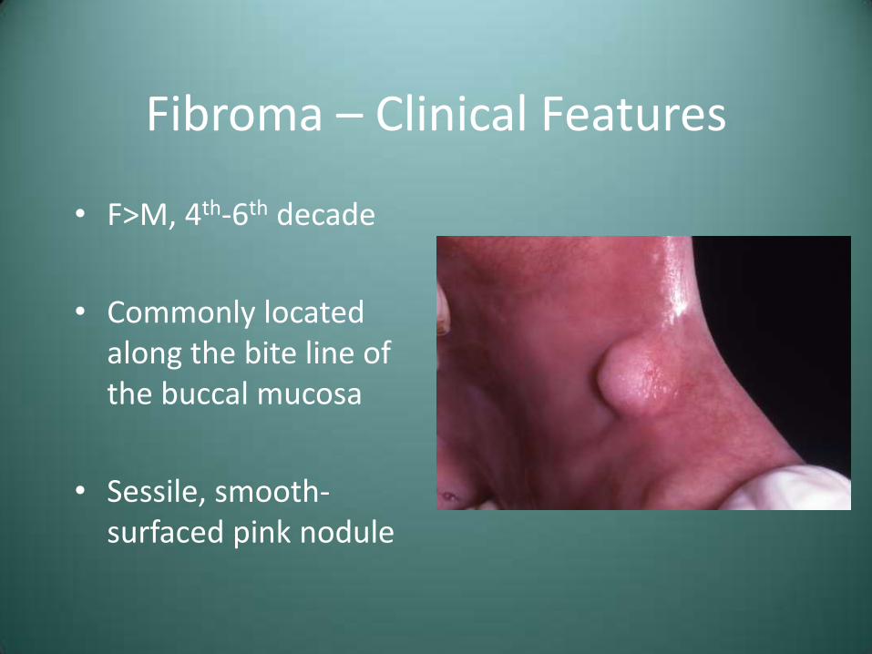
Fibroma – Clinical Features
• F>M, 4th-6th decade
• Commonly located along the bite line of the buccal mucosa
• Sessile, smooth-surfaced pink nodule
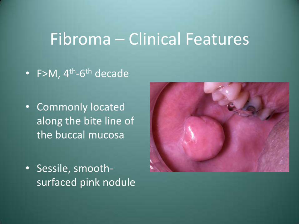
Fibroma – Clinical Features
• F>M, 4th-6th decade
• Commonly located along the bite line of the buccal mucosa
• Sessile, smooth-surfaced pink nodule
Fibroma – Clinical Features
• F>M, 4th-6th decade
• Commonly located along the bite line of the buccal mucosa
• Sessile, smooth-surfaced pink nodule
Fibroma – Clinical Features
• F>M, 4th-6th decade
• Commonly located along the bite line of the buccal mucosa
• Sessile, smooth-surfaced pink nodule
Fibroma – Treatment
• Conservative surgical excision
• Prognosis – Recurrence is rare

Differential Diagnosis
• Pyogenic Granuloma
• Peripheral Ossifying Fibroma
• Peripheral Giant Cell Granuloma

Diagnosis Case #1 – Pyogenic Granuloma

Diagnosis Case #2 – Peripheral Ossifying Fibroma
Other Soft Tissue Considerations
Lipoma
• Benign tumor of fat
• Although rare in the oral/maxillofacial area, the lipoma is the most common mesenchymal neoplasm
• Unrelated to metabolism/body fat

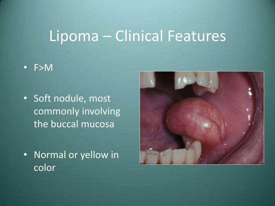
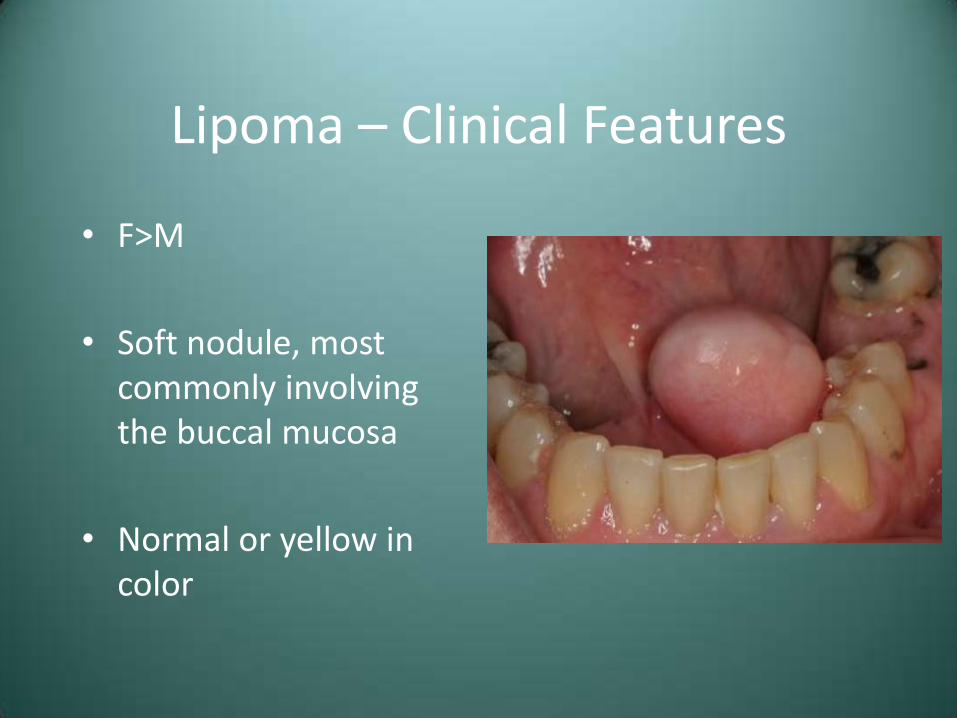
Lipoma – Clinical Features
• F>M
• Soft nodule, most commonly involving the buccal mucosa
• Normal or yellow in color
Lipoma – Clinical Features
• F>M
• Soft nodule, most commonly involving the buccal mucosa
• Normal or yellow in color
Lipoma – Clinical Features
• F>M
• Soft nodule, most commonly involving the buccal mucosa
• Normal or yellow in color

Lipoma – Clinical Features
• F>M
• Soft nodule, most commonly involving the buccal mucosa
• Normal or yellow in color
Lipoma – Clinical Features
• F>M
• Soft nodule, most commonly involving the buccal mucosa
• Normal or yellow in color

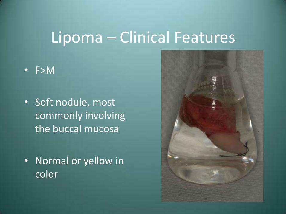
Lipoma – Treatment and Prognosis
• Conservative surgical excision
• Recurrence is rare
Granular Cell Tumor
• Uncommon tumor that appears to be of Schwann cell origin
• Significant predilection for the oral cavity
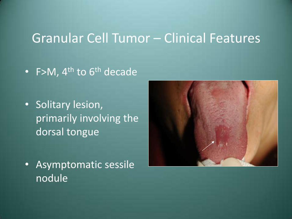
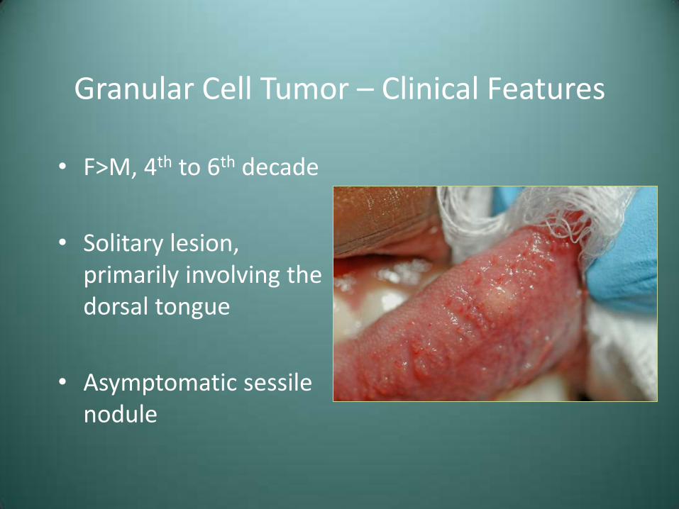
Granular Cell Tumor – Clinical Features
• F>M, 4th to 6th decade
• Solitary lesion, primarily involving the dorsal tongue
• Asymptomatic sessile nodule
Granular Cell Tumor – Clinical Features
• F>M, 4th to 6th decade
• Solitary lesion, primarily involving the dorsal tongue
• Asymptomatic sessile nodule
Granular Cell Tumor – Clinical Features
• F>M, 4th to 6th decade
• Solitary lesion, primarily involving the dorsal tongue
• Asymptomatic sessile nodule
Granular Cell Tumor – Treatment and Prognosis
• Conservative surgical excision
• Recurrence is rare, even with incomplete removal

Traumatic Neuroma
• Reactive proliferation of neural tissue
• Not necessarily a true neoplasm
• Secondary to disruption of Schwann cell tube
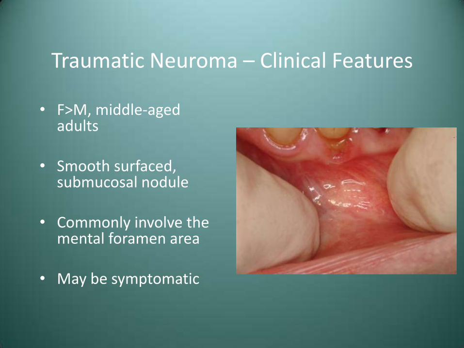
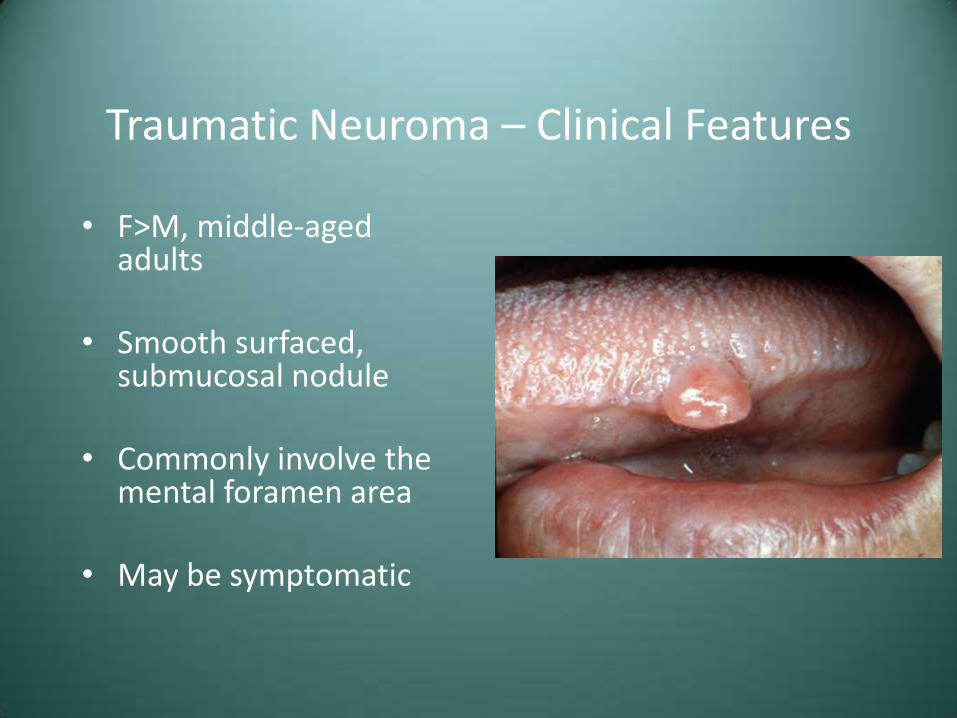
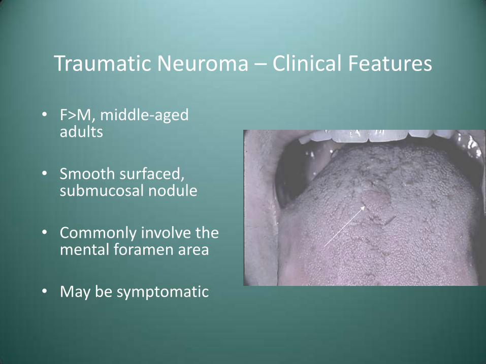
Traumatic Neuroma – Clinical Features
• F>M, middle-aged adults
• Smooth surfaced, submucosal nodule
• Commonly involve the mental foramen area
• May be symptomatic
Traumatic Neuroma – Clinical Features
• F>M, middle-aged adults
• Smooth surfaced, submucosal nodule
• Commonly involve the mental foramen area
• May be symptomatic
Traumatic Neuroma – Clinical Features
• F>M, middle-aged adults
• Smooth surfaced, submucosal nodule
• Commonly involve the mental foramen area
• May be symptomatic
Traumatic Neuroma – Clinical Features
• F>M, middle-aged adults
• Smooth surfaced, submucosal nodule
• Commonly involve the mental foramen area
• May be symptomatic

Traumatic Neuroma – Treatment and Prognosis
• Surgical excision, including a portion of the involved nerve bundle
• Recurrence is not expected
Schwannoma (Neurilomoma)
• Benign neural tumor of Schwann cell origin
• Uncommon, but often involve the head and neck
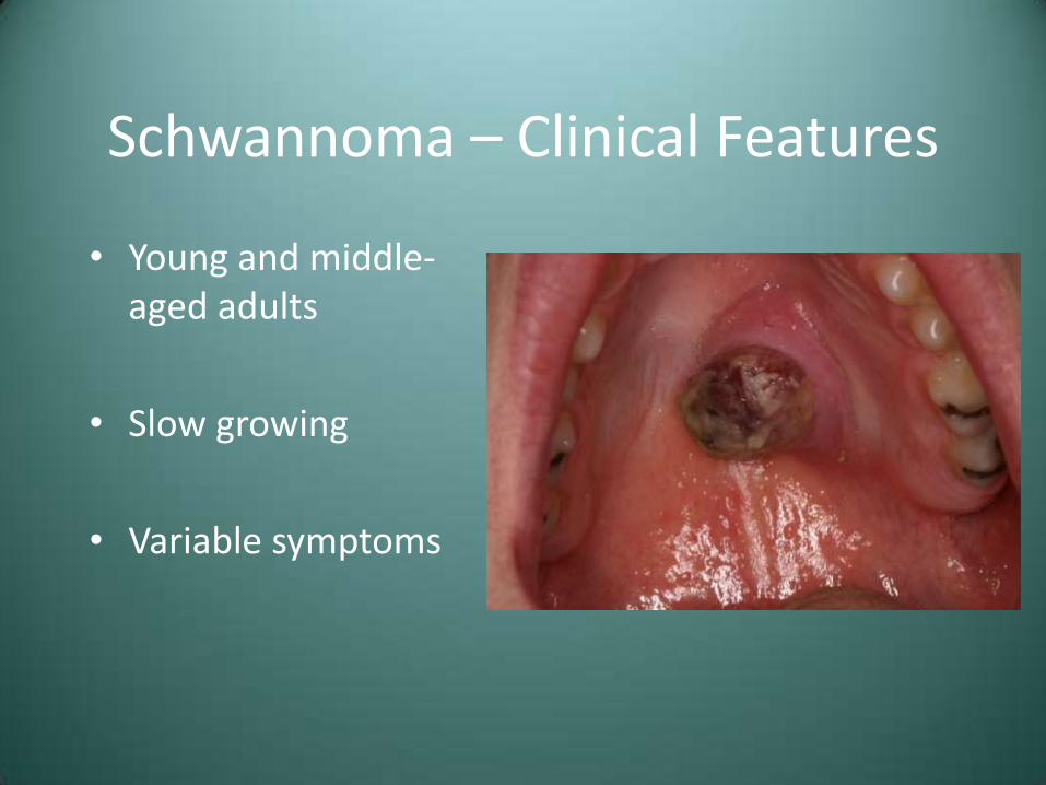
Schwannoma – Clinical Features
• Young and middle-aged adults
• Slow growing
• Variable symptoms

Schwannoma – Clinical Features
• Young and middle-aged adults
• Slow growing
• Variable symptoms
Schwannoma – Clinical Features
• Young and middle-aged adults
• Slow growing
• Variable symptoms

Schwannoma – Clinical Features
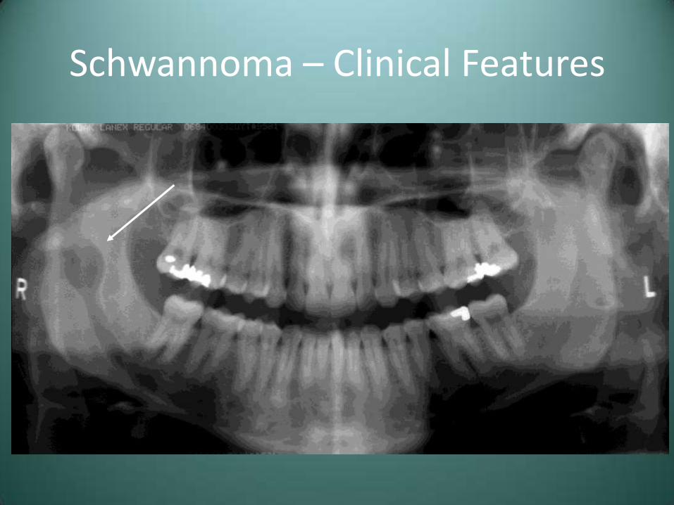
• Oral tumors most commonly involve the tongue
• May arise within bone, causing an expansile, unilocular radiolucency
Schwannoma – Clinical Features
Schwannoma – Treatment and Prognosis
• Surgical excision
• Recurrence is not expected
• Malignant transformation is rare
– Malignant peripheral nerve sheath tumor, malignant schwannoma, neurofibrosarcoma

Neurofibroma
• The most common peripheral nerve neoplasm
• Tumor cells are a mixture of Schwann cells and fibroblasts
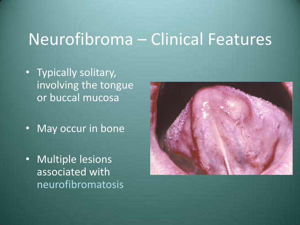
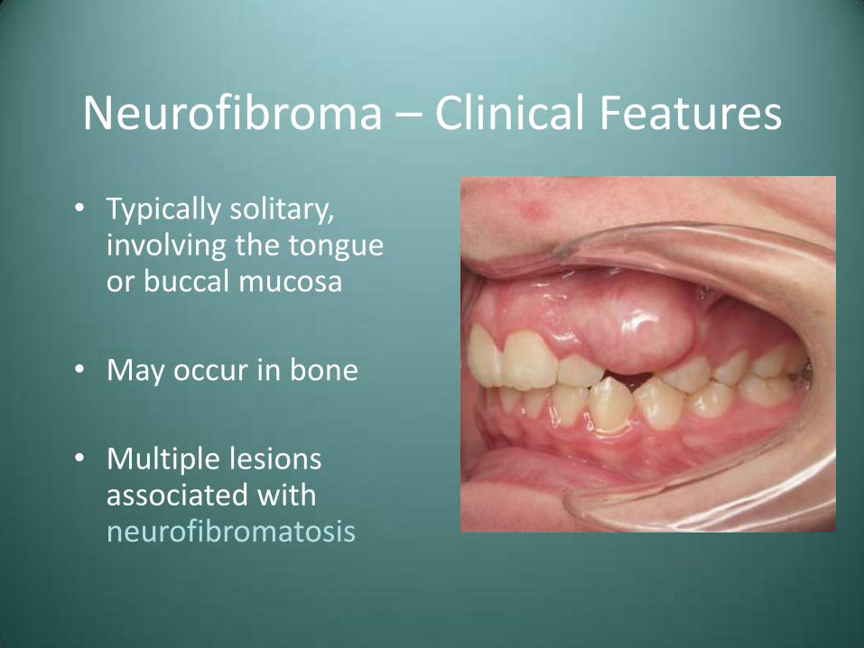
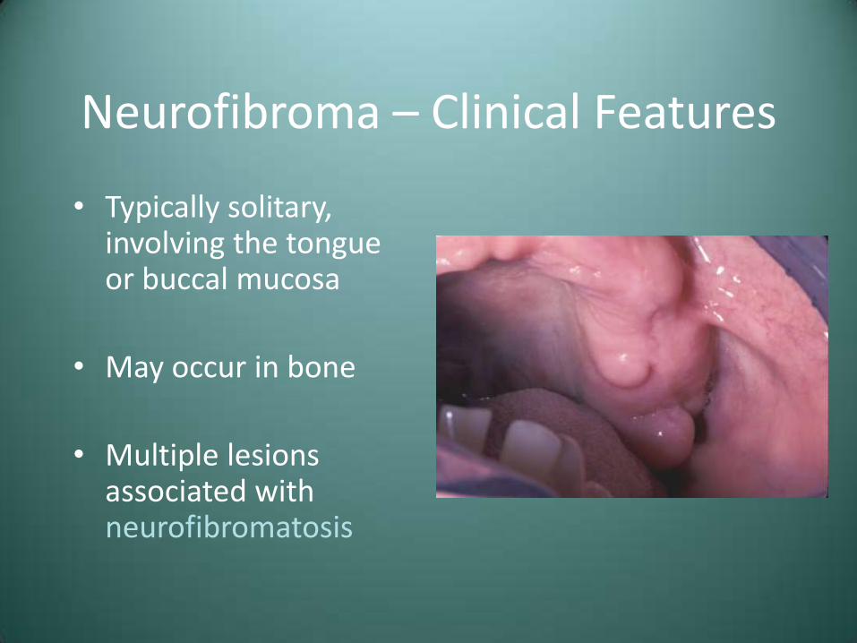
Neurofibroma – Clinical Features
• Typically solitary, involving the tongue or buccal mucosa
• May occur in bone
• Multiple lesions associated with neurofibromatosis
Neurofibroma – Clinical Features
• Typically solitary, involving the tongue or buccal mucosa
• May occur in bone
• Multiple lesions associated with neurofibromatosis
Neurofibroma – Clinical Features
• Typically solitary, involving the tongue or buccal mucosa
• May occur in bone
• Multiple lesions associated with neurofibromatosis
Neurofibroma – Clinical Features
• Typically solitary, involving the tongue or buccal mucosa
• May occur in bone
• Multiple lesions associated with neurofibromatosis

Neurofibroma – Clinical Features
• Typically solitary, involving the tongue or buccal mucosa
• May occur in bone
• Multiple lesions associated with neurofibromatosis

Neurofibroma – Clinical Features
• Typically solitary, involving the tongue or buccal mucosa
• May occur in bone
• Multiple lesions associated with neurofibromatosis
Neurofibroma – Treatment and Prognosis
• Solitary lesions – Surgical excision
• Multiple (neurofibromatosis) – Removal of symptomatic lesions
• Malignant transformation is possible, much more so
in patients with neurofibromatosis
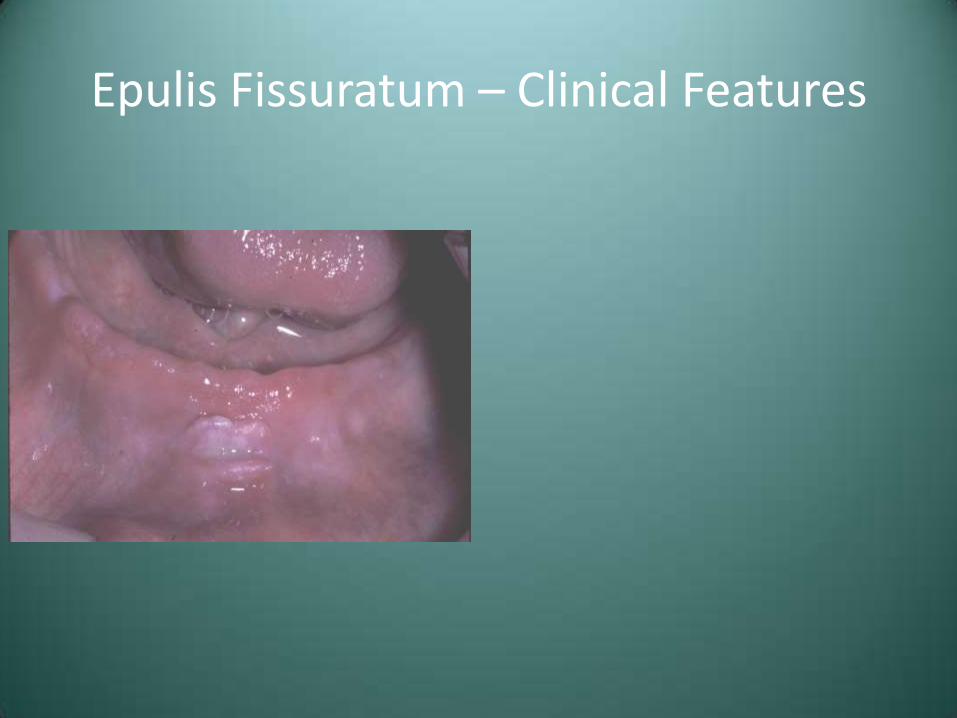
Epulis Fissuratum (Inflammatory Fibrous Hyperplasia, “Denture Epulis”)
• Reactive lesion that occurs secondary to irritation from an ill-fitting denture
• Epulis – Any tumor of the gingiva or alveolar mucosa
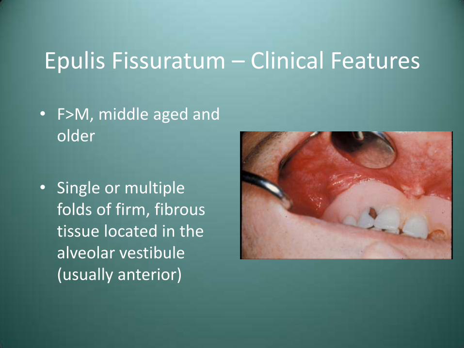
Epulis Fissuratum – Clinical Features
• F>M, middle aged and older
• Single or multiple folds of firm, fibrous tissue located in the alveolar vestibule (usually anterior)
Epulis Fissuratum – Clinical Features
• F>M, middle aged and older
• Single or multiple folds of firm, fibrous tissue located in the alveolar vestibule (usually anterior)
Epulis Fissuratum – Clinical Features
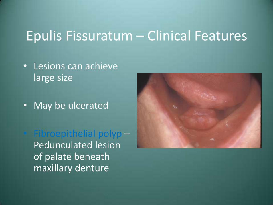
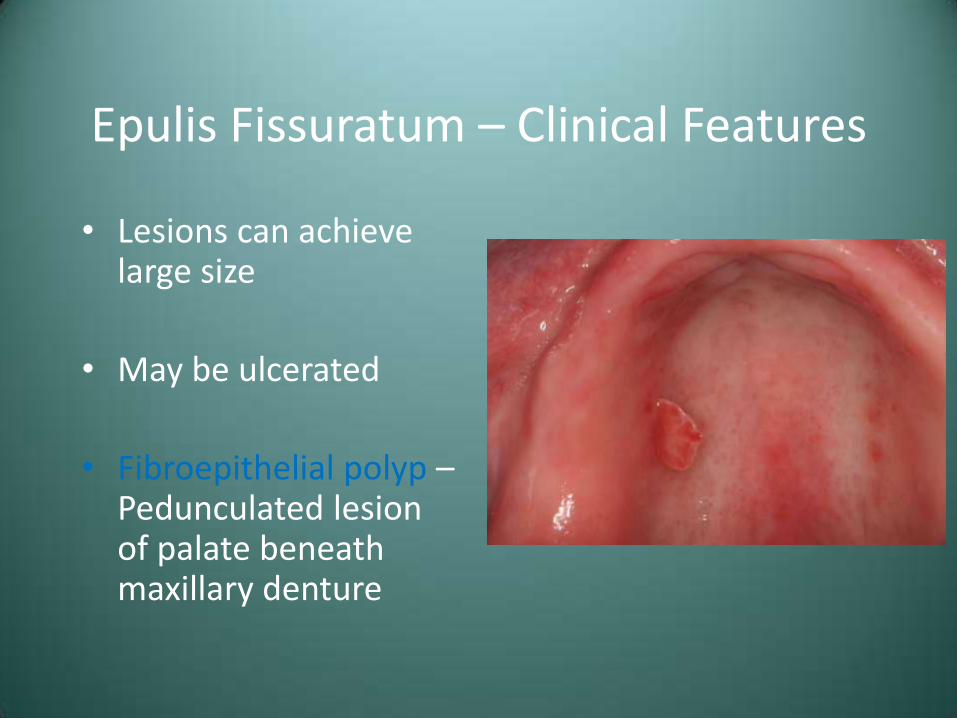
• Lesions can achieve large size
• May be ulcerated
• Fibroepithelial polyp – Pedunculated lesion of palate beneath maxillary denture

Epulis Fissuratum – Clinical Features
• Lesions can achieve large size
• May be ulcerated
• Fibroepithelial polyp – Pedunculated lesion of palate beneath maxillary denture
Epulis Fissuratum – Clinical Features
• Lesions can achieve large size
• May be ulcerated
• Fibroepithelial polyp – Pedunculated lesion of palate beneath maxillary denture
Epulis Fissuratum – Clinical Features

Epulis Fissuratum – Clinical Features
Epulis Fissuratum – Treatment and Prognosis
• Surgical removal
• Refabrication of the associated denture or relign
Erythematous Candidiasis - Denture Stomatitis
• Often referred to as “chronic atrophic candidiasis”
• Denture is often contaminated with candidal organisms, but no invasion of mucosa is seen
• Erythema of palatal denture-bearing area-typically asymptomatic
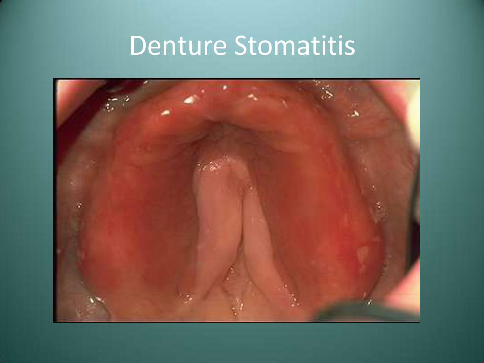
Denture Stomatitis
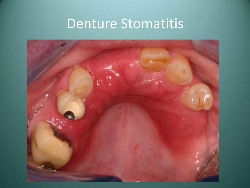
Denture Stomatitis
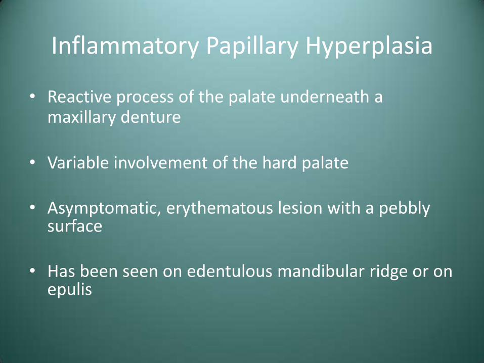
Inflammatory Papillary Hyperplasia
• Reactive process of the palate underneath a maxillary denture
• Variable involvement of the hard palate
• Asymptomatic, erythematous lesion with a pebbly surface
• Has been seen on edentulous mandibular ridge or on epulis
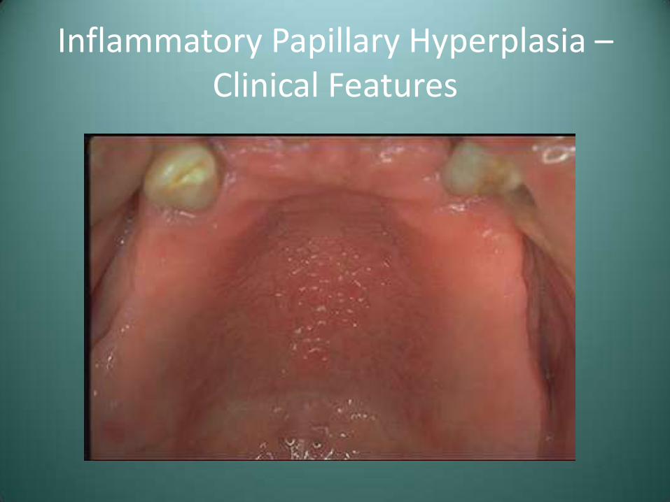
Inflammatory Papillary Hyperplasia – Clinical Features
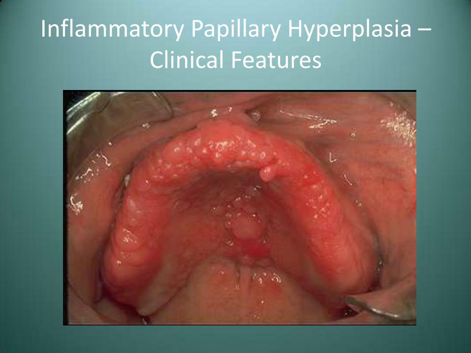
Inflammatory Papillary Hyperplasia – Clinical Features
Oral Squamous Papilloma
• Probably caused by human papillomavirus (HPV)
– Over 100 HPV types identified
– Types 6 and 11 are most commonly associated with oral papillomas
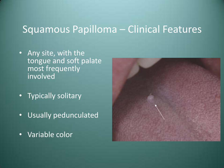
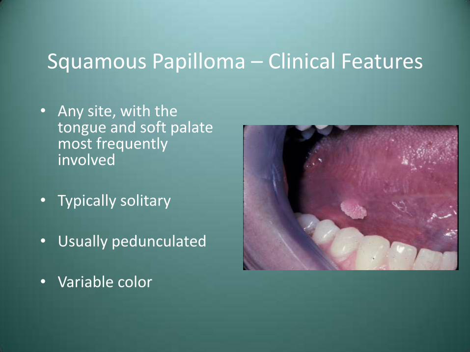
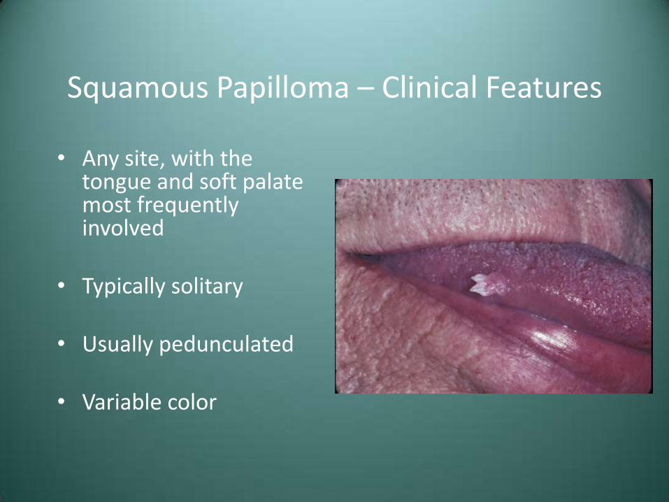
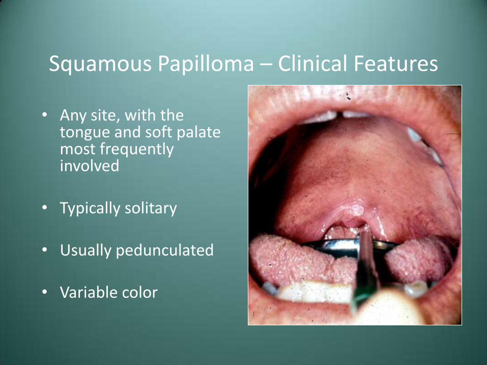
Squamous Papilloma – Clinical Features
• Any site, with the tongue and soft palate most frequently involved
• Typically solitary
• Usually pedunculated
• Variable color
Squamous Papilloma – Clinical Features
• Any site, with the tongue and soft palate most frequently involved
• Typically solitary
• Usually pedunculated
• Variable color

Squamous Papilloma – Clinical Features
• Any site, with the tongue and soft palate most frequently involved
• Typically solitary
• Usually pedunculated
• Variable color
Squamous Papilloma – Clinical Features
• Any site, with the tongue and soft palate most frequently involved
• Typically solitary
• Usually pedunculated
• Variable color
Squamous Papilloma – Clinical Features
• Any site, with the tongue and soft palate most frequently involved
• Typically solitary
• Usually pedunculated
• Variable color
Squamous Papilloma – Clinical Features
• Any site, with the tongue and soft palate most frequently involved
• Typically solitary
• Usually pedunculated
• Variable color

Squamous Papilloma - Treatment
• Surgical excision
• Recurrence is not expected, although lesions of the larynx may behave differently
– Laryngeal papillomatosis
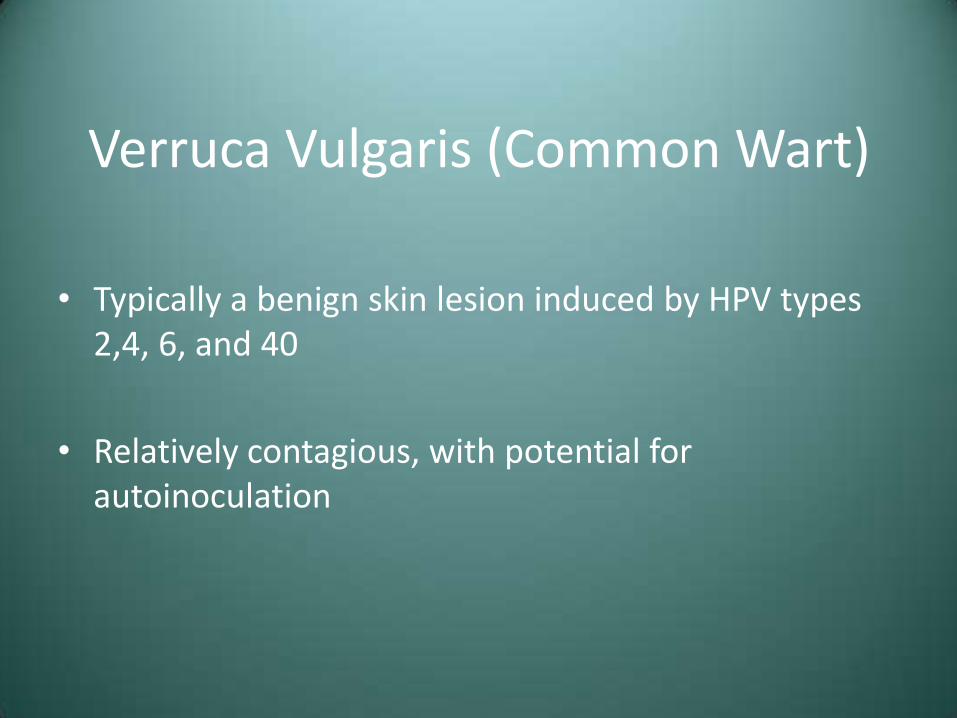
Verruca Vulgaris (Common Wart)
• Typically a benign skin lesion induced by HPV types 2,4, 6, and 40
• Relatively contagious, with potential for autoinoculation

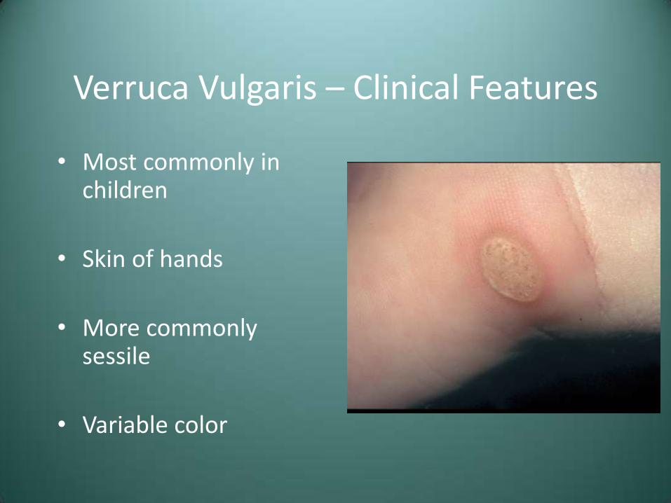
Verruca Vulgaris – Clinical Features
• Most commonly in children
• Skin of hands
• More commonly sessile
• Variable color
Verruca Vulgaris – Clinical Features
• Most commonly in children
• Skin of hands
• More commonly sessile
• Variable color

Verruca Vulgaris – Clinical Features
• Oral lesions uncommon
• Often indistinguishable from squamous papilloma
• Oral lesions typically appear white

Verruca Vulgaris - Treatment
• Surgical excision or curettage
• Liquid nitrogen, cryotherapy, or keratinolytic agents
• May spontaneously resolve
• Small rate of recurrence
Condyloma Acuminatum
• Also known as “venereal warts”
• Caused by several strains of HPV, including types 2, 6,11,16,18

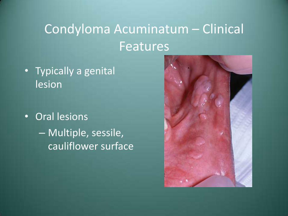
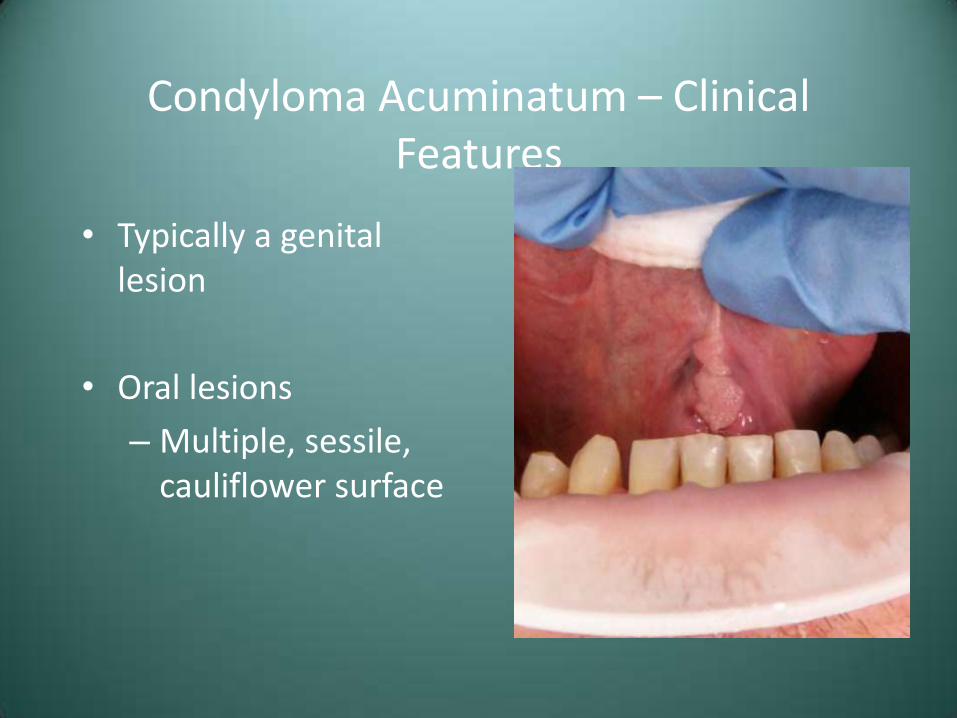
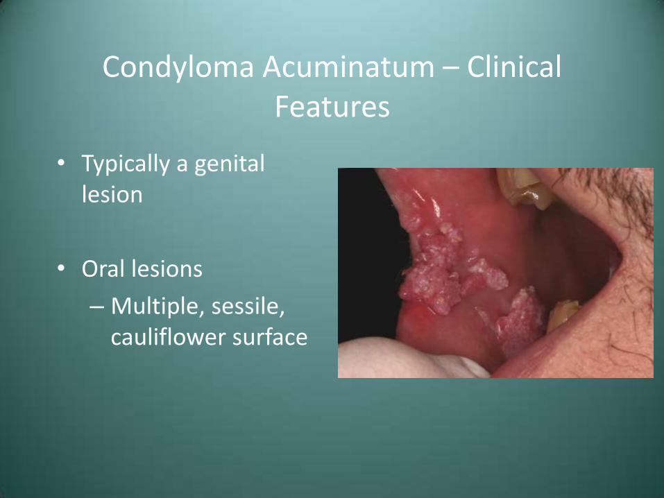
Condyloma Acuminatum – Clinical Features
• Typically a genital lesion
• Oral lesions
– Multiple, sessile, cauliflower surface
Condyloma Acuminatum – Clinical Features
• Typically a genital lesion
• Oral lesions
– Multiple, sessile, cauliflower surface
Condyloma Acuminatum – Clinical Features
• Typically a genital lesion
• Oral lesions
– Multiple, sessile, cauliflower surface
Condyloma Acuminatum – Clinical Features
• Typically a genital lesion
• Oral lesions
– Multiple, sessile, cauliflower surface
Condyloma Acuminatum
• Excision, cryotherapy, laser excision
• Recurrence is common-30% of patients have recurrent lesions after each treatment episode
• Associated with squamous cell carcinoma of the uterine cervix

Ulcerative Conditions of the Oral Regions

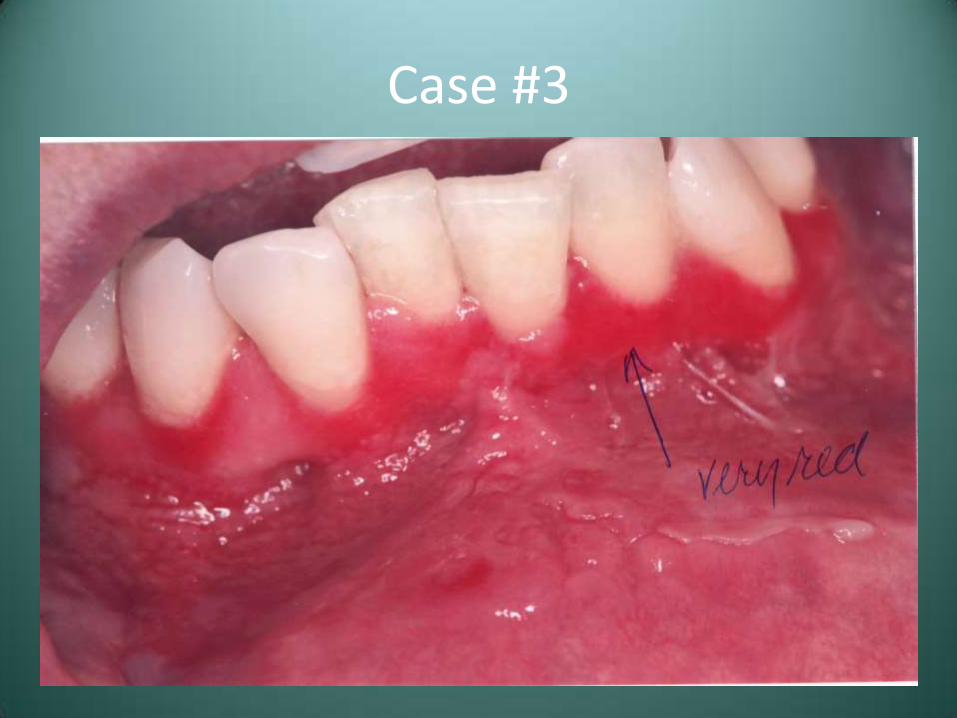
Case #3
• A 47 year old female presented with a history of these painful lesions
Case #3
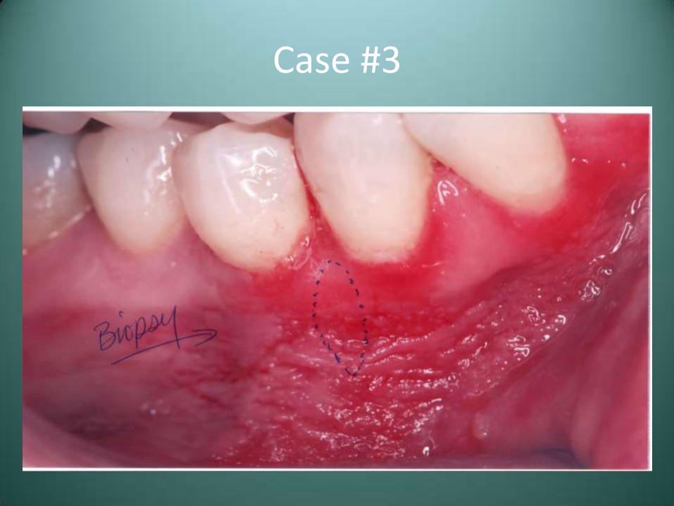
Case #3
Case #3
• Clinical Diagnosis – “Desquamative Gingivitis”
• Differential Diagnosis
– Lichen Planus
– Cicatricial Pemphigoid
– Pemphigus Vulgaris
Lichen Planus
• Common chronic mucocutaneous disease
• Probably immune-mediated
• May have only skin, only oral, or both
Lichen Planus – Clinical Features
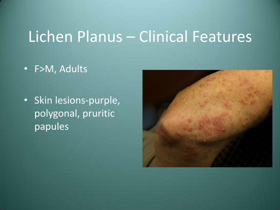
• F>M, Adults
• Skin lesions-purple, polygonal, pruritic papules
Lichen Planus – Clinical Features
• F>M, Adults
• Skin lesions-purple, polygonal, pruritic papules

Lichen Planus – Clinical Features
• Oral lesions-reticular or erosive
• Reticular-interlacing white lines, buccal mucosa
• Erosive-ulcers with erythema and white streaks

Lichen Planus – Clinical Features
• Oral lesions-reticular or erosive
• Reticular-interlacing white lines, buccal mucosa
• Erosive-ulcers with erythema and white streaks

Lichen Planus – Clinical Features
• Oral lesions-reticular or erosive
• Reticular-interlacing white lines, buccal mucosa
• Erosive-ulcers with erythema and white streaks
Lichen Planus – Clinical Features
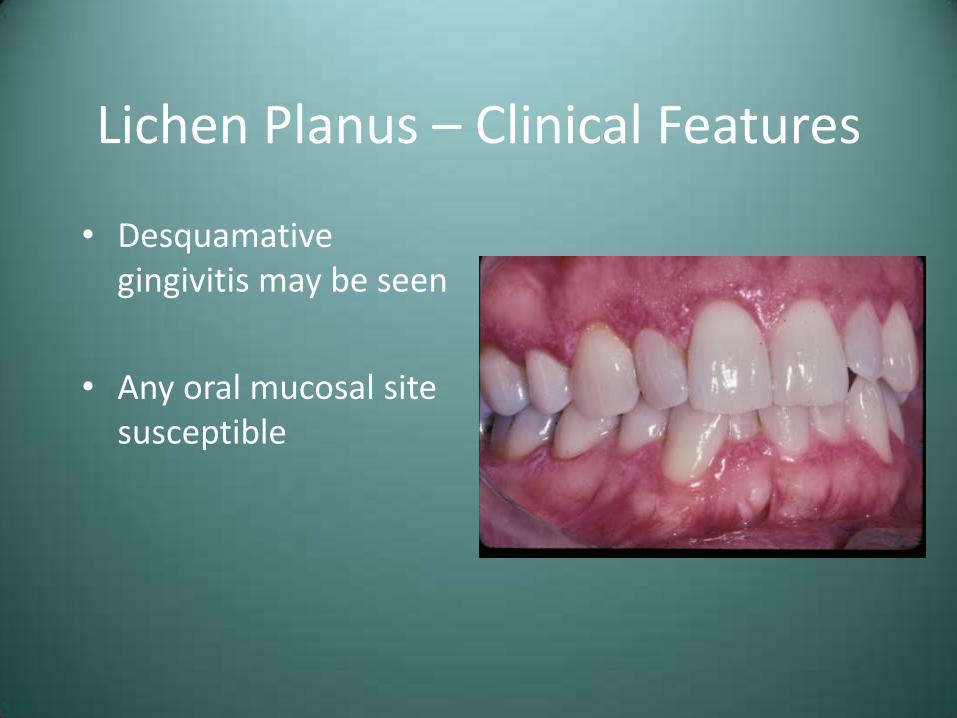
• Desquamative gingivitis may be seen
• Any oral mucosal site susceptible

Lichen Planus – Clinical Features
• Desquamative gingivitis may be seen
• Any oral mucosal site susceptible
Lichen Planus -Treatment
• 25% have superimposed candidiasis, so anti-fungal Tx may be necessary
• No treatment for reticular
• Topical corticosteroids for erosive
– Betemethasone Gel or Temovate (clobetasol) Gel
Lichen Planus -Prognosis
• Skin lesions may resolve spontaneously
• Oral lesions persist
• Malignant potential is controversial
• If premalignant, risk of transformation is probably small
Cicatricial Pemphigoid (Mucous Membrane Pemphigoid)
• Group of autoimmune disease characterized by antibodies directed against one or more components of the basement membrane
• Clinically resembles pemphigus due to blister formation
• About 2x more common than pemphigus

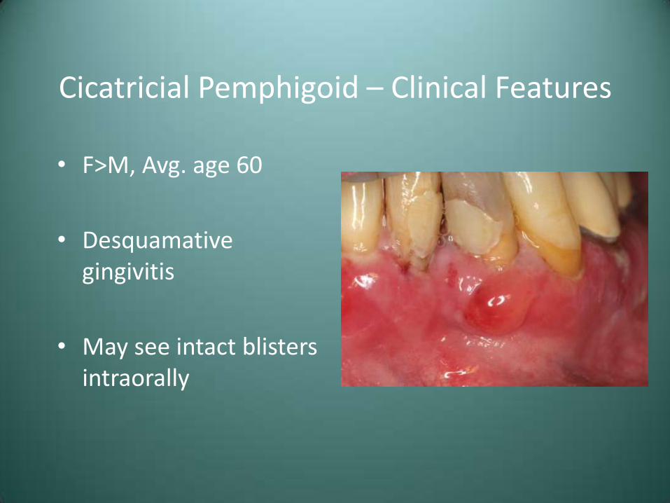
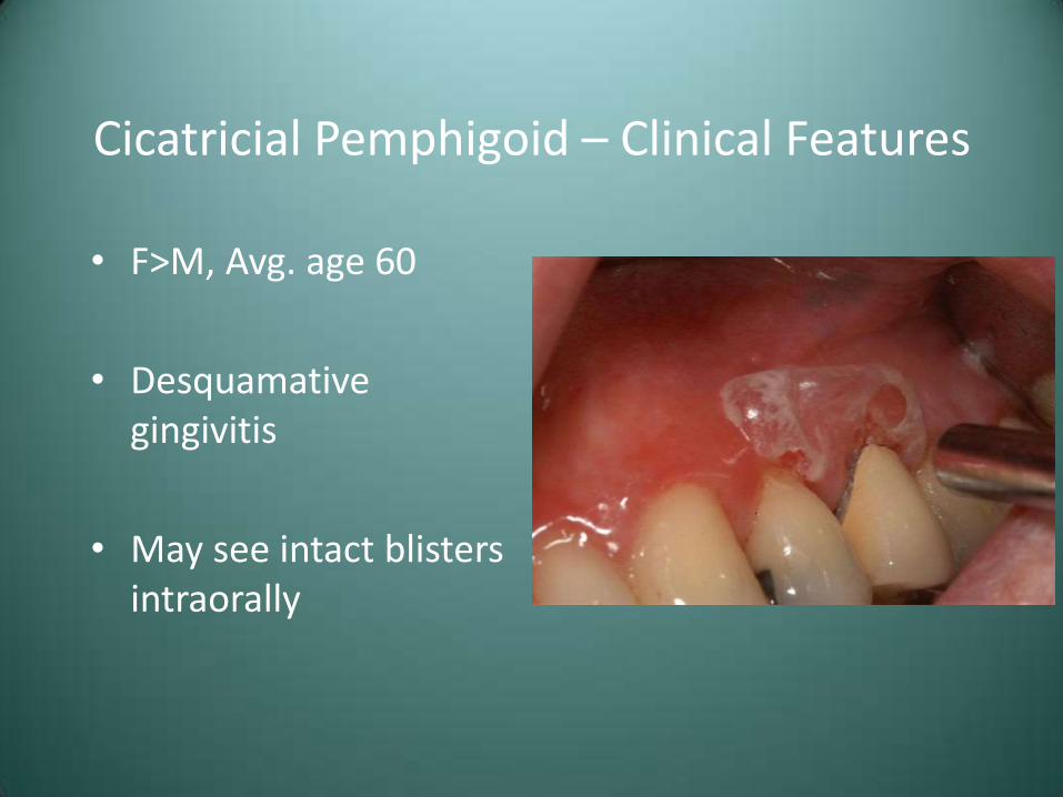
Cicatricial Pemphigoid – Clinical Features
• F>M, Avg. age 60
• Desquamative gingivitis
• May see intact blisters intraorally
Cicatricial Pemphigoid – Clinical Features
• F>M, Avg. age 60
• Desquamative gingivitis
• May see intact blisters intraorally
Cicatricial Pemphigoid – Clinical Features
• F>M, Avg. age 60
• Desquamative gingivitis
• May see intact blisters intraorally

Cicatricial Pemphigoid – Clinical Features
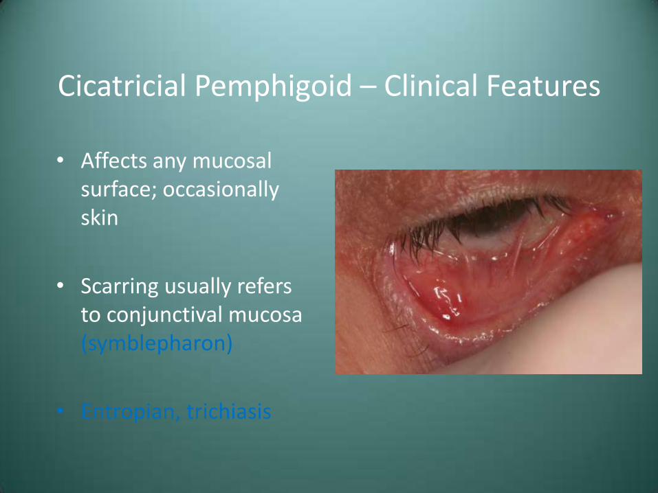
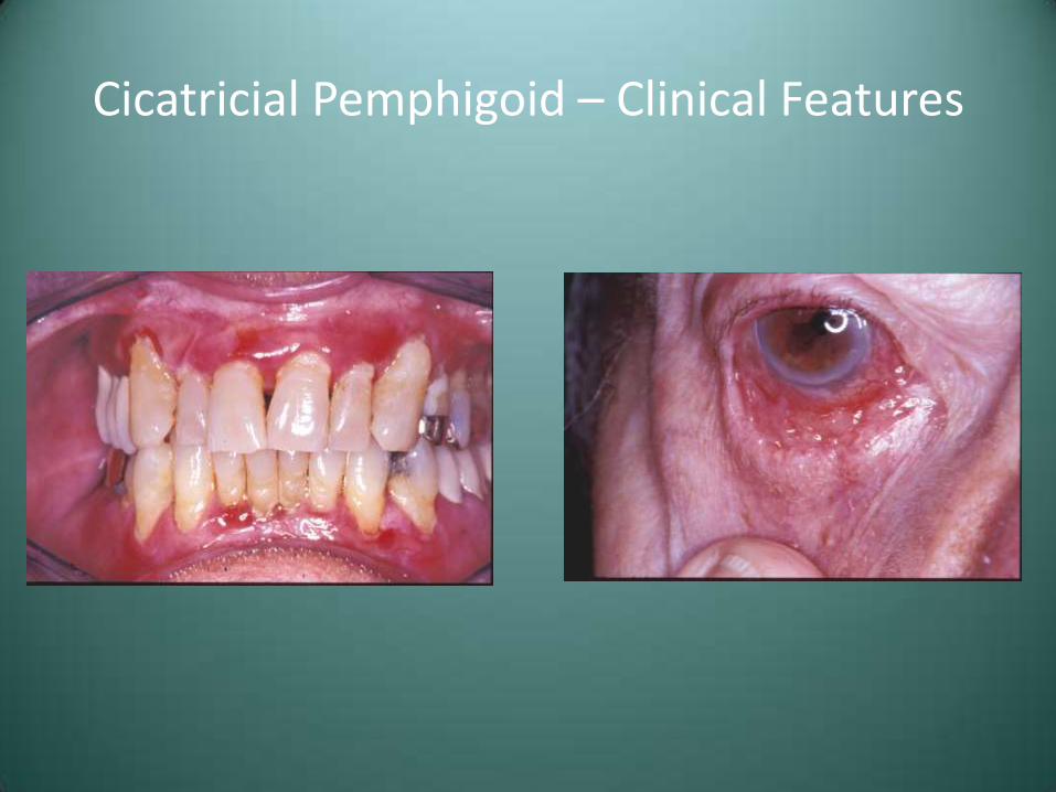
• Affects any mucosal surface; occasionally skin
• Scarring usually refers to conjunctival mucosa (symblepharon)
• Entropian, trichiasis
Cicatricial Pemphigoid – Clinical Features
• Affects any mucosal surface; occasionally skin
• Scarring usually refers to conjunctival mucosa (symblepharon)
• Entropian, trichiasis

Cicatricial Pemphigoid – Clinical Features
Cicatricial Pemphigoid – Clinical Features
Pemphigoid-Treatment
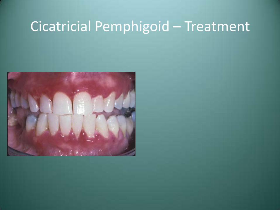
• Depends on extent of involvement
• Oral only-topical corticosteroids or dapsone
• Ocular lesions require systemic immunosuppressive therapy or human immunoglobulin therapy
Cicatricial Pemphigoid – Treatment

Cicatricial Pemphigoid – Treatment
Pemphigoid-Prognosis
• Rarely fatal
• Blindness results with untreated ocular disease
• Condition can usually be controlled
• Rarely undergoes spontaneous resolution

Pemphigus (Pemphigus Vulgaris)
• Autoimmune disorder characterized by antibodies directed against components of the epithelial desmosome complex
• Oral signs are often the first manifestations of the disease and the most difficult to resolve
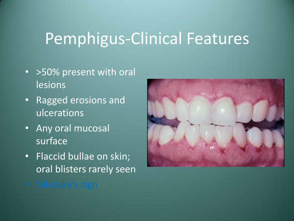
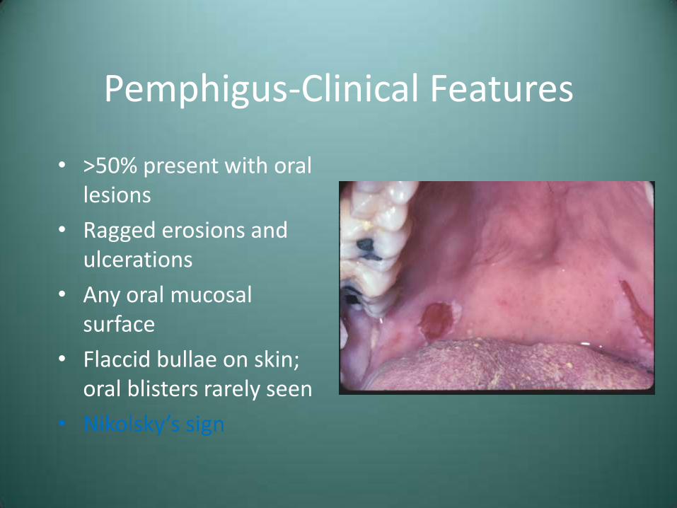
Pemphigus-Clinical Features
• >50% present with oral lesions
• Ragged erosions and ulcerations
• Any oral mucosal surface
• Flaccid bullae on skin; oral blisters rarely seen
• Nikolsky’s sign
Pemphigus-Clinical Features
• >50% present with oral lesions
• Ragged erosions and ulcerations
• Any oral mucosal surface
• Flaccid bullae on skin; oral blisters rarely seen
• Nikolsky’s sign

Pemphigus-Clinical Features
• >50% present with oral lesions
• Ragged erosions and ulcerations
• Any oral mucosal surface
• Flaccid bullae on skin; oral blisters rarely seen
• Nikolsky’s sign
Pemphigus-Clinical Features
• >50% present with oral lesions
• Ragged erosions and ulcerations
• Any oral mucosal surface
• Flaccid bullae on skin; oral blisters rarely seen
• Nikolsky’s sign
Pemphigus - Treatment and Prognosis
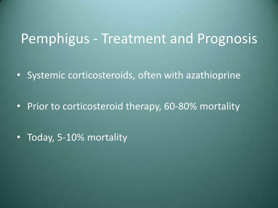
• Systemic corticosteroids, often with azathioprine
• Prior to corticosteroid therapy, 60-80% mortality
• Today, 5-10% mortality

Case #3
• Clinical Diagnosis – “Desquamative Gingivitis”
• Differential Diagnosis
– Lichen Planus
– Cicatricial Pemphigoid
– Pemphigus Vulgaris
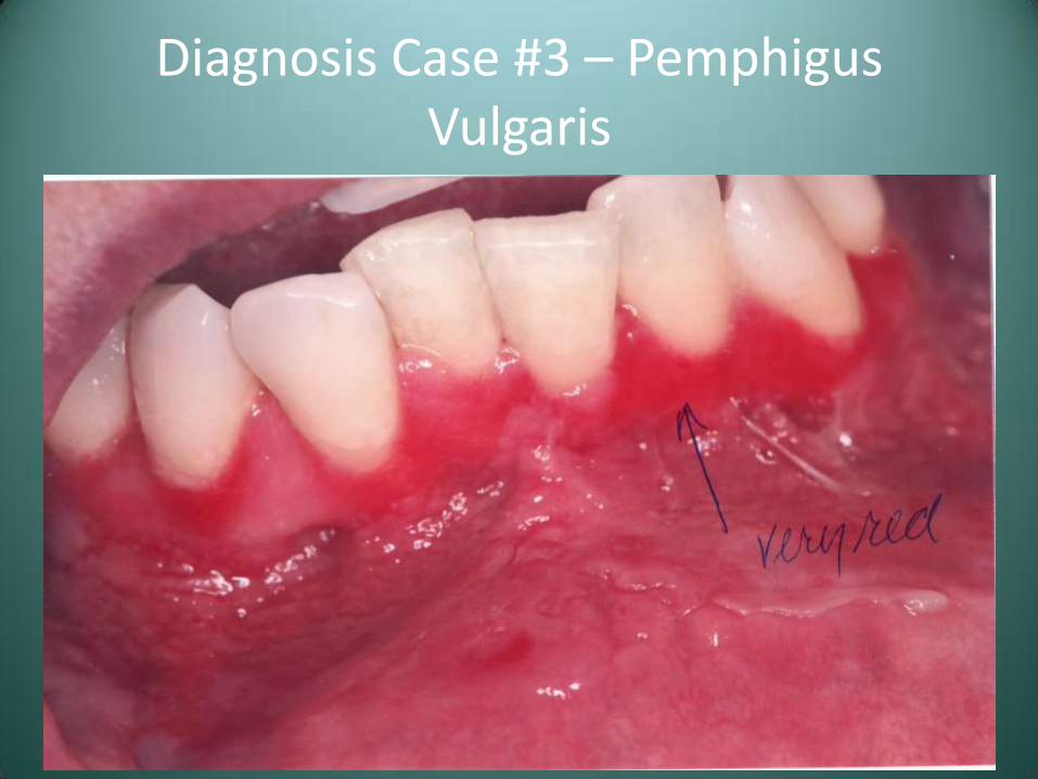
Diagnosis Case #3 – Pemphigus Vulgaris
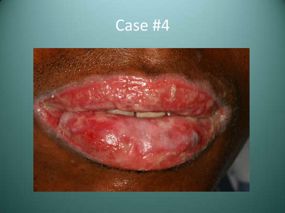
Case #4
• A 42 year old male presented with the lesions seen here as well as genital lesions
Case #4
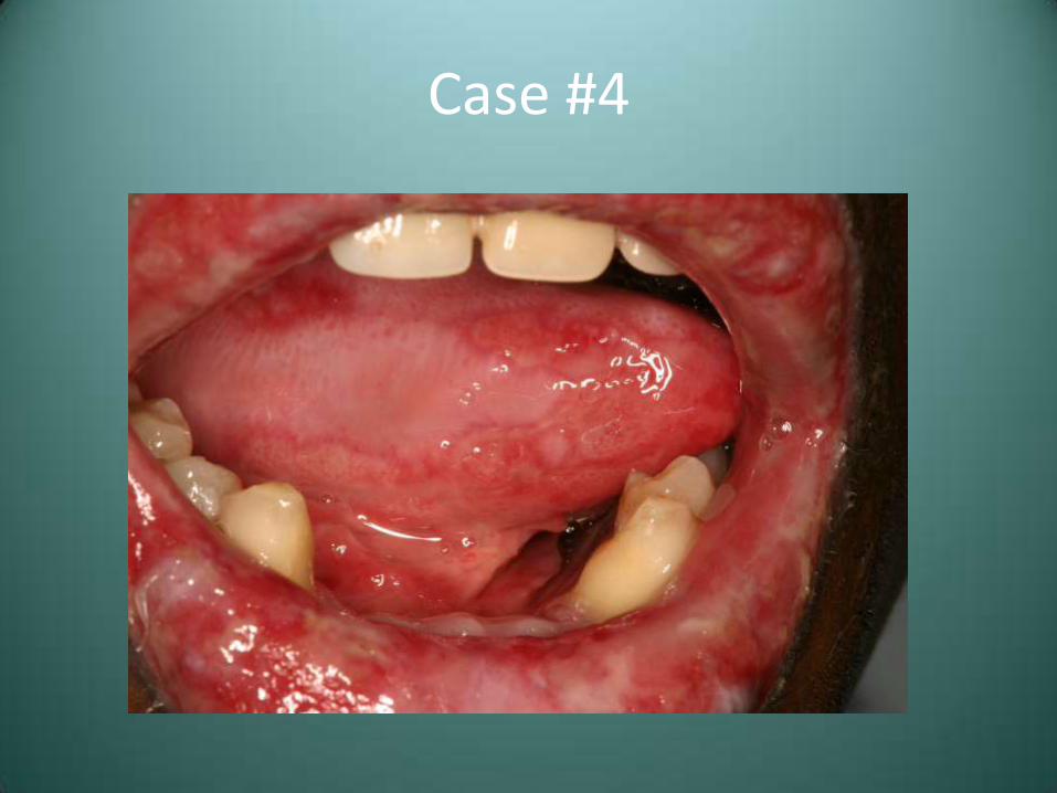
Case #4
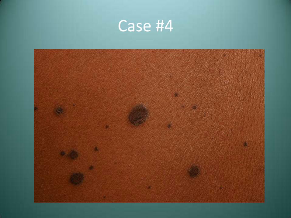
Case #4
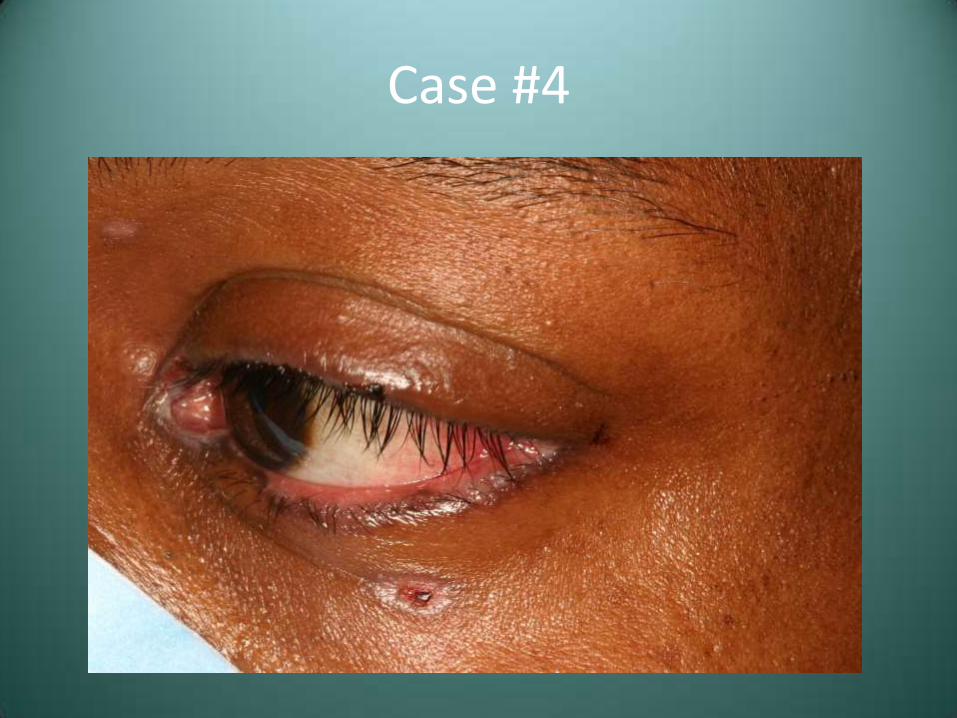
Case #4
Case #4 – Differential Diagnosis
• Erythema Multiforme
• Paraneoplastic Pemphigus
Erythema Multiforme (EM)
• Acute, self-limiting ulcerative disorder
• Probably immune-mediated
• 50%-unknown; 25%-drugs (particularly antibiotics or analgesics); 25%-infection (herpes/Mycoplasma)
EM - Spectrum of Clinical Disease
• Erythema multiforme minor - skin and/or mucosa only
• Erythema multiforme major (Stevens-Johnson syndrome)
– At least two mucosal sites plus skin involvement
• Toxic epidermal necrolysis (Lyell’s disease)
EM-Clinical Features
• M>F
• Young adults
• May experience prodrome
EM-Clinical Features
• M>F
• Young adults
• May experience prodrome
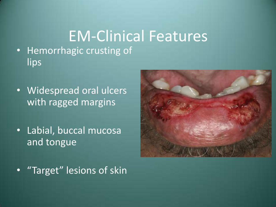
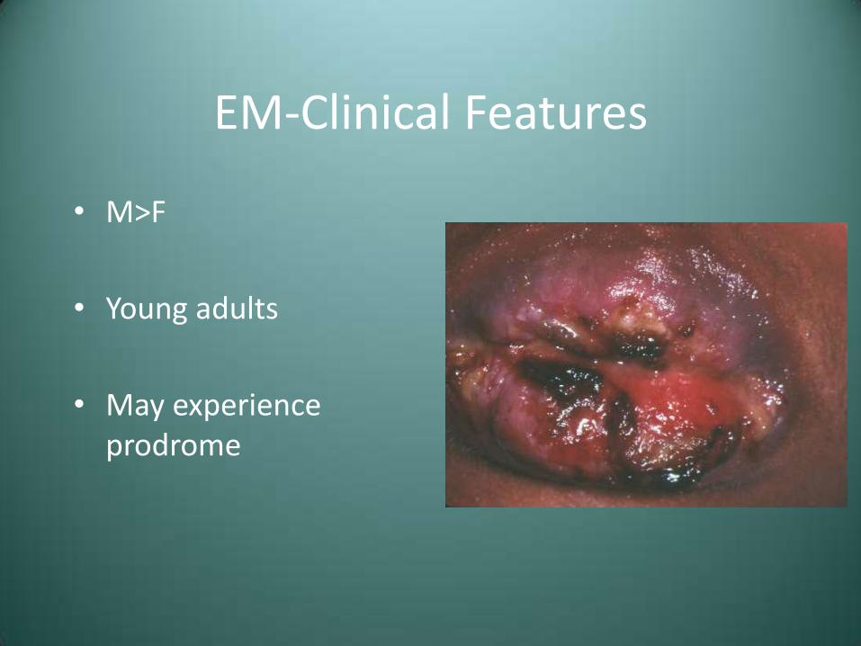
EM-Clinical Features • Hemorrhagic crusting of
lips
• Widespread oral ulcers with ragged margins
• Labial, buccal mucosa and tongue
• “Target” lesions of skin
EM-Clinical Features
• Outbreak typically clears in 2-6 weeks
• Often recurs in spring and fall
EM-Treatment
Supportive or topical corticosteroids for mild cases
Systemic corticosteroids for EM major
TEN managed in burn unit, possibly with pooled immunoglobulin
EM Prognosis
• Good for mild to moderate cases
• EM major-2-10% mortality
• TEN-34% mortality
Paraneoplastic Pemphigus • Serious vesiculobullous disorder affecting patients
with neoplastic disease, typically a lymphoreticular malignancy (CLL and lymphoma)
• Antibodies in response to the tumor probably cross react with components of the epithelial layer
• Cytotoxic T lymphocytes may also play a role in cutaneous and mucosal damage

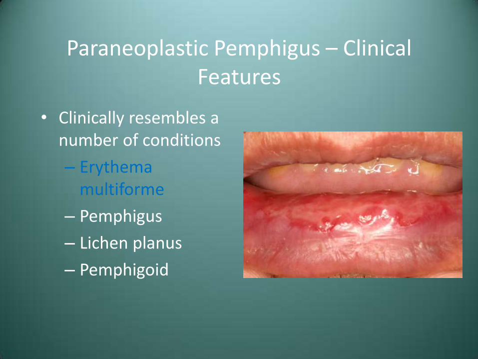
Paraneoplastic Pemphigus – Clinical Features
• Clinically resembles a number of conditions
– Erythema multiforme
– Pemphigus
– Lichen planus
– Pemphigoid
Paraneoplastic Pemphigus – Clinical Features
• Clinically resembles a number of conditions
– Erythema multiforme
– Pemphigus
– Lichen planus
– Pemphigoid
Paraneoplastic Pemphigus – Clinical Features
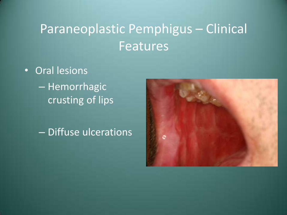
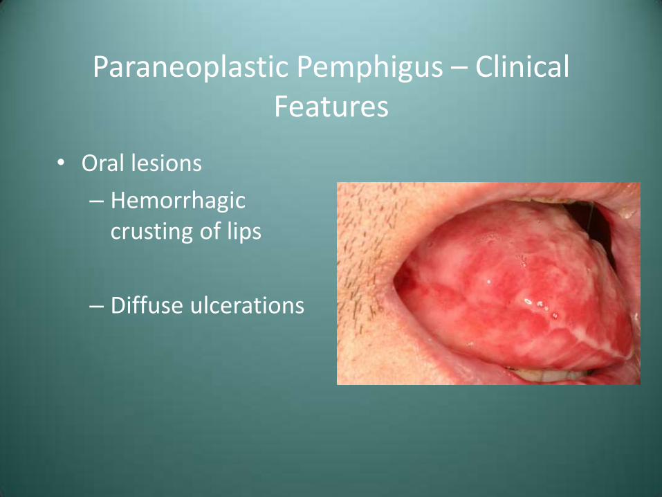
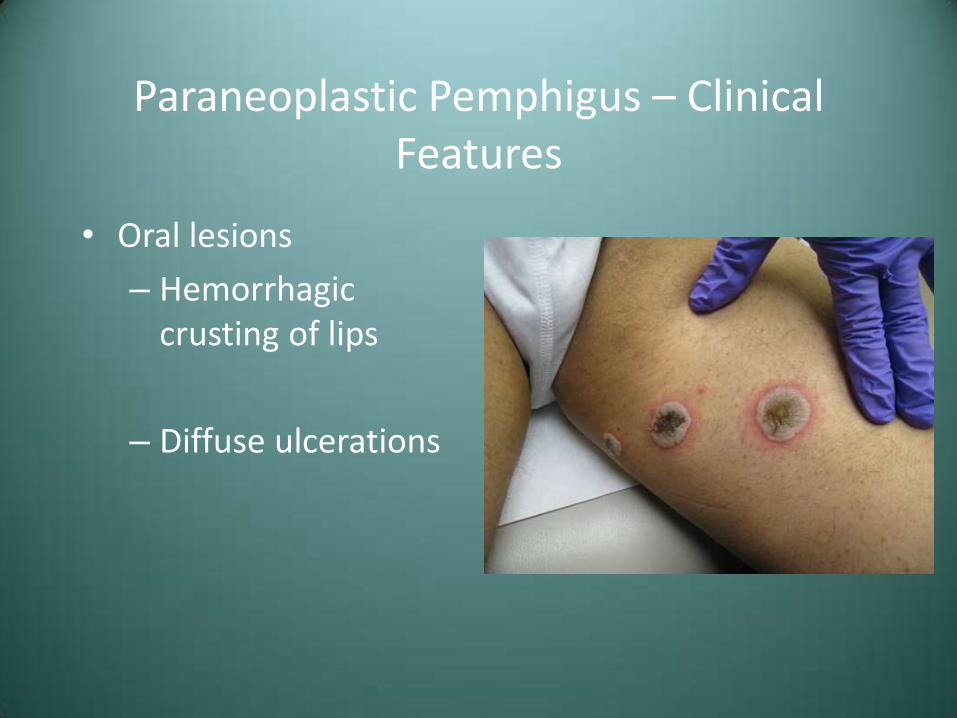
• Oral lesions
– Hemorrhagic crusting of lips
– Diffuse ulcerations

Paraneoplastic Pemphigus – Clinical Features
• Oral lesions
– Hemorrhagic crusting of lips
– Diffuse ulcerations
Paraneoplastic Pemphigus – Clinical Features
• Oral lesions
– Hemorrhagic crusting of lips
– Diffuse ulcerations
Paraneoplastic Pemphigus – Clinical Features
• Oral lesions
– Hemorrhagic crusting of lips
– Diffuse ulcerations
Paraneoplastic Pemphigus – Treatment and Prognosis
• Systemic corticosteroids plus azathioprine
• Topical corticosteroids
• Generally poor prognosis, high mortality due to sepsis or malignant progression

Case #4 – Differential Diagnosis
• Erythema Multiforme
• Paraneoplastic Pemphigus

Diagnosis Case #4 – Erythema Multiforme

Case #5
• An adult male presents with ulcerations distributed as seen

Case #5
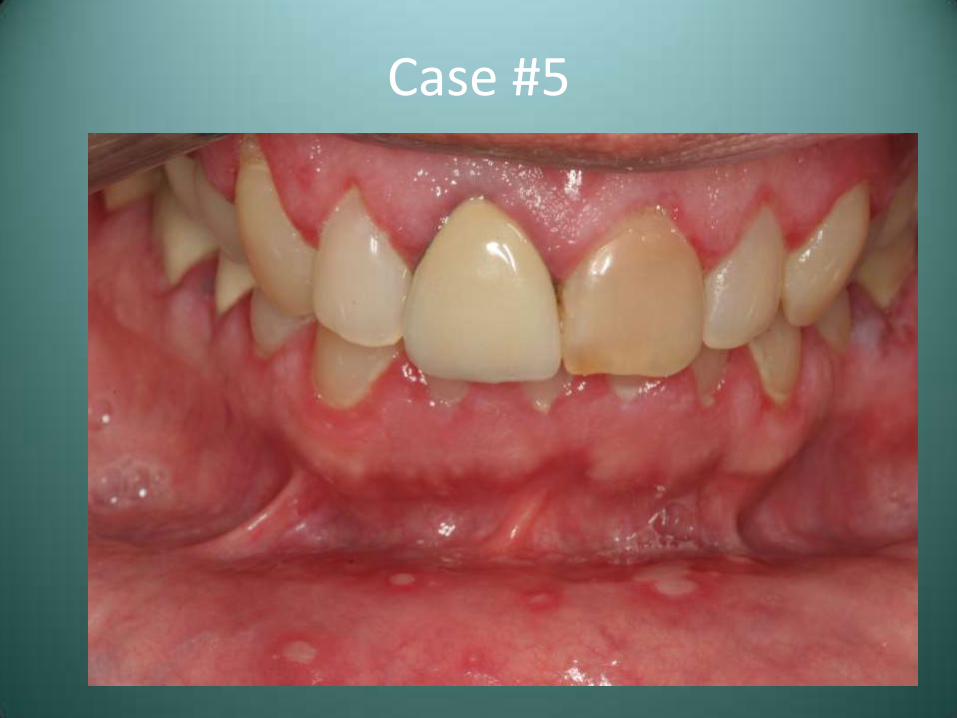
Case #5

Case #5
Case #7 – Differential Diagnosis
• Herpes Simplex Type 1
• Recurrent Aphthous Stomatitis
• Erythema multiforme

Herpes Simplex Virus (HSV)
• DNA virus in the herpesvirus family
– HHV-1 – oral herpes
– HHV-2 – genital herpes
– HHV-3 – chicken pox and shingles (Varicella-Zoster virus)
– HHV-4 – mononucleosis (Epstein-Barr virus)
– HHV-5 – cytomegalovirus (CMV)
– HHV-8 – Kaposi’s sarcoma-associated
Herpes Simplex Virus
• Two clinical patterns
– Primary herpetic infection
– Secondary or recurrent HSV
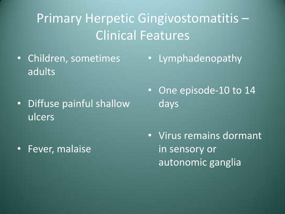
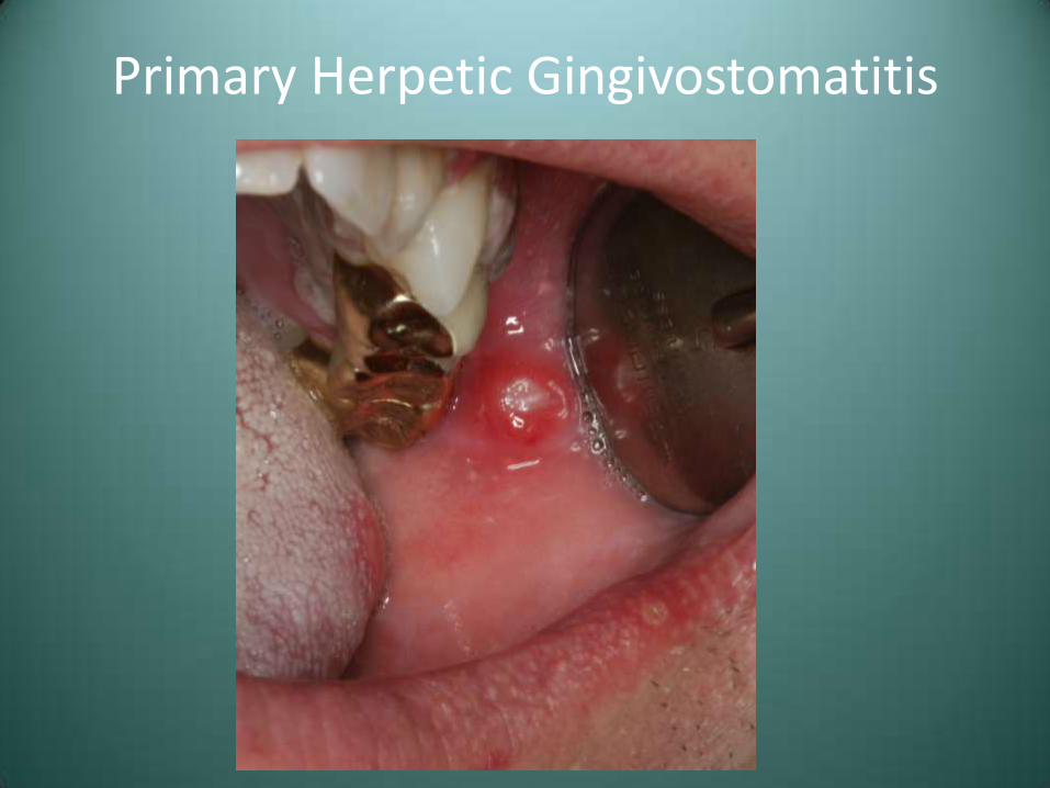
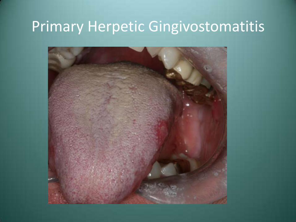
Primary Herpetic Gingivostomatitis – Clinical Features
• Children, sometimes adults
• Diffuse painful shallow ulcers
• Fever, malaise
• Lymphadenopathy
• One episode-10 to 14 days
• Virus remains dormant in sensory or autonomic ganglia
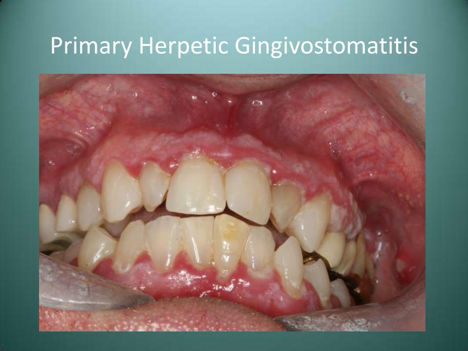
Primary Herpetic Gingivostomatitis

Primary Herpetic Gingivostomatitis

Primary Herpetic Gingivostomatitis
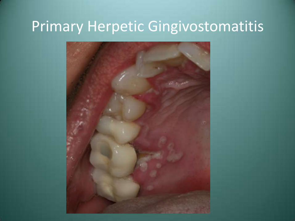
Primary Herpetic Gingivostomatitis
Primary Herpetic Gingivostomatitis
Primary Herpetic Gingivostomatitis
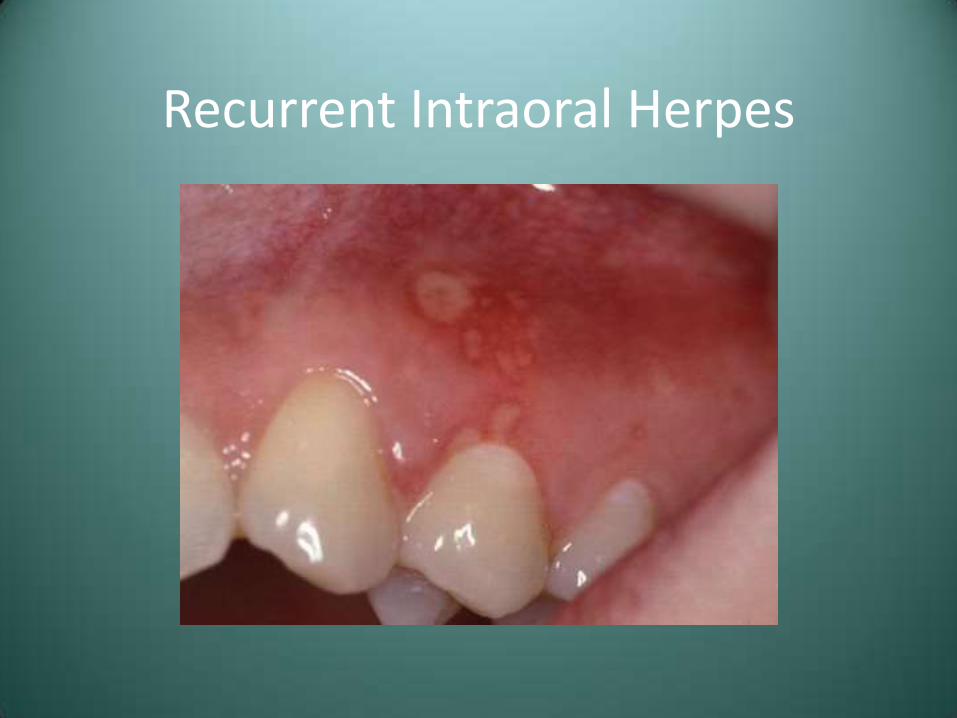
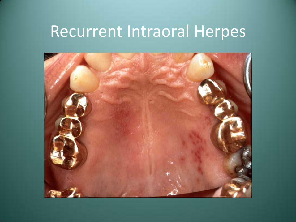
Recurrent Intraoral Herpes
• Relatively uncommon
• Usually few symptoms
• Cluster of shallow ulcers - intact vesicles rare
• Mucosa bound to periosteum
– Hard palate and attached gingiva
• Heal within one week
Recurrent Intraoral Herpes
Recurrent Intraoral Herpes

Primary Herpes-Treatment
• Restrict contact with lesions
• Topical anesthetics
– Dyclonine HCL or viscous lidocaine
• Ibuprofen or other NSAID’s
• Soft diet with fluids
• Antiviral medications of recognized early (1st 72 hours)
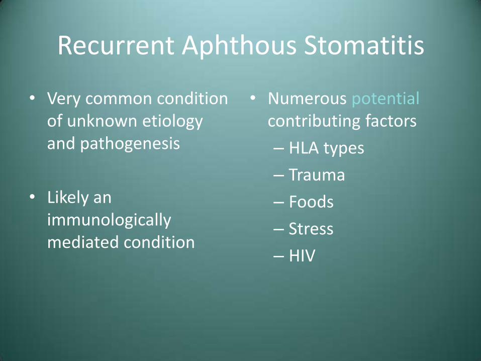
Recurrent Aphthous Stomatitis
• Very common condition of unknown etiology and pathogenesis
• Likely an immunologically mediated condition
• Numerous potential contributing factors
– HLA types
– Trauma
– Foods
– Stress
– HIV

Recurrent Aphthous Stomatitis – Clinical Features
• Three major forms
– Minor
– Major
– Herpetiform

Recurrent Aphthous Stomatitis – Clinical Features
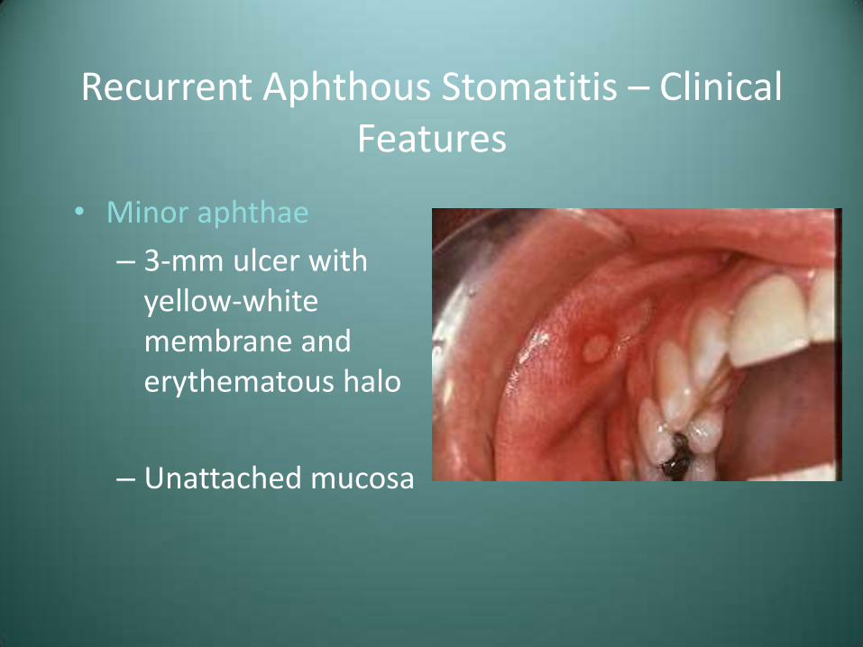
• Minor aphthae
– 3-mm ulcer with yellow-white membrane and erythematous halo
– Unattached mucosa
Recurrent Aphthous Stomatitis – Clinical Features
• Minor aphthae
– 3-mm ulcer with yellow-white membrane and erythematous halo
– Unattached mucosa

Recurrent Aphthous Stomatitis – Clinical Features
• Minor aphthae
– 3-mm ulcer with yellow-white membrane and erythematous halo
– Unattached mucosa
Recurrent Aphthous Stomatitis – Clinical Features
• Major aphthae
– Larger (up to 3cm) and longer duration (2-6 weeks)
– May heal with scar
– HIV
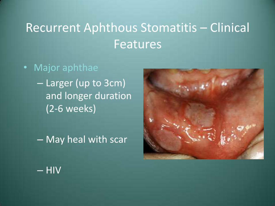
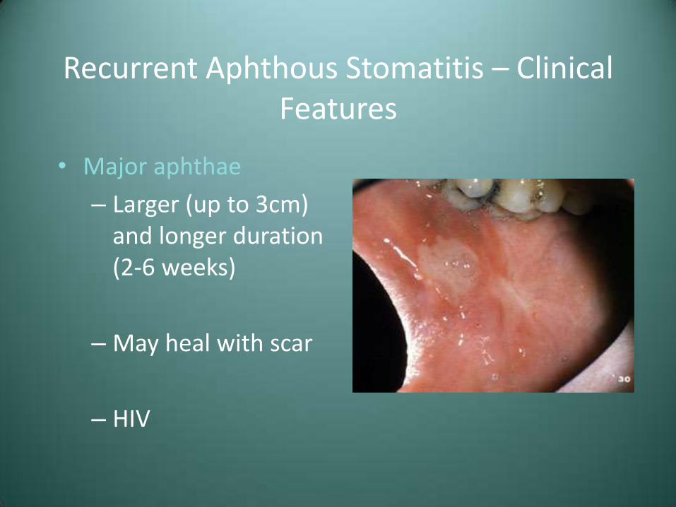
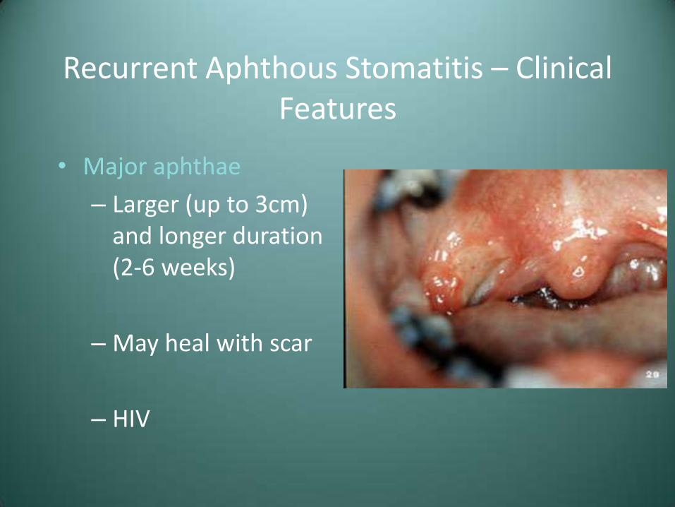
Recurrent Aphthous Stomatitis – Clinical Features
• Major aphthae
– Larger (up to 3cm) and longer duration (2-6 weeks)
– May heal with scar
– HIV
Recurrent Aphthous Stomatitis – Clinical Features
• Major aphthae
– Larger (up to 3cm) and longer duration (2-6 weeks)
– May heal with scar
– HIV
Recurrent Aphthous Stomatitis – Clinical Features
• Major aphthae
– Larger (up to 3cm) and longer duration (2-6 weeks)
– May heal with scar
– HIV
Recurrent Aphthous Stomatitis - Treatment
• Topical corticosteroids
– Betamethasone 0.05%
– Clobetasol propionate 0.05% (Temovate gel)
• Elixirs or syrup preparations for numerous and/or ulcerations in inaccessible areas
• If unresponsive, investigate possible underlying cause

Case #7 – Differential Diagnosis
• Herpes Simplex Type 1
• Recurrent Aphthous Stomatitis
• Erythema multiforme
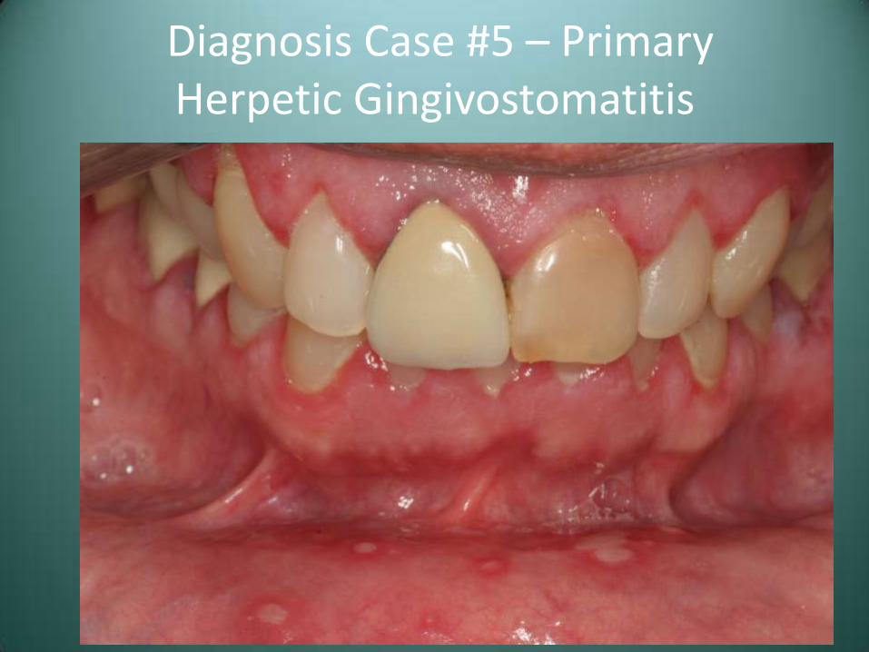
Diagnosis Case #5 – Primary Herpetic Gingivostomatitis

White, Red and Malignant Lesions

Smokeless Tobacco Use/Tobacco Pouch Keratosis
• Mucosal lesion secondary to the presence of chronic irritation from smokeless tobacco
• These products are currently used by approximately 4.5% of US males
• Also associated with gingival/periodontal destruction and tooth decay
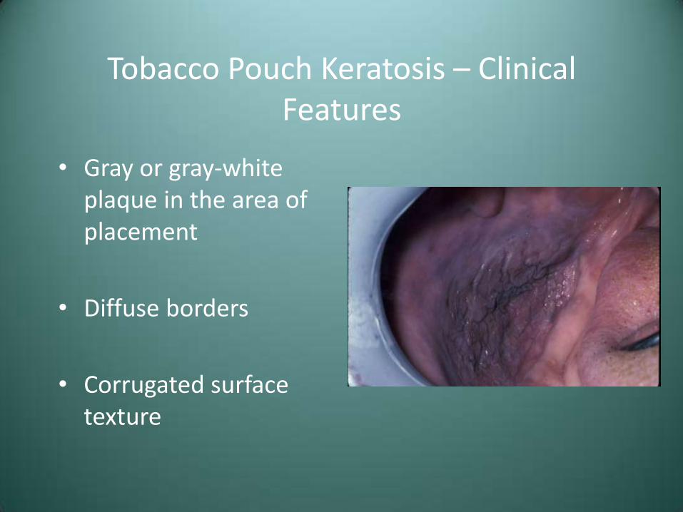
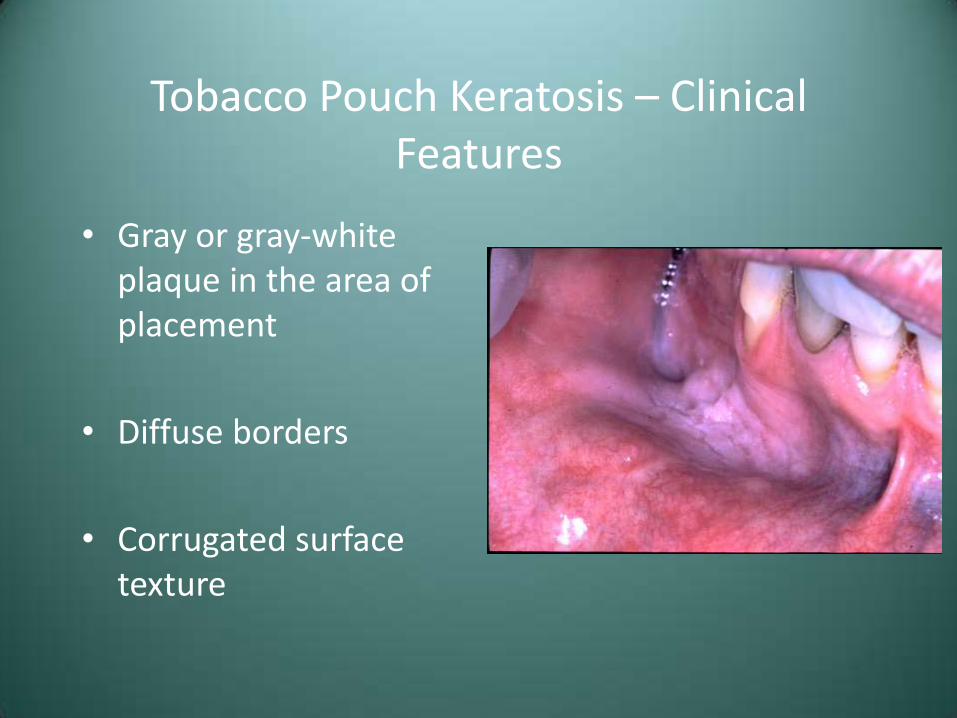
Tobacco Pouch Keratosis – Clinical Features
• Gray or gray-white plaque in the area of placement
• Diffuse borders
• Corrugated surface texture
Tobacco Pouch Keratosis – Clinical Features
• Gray or gray-white plaque in the area of placement
• Diffuse borders
• Corrugated surface texture
Tobacco Pouch Keratosis – Clinical Features
• Gray or gray-white plaque in the area of placement
• Diffuse borders
• Corrugated surface texture

Tobacco Pouch Keratosis – Clinical Features
• Gray or gray-white plaque in the area of placement
• Diffuse borders
• Corrugated surface texture
Tobacco Pouch Keratosis – Treatment and Prognosis
• Have patient stop or move the tobacco to another location to observe for resolution (2-4 weeks)
• If the lesion persists (after 6 weeks), biopsy for histologic diagnosis
• Controversy over true carcinogenicity of smokeless tobacco
Nicotine Stomatitis
• Benign hyperkeratotic change to the palatal mucosa secondary to tobacco smoking
• Most common in pipe and cigar smokers
• Similar changes may be induced by drinking hot beverages
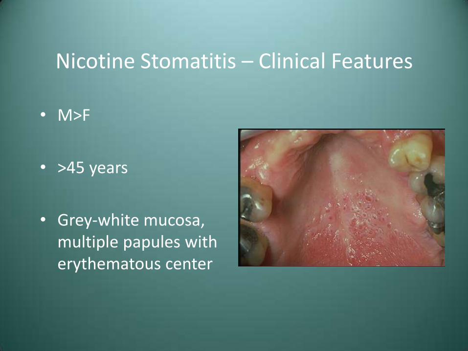
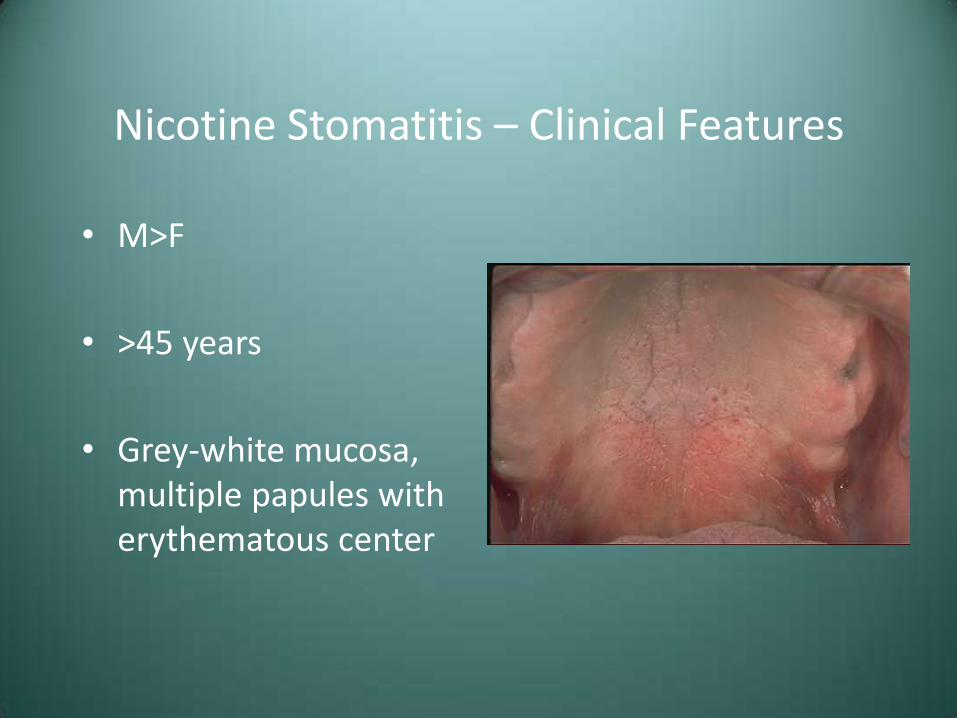
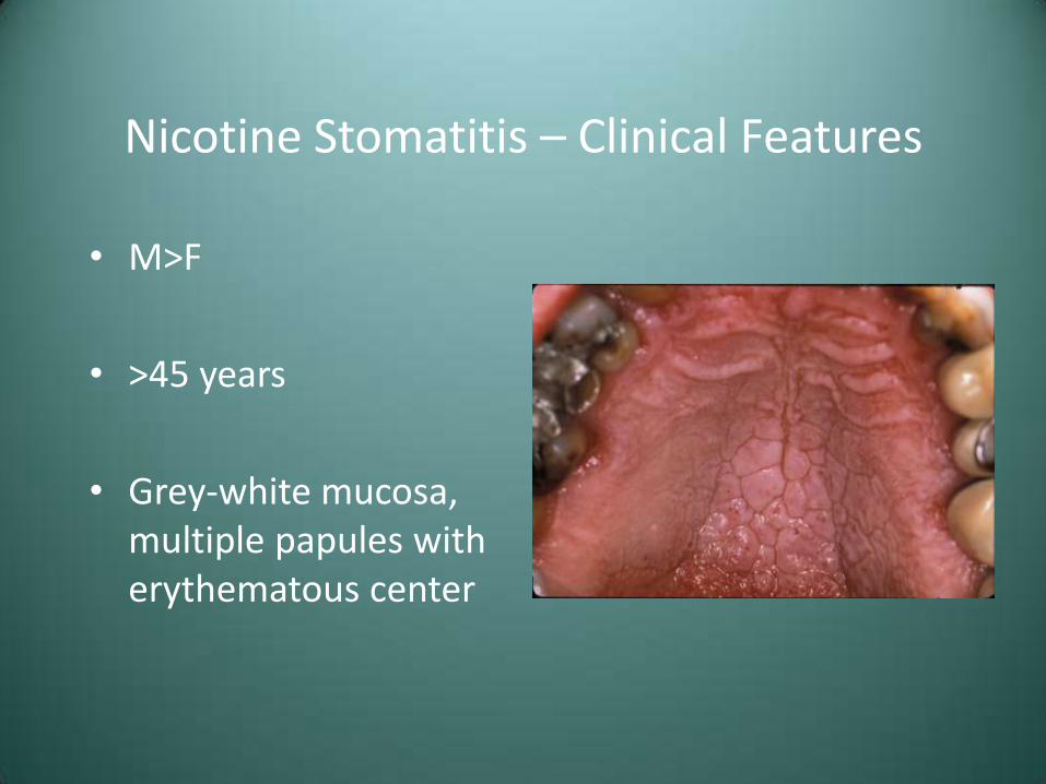
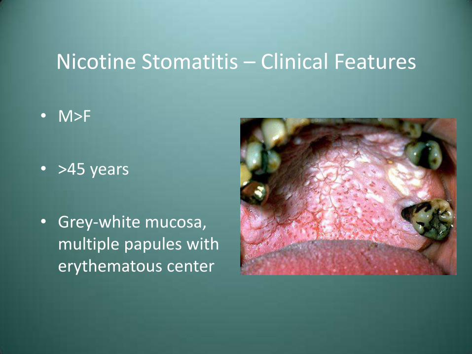
Nicotine Stomatitis – Clinical Features
• M>F,
• >45 years
• Grey-white mucosa, multiple papules with erythematous center
Nicotine Stomatitis – Clinical Features
• M>F
• >45 years
• Grey-white mucosa, multiple papules with erythematous center
Nicotine Stomatitis – Clinical Features
• M>F
• >45 years
• Grey-white mucosa, multiple papules with erythematous center
Nicotine Stomatitis – Clinical Features
• M>F
• >45 years
• Grey-white mucosa, multiple papules with erythematous center
Nicotine Stomatitis – Clinical Features
• M>F
• >45 years
• Grey-white mucosa, multiple papules with erythematous center
Nicotine Stomatitis – Treatment
• None
• If patient quits, changes will normally resolve within 1-2 weeks
• Persistent changes should be biopsied
Leukoplakia
• Definition (WHO)-A white patch or plaque which cannot be characterized clinically or pathologically as any other disease
• Considered premalignant
– Most common precancerous oral lesion
Leukoplakia
• Etiology-Technically unknown
– Tobacco smoking
– Alcohol is not necessarily associated with leukoplakia
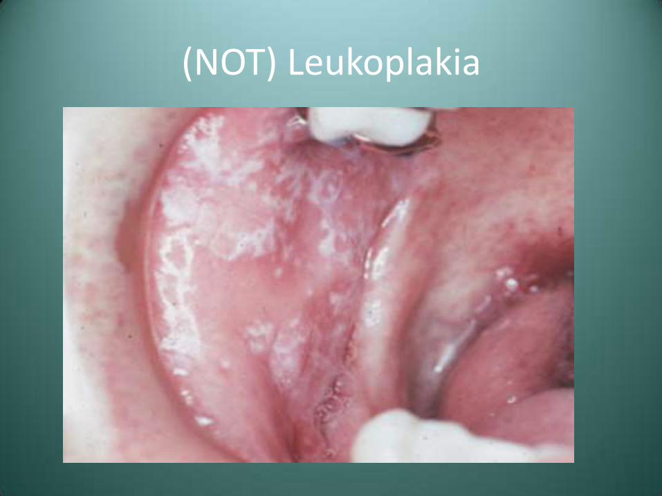
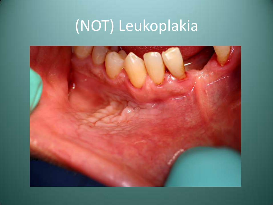
• Lesions that are not leukoplakia
– Nicotine stomatitis
– Frictional keratosis
– Lichen planus
– Amalgam reactions
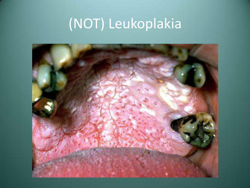
(NOT) Leukoplakia
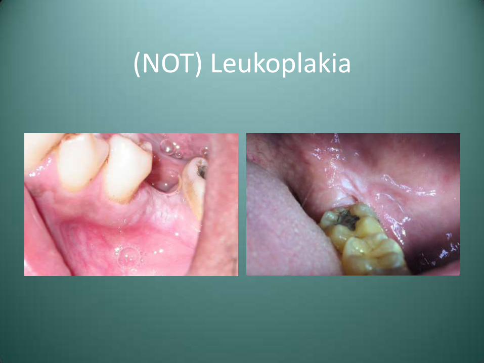
(NOT) Leukoplakia
(NOT) Leukoplakia
(NOT) Leukoplakia
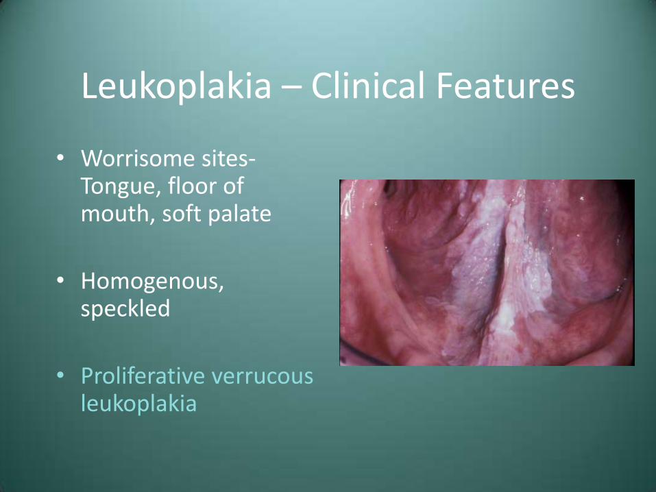
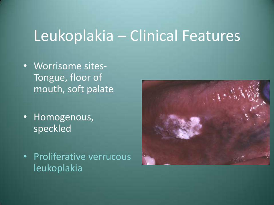
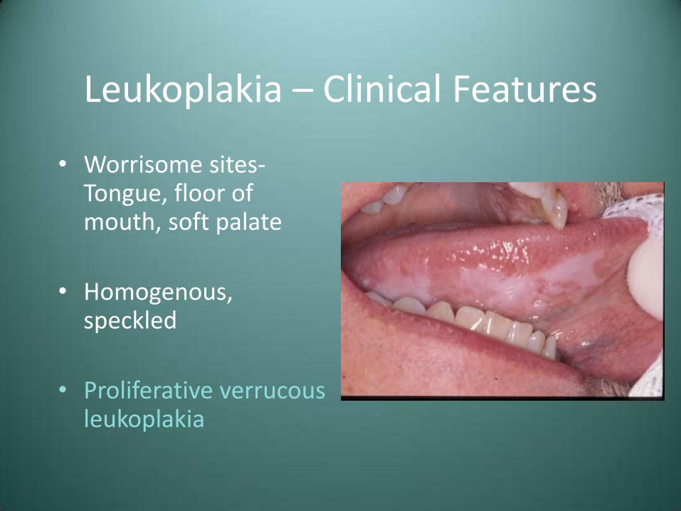
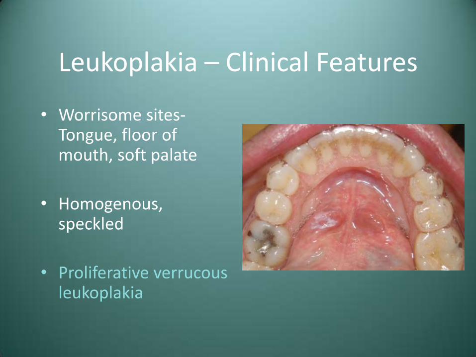
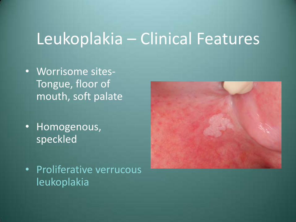
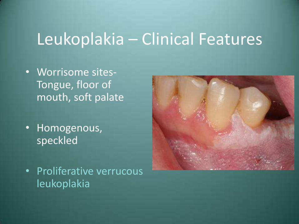
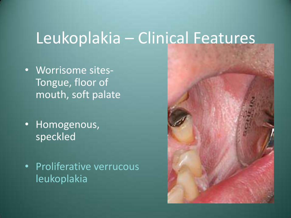
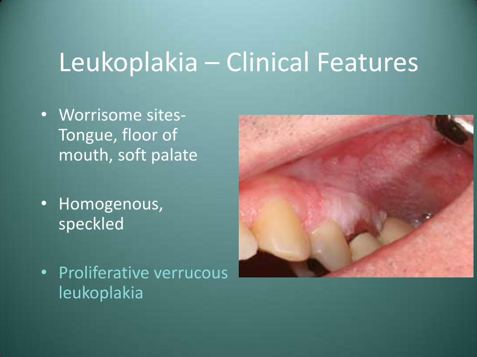
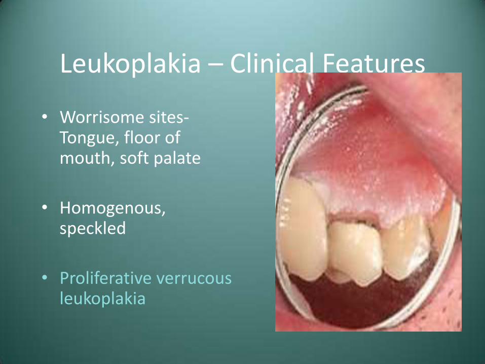
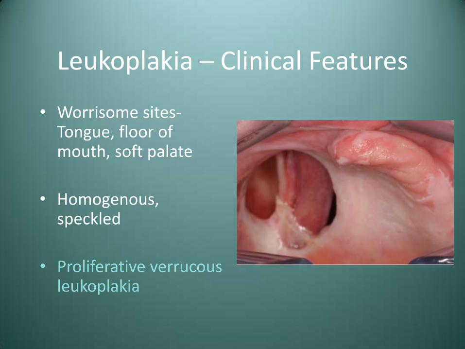
Leukoplakia – Clinical Features
• Worrisome sites-Tongue, floor of mouth, soft palate
• Homogenous, speckled
• Proliferative verrucous leukoplakia
Leukoplakia – Clinical Features
• Worrisome sites-Tongue, floor of mouth, soft palate
• Homogenous, speckled
• Proliferative verrucous leukoplakia

Leukoplakia – Clinical Features
• Worrisome sites-Tongue, floor of mouth, soft palate
• Homogenous, speckled
• Proliferative verrucous leukoplakia
Leukoplakia – Clinical Features
• Worrisome sites-Tongue, floor of mouth, soft palate
• Homogenous, speckled
• Proliferative verrucous leukoplakia
Leukoplakia – Clinical Features
• Worrisome sites-Tongue, floor of mouth, soft palate
• Homogenous, speckled
• Proliferative verrucous leukoplakia
Leukoplakia – Clinical Features
• Worrisome sites-Tongue, floor of mouth, soft palate
• Homogenous, speckled
• Proliferative verrucous leukoplakia
Leukoplakia – Clinical Features
• Worrisome sites-Tongue, floor of mouth, soft palate
• Homogenous, speckled
• Proliferative verrucous leukoplakia

Leukoplakia – Clinical Features
• Worrisome sites-Tongue, floor of mouth, soft palate
• Homogenous, speckled
• Proliferative verrucous leukoplakia

Leukoplakia – Clinical Features
• Worrisome sites-Tongue, floor of mouth, soft palate
• Homogenous, speckled
• Proliferative verrucous leukoplakia
Leukoplakia – Clinical Features
• Worrisome sites-Tongue, floor of mouth, soft palate
• Homogenous, speckled
• Proliferative verrucous leukoplakia
Leukoplakia – Clinical Features
• Worrisome sites-Tongue, floor of mouth, soft palate
• Homogenous, speckled
• Proliferative verrucous leukoplakia
Leukoplakia – Clinical Features
• Worrisome sites-Tongue, floor of mouth, soft palate
• Homogenous, speckled
• Proliferative verrucous leukoplakia
Leukoplakia – Clinical Features
• Worrisome sites-Tongue, floor of mouth, soft palate
• Homogenous, speckled
• Proliferative verrucous leukoplakia
Leukoplakia – Clinical Features
• Worrisome sites-Tongue, floor of mouth, soft palate
• Homogenous, speckled
• Proliferative verrucous leukoplakia
Leukoplakia – Treatment and Prognosis
• Biopsy is mandatory
• Treatment will then depend upon the histologic findings
• 4% risk of transformation to SCC
• With or without removal, follow-up is essential
• Recurrences are common (about 1/3)
Erythroplakia
• Red patch that cannot be clinically or pathologically diagnosed as any other condition
• Greater presence of dysplasia than leukoplakia
• Same etiology as SCC (tobacco, alcohol)
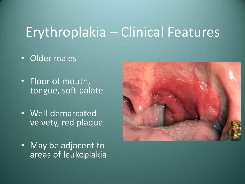
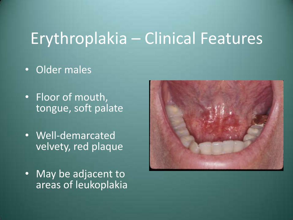
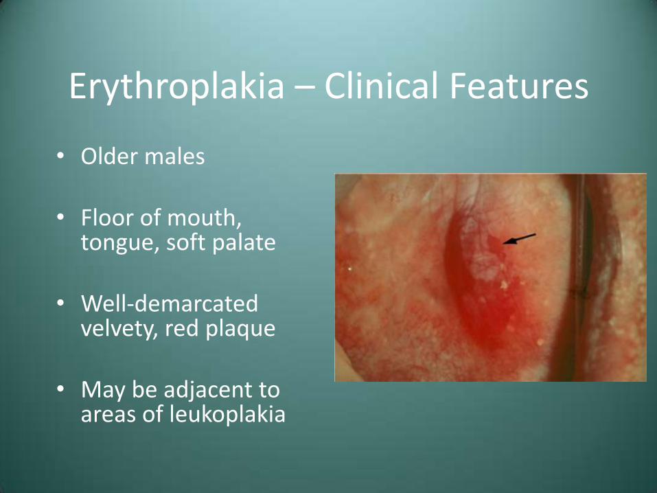
Erythroplakia – Clinical Features
• Older males
• Floor of mouth, tongue, soft palate
• Well-demarcated velvety, red plaque
• May be adjacent to areas of leukoplakia

Erythroplakia – Clinical Features
• Older males
• Floor of mouth, tongue, soft palate
• Well-demarcated velvety, red plaque
• May be adjacent to areas of leukoplakia
Erythroplakia – Clinical Features
• Older males
• Floor of mouth, tongue, soft palate
• Well-demarcated velvety, red plaque
• May be adjacent to areas of leukoplakia

Erythroplakia – Clinical Features
• Older males
• Floor of mouth, tongue, soft palate
• Well-demarcated velvety, red plaque
• May be adjacent to areas of leukoplakia
Erythroplakia – Clinical Features
• Older males
• Floor of mouth, tongue, soft palate
• Well-demarcated velvety, red plaque
• May be adjacent to areas of leukoplakia
Erythroplakia – Clinical Features
• Older males
• Floor of mouth, tongue, soft palate
• Well-demarcated velvety, red plaque
• May be adjacent to areas of leukoplakia

Erythroplakia - Histology
• 90% will show severe dysplasia or CIS
• Epithelial atrophy with lack of keratin production
• Chronic inflammation
Erythroplakia – Treatment and Prognosis
• Biopsy is mandatory, with treatment dependant upon the degree of dysplasia
• Close follow-up is necessary, since recurrence and the development of separate lesions are common
Oral Squamous Cell Carcinoma
• 22,000 cases per year, with about 1 in four dying of the disease
• Males-8th most common cancer (Females-15th)
• M>F
• Blacks>Whites
• Carcinoma of the lip should be considered in a different context
Oral Squamous Cell Carcinoma - Etiology
• Tobacco (especially combustible)
• Alcohol (works synergistically with tobacco)
• Radiation
• Plummer-Vinson syndrome (iron deficiency anemia, glossitis, dysphagia)
• Viruses (HPV)
• Immunosuppression
Oral Squamous Cell Carcinoma – Clinical Features
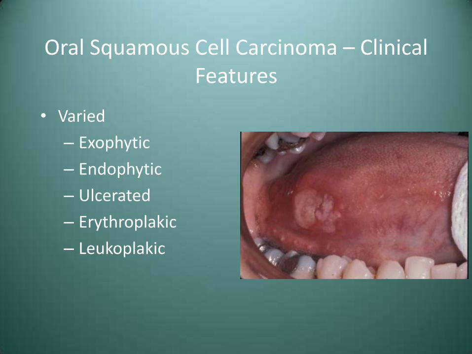
• Varied
– Exophytic
– Endophytic
– Ulcerated
– Erythroplakic
– Leukoplakic
Oral Squamous Cell Carcinoma – Clinical Features
• Varied
– Exophytic
– Endophytic
– Ulcerated
– Erythroplakic
– Leukoplakic
Oral Squamous Cell Carcinoma – Clinical Features
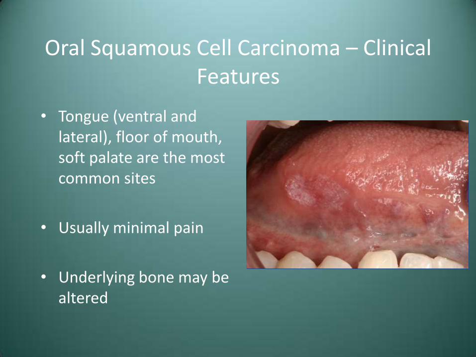
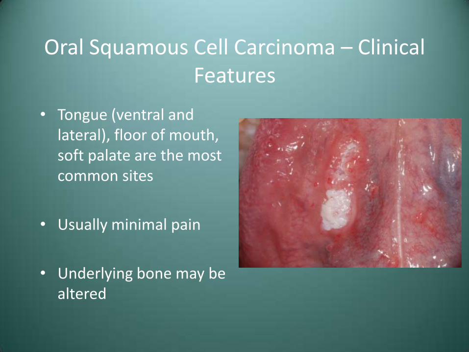
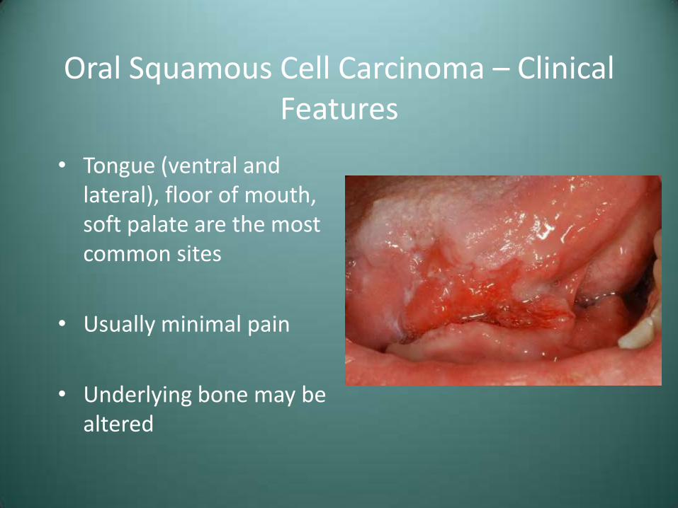
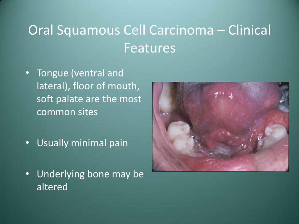
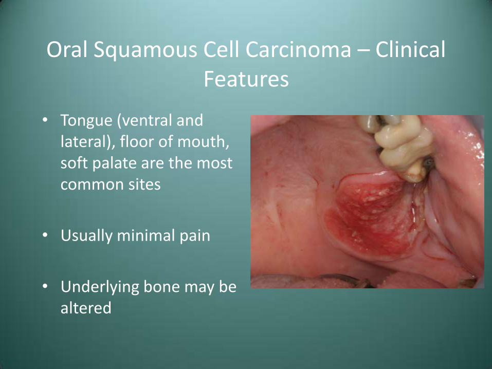
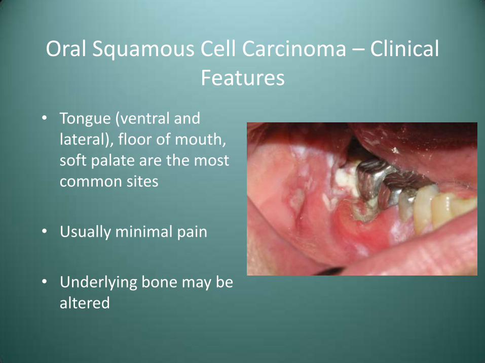
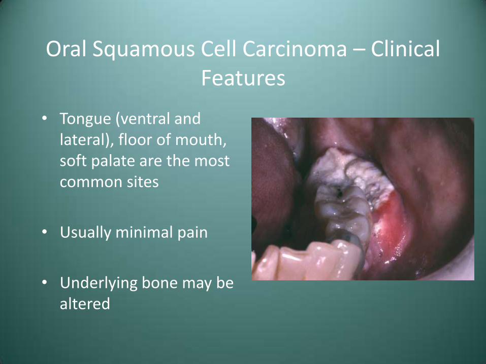
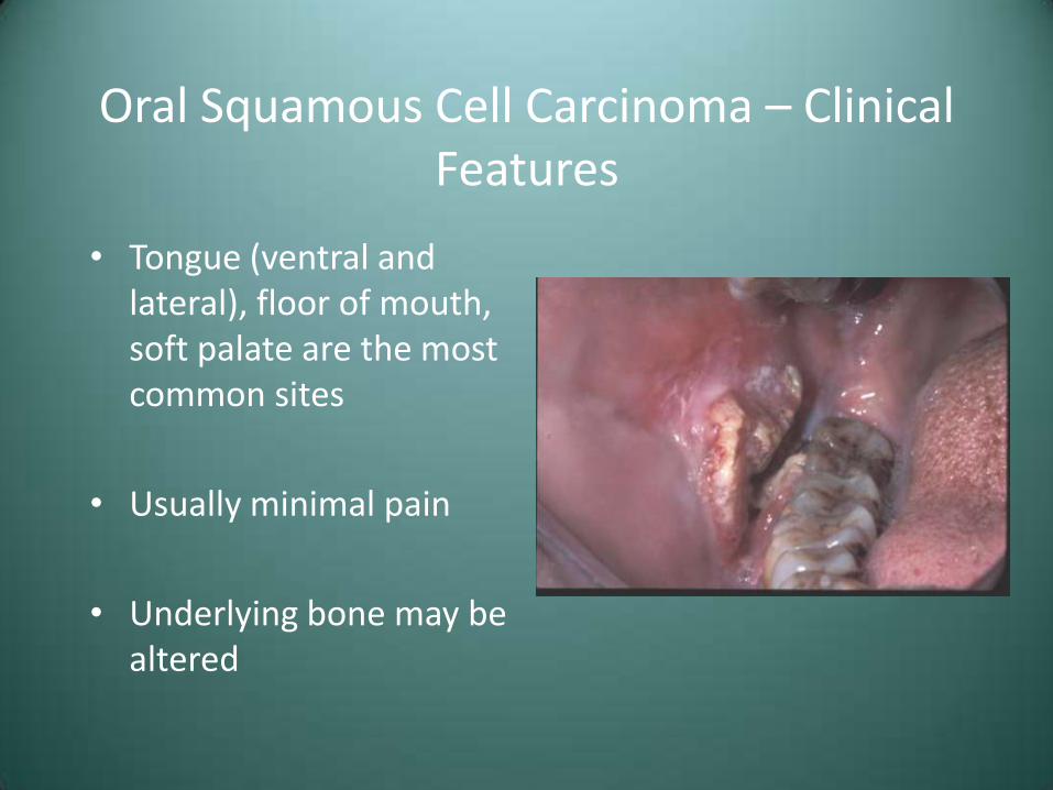
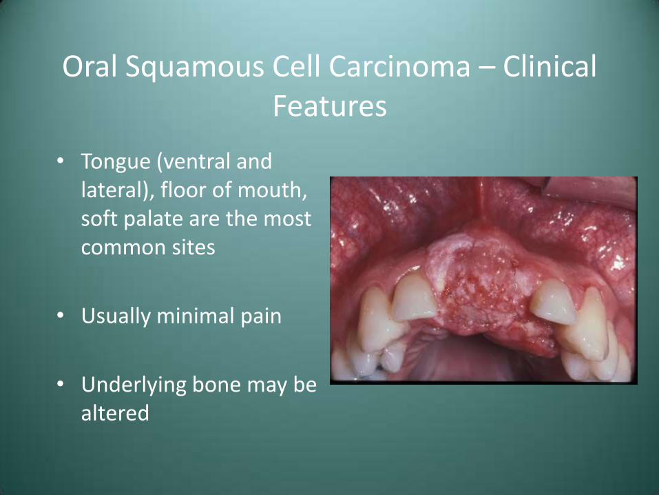
• Tongue (ventral and lateral), floor of mouth, soft palate are the most common sites
• Usually minimal pain
• Underlying bone may be altered
Oral Squamous Cell Carcinoma – Clinical Features
• Tongue (ventral and lateral), floor of mouth, soft palate are the most common sites
• Usually minimal pain
• Underlying bone may be altered
Oral Squamous Cell Carcinoma – Clinical Features
• Tongue (ventral and lateral), floor of mouth, soft palate are the most common sites
• Usually minimal pain
• Underlying bone may be altered

Oral Squamous Cell Carcinoma – Clinical Features
• Tongue (ventral and lateral), floor of mouth, soft palate are the most common sites
• Usually minimal pain
• Underlying bone may be altered
Oral Squamous Cell Carcinoma – Clinical Features
• Tongue (ventral and lateral), floor of mouth, soft palate are the most common sites
• Usually minimal pain
• Underlying bone may be altered

Oral Squamous Cell Carcinoma – Clinical Features
• Tongue (ventral and lateral), floor of mouth, soft palate are the most common sites
• Usually minimal pain
• Underlying bone may be altered
Oral Squamous Cell Carcinoma – Clinical Features
• Tongue (ventral and lateral), floor of mouth, soft palate are the most common sites
• Usually minimal pain
• Underlying bone may be altered
Oral Squamous Cell Carcinoma – Clinical Features
• Tongue (ventral and lateral), floor of mouth, soft palate are the most common sites
• Usually minimal pain
• Underlying bone may be altered
Oral Squamous Cell Carcinoma – Clinical Features
• Tongue (ventral and lateral), floor of mouth, soft palate are the most common sites
• Usually minimal pain
• Underlying bone may be altered

Oral Squamous Cell Carcinoma – Clinical Features
• Tongue (ventral and lateral), floor of mouth, soft palate are the most common sites
• Usually minimal pain
• Underlying bone may be altered

Oral Squamous Cell Carcinoma – Clinical Features
• Tongue (ventral and lateral), floor of mouth, soft palate are the most common sites
• Usually minimal pain
• Underlying bone may be altered
Oral Squamous Cell Carcinoma – Clinical Features
• Tongue (ventral and lateral), floor of mouth, soft palate are the most common sites
• Usually minimal pain
• Underlying bone may be altered
Oral Squamous Cell Carcinoma – Clinical Features
• Tongue (ventral and lateral), floor of mouth, soft palate are the most common sites
• Usually minimal pain
• Underlying bone may be altered
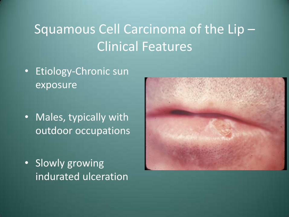
Squamous Cell Carcinoma of the Lip – Clinical Features
• Etiology-Chronic sun exposure
• Males, typically with outdoor occupations
• Slowly growing indurated ulceration
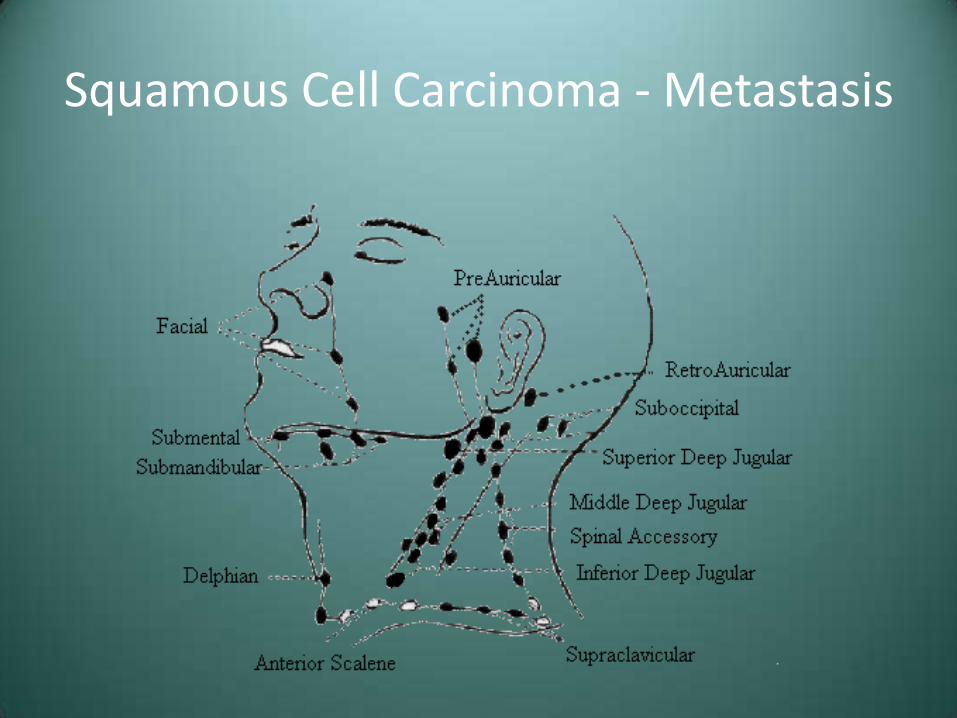
Squamous Cell Carcinoma - Metastasis
• Spread through lymphatics
• Firm nodes
• Movable or fixed
• Distant spread to lungs, liver, bones
• TNM staging
– Stage at diagnosis is the most important prognostic indicator
Squamous Cell Carcinoma - Metastasis
• TNM staging system
– T-Tumor size
– N-Local node involvement
– M-Distant metastasis
Squamous Cell Carcinoma - Metastasis

Squamous Cell Carcinoma – Treatment and Prognosis
• Surgical excision/resection
• Radiation
• Chemotherapy – Squamous cell carcinoma rarely responds well
• Stage I – 85% 5 year survival
• Stage II – 66%
• Stage III – 41%
• Stage IV – 9%
Squamous Cell Carcinoma – Treatment and Prognosis
• National Comprehensive Cancer Network
• http://www.nccn.org/professionals/physician_gls/f_guidelines.asp
Squamous Cell Carcinoma – Treatment and Prognosis
• Carcinoma of the lip carries a much better prognosis
• Prognosis is better for Whites than Blacks
• “Field cancerization” – Persons with one carcinoma are at increased risk of developing a second mucosal tumor

Odds and Ends

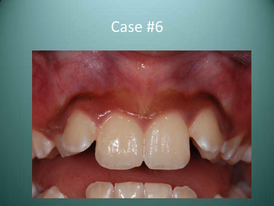
Case #6
• This patient presented with recent onset of the pigmentation seen here
Case #6
Case #5 – Differential Diagnosis
• Normal Physiologic Pigmentation
• Smoker’s Melanosis
• Medication-Associated
• Addison’s Disease
Smoker’s Melanosis
• Rather common melanocytic response found in heavy smokers
• Probably a protective response to the harmful aspects (polycyclic aromatic hydrocarbons) of tobacco smoke
Smoker’s Melanosis – Clinical Features
• F>M
• Frequently on anterior facial gingiva
• “Reverse smokers” show involvement of the palate

Smoker’s Melanosis – Clinical Features
• F>M
• Frequently on anterior facial gingiva
• “Reverse smokers” show involvement of the palate

Smoker’s Melanosis – Clinical Features
• F>M
• Frequently on anterior facial gingiva
• “Reverse smokers” show involvement of the palate
Smoker’s Melanosis – Diagnosis and Treatment
• Clinical, tobacco, and medical history
• May need to rule out systemic cause
• Cessation of smoking will result in gradual resolution
Drug-Related Discolorations of the Oral Mucosa
• Discoloration secondary to melanocytic stimulation or direct deposition into tissue
• Antimalarial meds, minocycline, estrogen, chemotherapeutic agents, AIDS medications

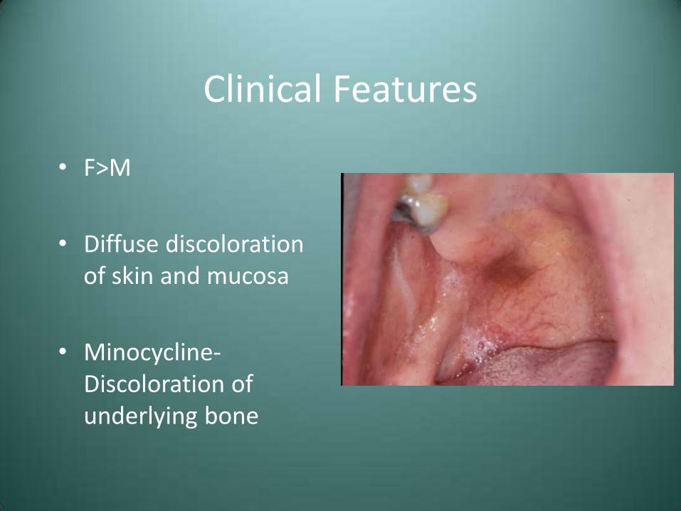
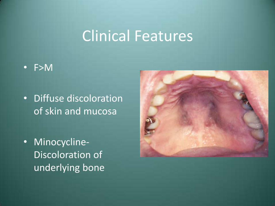
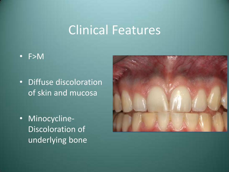
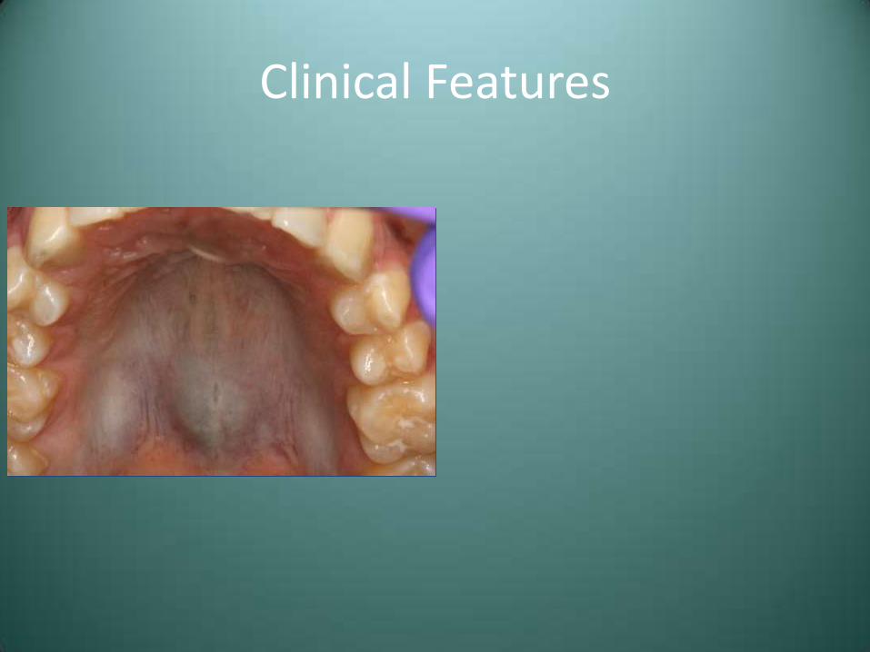
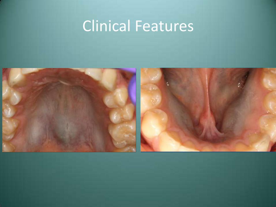
Clinical Features
• F>M
• Diffuse discoloration of skin and mucosa
• Minocycline-Discoloration of underlying bone
Clinical Features
• F>M
• Diffuse discoloration of skin and mucosa
• Minocycline-Discoloration of underlying bone
Clinical Features
• F>M
• Diffuse discoloration of skin and mucosa
• Minocycline-Discoloration of underlying bone
Clinical Features
• F>M
• Diffuse discoloration of skin and mucosa
• Minocycline-Discoloration of underlying bone
Clinical Features
Clinical Features
Treatment
• Gradual resolution upon discontinuation of medication
• Strictly and esthetic issue
• No long term complications
Addison’s Disease (Hypoadrenocorticism)
• Insufficient production of adrenal corticosteroid hormones
• Primary – Secondary to adrenal destruction
• Secondary – Due to malfunctioning pituitary gland
Addison’s Disease – Clinical Features
• Fatigue, irritability, depression, weakness, and hypotension
• Hyperpigmentation (may be seen intraorally)
• GI symptoms, salt-craving

Addison’s Disease – Lab Findings
• Primary – High plasma ACTH
• Secondary – Low plasma ACTH

Addison’s Disease - Treatment
• Corticosteroid replacement therapy
• Preplan dental and oral surgical procedures
• Good prognosis, with patients typically living a normal life span
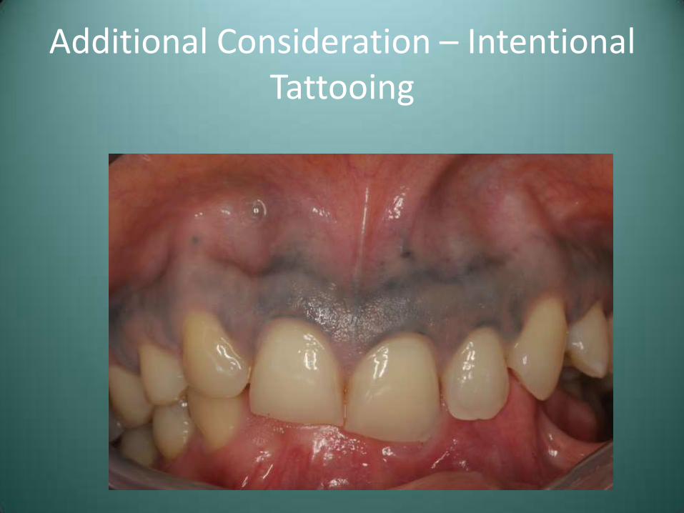
Additional Consideration – Intentional Tattooing

Case #6 – Differential Diagnosis
• Normal Physiologic Pigmentation
• Smoker’s Melanosis
• Medication-Associated
• Addison’s Disease
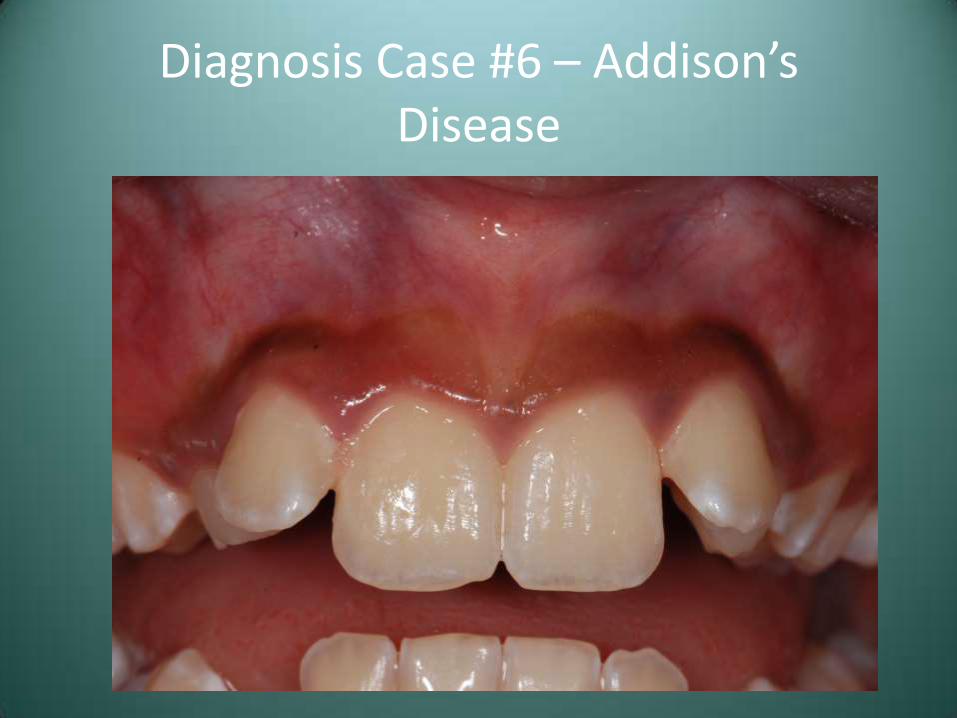
Diagnosis Case #6 – Addison’s Disease
Diagnosis Case #6 – Addison’s Disease
• Further questioning revealed a one month history of nausea, vomiting and intermittent weakness
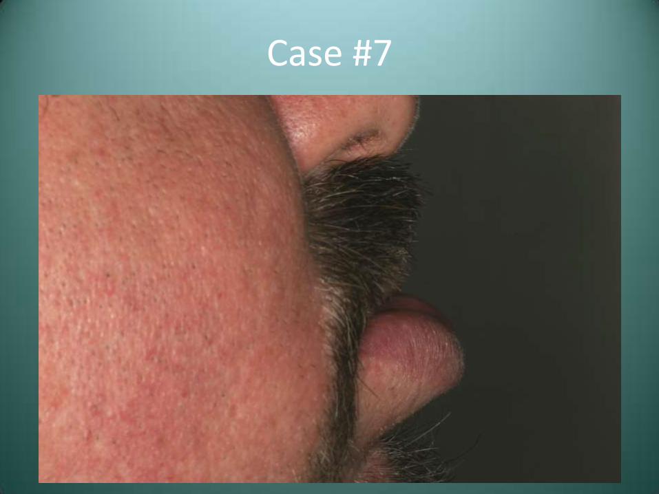
Case #7
• This patient presents with the abnormality seen

Case #7
Case #7
Case #7 – Differential Diagnosis
• Angioedema
• Cheilitis Granulomatosis (Orofacial Granulomatosis)
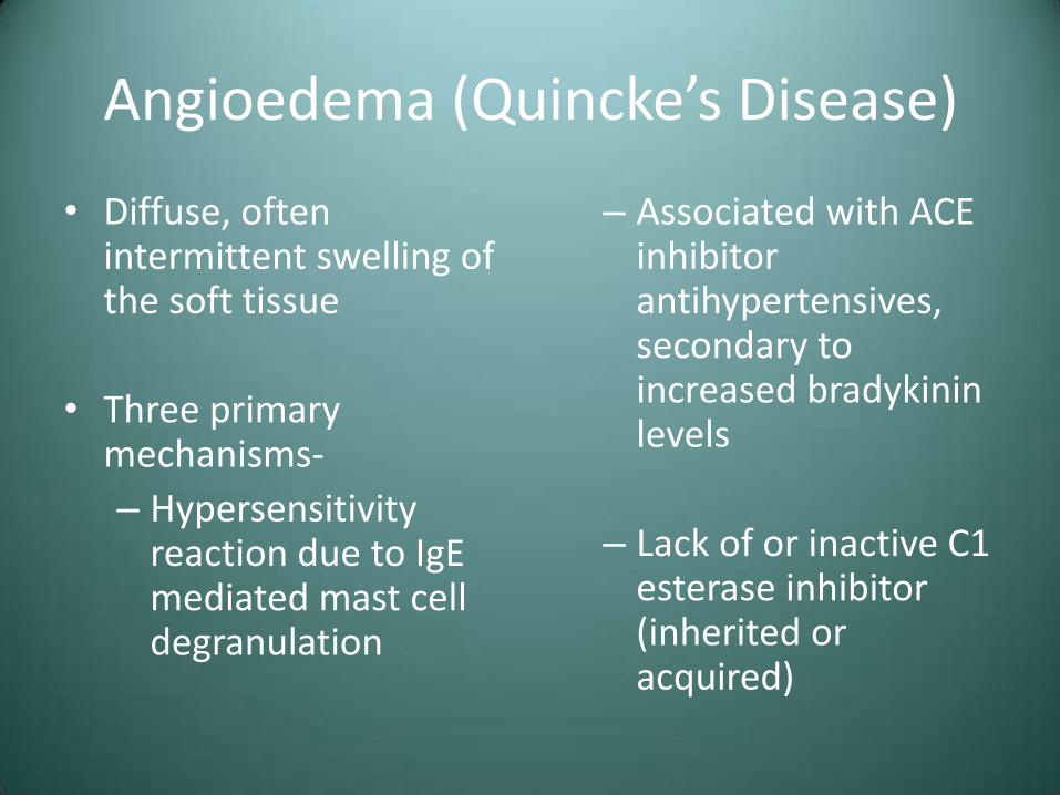
Angioedema (Quincke’s Disease)
• Diffuse, often intermittent swelling of the soft tissue
• Three primary mechanisms-
– Hypersensitivity reaction due to IgE mediated mast cell degranulation
– Associated with ACE inhibitor antihypertensives, secondary to increased bradykinin levels
– Lack of or inactive C1 esterase inhibitor (inherited or acquired)
Angioedema – Clinical Features
• Enlargement of relatively rapid onset
• Pruritis, erythema
• Respiratory involvement may be life threatening
Angioedema – Diagnosis
• Allergic - Clinical presentation in association with suspected antigen
• Inciting cause often not determined
• Evaluate functional C1-INH

Angioedema - Treatment
• Antihistamines for allergic form
• IM epinephrine
• ACE inhibitor-related and C1-INH deficient do not respond to antihistamines
– C1-INH concentrate administration or esterase inhibiting drugs
Cheilitis Granulomatosis (Orofacial Granulomatosis)
• Granulomatous inflammation of unknown etiology or the orofacial presentation of Crohn’s, sarcoidosis, TB, or any other granulomatous process
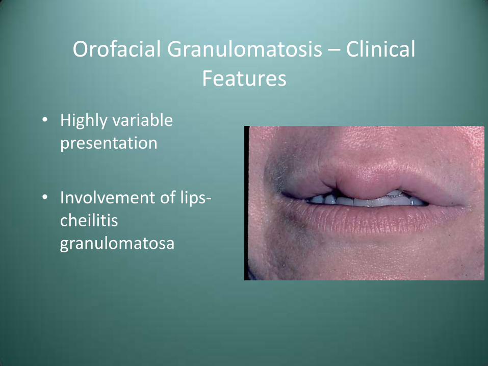
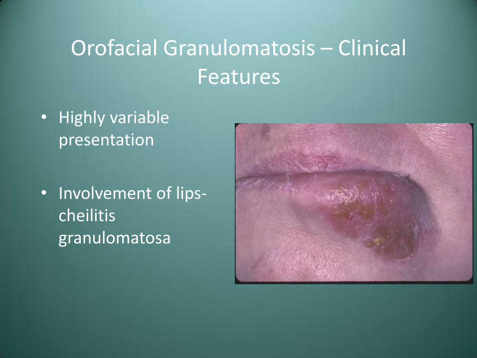
Orofacial Granulomatosis – Clinical Features
• Highly variable presentation
• Involvement of lips-cheilitis granulomatosa
Orofacial Granulomatosis – Clinical Features
• Highly variable presentation
• Involvement of lips-cheilitis granulomatosa
Orofacial Granulomatosis – Clinical Features
• Highly variable presentation
• Involvement of lips-cheilitis granulomatosa
Treatment and Prognosis
• Intralesional corticosteroids
• Multiple treatments
• Good prognosis; requires thorough work-up
• Primarily a cosmetic problem

Case #7 – Differential Diagnosis
• Angioedema
• Cheilitis Granulomatosis (Orofacial Granulomatosis)
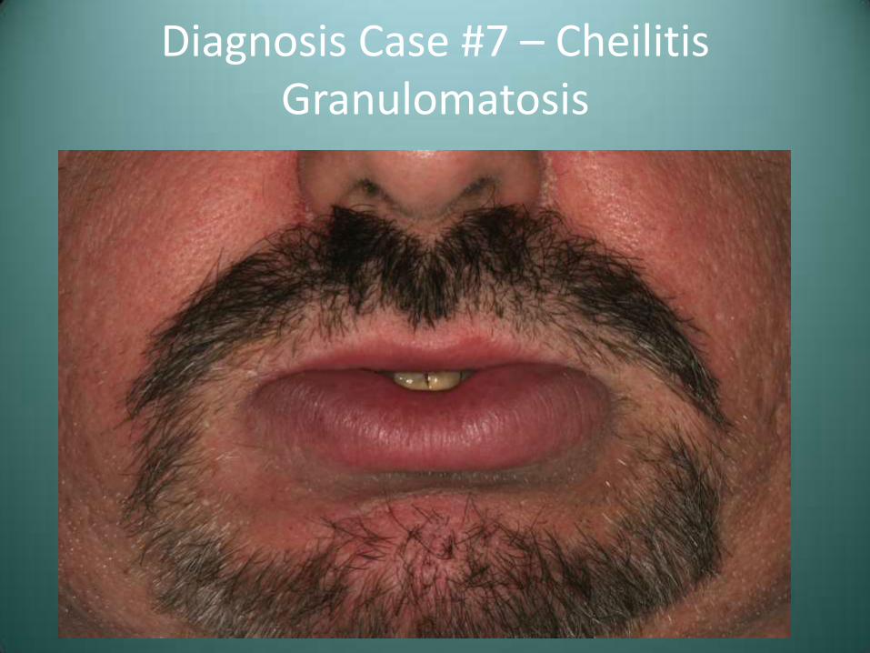
Diagnosis Case #7 – Cheilitis Granulomatosis