cong.heart desiases - elearning.sumdu.edu.uac4b16c5d... · acyanotic congenital heart disease heart...
TRANSCRIPT
Cong.heart_desiases.docxОлена Костянтинівна Редько
2015

3
3
3
20
22
ЗмістКлючові терміни:Acyanotic Congenital Heart DiseaseHeart and Main VesselsAtrioventricular Septal Defects (Ostium Primum and Atrioventricular Canal orEndocardial Cushion Defects)Cyanotic Congenital Heart Disease
Ключові терміни: 3
Acyanotic Congenital Heart DiseaseHeart and Main VesselsAtrioventricular Septal Defects (Ostium Primum and Atrioventricular Canal or Endocardial CushionDefects)Cyanotic Congenital Heart Disease
Ключові терміни:Atrial Septal Defect, Atrioventricular Septal Defects (Ostium Primum and Atrioventricular Canal orEndocardial Cushion Defects), Coarctation of the aorta, Ebstein anomaly, Hypoplastic left heartsyndrome, PATHOPHYSIOLOGY, Questions, Teaching Points:, Tetralogy of Fallot, The EKG, Thechest radiograph, The clinical features, Total anomalous pulmonary venous return (TAPVR),Transposition of the great vessels, Treatment, Tricuspid atresia, Truncus arteriosus, Ventricularseptal defects (VSD), atrial septal defects (ASD), and patent ductus arteriosus (PDA), patentductus arteriosus (PDA)
Acyanotic Congenital HeartDisease
Heart and Main Vessels
A 4 year old male presents in the office for a preschool physical examination. In the course of theinterview, his mother mentions that he seems to get short of breath with exercise recently. It isespecially noticeable during his swimming lessons when he tires before the other children do in his class.He has otherwise been in good health since his last physical exam in the previous year. His records forthe past year show 3 office visits for minor upper respiratory illnesses, and no emergency room visits.He has never had wheezing during his colds.
Exam: T37.5, P92, R25, BP right arm 97/70, oxygen saturation 98% in room air. Height and weight are
Ключові терміни: 4
Exam: T37.5, P92, R25, BP right arm 97/70, oxygen saturation 98% in room air. Height and weight areat the 25th percentile. He is cooperative and well nourished in no distress. HEENT and neck exams arenormal. His chest is symmetrical. Heart: No palpable thrill, normal 1st and 2nd heart sounds; no clicks orrubs; grade 1/6 ejection systolic murmur heard along the left sternal border with radiation to the backbetween the scapulae; no diastolic murmur. Lungs are clear to auscultation. Abdomen without noorganomegaly or masses palpable. Genitalia: normal male. Extremities: Femoral pulses are slightlydiminished to palpation; no peripheral edema, clubbing or cyanosis of the nail beds. His neurological isnormal.
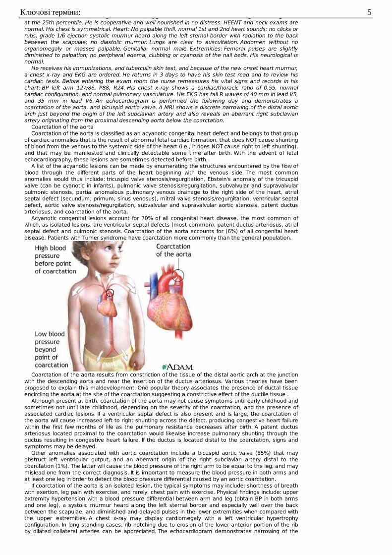
He receives his immunizations, and tuberculin skin test, and because of the new onset heart murmur,a chest x-ray and EKG are ordered. He returns in 3 days to have his skin test read and to review hiscardiac tests. Before entering the exam room the nurse remeasures his vital signs and records in hischart: BP left arm 127/86, P88, R24. His chest x-ray shows a cardiac/thoracic ratio of 0.55, normalcardiac configuration, and normal pulmonary vasculature. His EKG has tall R waves of 40 mm in lead V5,and 35 mm in lead V6. An echocardiogram is performed the following day and demonstrates acoarctation of the aorta, and bicuspid aortic valve. A MRI shows a discrete narrowing of the distal aorticarch just beyond the origin of the left subclavian artery and also reveals an aberrant right subclavianartery originating from the proximal descending aorta below the coarctation.
Coarctation of the aortaCoarctation of the aorta is classified as an acyanotic congenital heart defect and belongs to that group
of cardiac anomalies that is the result of abnormal fetal cardiac formation, that does NOT cause shuntingof blood from the venous to the systemic side of the heart (i.e., it does NOT cause right to left shunting),and that may be manifested and clinically detectable some time after birth. With the advent of fetalechocardiography, these lesions are sometimes detected before birth.
A list of the acyanotic lesions can be made by enumerating the structures encountered by the flow ofblood through the different parts of the heart beginning with the venous side. The most commonanomalies would thus include: tricuspid valve stenosis/regurgitation, Ebstein's anomaly of the tricuspidvalve (can be cyanotic in infants), pulmonic valve stenosis/regurgitation, subvalvular and supravalvularpulmonic stenosis, partial anomalous pulmonary venous drainage to the right side of the heart, atrialseptal defect (secundum, primum, sinus venosus), mitral valve stenosis/regurgitation, ventricular septaldefect, aortic valve stenosis/regurgitation, subvalvular and supravalvular aortic stenosis, patent ductusarteriosus, and coarctation of the aorta.
Acyanotic congenital lesions account for 70% of all congenital heart disease, the most common ofwhich, as isolated lesions, are ventricular septal defects (most common), patent ductus arteriosus, atrialseptal defect and pulmonic stenosis. Coarctation of the aorta accounts for (6%) of all congenital heartdisease. Patients with Turner syndrome have coarctation more commonly than the general population.
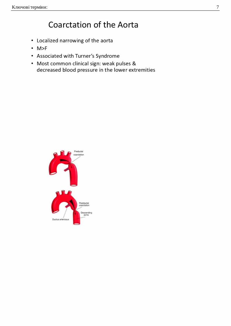
Coarctation of the aorta results from constriction of the tissue of the distal aortic arch at the junctionwith the descending aorta and near the insertion of the ductus arteriosus. Various theories have beenproposed to explain this maldevelopment. One popular theory associates the presence of ductal tissueencircling the aorta at the site of the coarctation suggesting a constrictive effect of the ductile tissue .
Although present at birth, coarctation of the aorta may not cause symptoms until early childhood andsometimes not until late childhood, depending on the severity of the coarctation, and the presence ofassociated cardiac lesions. If a ventricular septal defect is also present and is large, the coarctation ofthe aorta will cause increased left to right shunting across the defect, producing congestive heart failurewithin the first few months of life as the pulmonary resistance decreases after birth. A patent ductusarteriosus located proximal to the coarctation would likewise increase pulmonary shunting through theductus resulting in congestive heart failure. If the ductus is located distal to the coarctation, signs andsymptoms may be delayed.
Other anomalies associated with aortic coarctation include a bicuspid aortic valve (85%) that mayobstruct left ventricular output, and an aberrant origin of the right subclavian artery distal to thecoarctation (1%). The latter will cause the blood pressure of the right arm to be equal to the leg, and maymislead one from the correct diagnosis. It is important to measure the blood pressure in both arms andat least one leg in order to detect the blood pressure differential caused by an aortic coarctation.
If coarctation of the aorta is an isolated lesion, the typical symptoms may include: shortness of breathwith exertion, leg pain with exercise, and rarely, chest pain with exercise. Physical findings include: upperextremity hypertension with a blood pressure differential between arm and leg (obtain BP in both armsand one leg), a systolic murmur heard along the left sternal border and especially well over the backbetween the scapulae, and diminished and delayed pulses in the lower extremities when compared withthe upper extremities. A chest x-ray may display cardiomegaly with a left ventricular hypertrophyconfiguration. In long standing cases, rib notching due to erosion of the lower anterior portion of the ribby dilated collateral arteries can be appreciated. The echocardiogram demonstrates narrowing of the
Ключові терміни: 5
distal aortic arch with increased velocities on pulsed and color Doppler. The pulsed Doppler waveform hasa typical prolonged systolic phase extending throughout systole. The MRI produces a static but clearerpicture, than the echocardiogram, of the anatomy of the coarctation. An angiogram is sometimesnecessary to clarify associated cardiac lesions.
There are several surgical techniques used to repair a coarctation of the aorta. Each technique hashad its own proponents at one time or another. If the coarcted segment is short and discrete, resectionand end to end anastomosis of the proximal and distal ends is possible. If the coarctation is a longtubular obstruction, resection with interposition of a tube graft would be necessary. Some surgeons favora longitudinal incision with insertion of a synthetic graft to enlarge the diameter. In the young infant,sacrificing the left subclavian artery, and using the transected blood vessel as a graft by turning it downand sewing it into the aortic wall was popular at one time.
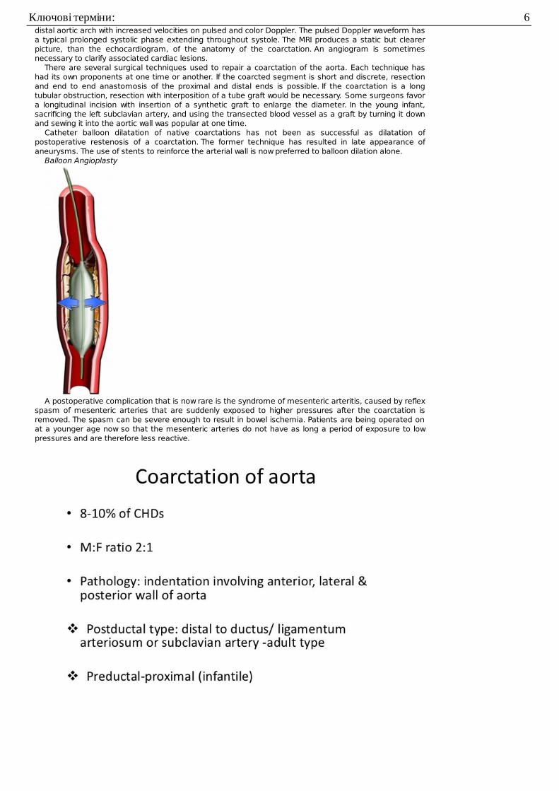
Catheter balloon dilatation of native coarctations has not been as successful as dilatation ofpostoperative restenosis of a coarctation. The former technique has resulted in late appearance ofaneurysms. The use of stents to reinforce the arterial wall is now preferred to balloon dilation alone.
Balloon Angioplasty
A postoperative complication that is now rare is the syndrome of mesenteric arteritis, caused by reflexspasm of mesenteric arteries that are suddenly exposed to higher pressures after the coarctation isremoved. The spasm can be severe enough to result in bowel ischemia. Patients are being operated onat a younger age now so that the mesenteric arteries do not have as long a period of exposure to lowpressures and are therefore less reactive.
Ключові терміни: 6
Ключові терміни: 7
Ключові терміни: 8
Ключові терміни: 9
Ventricular septal defects (VSD), atrial septal defects (ASD), and patent ductus arteriosus (PDA)account for a large percentage of all congenital heart defects. They share common physiologichemodynamics and will be discussed together.
Ventricular septal defects (VSD)
Ключові терміни: 10
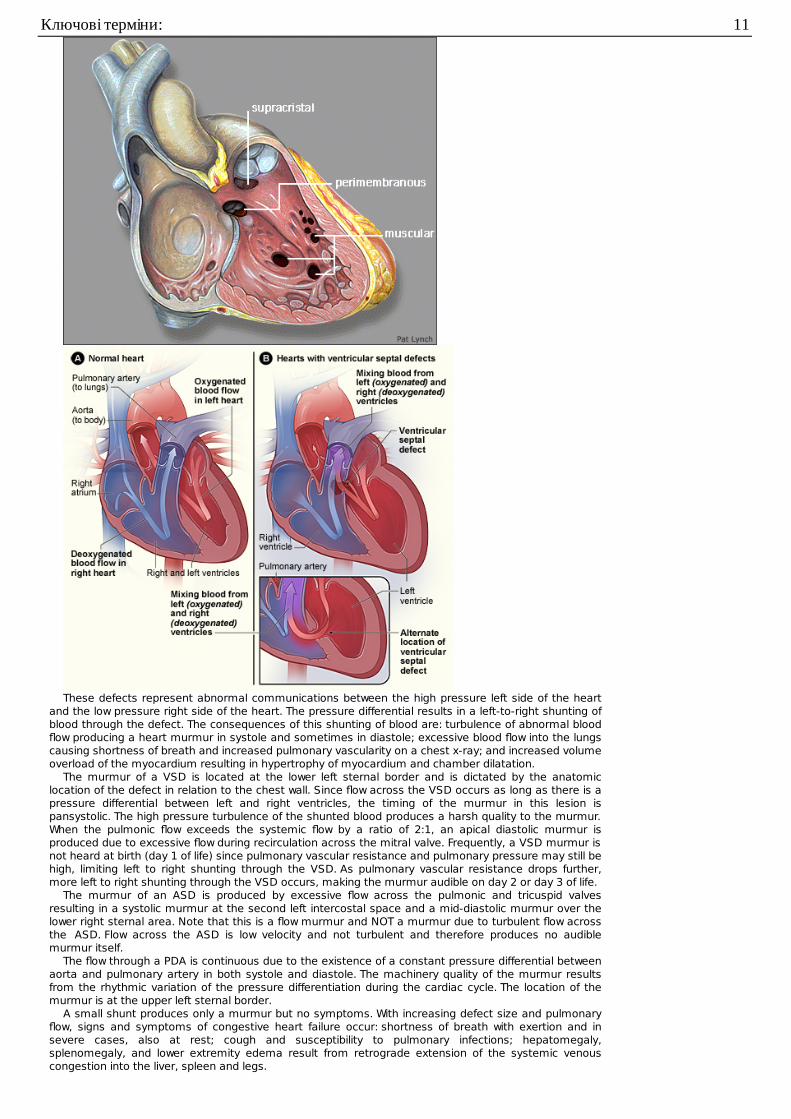
These defects represent abnormal communications between the high pressure left side of the heartand the low pressure right side of the heart. The pressure differential results in a left-to-right shunting ofblood through the defect. The consequences of this shunting of blood are: turbulence of abnormal bloodflow producing a heart murmur in systole and sometimes in diastole; excessive blood flow into the lungscausing shortness of breath and increased pulmonary vascularity on a chest x-ray; and increased volumeoverload of the myocardium resulting in hypertrophy of myocardium and chamber dilatation.
The murmur of a VSD is located at the lower left sternal border and is dictated by the anatomiclocation of the defect in relation to the chest wall. Since flow across the VSD occurs as long as there is apressure differential between left and right ventricles, the timing of the murmur in this lesion ispansystolic. The high pressure turbulence of the shunted blood produces a harsh quality to the murmur.When the pulmonic flow exceeds the systemic flow by a ratio of 2:1, an apical diastolic murmur isproduced due to excessive flow during recirculation across the mitral valve. Frequently, a VSD murmur isnot heard at birth (day 1 of life) since pulmonary vascular resistance and pulmonary pressure may still behigh, limiting left to right shunting through the VSD. As pulmonary vascular resistance drops further,more left to right shunting through the VSD occurs, making the murmur audible on day 2 or day 3 of life.
The murmur of an ASD is produced by excessive flow across the pulmonic and tricuspid valvesresulting in a systolic murmur at the second left intercostal space and a mid-diastolic murmur over thelower right sternal area. Note that this is a flow murmur and NOT a murmur due to turbulent flow acrossthe ASD. Flow across the ASD is low velocity and not turbulent and therefore produces no audiblemurmur itself.
The flow through a PDA is continuous due to the existence of a constant pressure differential betweenaorta and pulmonary artery in both systole and diastole. The machinery quality of the murmur resultsfrom the rhythmic variation of the pressure differentiation during the cardiac cycle. The location of themurmur is at the upper left sternal border.
A small shunt produces only a murmur but no symptoms. With increasing defect size and pulmonaryflow, signs and symptoms of congestive heart failure occur: shortness of breath with exertion and insevere cases, also at rest; cough and susceptibility to pulmonary infections; hepatomegaly,splenomegaly, and lower extremity edema result from retrograde extension of the systemic venouscongestion into the liver, spleen and legs.
Ключові терміни: 11

The chest x-ray in a left-to-right shunt lesion will demonstrate congested pulmonary vessels.Enlargement of specific cardiac chambers is due to excessive volume overload. The left atrium andventricle are dilated in VSD and PDA, and the right heart chamber is dilated in ASD. The EKG revealshypertrophy of the corresponding cardiac chambers.
patent ductus arteriosus (PDA)
Untreated defects with large shunts will eventually result in injury to the pulmonary arterioles, vascularobstruction, and pulmonary hypertension. The development of permanent injury to the pulmonaryvessels is a function of the duration of the exposure to excessive blood flow and the anatomy, occurringmore rapidly in VSD and PDA than in ASD. If this process is not reversed, Eisenmenger's complex of rightto left shunting may occur as the right sided pressures (pulmonary hypertension) exceeds left sidedpressures.
Intracardiac repair of a VSD and ASD require cardiopulmonary bypass. Repair of a PDA is extracardiacand is achieved without cardiopulmonary bypass. The intracardiac defects can be closed by primarysuturing of the edges of the defect if small, or by covering with a patch material if large. The PDA isusually tied off and divided.
Complete heart block secondary to injury to the conduction system during repair of a VSD may requirea pacemaker in the postoperative period. The knowledge of the location of the conduction system inrelationship to the defect now makes this a rare complication. The mortality rate in experienced handsshould be less than 5% if all ages are considered, with infants carrying a higher mortality rate especially ifpulmonary hypertension is present.
Questions
1. 1. True/False: Congenital heart disease is always detectable at birth.2. 2. True/False: Equal blood pressures in the right arm and left leg rule out the diagnosis of
coarctation of the aorta.3. 3. Which are the three most common acyanotic congenital heart lesions?4. 4. True/False: The presence of palpable femoral pulses rules out the diagnosis of aortic coarctation.5. 5. True/False: Surgical repair of PDA does not require cardiopulmonary bypass.6. 6. Explain how a child with an isolated VSD (classified as an acyanotic lesion) could become
cyanotic?
Ключові терміни: 12
7. 7. True/False: Medical students and residents will typically not hear the murmur of a VSD duringthe initial newborn assessment in the nursery because the murmur of a VSD is subtle and lowpitched.
Answers to questions
1. 1. False. The physiologic pulmonary hypertension present in a newborn can prevent blood flowacross a septal defect or PDA. These can be detected several hours after birth or several daysafter birth. Other congenital heart disease lesions may remain occult for longer period of time.
2. 2. False. An aberrant right subclavian artery originating below a coarctation will produce equalpressures in the right arm and leg.
3. 3. VSD, ASD, PDA. Of these, VSD is the most common.4. 4. False. Development of collateral vessels to the lower body can produce palpable femoral
pulses.5. 5. True.6. 6. Congestive heart failure and pulmonary edema may cause hypoxia. If the hypoxia is severe
enough, visible cyanosis will result, although this can be overcome with oxygen and othertreatments for pulmonary edema and congestive heart failure. Long standing excessivepulmonary blood flow leads to pulmonary hypertension and Eisenmenger's complex, right to leftshunting and cyanosis.
7. 7. False. They cannot hear the murmur of a VSD on day 1 because on day 1, pulmonary vascularresistance is still high, which restricts left to right flow through the VSD. On day 2, pulmonaryvascular resistance is lower, so left to right shunting through the VSD increases making themurmur louder.
This is a 7 week old term female infant who presented with wheezing, coughing, and two episodes of non-bilious emesis. She was seen by her pediatrician, who suspected that she had bronchiolitis, and she wastreated with oral albuterol syrup. An RSV (Respiratory Syncytial Virus) nasal prep
was done, and this was negative; however, the patient's condition worsened, and she was brought tothe Emergency Department later that evening.
Exam: VS T37.2R, P168, R70, BP126/86, oxygen saturation 96% on room air. The infant was fussy,though consolable, with moderate respiratory distress. She was non-toxic in appearance. The anteriorfontanelle was soft and flat. The pupils were equal and reactive and the mucus membranes were moist.The neck was supple. The lungs had diffuse wheezes and crackles bilaterally with intercostal retractions.Heart sounds were difficult to auscultate due to the noisy breathing, but no obvious loud pathologicmurmur was heard. The abdomen was soft and nontender with active bowel sounds. The liver edge waspalpated 3-4 cm below the right costal margin. Capillary refill time in the extremities was 3 seconds.
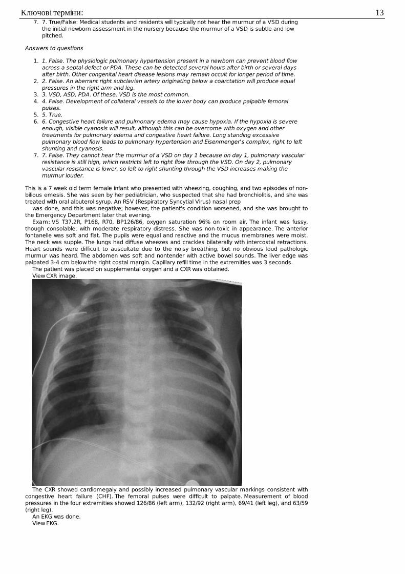
The patient was placed on supplemental oxygen and a CXR was obtained.View CXR image.
The CXR showed cardiomegaly and possibly increased pulmonary vascular markings consistent withcongestive heart failure (CHF). The femoral pulses were difficult to palpate. Measurement of bloodpressures in the four extremities showed 126/86 (left arm), 132/92 (right arm), 69/41 (left leg), and 63/59(right leg).
An EKG was done.View EKG.
Ключові терміни: 13
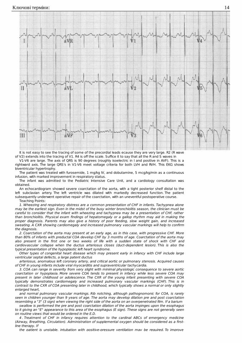
It is not easy to see the tracing of some of the precordial leads ecause they are very large. R2 (R waveof V2) extends into the tracing of V1. R4 is off the scale. Suffice it to say that all the R and S waves in
V1-V6 are large. The axis of QRS is 90 degrees (roughly isoelectric in I and positive in AVF). This is arightward axis. The large QRS's in V1-V6 meet voltage criteria for both LVH and RVH. This EKG showsbiventricular hypertrophy.
The patient was treated with furosemide, 1 mg/kg IV, and dobutamine, 5 mcg/kg/min as a continuousinfusion, with marked improvement in respiratory status.
The infant was admitted to the Pediatric Intensive Care Unit, and a cardiology consultation wasobtained.
An echocardiogram showed severe coarctation of the aorta, with a tight posterior shelf distal to theleft subclavian artery. The left ventricle was dilated with markedly decreased function. The patientsubsequently underwent operative repair of the coarctation, with an uneventful postoperative course.
Teaching Points:1. Wheezing and respiratory distress are a common presentation of CHF in infants. Tachypnea alone
may be the earliest sign. Even in the midst of the busy winter bronchiolitis season, the clinician must becareful to consider that the infant with wheezing and tachypnea may be a presentation of CHF, ratherthan bronchiolitis. Physical exam findings of hepatomegaly or a gallop rhythm may aid in making theproper diagnosis. Parents may also give a history of poor feeding, slow weight gain, and increasedsweating. A CXR showing cardiomegaly and increased pulmonary vascular markings will help to confirmthe diagnosis.
2. Coarctation of the aorta may present at an early age, as in this case, with progressive CHF. Morethan 80% of infants with preductal COA develop CHF by 3 months of age. Coarctation of the aorta mayalso present in the first one or two weeks of life with a sudden state of shock with CHF andcardiovascular collapse when the ductus arteriosus closes (duct-dependent lesion). This is also thetypical presentation of the hypoplastic left heart syndrome.
Other types of congenital heart disease which may present early in infancy with CHF include largeventricular septal defects, a large patent ductus
arteriosus, anomalous left coronary artery, and critical aortic or pulmonary stenosis. Acquired causesof CHF in young infants include viral myocarditis and supraventricular tachycardia.
3. COA can range in severity from very slight with minimal physiologic consequence to severe aorticcoarctation or hypoplasia. More severe COA tends to present in infancy while less severe COA maypresent in later childhood or adolescence. The CXR of the young infant presenting with severe COAtypically demonstrates cardiomegaly and increased pulmonary vascular markings (CHF). This is incontrast to the CXR of COA presenting later in childhood, which typically shows a normal or only slightlyenlarged heart,
and normal pulmonary vascular markings. Rib notching, although pathognomonic for COA, is rarelyseen in children younger than 9 years of age. The aorta may develop dilation pre and post coarctationresembling a "3" (3 sign) when viewing the right side of the aorta on an overpenetrated film. If a barium
swallow is performed the pre and post coarctation dilation of the aorta impinges upon the esophagusto it giving an "E" appearance to this area of the esophagus (E sign). These signs are not generally seenon routine views that would be ordered in the E.D.
4 . Treatment of CHF in infancy requires attention to the cardinal ABCs of emergency medicine(Airway, Breathing, Circulation). Administration of supplemental oxygen should be considered as a first-line therapy. If
the patient is unstable, intubation with positive-pressure ventilation may be required. To improve
Ключові терміни: 14
the patient is unstable, intubation with positive-pressure ventilation may be required. To improvecardiac contractility, inotropes such as digoxin or dobutamine may be needed. Dobutamine also has theadvantage of decreasing afterload. Diuretics such as furosemide aid by decreasing preload. In severecases, other vasoactive or inotropic agents such as sodium nitroprusside and amrinone may beconsidered. These agents should be used in the intensive care unit, using invasive hemodynamicmonitoring. If a duct-dependent lesion is suspected, prostaglandin E1 should be started
as a continuous infusion at 0.1 mcg/kg/min.This is 2-month old male who presents to the emergency department with a five day history of funnybreathing. He was well until 5 days prior when his mother noted noisy, rapid breathing and a tactiletemperature. Four days prior, he was taken to his private physician and was started on amoxicillin forotitis media. His lung exam at that time was normal. Two days prior he was taken to the emergencydepartment and was noted to be wheezing. He was given an albuterol aerosol and was discharged on
oral albuterol. He continued to have breathing problems at home and now returns to the emergencydepartment since his condition has not improved. His birth history is unremarkable, and he has shownadequate weight gain since birth. There are no reported feeding problems according to his mother. Hisfamily history is significant for two siblings with asthma.
Exam: T36.9, P168, BP 98/60. His respiratory rate varies between 60 and 80 per minute. His oxygensaturation is 97% in room air. His oxygen saturation improves to 100% on oxygen by nasal cannula at
2 liters per minute. He is a fussy infant with modest achypnea. Despite this, he does not appear to be insignificant distress. He is not toxic. He is noted to have mild retractions when crying, with bilaterallycoarse breath sounds without wheezes. Heart regular
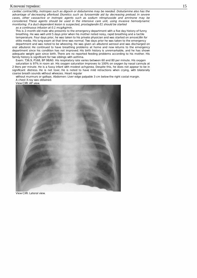
without murmurs or gallops. Abdomen: Liver edge palpable 3 cm below the right costal margin.A chest X-ray was obtained.View CXR: AP view.
View CXR: Lateral view.
Ключові терміни: 15
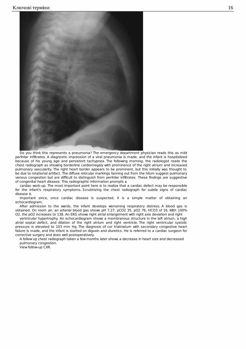
Do you think this represents a pneumonia? The emergency department physician reads this as mildperihilar infiltrates. A diagnostic impression of a viral pneumonia is made, and the infant is hospitalizedbecause of his young age and persistent tachypnea. The following morning, the radiologist reads thechest radiograph as showing borderline cardiomegaly with prominence of the right atrium and increasedpulmonary vascularity. The right heart border appears to be prominent, but this initially was thought tobe due to rotational artifact. The diffuse reticular markings fanning out from the hilum suggest pulmonaryvenous congestion but are difficult to distinguish from perihilar infiltrates. These findings are suggestiveof congenital heart disease. This radiographic information prompts a
cardiac work-up. The most important point here is to realize that a cardiac defect may be responsiblefor the infant's respiratory symptoms. Scrutinizing the chest radiograph for subtle signs of cardiacdisease is
important since, once cardiac disease is suspected, it is a simple matter of obtaining anechocardiogram.
After admission to the wards, the infant develops worsening respiratory distress. A blood gas isobtained. On room air, an arterial blood gas shows pH 7.27, pCO2 35, pO2 76, HCO3 of 16. With 100%O2, the pO2 increases to 138. An EKG shows right atrial enlargement with right axis deviation and right
ventricular hypertrophy. An echocardiogram shows a membranous structure in the left atrium, a highatrial septal defect, and dilation of the right atrium and right ventricle. The right ventricular systolicpressure is elevated to 103 mm Hg. The diagnosis of cor triatriatum with secondary congestive heartfailure is made, and the infant is started on digoxin and diuretics. He is referred to a cardiac surgeon forcorrective surgery and does well postoperatively.
A follow up chest radiograph taken a few months later shows a decrease in heart size and decreasedpulmonary congestion.View follow-up CXR.
Ключові терміни: 16
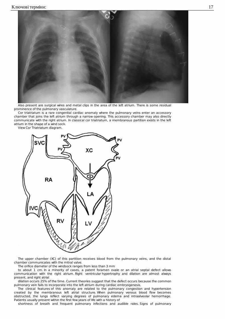
Also present are surgical wires and metal clips in the area of the left atrium. There is some residualprominence of the pulmonary vasculature.
Cor triatriatum is a rare congenital cardiac anomaly where the pulmonary veins enter an accessorychamber that joins the left atrium through a narrow opening. This accessory chamber may also directlycommunicate with the right atrium. In classical cor triatriatum, a membranous partition exists in the leftatrium in the shape of a wind sock.
View Cor Triatriatum diagram.
The upper chamber (XC) of this partition receives blood from the pulmonary veins, and the distalchamber communicates with the mitral valve.
The orifice diameter of the windsock ranges from less than 3 mmto about 1 cm. In a minority of cases, a patent foramen ovale or an atrial septal defect allows
communication with the right atrium. Right ventricular hypertrophy and dilation are almost alwayspresent, and right atrial
dilation occurs 25% of the time. Current theories suggest that the defect occurs because the commonpulmonary vein fails to incorporate into the left atrium during cardiac embryogenesis.
The clinical features of this anomaly are related to the pulmonary congestion and hypertensioncreated by the membranous left atrial structure. When pulmonary venous blood flow becomesobstructed, the lungs reflect varying degrees of pulmonary edema and intraalveolar hemorrhage.Patients usually present within the first few years of life with a history of
shortness of breath and frequent pulmonary infections and audible rales. Signs of pulmonary
Ключові терміни: 17
hypertension, including a loud pulmonic component of the second heart sound, right ventricular heaveand pulmonary systolic ejection clicks are often present. The usual heart murmur is a soft,blowing,systolic murmur heard best at the left sternal border.
The EKG usually reveals signs of right-sided heart overload such as right ventricular hypertrophy andright atrial enlargement.
The chest radiograph often reveals pulmonary venous obstruction. Diffuse reticular pulmonarymarkings fan out from the hilum to involve the lower lung fields. Kerley B lines may also be present. Theright heart border may reveal a double density suggestive of left atrial enlargement. Other findingsinclude enlargement of the main pulmonary artery and
right ventricular hypertrophy.Treatment of this disease involves management of congestive heart failure. Once patients reach this
stage, they usually deteriorate fairly quickly despite medical management. Surgical intervention shouldbe planned as soon as possible in symptomatic patients once the diagnosis is made. The operation ofchoice is usually correction under direct vision with cardiopulmonary bypass. The prognosis of cortriatriatum is related to the size of the orifice in the obstructing membrane. Without surgical correction,the average survival is about 3 months when the opening is less than 3 mm, and 16 years when theopening is greater than 3 mm. In those patients surviving operative correction, the prognosis is excellent.
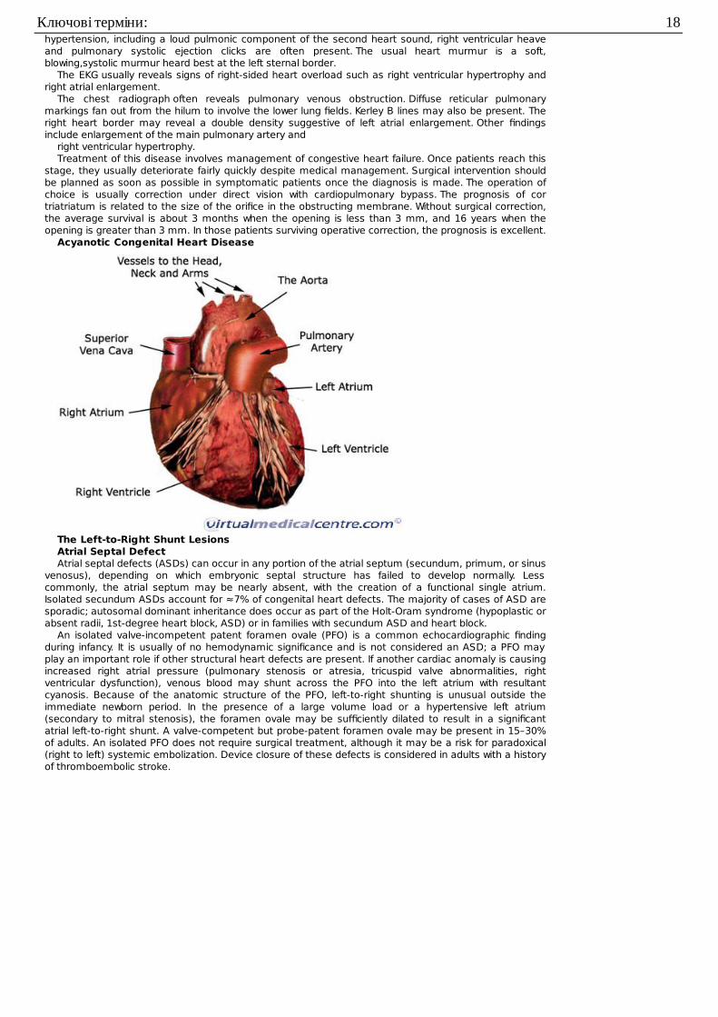
Acyanotic Congenital Heart Disease
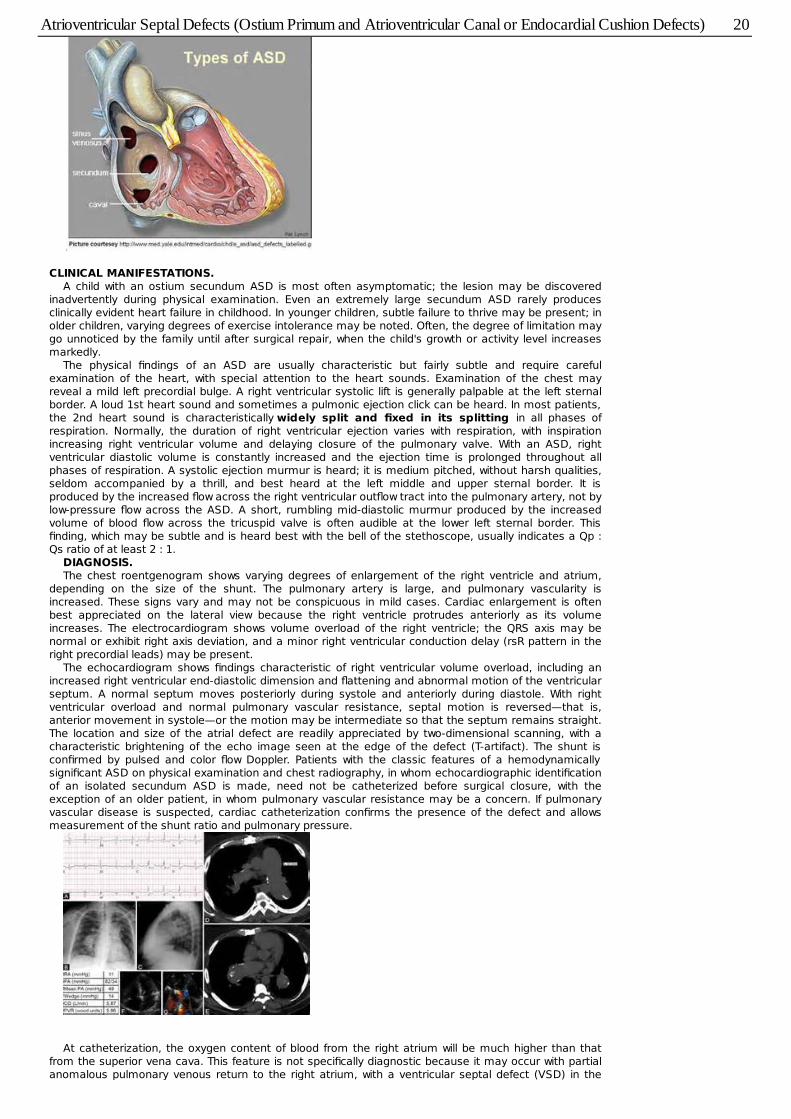
The Left-to-Right Shunt LesionsAtrial Septal DefectAtrial septal defects (ASDs) can occur in any portion of the atrial septum (secundum, primum, or sinus
venosus), depending on which embryonic septal structure has failed to develop normally. Lesscommonly, the atrial septum may be nearly absent, with the creation of a functional single atrium.Isolated secundum ASDs account for ≈7% of congenital heart defects. The majority of cases of ASD aresporadic; autosomal dominant inheritance does occur as part of the Holt-Oram syndrome (hypoplastic orabsent radii, 1st-degree heart block, ASD) or in families with secundum ASD and heart block.
An isolated valve-incompetent patent foramen ovale (PFO) is a common echocardiographic findingduring infancy. It is usually of no hemodynamic significance and is not considered an ASD; a PFO mayplay an important role if other structural heart defects are present. If another cardiac anomaly is causingincreased right atrial pressure (pulmonary stenosis or atresia, tricuspid valve abnormalities, rightventricular dysfunction), venous blood may shunt across the PFO into the left atrium with resultantcyanosis. Because of the anatomic structure of the PFO, left-to-right shunting is unusual outside theimmediate newborn period. In the presence of a large volume load or a hypertensive left atrium(secondary to mitral stenosis), the foramen ovale may be sufficiently dilated to result in a significantatrial left-to-right shunt. A valve-competent but probe-patent foramen ovale may be present in 15–30%of adults. An isolated PFO does not require surgical treatment, although it may be a risk for paradoxical(right to left) systemic embolization. Device closure of these defects is considered in adults with a historyof thromboembolic stroke.
Ключові терміни: 18
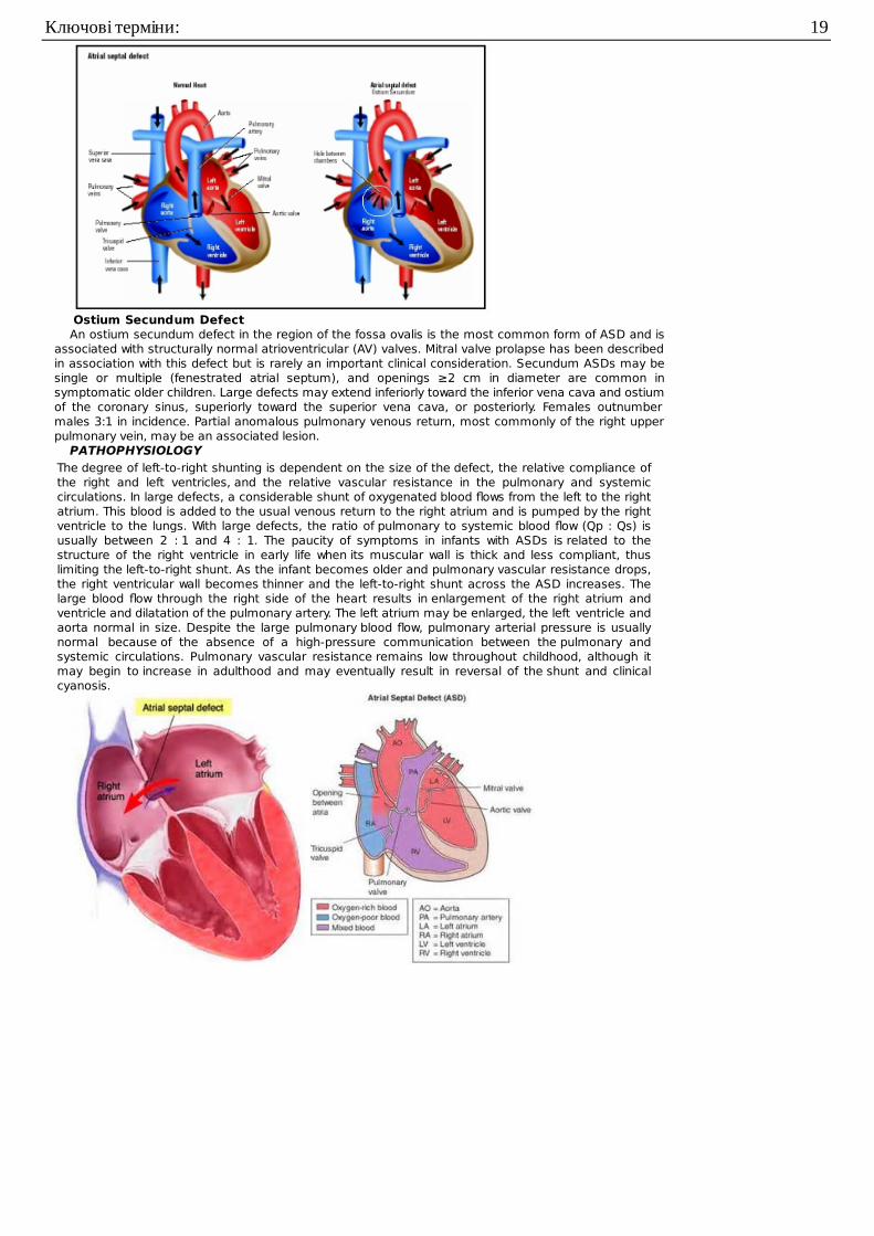
Ostium Secundum DefectAn ostium secundum defect in the region of the fossa ovalis is the most common form of ASD and is
associated with structurally normal atrioventricular (AV) valves. Mitral valve prolapse has been describedin association with this defect but is rarely an important clinical consideration. Secundum ASDs may besingle or multiple (fenestrated atrial septum), and openings ≥2 cm in diameter are common insymptomatic older children. Large defects may extend inferiorly toward the inferior vena cava and ostiumof the coronary sinus, superiorly toward the superior vena cava, or posteriorly. Females outnumbermales 3:1 in incidence. Partial anomalous pulmonary venous return, most commonly of the right upperpulmonary vein, may be an associated lesion.
PATHOPHYSIOLOGYThe degree of left-to-right shunting is dependent on the size of the defect, the relative compliance ofthe right and left ventricles, and the relative vascular resistance in the pulmonary and systemiccirculations. In large defects, a considerable shunt of oxygenated blood flows from the left to the rightatrium. This blood is added to the usual venous return to the right atrium and is pumped by the rightventricle to the lungs. With large defects, the ratio of pulmonary to systemic blood flow (Qp : Qs) isusually between 2 : 1 and 4 : 1. The paucity of symptoms in infants with ASDs is related to thestructure of the right ventricle in early life when its muscular wall is thick and less compliant, thuslimiting the left-to-right shunt. As the infant becomes older and pulmonary vascular resistance drops,the right ventricular wall becomes thinner and the left-to-right shunt across the ASD increases. Thelarge blood flow through the right side of the heart results in enlargement of the right atrium andventricle and dilatation of the pulmonary artery. The left atrium may be enlarged, the left ventricle andaorta normal in size. Despite the large pulmonary blood flow, pulmonary arterial pressure is usuallynormal because of the absence of a high-pressure communication between the pulmonary andsystemic circulations. Pulmonary vascular resistance remains low throughout childhood, although itmay begin to increase in adulthood and may eventually result in reversal of the shunt and clinicalcyanosis.
Ключові терміни: 19
CLINICAL MANIFESTATIONS.A child with an ostium secundum ASD is most often asymptomatic; the lesion may be discovered
inadvertently during physical examination. Even an extremely large secundum ASD rarely producesclinically evident heart failure in childhood. In younger children, subtle failure to thrive may be present; inolder children, varying degrees of exercise intolerance may be noted. Often, the degree of limitation maygo unnoticed by the family until after surgical repair, when the child's growth or activity level increasesmarkedly.
The physical findings of an ASD are usually characteristic but fairly subtle and require carefulexamination of the heart, with special attention to the heart sounds. Examination of the chest mayreveal a mild left precordial bulge. A right ventricular systolic lift is generally palpable at the left sternalborder. A loud 1st heart sound and sometimes a pulmonic ejection click can be heard. In most patients,the 2nd heart sound is characteristically widely split and fixed in its splitting in all phases ofrespiration. Normally, the duration of right ventricular ejection varies with respiration, with inspirationincreasing right ventricular volume and delaying closure of the pulmonary valve. With an ASD, rightventricular diastolic volume is constantly increased and the ejection time is prolonged throughout allphases of respiration. A systolic ejection murmur is heard; it is medium pitched, without harsh qualities,seldom accompanied by a thrill, and best heard at the left middle and upper sternal border. It isproduced by the increased flow across the right ventricular outflow tract into the pulmonary artery, not bylow-pressure flow across the ASD. A short, rumbling mid-diastolic murmur produced by the increasedvolume of blood flow across the tricuspid valve is often audible at the lower left sternal border. Thisfinding, which may be subtle and is heard best with the bell of the stethoscope, usually indicates a Qp :Qs ratio of at least 2 : 1.
DIAGNOSIS.The chest roentgenogram shows varying degrees of enlargement of the right ventricle and atrium,
depending on the size of the shunt. The pulmonary artery is large, and pulmonary vascularity isincreased. These signs vary and may not be conspicuous in mild cases. Cardiac enlargement is oftenbest appreciated on the lateral view because the right ventricle protrudes anteriorly as its volumeincreases. The electrocardiogram shows volume overload of the right ventricle; the QRS axis may benormal or exhibit right axis deviation, and a minor right ventricular conduction delay (rsR pattern in theright precordial leads) may be present.
The echocardiogram shows findings characteristic of right ventricular volume overload, including anincreased right ventricular end-diastolic dimension and flattening and abnormal motion of the ventricularseptum. A normal septum moves posteriorly during systole and anteriorly during diastole. With rightventricular overload and normal pulmonary vascular resistance, septal motion is reversed—that is,anterior movement in systole—or the motion may be intermediate so that the septum remains straight.The location and size of the atrial defect are readily appreciated by two-dimensional scanning, with acharacteristic brightening of the echo image seen at the edge of the defect (T-artifact). The shunt isconfirmed by pulsed and color flow Doppler. Patients with the classic features of a hemodynamicallysignificant ASD on physical examination and chest radiography, in whom echocardiographic identificationof an isolated secundum ASD is made, need not be catheterized before surgical closure, with theexception of an older patient, in whom pulmonary vascular resistance may be a concern. If pulmonaryvascular disease is suspected, cardiac catheterization confirms the presence of the defect and allowsmeasurement of the shunt ratio and pulmonary pressure.
At catheterization, the oxygen content of blood from the right atrium will be much higher than thatfrom the superior vena cava. This feature is not specifically diagnostic because it may occur with partialanomalous pulmonary venous return to the right atrium, with a ventricular septal defect (VSD) in the
Atrioventricular Septal Defects (Ostium Primum and Atrioventricular Canal or Endocardial Cushion Defects) 20
presence of tricuspid insufficiency, with AV septal defects associated with left ventricular to right atrialshunts, and with aorta to right atrial communications (ruptured sinus of Valsalva aneurysm). Pressure inthe right side of the heart is usually normal, but small to moderate pressure gradients (<25 mm Hg)may be measured across the right ventricular outflow tract because of functional stenosis related toexcessive blood flow. In children and adolescents, the pulmonary vascular resistance is almost alwaysnormal. The shunt is variable and depends on the size of the defect, but it may be of considerablevolume (as high as 20 L/min/m2). Cineangiography, performed with the catheter through the defect andin the right upper pulmonary vein, demonstrates the defect and the location of the right upperpulmonary venous drainage. Alternatively, pulmonary angiography demonstrates the defect on thelevophase (return of contrast to the left side of the heart after passing through the lungs).
COMPLICATIONS.Secundum ASDs are usually isolated, although they may be associated with partial anomalous
pulmonary venous return, pulmonary valvular stenosis, VSD, pulmonary artery branch stenosis, andpersistent left superior vena cava, as well as mitral valve prolapse and insufficiency. Secundum ASDs areassociated with the autosomal dominant Holt-Oram syndrome. The gene responsible for this syndrome,situated in the region 12q21–q22 of chromosome 12, is TBX5, a member of the T-box transcriptionalfamily. A familial form of secundum ASD associated with AV conduction delay has been linked tomutations in another transcription factor, Nkx2.5. Patients with familial ASD without heart block maycarry a mutation in the transcription factor GATA4, located on chromosome 8p22–23.
TREATMENT.Surgical or transcatheter device closure is advised for all symptomatic patients and also for
asymptomatic patients with a Qp : Qs ratio of at least 2 : 1. The timing for elective closure is usually afterthe 1st yr and before entry into school. Closure carried out at open heart surgery is associated with amortality rate of <1%. Repair is preferred during early childhood because surgical mortality and morbidityare significantly greater in adulthood; the long-term risk of arrhythmia is also greater after ASD repair inadults. Atrial septal occlusion devices are implanted transvenously in the cardiac catheterizationlaboratory ( Fig. 426-3 ). The results are excellent and patients are discharged the following day. With thelatest generation of devices, the incidence of serious complications such as device erosion is 0.1% andcan be decreased by identifying high-risk patients such as those with a deficient rim of septum aroundthe device. In patients with small secundum ASDs and minimal left-to-right shunts, the consensus is thatclosure is not required. It is unclear at present whether the persistence of a small ASD into adulthoodincreases the risk for stroke enough to warrant prophylactic closure of all these defects.
PROGNOSIS.ASDs detected in term infants may close spontaneously. Secundum ASDs are well tolerated during
childhood, and symptoms do not usually appear until the 3rd decade or later. Pulmonary hypertension,atrial dysrhythmias, tricuspid or mitral insufficiency, and heart failure are late manifestations; thesesymptoms may initially appear during the increased volume load of pregnancy. Infective endocarditis isextremely rare, and antibiotic prophylaxis for isolated secundum ASDs is not recommended.
The results after surgical or device closure in children with moderate to large shunts are excellent.Symptoms disappear rapidly, and growth is frequently enhanced. Heart size decreases to normal, andthe electrocardiogram shows decreased right ventricular forces. Late right heart failure and arrhythmiasare less frequent in patients who have had early surgical repair, becoming more common in patients whoundergo surgery after 20 yr of age. Although early and midterm results with device closure are excellent,the long-term effects are not yet known. Reports of resolution of migraine headaches in patients afterdevice closure of ASD or PFO are intriguing, suggesting a possible thromboembolic etiology; however,there are also paradoxical reports of patients whose migraines began or worsened after placement ofone of these devices.
Atrioventricular Septal Defects(Ostium Primum andAtrioventricular Canal or
Atrioventricular Septal Defects (Ostium Primum and Atrioventricular Canal or Endocardial Cushion Defects) 21
Endocardial Cushion Defects)
The abnormalities encompassed by AV septal defects are grouped together because they represent aspectrum of a basic embryologic abnormality, a deficiency of the AV septum. An ostium primum defectis situated in the lower portion of the atrial septum and overlies the mitral and tricuspid valves. In mostinstances, a cleft in the anterior leaflet of the mitral valve is also noted. The tricuspid valve is usuallyfunctionally normal, although some anatomic abnormality of the septal leaflet is generally present. Theventricular septum is intact.
An AV septal defect, also known as an AV canal defect or an endocardial cushion defect, consistsof contiguous atrial and ventricular septal defects with markedly abnormal AV valves. The severity of thevalve abnormalities varies considerably; in the complete form of AV septal defect, a single AV valve iscommon to both ventricles and consists of an anterior and a posterior bridging leaflet related to theventricular septum, with a lateral leaflet in each ventricle. The lesion is common in children with Downsyndrome and may occasionally occur with pulmonary stenosis.
Transitional varieties of these defects also occur and include ostium primum defects with clefts in theanterior mitral and septal tricuspid valve leaflets, minor ventricular septal deficiencies, and, lesscommonly, ostium primum defects with normal AV valves. In some patients, the atrial septum is intact,but the inlet VSD simulates that found in the full AV septal defect. These defects are also commonlyassociated with deformities of the AV valves. Sometimes AV septal defects are associated with varyingdegrees of hypoplasia of one of the ventricles, known as either left- or right-dominant AVSD. If theaffected ventricular chamber is too small, then surgical palliation, aiming for an eventual Fontanprocedure, is similar to that for hypoplastic left or right heart syndromes.
PATHOPHYSIOLOGY.The basic abnormality in patients with ostium primum defects is the combination of a left-to-right
shunt across the atrial defect and mitral (or occasionally tricuspid) insufficiency. The shunt is usuallymoderate to large, the degree of mitral insufficiency is generally mild to moderate, and pulmonaryarterial pressure is typically normal or only mildly increased. The physiology of this lesion is thereforesimilar to that of an ostium secundum ASD.
In AV septal defects, the left-to-right shunt occurs at both the atrial and ventricular levels. Additionalshunting may occur directly from the left ventricle to the right atrium because of absence of the AVseptum. Pulmonary hypertension and an early tendency to increase pulmonary vascular resistance arecommon. AV valvular insufficiency increases the volume load on one or both ventricles. Some right-to-left shunting may also occur at both the atrial and ventricular levels and lead to mild but significantarterial desaturation. With time, progressive pulmonary vascular disease increases the right-to-left shuntso that clinical cyanosis develops.
CLINICAL MANIFESTATIONS.Many children with ostium primum defects are asymptomatic, and the anomaly is discovered during a
general physical examination. In patients with moderate shunts and mild mitral insufficiency, the physicalsigns are similar to those of the secundum ASD, but with an additional apical murmur caused by mitralinsufficiency.
A history of exercise intolerance, easy fatigability, and recurrent pneumonia may be obtained,especially in infants with large left-to-right shunts and severe mitral insufficiency. In these patients,cardiac enlargement is moderate or marked, and the precordium is hyperdynamic. Auscultatory signsproduced by the left-to-right shunt include a normal or accentuated 1st sound; wide, fixed splitting of the2nd sound; a pulmonary systolic ejection murmur sometimes preceded by a click; and a low-pitched,mid-diastolic rumbling murmur at the lower left sternal edge or apex, or both, as a result of increasedflow through the AV valves. Mitral insufficiency may be manifested by a harsh (occasionally very highpitched) apical holosystolic murmur that radiates to the left axilla.
With complete AV septal defects, heart failure and intercurrent pulmonary infection usually appear ininfancy. During these episodes, minimal cyanosis may be evident. The liver is enlarged and the infantshows signs of failure to thrive. Cardiac enlargement is moderate to marked, and a systolic thrill isfrequently palpable at the lower left sternal border. A precordial bulge and lift may be present as well. The1st heart sound is normal or accentuated. The 2nd heart sound is widely split if the pulmonary flow ismassive. A low-pitched, mid-diastolic rumbling murmur is audible at the lower left sternal border, and apulmonary systolic ejection murmur is produced by the large pulmonary flow. The harsh apicalholosystolic murmur of mitral insufficiency may also be present.
DIAGNOSIS.Chest radiographs of children with complete AV septal defects often show moderate to severe cardiac
enlargement caused by the prominence of both ventricles and atria. The pulmonary artery is large, andpulmonary vascularity is increased.
The electrocardiogram in patients with a complete AV septal defect is distinctive. The principalabnormalities are (1) superior orientation of the mean frontal QRS axis with left axis deviation to the leftupper or right upper quadrant, (2) counterclockwise inscription of the superiorly oriented QRS vector loop,(3) signs of biventricular hypertrophy or isolated right ventricular hypertrophy, (4) right ventricularconduction delay (RSR′ pattern in leads V3R and V1), (5) normal or tall P waves, and (6) occasionalprolongation of the P-R interval. shows signs of right ventricular enlargement with encroachment of themitral valve echo on the left ventricular outflow tract; the abnormally low position of the AV valves resultsin a “gooseneck” deformity of the left ventricular outflow tract on both echocardiography andangiography. In normal hearts, the tricuspid valve inserts slightly more toward the apex than the mitralvalve does. In AV septal defects, both valves insert at the same level because of absence of the AVseptum. In complete AV septal defects, the ventricular septal echo is also deficient and the common AVvalve is readily appreciated. Pulsed and color flow Doppler echocardiography will demonstrate left-to-rightshunting at the atrial, ventricular, or ventricular to atrial levels and semiquantitate the degree of AV valve
Cyanotic Congenital Heart Disease 22
insufficiency. Echocardiography is useful for determining the insertion points of the chordae of thecommon AV valve and for evaluating the presence of associated lesions such as patent ductusarteriosus (PDA) or coarctation of the aorta.
Selective left ventriculography will demonstrate deformity of the mitral or common AV valve and thedistortion of the left ventricular outflow tract caused by this valve (“gooseneck” deformity). The abnormalanterior leaflet of the mitral valve is serrated, and mitral insufficiency is noted, usually with regurgitationof blood into both the left and right atria. Direct shunting of blood from the left ventricle to the rightatrium may also be demonstrated.
TREATMENT.Ostium primum defects are approached surgically from an incision in the right atrium. The cleft in the
mitral valve is located through the atrial defect and is repaired by direct suture. The defect in the atrialseptum is usually closed by insertion of a patch prosthesis. The surgical mortality rate for ostium primumdefects is very low. Surgical treatment of complete AV septal defects is more difficult, especially ininfants with cardiac failure and pulmonary hypertension. Because of the risk of pulmonary vasculardisease developing as early as 6–12 mo of age, surgical intervention must be performed during infancy.Correction of these defects can be accomplished in infancy, and palliation with pulmonary arterialbanding is reserved for the subset of patients who have other associated lesions that make earlycorrective surgery too risky. The atrial and ventricular defects are patched and the AV valvesreconstructed. Complications include surgically induced heart block requiring placement of a permanentpacemaker, excessive narrowing of the left ventricular outflow tract requiring surgical revision, andeventual worsening of mitral regurgitation requiring replacement with a prosthetic valve.
PROGNOSIS.The prognosis for unrepaired complete AV septal defects depends on the magnitude of the left-to-
right shunt, the degree of elevation of pulmonary vascular resistance, and the severity of AV valveinsufficiency. Death from cardiac failure during infancy used to be frequent before the advent of earlycorrective surgery. In patients who survived without surgery, pulmonary vascular obstructive disease or,more rarely, pulmonic stenosis usually developed. Most patients with ostium primum defects andminimal AV valve involvement are asymptomatic or have only minor, nonprogressive symptoms untilthey reach the 3rd–4th decade of life, similar to the course of patients with secundum ASDs. Latepostoperative complications include atrial arrhythmias and heart block, progressive narrowing of the leftventricular outflow tract requiring surgical revision, and eventual worsening of atrioventricular valveregurgitation (usually on the left side) requiring replacement with a prosthetic valve.
Cyanotic Congenital Heart Disease
This is a 3 month old male infant who presents to the emergency department with a history of havingepisodes of excessive crying followed by limpness, cyanosis and fainting. He was born at 41 weeks ofgestation by C-section because of failure to progress to a 23 year old mother G1P0. Apgar scores of 7and 8 at 1 and 5 minutes, respectively. He had a two vessel cord and acrocyanosis. His cyanosisincreased with crying and he had a grade 3/6 ejection systolic murmur along the upper left sternalborder (ULSB). His oxygen saturations were 95% and stable. He was discharged from the hospital andfollowed in the office until this episode. He is now being hospitalized.
Exam: VS T 37, P164, RR 64, oxygen saturation 83% on oxygen by nasal prongs. Weight 50thpercentile. He is alert and active in mild respiratory distress, with visible cyanosis. HEENT exam isnegative. His heart rhythm is tachycardic. He has a mild right precordial heave with a grade 3/6 ejectionmurmur at ULSB and a diminished 2nd heart sound. His lungs are clear. Liver and spleen are notenlarged. He has normal peripheral pulses with cyanotic nail beds and mucous membranes.
An echocardiogram is obtained which identifies cyanotic congenital heart disease. This is confirmed atcardiac catheterization. He subsequently undergoes palliative surgery with improved oxygenation andappearance of a continuous murmur. He is discharged in stable condition to be followed on an outpatientbasis and to undergo further corrective surgery at a later date.
Cyanotic Congenital Heart Disease 23
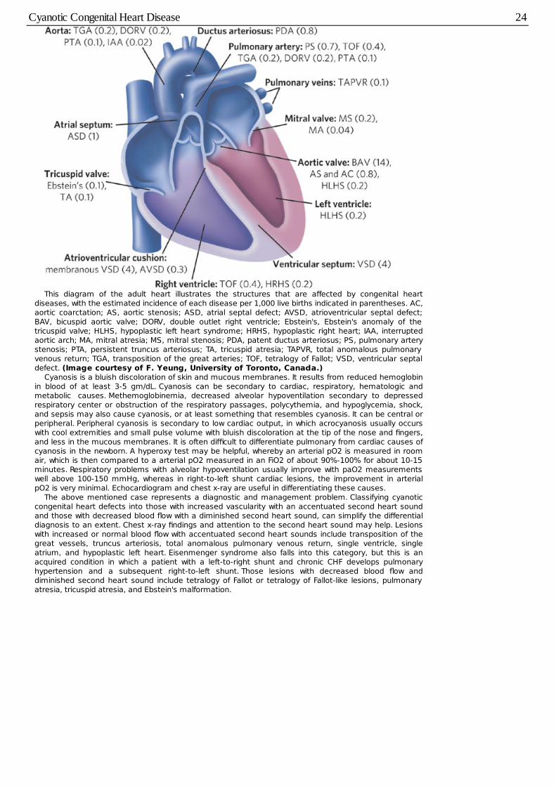
This diagram of the adult heart illustrates the structures that are affected by congenital heartdiseases, with the estimated incidence of each disease per 1,000 live births indicated in parentheses. AC,aortic coarctation; AS, aortic stenosis; ASD, atrial septal defect; AVSD, atrioventricular septal defect;BAV, bicuspid aortic valve; DORV, double outlet right ventricle; Ebstein's, Ebstein's anomaly of thetricuspid valve; HLHS, hypoplastic left heart syndrome; HRHS, hypoplastic right heart; IAA, interruptedaortic arch; MA, mitral atresia; MS, mitral stenosis; PDA, patent ductus arteriosus; PS, pulmonary arterystenosis; PTA, persistent truncus arteriosus; TA, tricuspid atresia; TAPVR, total anomalous pulmonaryvenous return; TGA, transposition of the great arteries; TOF, tetralogy of Fallot; VSD, ventricular septaldefect. (Image courtesy of F. Yeung, University of Toronto, Canada.)
Cyanosis is a bluish discoloration of skin and mucous membranes. It results from reduced hemoglobinin blood of at least 3-5 gm/dL. Cyanosis can be secondary to cardiac, respiratory, hematologic andmetabolic causes. Methemoglobinemia, decreased alveolar hypoventilation secondary to depressedrespiratory center or obstruction of the respiratory passages, polycythemia, and hypoglycemia, shock,and sepsis may also cause cyanosis, or at least something that resembles cyanosis. It can be central orperipheral. Peripheral cyanosis is secondary to low cardiac output, in which acrocyanosis usually occurswith cool extremities and small pulse volume with bluish discoloration at the tip of the nose and fingers,and less in the mucous membranes. It is often difficult to differentiate pulmonary from cardiac causes ofcyanosis in the newborn. A hyperoxy test may be helpful, whereby an arterial pO2 is measured in roomair, which is then compared to a arterial pO2 measured in an FiO2 of about 90%-100% for about 10-15minutes. Respiratory problems with alveolar hypoventilation usually improve with paO2 measurementswell above 100-150 mmHg, whereas in right-to-left shunt cardiac lesions, the improvement in arterialpO2 is very minimal. Echocardiogram and chest x-ray are useful in differentiating these causes.
The above mentioned case represents a diagnostic and management problem. Classifying cyanoticcongenital heart defects into those with increased vascularity with an accentuated second heart soundand those with decreased blood flow with a diminished second heart sound, can simplify the differentialdiagnosis to an extent. Chest x-ray findings and attention to the second heart sound may help. Lesionswith increased or normal blood flow with accentuated second heart sounds include transposition of thegreat vessels, truncus arteriosis, total anomalous pulmonary venous return, single ventricle, singleatrium, and hypoplastic left heart. Eisenmenger syndrome also falls into this category, but this is anacquired condition in which a patient with a left-to-right shunt and chronic CHF develops pulmonaryhypertension and a subsequent right-to-left shunt. Those lesions with decreased blood flow anddiminished second heart sound include tetralogy of Fallot or tetralogy of Fallot-like lesions, pulmonaryatresia, tricuspid atresia, and Ebstein's malformation.
Cyanotic Congenital Heart Disease 24
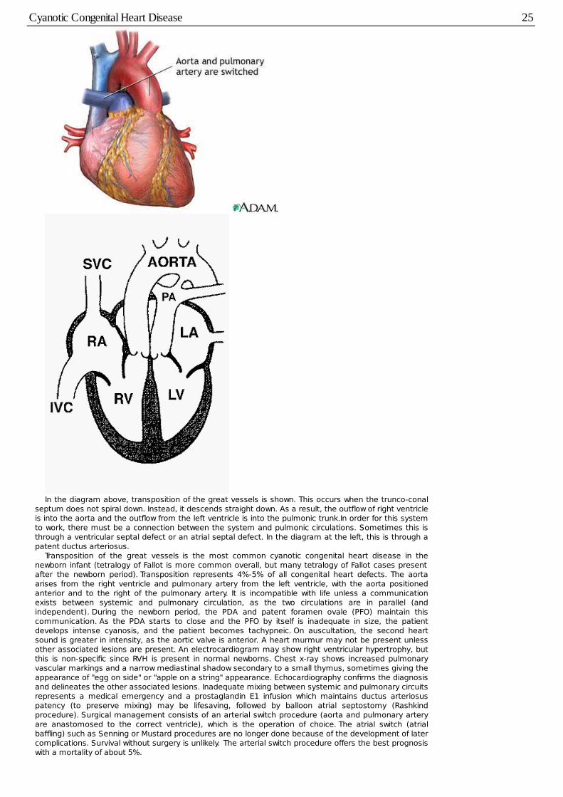
In the diagram above, transposition of the great vessels is shown. This occurs when the trunco-conalseptum does not spiral down. Instead, it descends straight down. As a result, the outflow of right ventricleis into the aorta and the outflow from the left ventricle is into the pulmonic trunk.In order for this systemto work, there must be a connection between the system and pulmonic circulations. Sometimes this isthrough a ventricular septal defect or an atrial septal defect. In the diagram at the left, this is through apatent ductus arteriosus.
Transposition of the great vessels is the most common cyanotic congenital heart disease in thenewborn infant (tetralogy of Fallot is more common overall, but many tetralogy of Fallot cases presentafter the newborn period). Transposition represents 4%-5% of all congenital heart defects. The aortaarises from the right ventricle and pulmonary artery from the left ventricle, with the aorta positionedanterior and to the right of the pulmonary artery. It is incompatible with life unless a communicationexists between systemic and pulmonary circulation, as the two circulations are in parallel (andindependent). During the newborn period, the PDA and patent foramen ovale (PFO) maintain thiscommunication. As the PDA starts to close and the PFO by itself is inadequate in size, the patientdevelops intense cyanosis, and the patient becomes tachypneic. On auscultation, the second heartsound is greater in intensity, as the aortic valve is anterior. A heart murmur may not be present unlessother associated lesions are present. An electrocardiogram may show right ventricular hypertrophy, butthis is non-specific since RVH is present in normal newborns. Chest x-ray shows increased pulmonaryvascular markings and a narrow mediastinal shadow secondary to a small thymus, sometimes giving theappearance of "egg on side" or "apple on a string" appearance. Echocardiography confirms the diagnosisand delineates the other associated lesions. Inadequate mixing between systemic and pulmonary circuitsrepresents a medical emergency and a prostaglandin E1 infusion which maintains ductus arteriosuspatency (to preserve mixing) may be lifesaving, followed by balloon atrial septostomy (Rashkindprocedure). Surgical management consists of an arterial switch procedure (aorta and pulmonary arteryare anastomosed to the correct ventricle), which is the operation of choice. The atrial switch (atrialbaffling) such as Senning or Mustard procedures are no longer done because of the development of latercomplications. Survival without surgery is unlikely. The arterial switch procedure offers the best prognosiswith a mortality of about 5%.
Cyanotic Congenital Heart Disease 25
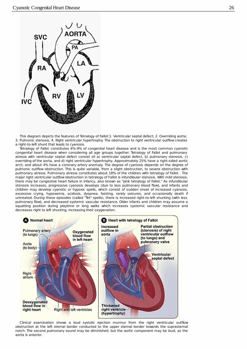
This diagram depicts the features of Tetralogy of Fallot:1. Ventricular septal defect; 2. Overriding aorta;3. Pulmonic stenosis; 4. Right ventricular hypertrophy. The obstruction to right ventricular outflow createsa right-to-left shunt that leads to cyanosis.
Tetralogy of Fallot constitutes 4%-9% of congenital heart disease and is the most common cyanoticcongenital heart disease when considering all age groups together. Tetralogy of Fallot and pulmonaryatresia with ventricular septal defect consist of: a) ventricular septal defect, b) pulmonary stenosis, c)overriding of the aorta, and d) right ventricular hypertrophy. Approximately 25% have a right-sided aorticarch, and about 4% have a coronary artery anomaly. The degree of cyanosis depends on the degree ofpulmonic outflow obstruction. This is quite variable, from a slight obstruction, to severe obstruction withpulmonary atresia. Pulmonary atresia constitutes about 18% of the children with tetralogy of Fallot . Themajor right ventricular outflow obstruction in tetralogy of Fallot is infundibular stenosis. With mild stenosis,there may be congestive heart failure in infancy, also known as "pink tetralogy of Fallot." As infundibularstenosis increases, progressive cyanosis develops (due to less pulmonary blood flow), and infants andchildren may develop cyanotic or hypoxic spells, which consist of sudden onset of increased cyanosis,excessive crying, hypoxemia, acidosis, dyspnea, fainting, rarely seizures, and occasionally death ifuntreated. During these episodes (called "Tet" spells), there is increased right-to-left shunting (with lesspulmonary flow), and decreased systemic vascular resistance. Older infants and children may assume asquatting position during playtime or long walks which increases systemic vascular resistance anddecreases right to left shunting, increasing their oxygenation.
Clinical examination shows a loud systolic ejection murmur from the right ventricular outflowobstruction at the left sternal border conducted to the upper sternal border towards the suprasternalnotch. The second pulmonary sound may be diminished, but the aortic component may be loud, as theaorta is anterior.
Cyanotic Congenital Heart Disease 26
The electrocardiogram shows the non-specific right ventricular hypertrophy. Chest x-ray showsdecreased pulmonary vascular markings (reduced pulmonary perfusion) and right ventricularhypertrophy with a leftward apex. There is an absence or decreased main pulmonary artery segment,which may give the appearance of a "boot shaped heart." Echocardiography demonstrates a ventricularseptal defect with an overriding of the aorta, pulmonic stenosis, right ventricular hypertrophy, and inabout 25% of cases, a right aortic arch (i.e., the aorta goes over the right mainstem bronchus instead ofthe left) is also present. Cardiac catheterization is done in cases in which the anatomy of the defect isnot clear on echocardiogram.
Management during the newborn period consists of administration of prostaglandin E1 when the infantis markedly cyanotic and pulmonary blood flow is ductus dependent. This is followed by a systemic arteryto pulmonary artery shunt (Blalock-Taussig shunt). Treatment of hypercyanotic spells is directed towardsimproving pulmonary blood flow. These include oxygen, knee/chest position, morphine, intravenous fluids,sodium bicarbonate, propranolol (beta-blocker), or increasing systemic vascular resistance byadministration of drugs, such as phenylephrine. Total surgical correction of the defect is performed undercardiopulmonary bypass, and it can now be performed in young infants from 3-6 months of age orearlier. Prognosis is good with total correction. However, the majority of them still have residual defectsand some of them may need reoperation and life long medical follow up.
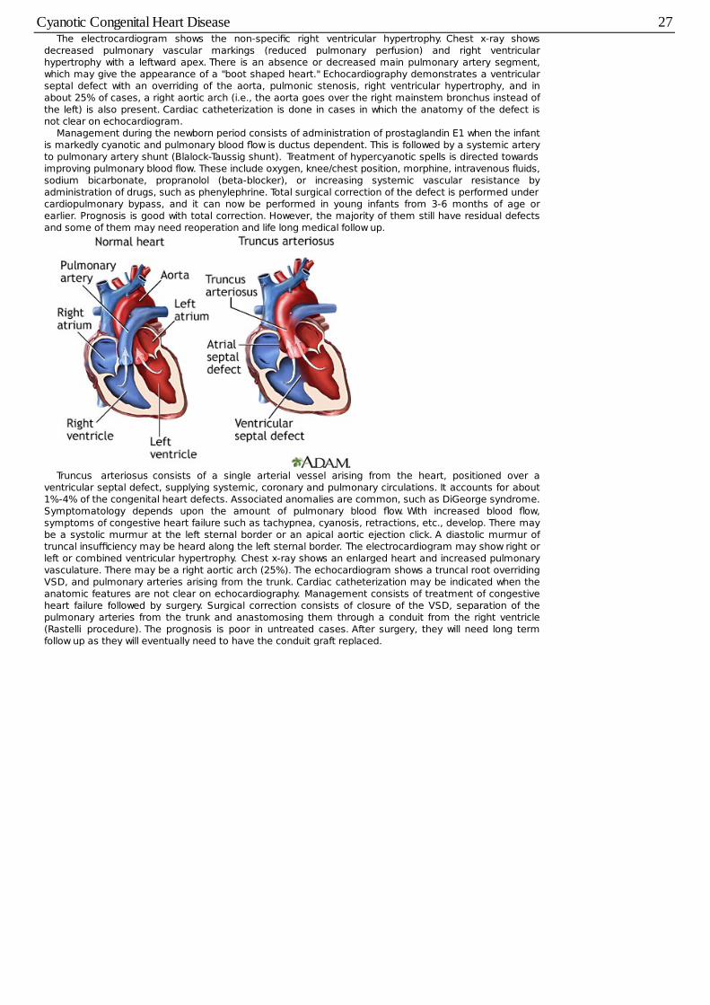
Truncus arteriosus consists of a single arterial vessel arising from the heart, positioned over aventricular septal defect, supplying systemic, coronary and pulmonary circulations. It accounts for about1%-4% of the congenital heart defects. Associated anomalies are common, such as DiGeorge syndrome.Symptomatology depends upon the amount of pulmonary blood flow. With increased blood flow,symptoms of congestive heart failure such as tachypnea, cyanosis, retractions, etc., develop. There maybe a systolic murmur at the left sternal border or an apical aortic ejection click. A diastolic murmur oftruncal insufficiency may be heard along the left sternal border. The electrocardiogram may show right orleft or combined ventricular hypertrophy. Chest x-ray shows an enlarged heart and increased pulmonaryvasculature. There may be a right aortic arch (25%). The echocardiogram shows a truncal root overridingVSD, and pulmonary arteries arising from the trunk. Cardiac catheterization may be indicated when theanatomic features are not clear on echocardiography. Management consists of treatment of congestiveheart failure followed by surgery. Surgical correction consists of closure of the VSD, separation of thepulmonary arteries from the trunk and anastomosing them through a conduit from the right ventricle(Rastelli procedure). The prognosis is poor in untreated cases. After surgery, they will need long termfollow up as they will eventually need to have the conduit graft replaced.
Cyanotic Congenital Heart Disease 27
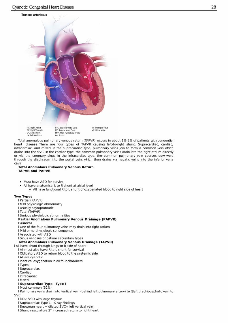
Total anomalous pulmonary venous return (TAPVR) occurs in about 1%-2% of patients with congentialheart disease. There are four types of TAPVR causing left-to-right shunt: Supracardiac, cardiac,infracardiac, and mixed. In the supracardiac type, pulmonary veins join to form a common vein whichdrains into the SVC. In the cardiac type, the common pulmonary veins drain into the right atrium directlyor via the coronary sinus. In the infracardiac type, the common pulmonary vein courses downwardthrough the diaphragm into the portal vein, which then drains via hepatic veins into the inferior venacava.
Total Anomalous Pulmonary Venous ReturnTAPVR and PAPVR
Must have ASD for survivalAll have anatomical L to R shunt at atrial level
All have functional R to L shunt of oxygenated blood to right side of heart
Two Typesl Partial (PAPVR)l Mild physiologic abnormalityl Usually asymptomaticl Total (TAPVR)l Serious physiologic abnormalitiesPartial Anomalous Pulmonary Venous Drainage (PAPVR)Generall One of the four pulmonary veins may drain into right atriuml Mild or no physiologic consequencel Associated with ASDl Sinus venosus or ostium secundum typesTotal Anomalous Pulmonary Venous Drainage (TAPVR)
l All have shunt through lungs to R side of heartl All must also have R to L shunt for survivall Obligatory ASD to return blood to the systemic sidel All are cyanoticl Identical oxygenation in all four chambersl Typesl Supracardiacl Cardiacl Infracardiacl Mixedl Supracardiac Type—Type Il Most common (52%)l Pulmonary veins drain into vertical vein (behind left pulmonary artery) to �left brachiocephalic vein to
SVCl DDx: VSD with large thymusl Supracardiac Type 1—X-ray Findingsl Snowman heart = dilated SVC+ left vertical veinl Shunt vasculature 2° increased return to right heart
Cyanotic Congenital Heart Disease 28
l Enlargement of right heart 2° volume overloadl Cardiac Type—Type IIl Second most common: 30%l Drains into coronary sinus or RAl Coronary sinus more commonl Increased pulmonary vasculaturel Overload of RV leads to CHF after birthl 20% of I’s and II’s survive to adulthoodl Remainder expire in first yearl Infracardiac Type—Type IIIl Percent of total: 12%l Long pulmonary veins course down along esophagusl Empty into IVC or portal vein (more common)l Vein constricted by diaphragm as it passes through esophageal hiatusl Severe CHF (90%) 2° obstruction to venous returnl Cyanotic 2° right to left shunt through ASDl Associated with asplenia (80%), or polysplenial Prognosis = death within a few daysl Mixed Type—Type IVl Percent of total: 6%l Mixtures of types I – IIIAnomalous pulmonary venous return could be total or partial. An atrial septal defect is necessary for
survival, since the oxygenated blood (from the pulmonary veins) must somehow reach the left side of theheart. Symptomatology depends on the amount of mixing and whether or not the pulmonary veins areobstructed. Cyanosis and signs and symptoms of congestive heart failure develop and progress rapidly.There may be a grade 2/6 systolic ejection flow murmur heard along the left sternal border, or it may beabsent. The electrocardiogram shows right ventricular hypertrophy and right atrial hypertrophy. Chest x-ray shows increased pulmonary vascular markings or even edema, and the heart may be normal in sizeor minimally enlarged.
The echocardiogram may show right ventricular volume overload, and a color-flow Doppler study mayhelp in locating the common pulmonary venous channel and its drainage. If the resolution is poor,cardiac catheterization and angiocardiography may help in delineating the anomaly further. Treatmentconsists of correction of the defect by surgery. If surgery is delayed and there is inadequate mixing,palliative balloon septostomy may be performed. Prognosis is good after surgery. Prognosis is poor inneonates with obstructive TAPVR. Long term follow up is needed to assess restenosis and latearrhythmias.
Cyanotic Congenital Heart Disease 29
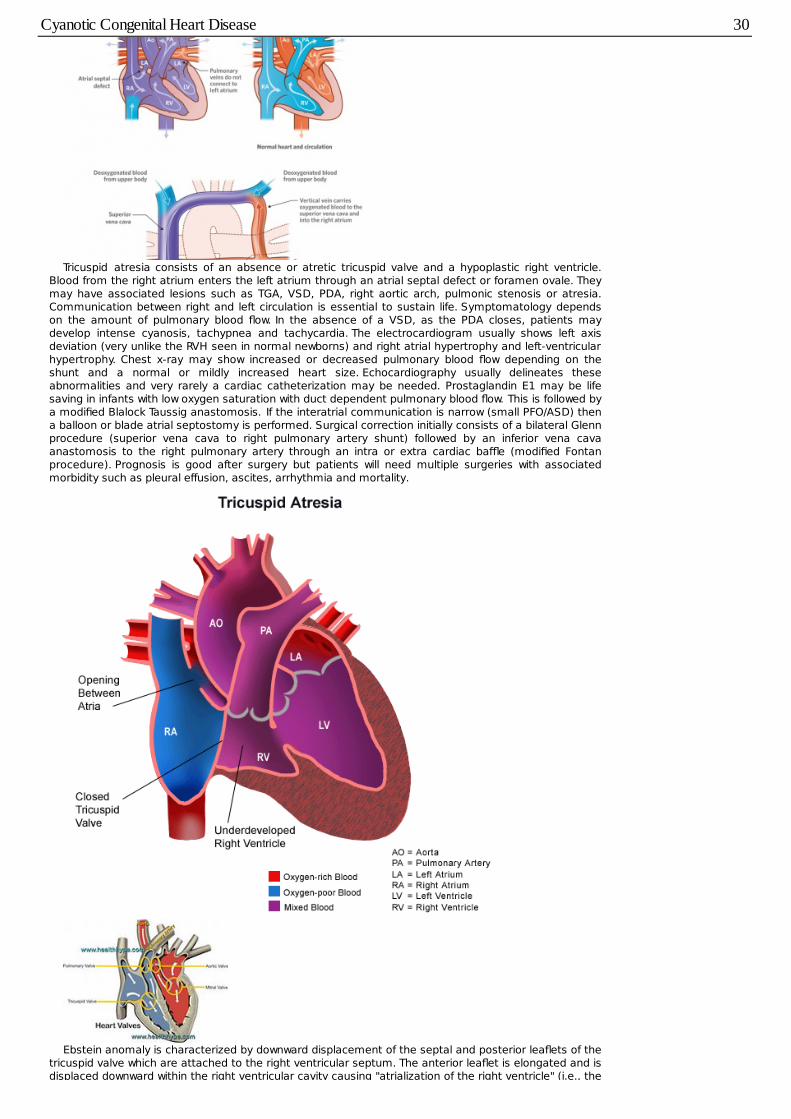
Tricuspid atresia consists of an absence or atretic tricuspid valve and a hypoplastic right ventricle.Blood from the right atrium enters the left atrium through an atrial septal defect or foramen ovale. Theymay have associated lesions such as TGA, VSD, PDA, right aortic arch, pulmonic stenosis or atresia.Communication between right and left circulation is essential to sustain life. Symptomatology dependson the amount of pulmonary blood flow. In the absence of a VSD, as the PDA closes, patients maydevelop intense cyanosis, tachypnea and tachycardia. The electrocardiogram usually shows left axisdeviation (very unlike the RVH seen in normal newborns) and right atrial hypertrophy and left-ventricularhypertrophy. Chest x-ray may show increased or decreased pulmonary blood flow depending on theshunt and a normal or mildly increased heart size. Echocardiography usually delineates theseabnormalities and very rarely a cardiac catheterization may be needed. Prostaglandin E1 may be lifesaving in infants with low oxygen saturation with duct dependent pulmonary blood flow. This is followed bya modified Blalock Taussig anastomosis. If the interatrial communication is narrow (small PFO/ASD) thena balloon or blade atrial septostomy is performed. Surgical correction initially consists of a bilateral Glennprocedure (superior vena cava to right pulmonary artery shunt) followed by an inferior vena cavaanastomosis to the right pulmonary artery through an intra or extra cardiac baffle (modified Fontanprocedure). Prognosis is good after surgery but patients will need multiple surgeries with associatedmorbidity such as pleural effusion, ascites, arrhythmia and mortality.
Ebstein anomaly is characterized by downward displacement of the septal and posterior leaflets of thetricuspid valve which are attached to the right ventricular septum. The anterior leaflet is elongated and isdisplaced downward within the right ventricular cavity causing "atrialization of the right ventricle" (i.e., the
Cyanotic Congenital Heart Disease 30
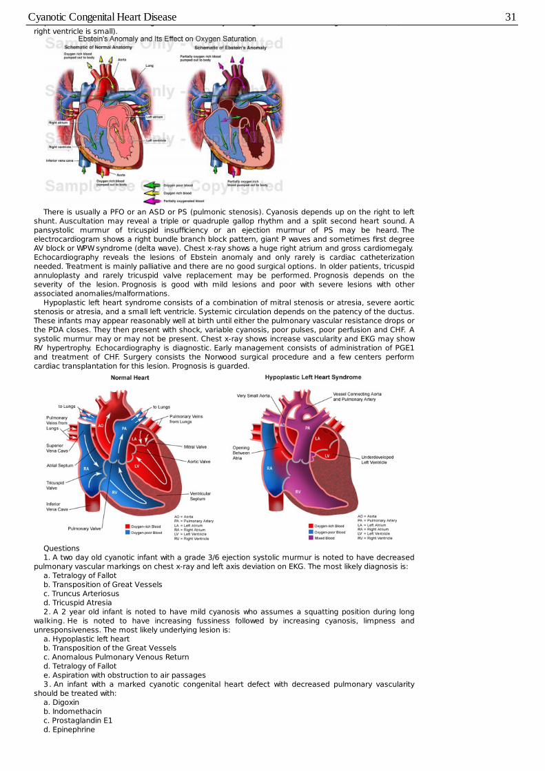
displaced downward within the right ventricular cavity causing "atrialization of the right ventricle" (i.e., theright ventricle is small).
There is usually a PFO or an ASD or PS (pulmonic stenosis). Cyanosis depends up on the right to leftshunt. Auscultation may reveal a triple or quadruple gallop rhythm and a split second heart sound. Apansystolic murmur of tricuspid insufficiency or an ejection murmur of PS may be heard. Theelectrocardiogram shows a right bundle branch block pattern, giant P waves and sometimes first degreeAV block or WPW syndrome (delta wave). Chest x-ray shows a huge right atrium and gross cardiomegaly.Echocardiography reveals the lesions of Ebstein anomaly and only rarely is cardiac catheterizationneeded. Treatment is mainly palliative and there are no good surgical options. In older patients, tricuspidannuloplasty and rarely tricuspid valve replacement may be performed. Prognosis depends on theseverity of the lesion. Prognosis is good with mild lesions and poor with severe lesions with otherassociated anomalies/malformations.
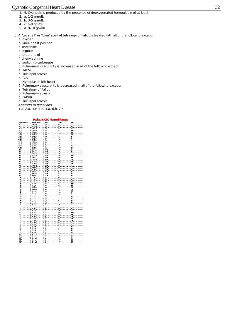
Hypoplastic left heart syndrome consists of a combination of mitral stenosis or atresia, severe aorticstenosis or atresia, and a small left ventricle. Systemic circulation depends on the patency of the ductus.These infants may appear reasonably well at birth until either the pulmonary vascular resistance drops orthe PDA closes. They then present with shock, variable cyanosis, poor pulses, poor perfusion and CHF. Asystolic murmur may or may not be present. Chest x-ray shows increase vascularity and EKG may showRV hypertrophy. Echocardiography is diagnostic. Early management consists of administration of PGE1and treatment of CHF. Surgery consists the Norwood surgical procedure and a few centers performcardiac transplantation for this lesion. Prognosis is guarded.
Questions1. A two day old cyanotic infant with a grade 3/6 ejection systolic murmur is noted to have decreased
pulmonary vascular markings on chest x-ray and left axis deviation on EKG. The most likely diagnosis is:a. Tetralogy of Fallotb. Transposition of Great Vesselsc. Truncus Arteriosusd. Tricuspid Atresia2. A 2 year old infant is noted to have mild cyanosis who assumes a squatting position during long
walking. He is noted to have increasing fussiness followed by increasing cyanosis, limpness andunresponsiveness. The most likely underlying lesion is:
a. Hypoplastic left heartb. Transposition of the Great Vesselsc. Anomalous Pulmonary Venous Returnd. Tetralogy of Fallote. Aspiration with obstruction to air passages3 . An infant with a marked cyanotic congenital heart defect with decreased pulmonary vascularity
should be treated with:a. Digoxinb. Indomethacinc. Prostaglandin E1d. Epinephrine
Cyanotic Congenital Heart Disease 31
1. 4. Cyanosis is produced by the presence of deoxygenated hemoglobin of at least:2. a. 1-2 gm/dL3. b. 3-5 gm/dL4. c. 6-8 gm/dL5. d. 9-10 gm/dL
5. A "tet spell" or "blue" spell of tetralogy of Fallot is treated with all of the following except:a. oxygenb. knee chest positionc. morphined. digoxine. propranololf. phenylephrineg. sodium bicarbonate6. Pulmonary vascularity is increased in all of the following except:a. TAPVRb. Tricuspid atresiac. TGVd. Hypoplastic left heart7. Pulmonary vascularity is decreased in all of the following except:a. Tetralogy of Fallotb. Pulmonary atresiac. TAPVRd. Tricuspid atresiaAnswers to questions1.d, 2.d, 3.c, 4.b, 5.d, 6.b, 7.c
Cyanotic Congenital Heart Disease 32