diagnosis of coronavirus disease 2019 (covid-19) with
TRANSCRIPT

1
Diagnosis of Coronavirus Disease 2019(COVID-19) with Structured Latent Multi-View
Representation LearningHengyuan Kang†, Liming Xia†, Fuhua Yan†, Zhibin Wan, Feng Shi, Huan Yuan, Huiting Jiang, Dijia Wu,
He Sui, Changqing Zhang* and Dinggang Shen*
Abstract—Recently, the outbreak of Coronavirus Disease 2019(COVID-19) has spread rapidly across the world. Due to thelarge number of infected patients and heavy labor for doctors,computer-aided diagnosis with machine learning algorithm isurgently needed, and could largely reduce the efforts of clin-icians and accelerate the diagnosis process. Chest computedtomography (CT) has been recognized as an informative toolfor diagnosis of the disease. In this study, we propose to conductthe diagnosis of COVID-19 with a series of features extractedfrom CT images. To fully explore multiple features describingCT images from different views, a unified latent representation islearned which can completely encode information from differentaspects of features and is endowed with promising class structurefor separability. Specifically, the completeness is guaranteed witha group of backward neural networks (each for one type offeatures), while by using class labels the representation is enforcedto be compact within COVID-19/community-acquired pneumonia(CAP) and also a large margin is guaranteed between differenttypes of pneumonia. In this way, our model can well avoidoverfitting compared to the case of directly projecting high-dimensional features into classes. Extensive experimental resultsshow that the proposed method outperforms all comparisonmethods, and rather stable performances are observed whenvarying the number of training data.
Index Terms—COVID-19, Pneumonia, Chest computed tomog-raphy (CT), Multi-view representation learning
I. INTRODUCTION
ANovel coronavirus disease 2019 (COVID-19) wasrecognized in December 2019, in Wuhan, China and has
Manuscript received April 12, 2020; accepted April 26, 2020. This workwas supported in part by National Natural Science Foundation of China(61976151 and 61732011), and the National Key Research and DevelopmentProgram of China under Grant 2018YFC0116400. (H. Kang, L. Xia and F.Yan contributed equally to this work.) (Corresponding authors: ChangqingZhang and Dinggang Shen.)
H. Kang, Z. Wan and C. Zhang are with the College of Intelli-gence and Computing, Tianjin University, Tianjin 300350, China (e-mail:{kanghengyuan, wanzhibin, zhangchangqing}@tju.edu.cn).
L. Xia is with Department of Radiology, Tongji Hospital, Tongji MedicalCollege, Huazhong University of Science and Technology, Wuhan, Hubei,China (e-mail: [email protected]).
F. Yan is with Department of Radiology, Ruijin Hospital, Shang-hai Jiao Tong University School of Medicine, Shanghai, China (e-mail:[email protected]).
F. Shi, H. Yuan, H. Jiang, D. Wu and D. Shen are with the Depart-ment of Research and Development, Shanghai United Imaging IntelligenceCo., Ltd., Shanghai, China (e-mail: {feng.shi, huan.yuan, huiting.jiang,dijia.wu}@united-imaging.com, [email protected]).
H. Sui is with Department of Radiology, China-Japan Union Hospital ofJilin University, Changchun, China (e-mail: [email protected]).
COVID-19 CAP
Fig. 1. Examples of chest CT images with infection of COVID-19 (left)and community-acquired pneumonia (right). The pneumonia becomes moreserious from top to bottom, and the yellow arrows indicate the representativeinfection areas. It can be observed that it is quite similar for the CT imagesof high-severity CAP and mild-severity COVID-19.
rapidly spread over the world [1]–[5]. Recently, COVID-19has been threatening all the world and the world healthorganization (WHO) has declared that COVID-19 becomesa global pandemic. The current clinical experience impliesthat the RT-PCR detection of viral RNA has a low sensitivityespecially in the early stage [6]–[9]. As a form of pneumonia,inflammation of air sacs in lungs has been found, and it hasshown that bilateral lung involvement could be observed forearly, intermediate, and late stage patients. Accordingly, ahigh proportion of abnormal chest CT images were obtainedfrom patients with this disease [10]–[13]. Then, it is necessaryto complement nucleic acid testing with automatic techniquebased on lung CT as one of the early diagnostic criteria forthis new type of pneumonia as soon as possible. In this study,
©2020 IEEE. Personal use of this material is permitted. Permission from IEEE must be obtained for all other uses, in any current orfuture media, including reprinting/republishing this material for advertising or promotional purposes, creating new collective works,
for resale or redistribution to servers or lists, or reuse of any copyrighted component of this work in other works.
arX
iv:2
005.
0322
7v1
[ee
ss.I
V]
6 M
ay 2
020

2
we focus on conducting diagnosis for COVID-19 andcommunity-acquired pneumonia [14]–[16], i.e., characterizingthe relationships between multiple types of features from CTimages and these diseases, which provides a possible pipelinefor automatic diagnosis and investigation. Specifically,multiple types of features are extracted and the correlations todiagnosis are extensively evaluated by conducting experimentswith multiple baseline methods. The experiment results showthat both radiomic features and handcrafted features arehelpful for classifying these two different categories of lungdiseases. Therefore, we propose a novel pipeline which caneffectively integrate information from different views. We alsonote that, although deep-learning methods [17], especiallyCNN-based models, have shown the power in imageclassification, they usually need large-scale training data andhave difficulty in exploiting expert prior. Although there arethousands of CT images available which is a quite largedataset for medical image analysis, it is still not comparablewith the large-scale image dataset in the computer visionfield, i.e., ImageNet [18]. Therefore, extracting manuallydesigned features to incorporate expert prior is a reasonableand probably preferred approach which could alleviate theoverfitting problem in machine learning.
For computer-assisted medical diagnosis, a number of meth-ods automatically learn classification models based on featuresextracted with expert prior [19], [20]. However, some of themare only applicable to single type of features thus cannot wellexplore the complementary information from multiple typesof features. Fortunately, multi-view representation learningprovides tools for exploiting multiple types of heterogeneousfeatures [21], [22]. Although significant progress achieved,existing multi-view representation learning methods cannotguarantee the information completeness and promising struc-ture for separability. This makes them tend to overfit trainingdata thus harm the performance in the testing stage.
Based on the above analysis, we propose a new classifi-cation framework to diagnose these diseases, where the mainaim is to identify the COVID-19 from CAP. It is worth notingthat these different types of features are not directly used asinput for a classifier to output the final decision [23]–[25] inour method. Instead, both the training and testing samples aremapped into a promising latent space [26], where these latentrepresentations are expected to encode complementary infor-mation from different types of features and have promisingstructures revealing the underlying class distribution.
First, since there are different types of features which arequite different in distribution and quality as shown in Fig. 5, itis very challenging and important to effectively integrate thesedifferent types of features. For this issue, a novel integrationstrategy is proposed with a group of neural networks, whereeach one encodes the information from one type of featuresinto a latent representation. Second, we conduct projectionlearning to build an accurate model to map a subject withthese multiple types of features into a latent representation,thus a latent-representation regressor is obtained which can beapplied on new subjects. Third, a final classifier is trainedbased on the latent representation, instead of the originalfeatures. We should emphasize the advantages of our model
over existing methods that often directly learn projectionsfrom original features into class labels. The first advantageis the latent representation, which is usually compact and thusmay be more effective since it can avoid to overfit the high-dimensional data and has better generalization in the testingstage. Second, the proposed pipeline can encode informationfrom different types of features and produce a structured rep-resentation which imposes simple bias on the model to furtherenhance the generalization performance. Moreover, the learnedrepresentation could be used in different classification models,and the performance with the learned latent representationsclearly outperforms that of original features for all baselineclassifiers used in experiments. The main contributions of thisstudy are summarized as follows:• We propose to conduct diagnosis of COVID-19 with
multi-view representation learning, where the comple-mentarity among different types of features is well ex-plored, achieving clear performance gain in classification.
• We propose a full pipeline for diagnosis COVID-19 fromcommunity-acquired pneumonia, which is quite differentfrom existing models that directly project features intothe class space. The key component is the structuredlatent representation learning which brings robustness,generalization and stability into the pipeline.
• The learned latent representation can be widely used fordifferent classifiers to promote the diagnosis accuracy.Specifically, the latent representations are adopted by sev-eral baseline models, and the results clearly demonstratethe effectiveness of the latent representation comparedwith original features.
• Extensive experiments on the CT images validate thatthe proposed model can obtain a well-structured latentrepresentation, thus significantly promoting the diagnosisin terms of accuracy, sensitivity and specificity comparedwith different methods.
II. MATERIAL
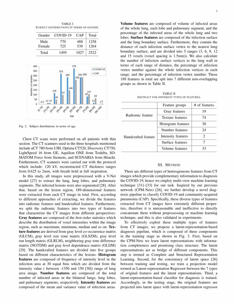
There are 2522 CT images involved in this study, where1495 cases are from COVID-19 patients and the left 1027cases are from community-acquired pneumonia (CAP) pa-tients. These COVID-19 infected subjects were confirmed withpositive nucleic acid testing and confirmed by Chinese Centersfor Disease Control and Prevention (CDC). The distributionsof subjects in terms of gender and age are shown in Table Iand Fig. 2. Specifically, according to Table I, we can findthat the number of males with COVID-19 is slightly largerthan that of females, while, for CAP, the contrary is the case.These subjects cover patients from 12-year old to 98-year old.In addition, basically, the average age of patients infectedwith COVID-19 is younger than that of CAP according toFig. 2. These CT images were provided by Tongji Hospitalof Huazhong University of Science and Technology, China-Japan Union Hospital of Jilin University, Ruijin Hospital ofShanghai Jiao Tong University, and their collaborators. TheCOVID-19 images were acquired from Jan. 9, 2020 to Feb.14, 2020, and CAP images were obtained from Jul. 30, 2018to Feb. 22, 2020.
3
TABLE ISUBJECT DISTRIBUTIONS IN TERMS OF GENDER.
Gender COVID-19 CAP Total
Male 770 488 1258Female 725 539 1264
Total 1495 1027 2522
0 12
118
323
285
378
283
82
13 1014
49
97
146
258
286
107
64
60
50
100
150
200
250
300
350
400
Num
be
r o
f sa
mp
les
Age
COVID-19
CAP
Fig. 2. Subject distributions in terms of age.
Chest CT scans were performed on all patients with thinsection. The CT scanners used in the three hospitals mentionedinclude uCT 780 from UIH, Optima CT520, Discovery CT750,LightSpeed 16 from GE, Aquilion ONE from Toshiba, SO-MATOM Force from Siemens, and SCENARIA from Hitachi.Furthermore, CT scanners were carried out with the protocolwhich include: 120 kV, reconstructed CT thickness rangesfrom 0.625 to 2mm, with breath hold at full inspiration.
In this study, all images were preprocessed with a V-Netmodel [27] to extract the lung, lung lobes, and pulmonarysegments. The infected lesions were also segmented [28]. Afterthat, based on the lesion region, 189-dimensional featureswere extracted from each CT image in total. First, accordingto different approaches of extracting, we divide the featuresinto radiomic features and handcrafted features. Furthermore,we split the radiomic features into two types of featuresthat characterize the CT images from different perspectives:Gray features are composed of the first-order statistics whichdescribe the distribution of voxel intensities within the imageregion, such as maximum, minimum, median and so on. Tex-ture features are derived from gray level co-occurrence matrix(GLCM), gray level size zone matrix (GLSZM), gray levelrun length matrix (GLRLM), neighboring gray tone differencematrix (NGTDM) and gray level dependence matrix (GLDM)[29]. The handcrafted features are divided into five groupsbased on different characteristics of the lesions: Histogramfeatures are composed of frequency of intensity level in theinfection area at 30 equal bins which are divided from theintensity value ( between -1350 and 150 [30]) range of lungarea image. Number features are composed of the totalnumber of infected areas in the bilateral lungs, lung lobes,and pulmonary segments, respectively. Intensity features arecomposed of the mean and variance value of infection areas.
Volume features are composed of volume of infected areasof the whole lung, each lobe and pulmonary segment, and thepercentage of the infected areas of the whole lung and twolobes. Surface features are composed of the infection surfaceand the lung boundary surface. Furthermore, they contain thedistance of each infection surface vertex to the nearest lungboundary surface, and are divided into 5 ranges (3, 6, 9, 12and 15 voxels (voxel spacing is 1.5mm)). We also calculatethe number of infection surface vertices to the lung wall interms of each range of distance, the percentage of infectionvertex number against the whole infection vertices in eachrange, and the percentage of infection vertex number. These189 features in total are spit into 7 different non-overlappinggroups as shown in Table II.
TABLE IIABSTRACT FOR DIFFERENT TYPES OF FEATURES.
Feature groups # of features
Radiomic featureGray features 19
Texture features 74
Handcrafted feature
Histogram features 30
Number features 24
Intensity features 2
Surface features 7
Volume features 33
III. METHOD
There are different types of heterogeneous features from CTimages which provide complementary information to diagnosisthe COVID-19, hence we employ multi-view machine learningtechnique [31]–[33] for our task. Inspired by our previousnetwork (CPM-Nets) [26], we further develop a novel diag-nosis pipeline to classify COVID-19 and community-acquiredpneumonia (CAP). Specifically, these diverse types of featuresextracted from CT images have extremely different proper-ties, therefore it is unreasonable and ineffective to directlyconcatenate them without prepossessing or machine learningtechnique, and this is also validated in experiments.
To effectively exploit these multiple types of featuresfrom CT images, we propose a latent-representation-baseddiagnosis pipeline, which is composed of three componentsin the training stage as shown in Fig. 3. First, based onthe CPM-Nets we learn latent representations with informa-tion completeness and promising class structure. The latentrepresentations act as bridge of different components. Thisstep is termed as Complete and Structured RepresentationLearning. Second, for the consistency of latent space [26]between training and testing, we train a projection modeltermed as Latent-representation Regressor between the 7 typesof original features and the latent representations. Third, alatent-representation-based classifier for diagnosis is trained.Accordingly, in the testing stage, the original features areprojected into latent space with latent-representation regressor
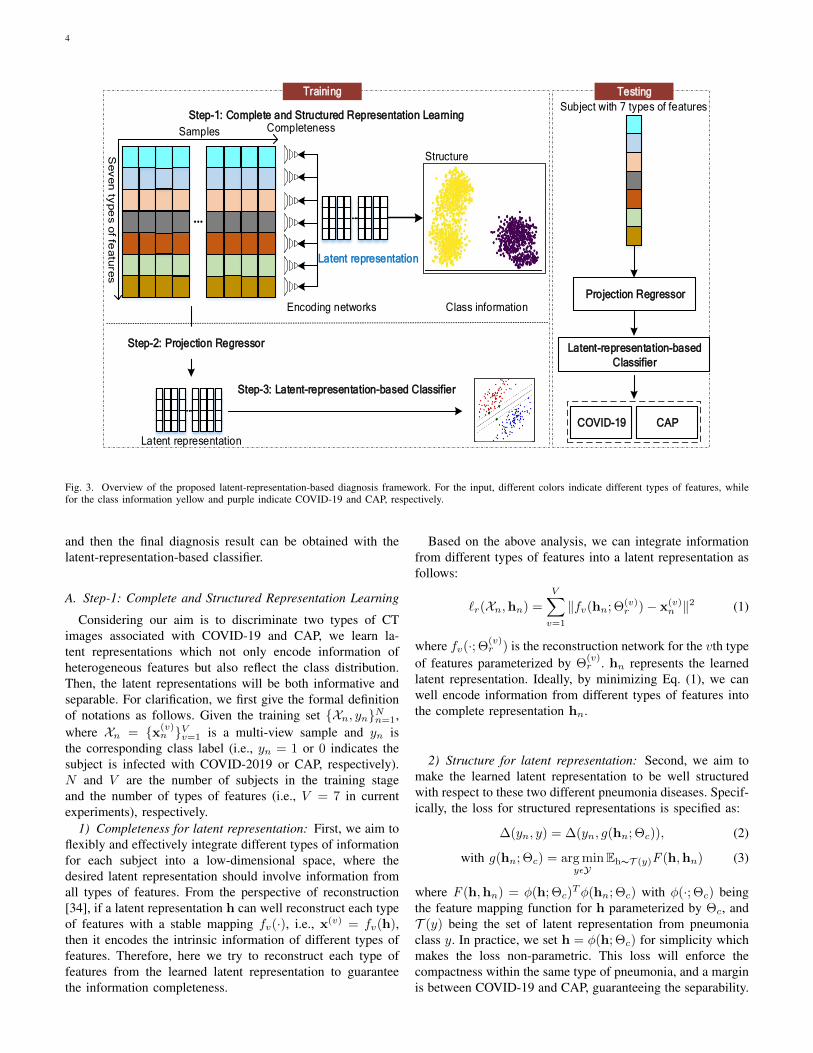
4
Step-1: Complete and Structured Representation Learning
Encoding networks Class information
Se
ve
n ty
pe
s o
f fea
ture
s
Completeness
Structure
Step-3: Latent-representation-based Classifier
Step-2: Projection Regressor
Projection Regressor
Latent-representation-based
Classifier
Training Testing
Latent representation
Latent representation
Subject with 7 types of features
...
...
...
...
...
COVID-19 CAPCOVID-19 CAP
Samples
Fig. 3. Overview of the proposed latent-representation-based diagnosis framework. For the input, different colors indicate different types of features, whilefor the class information yellow and purple indicate COVID-19 and CAP, respectively.
and then the final diagnosis result can be obtained with thelatent-representation-based classifier.
A. Step-1: Complete and Structured Representation Learning
Considering our aim is to discriminate two types of CTimages associated with COVID-19 and CAP, we learn la-tent representations which not only encode information ofheterogeneous features but also reflect the class distribution.Then, the latent representations will be both informative andseparable. For clarification, we first give the formal definitionof notations as follows. Given the training set {Xn, yn}Nn=1,where Xn = {x(v)
n }Vv=1 is a multi-view sample and yn isthe corresponding class label (i.e., yn = 1 or 0 indicates thesubject is infected with COVID-2019 or CAP, respectively).N and V are the number of subjects in the training stageand the number of types of features (i.e., V = 7 in currentexperiments), respectively.
1) Completeness for latent representation: First, we aim toflexibly and effectively integrate different types of informationfor each subject into a low-dimensional space, where thedesired latent representation should involve information fromall types of features. From the perspective of reconstruction[34], if a latent representation h can well reconstruct each typeof features with a stable mapping fv(·), i.e., x(v) = fv(h),then it encodes the intrinsic information of different types offeatures. Therefore, here we try to reconstruct each type offeatures from the learned latent representation to guaranteethe information completeness.
Based on the above analysis, we can integrate informationfrom different types of features into a latent representation asfollows:
`r(Xn,hn) =
V∑v=1
‖fv(hn; Θ(v)r )− x(v)
n ‖2 (1)
where fv(·; Θ(v)r ) is the reconstruction network for the vth type
of features parameterized by Θ(v)r . hn represents the learned
latent representation. Ideally, by minimizing Eq. (1), we canwell encode information from different types of features intothe complete representation hn.
2) Structure for latent representation: Second, we aim tomake the learned latent representation to be well structuredwith respect to these two different pneumonia diseases. Specif-ically, the loss for structured representations is specified as:
∆(yn, y) = ∆(yn, g(hn; Θc)), (2)
with g(hn; Θc) = arg minyεY
Eh∼T (y)F (h,hn) (3)
where F (h,hn) = φ(h; Θc)Tφ(hn; Θc) with φ(·; Θc) being
the feature mapping function for h parameterized by Θc, andT (y) being the set of latent representation from pneumoniaclass y. In practice, we set h = φ(h; Θc) for simplicity whichmakes the loss non-parametric. This loss will enforce thecompactness within the same type of pneumonia, and a marginis between COVID-19 and CAP, guaranteeing the separability.
5
(a) (b)
(c) (d)
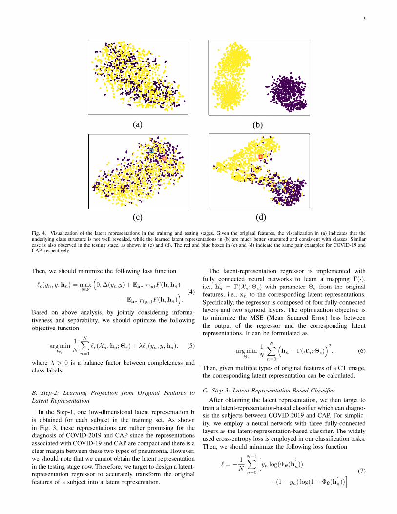
Fig. 4. Visualization of the latent representations in the training and testing stages. Given the original features, the visualization in (a) indicates that theunderlying class structure is not well revealed, while the learned latent representations in (b) are much better structured and consistent with classes. Similarcase is also observed in the testing stage, as shown in (c) and (d). The red and blue boxes in (c) and (d) indicate the same pair examples for COVID-19 andCAP, respectively.
Then, we should minimize the following loss function
`c(yn, y,hn) = maxyεY
(0,∆(yn,y) + Eh∼T (y)F (h,hn)
− Eh∼T (yn)F (h,hn)).
(4)
Based on above analysis, by jointly considering informa-tiveness and separability, we should optimize the followingobjective function
arg minΘr
1
N
N∑n=1
`r(Xn,hn; Θr) + λ`c(yn, y,hn). (5)
where λ > 0 is a balance factor between completeness andclass labels.
B. Step-2: Learning Projection from Original Features toLatent Representation
In the Step-1, one low-dimensional latent representation his obtained for each subject in the training set. As shownin Fig. 3, these representations are rather promising for thediagnosis of COVID-2019 and CAP since the representationsassociated with COVID-19 and CAP are compact and there is aclear margin between these two types of pneumonia. However,we should note that we cannot obtain the latent representationin the testing stage now. Therefore, we target to design a latent-representation regressor to accurately transform the originalfeatures of a subject into a latent representation.
The latent-representation regressor is implemented withfully connected neural networks to learn a mapping Γ(·),i.e., h
′
n = Γ(Xn; Θe) with parameter Θe from the originalfeatures, i.e., xn to the corresponding latent representations.Specifically, the regressor is composed of four fully-connectedlayers and two sigmoid layers. The optimization objective isto minimize the MSE (Mean Squared Error) loss betweenthe output of the regressor and the corresponding latentrepresentations. It can be formulated as
arg minΘe
1
N
N∑n=0
(hn − Γ(Xn; Θe)
)2
. (6)
Then, given multiple types of original features of a CT image,the corresponding latent representation can be calculated.
C. Step-3: Latent-Representation-Based ClassifierAfter obtaining the latent representation, we then target to
train a latent-representation-based classifier which can diagno-sis the subjects between COVID-2019 and CAP. For simplic-ity, we employ a neural network with three fully-connectedlayers as the latent-representation-based classifier. The widelyused cross-entropy loss is employed in our classification tasks.Then, we should minimize the following loss function
` = − 1
N
N−1∑n=0
[yn log(Φθ(h
′
n))
+ (1− yn) log(1− Φθ(h′
n))] (7)

6
where Φθ(h′
n) indicates the prediction of pneumonia type fromthe classifier with h
′as the input.
D. Testing Stage
After training, a full pipeline for diagnosis of COVID-19 and CAP is available. The latent-representation regressorand latent-representation-based classifier play an essential rolein the testing phase, as shown on the right side of Fig. 3.Specifically, subjects with different types of features are firsttransformed into latent representation and then the diagnosisresult can be obtained with the latent-representation-basedclassifier.
IV. EXPERIMENT AND RESULT
A. Experimental Setting
We conduct extensive experiments on the CT images datato evaluate the proposed pipeline. The dataset is randomlydivided into 70% and 30% for training and testing, respec-tively. Furthermore, we adopt 5-fold cross-validation strategyon the training data to tune the parameter λ from the set{0.1, 1, 10, 100}. In practice, promising performance can beexpected given a relatively large value for λ, which is fixedas 100 in our experiments.
TABLE IIIEVALUATION OF PREPROCESSING OF FEATURES.
Method Original Normalized Standardized
LRACC 63.50±3.88 89.01±1.21 89.19±1.21SEN 1.00±0.00 90.64±1.04 90.66±2.08SPE 0.00±0.00 86.15±2.05 86.82±2.38
SVMACC 61.26±5.21 86.40±2.73 89.40±1.21SEN 1.00±0.00 90.51±2.31 89.90±1.92SPE 0.00±0.00 74.52±4.12 88.59±2.23
GNBACC 71.69±3.79 73.01±2.97 75.60±2.13SEN 96.42±1.98 89.32±2.14 85.22±2.75SPE 23.31±6.92 71.22±3.46 71.98±5.26
KNNACC 64.62±3.24 87.10±2.03 86.40±2.44SEN 1.00±0.00 89.44±2.78 89.22±3.35SPE 0.00±0.00 78.33±3.36 76.00±4.75
NNACC 70.43±3.23 92.31±1.87 93.90±2.04SEN 98.21±0.56 91.76±1.52 94.60±2.16SPE 21.79±4.82 82.01±3.11 91.70±2.53
Data preprocessing. The original features extracted fromCT images are of rather different scales. Accordingly, datapreprocessing is necessary before using them as input forlearning algorithm. There are several data preprocessing strate-gies, e.g., normalization and standardization. Specifically, thestandardization for K features is computed as:
xi =xi − µi
σi, i = 1, 2, ...,K (8)
where µi and σi are mean value and standard deviation ofthe feature xi, respectively. xi denotes the standardization
feature of original feature xi. The features of normalizationare calculated as:
xi =xi − ximin
ximax − ximin, i = 1, 2, ...,K (9)
where ximin and ximax are minimum and maximum valuesof the feature xi respectively. Accordingly, xi indicates thenormalization feature of original feature xi.
We conduct experiments on several baseline models byconcatenating all the original features, normalized features andstandardized features, respectively. The effects of the data pre-processing for diagnosis are shown in Table III. Specifically,the performance of using the original features is relatively low,the main reason of which may be the large scale differenceamong different features. Fortunately, in terms of accuracy,both of these preprocessing methods obtain a significantimprovement (1.32% ∼ 25.69%) on all classification models.For clarification and comparison fairness, we employ the stan-dardized data for all methods in the following experiments. Wecompare the proposed method with the following methods inthe diagnosis task, including SVM, Logistic-Regression (LR),Gaussian-Naive-Bayes (GNB), K-Nearest-Neighbors (KNN),and Fully-Connected-Neural-Networks (NN). For all thesemethods, we repeat 10 times and report the mean and standarddeviation performance. Diagnostic performance is evaluatedin terms of accuracy (ACC), sensitivity (SEN) and specificity(SPC).
B. Performance Evaluation1) Discrimination power of these different types of features:
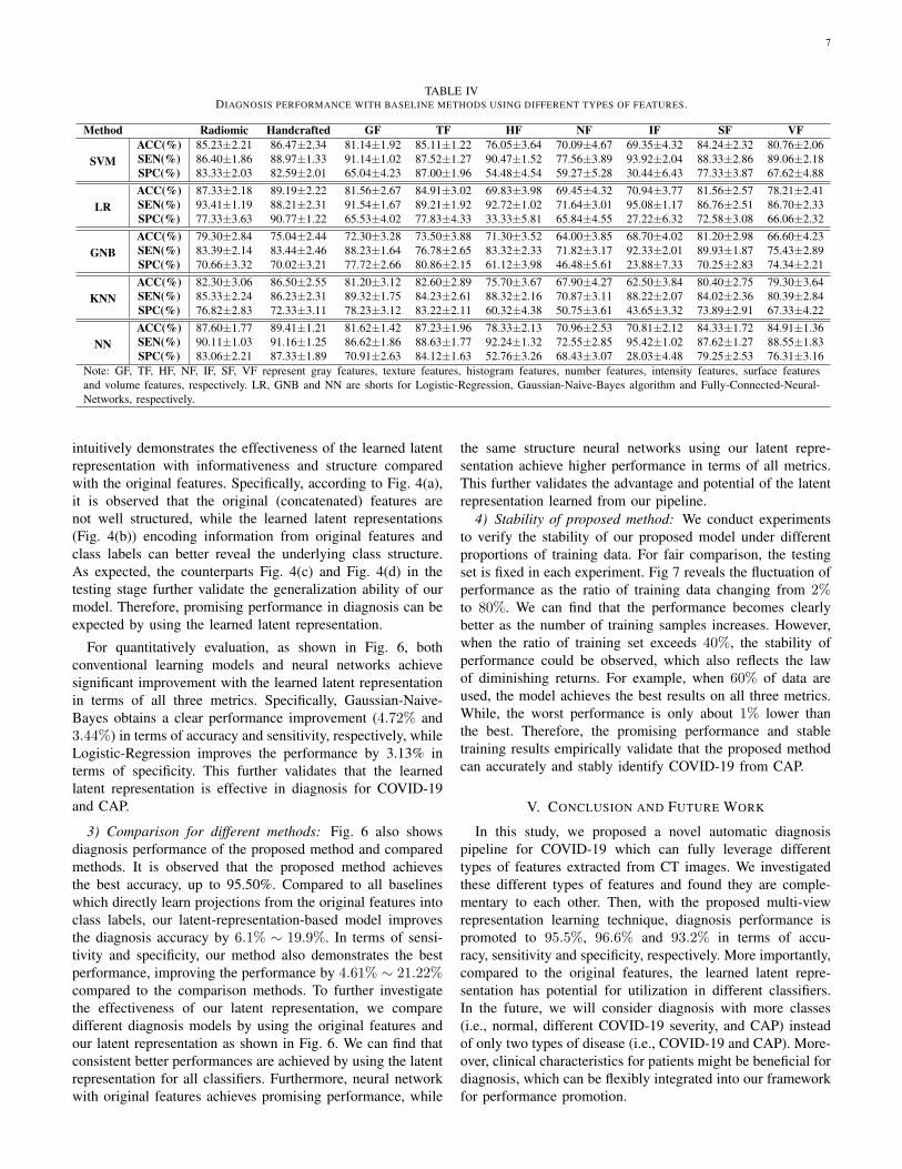
In order to investigate the discrimination power (related todiagnosis) of different types of features, we visualize themwith t-distributed stochastic neighbor embedding (t-SNE) [35].Fig. 5 demonstrates different distributions for these 7 types offeatures and concatenated features (7 types). Furthermore, toquantitatively evaluate these features, we conduct experimentson each type of features for diagnosis task with baseline algo-rithms. Table IV presents the diagnostic performance. First, wecan find that large performance gaps exist between differenttypes of features. For example, the baselines with gray featuresand texture features achieve clearly better performances thannumber features and intensity features. There are differentmanifestations reported between COVID-19 and other typesof pneumonia, such as Influenza-A viral pneumonia [24]. Asexpected, radiomic features including gray and texture fea-tures have better discrimination ability. However, the numberfeatures and intensity features are a little less discriminative,and the possible reason is that the number of lesions and theintensity in lung may be not quite different for COVID-19and CAP. Note that, although different types of features havedifferent power in diagnosis, they are complementary to eachother. As shown in Table IV, the concatenated features (i.e.,radiomic and handcrafted features) perform much better thanthe case of using each individual type of features in termsof accuracy, which strongly supports the necessity of jointlyusing different types of features.
2) Effectiveness of latent representation compared with theoriginal features: With latent-representation regressor, Fig. 4
7
TABLE IVDIAGNOSIS PERFORMANCE WITH BASELINE METHODS USING DIFFERENT TYPES OF FEATURES.
Method Radiomic Handcrafted GF TF HF NF IF SF VF
SVMACC(%) 85.23±2.21 86.47±2.34 81.14±1.92 85.11±1.22 76.05±3.64 70.09±4.67 69.35±4.32 84.24±2.32 80.76±2.06SEN(%) 86.40±1.86 88.97±1.33 91.14±1.02 87.52±1.27 90.47±1.52 77.56±3.89 93.92±2.04 88.33±2.86 89.06±2.18SPC(%) 83.33±2.03 82.59±2.01 65.04±4.23 87.00±1.96 54.48±4.54 59.27±5.28 30.44±6.43 77.33±3.87 67.62±4.88
LRACC(%) 87.33±2.18 89.19±2.22 81.56±2.67 84.91±3.02 69.83±3.98 69.45±4.32 70.94±3.77 81.56±2.57 78.21±2.41SEN(%) 93.41±1.19 88.21±2.31 91.54±1.67 89.21±1.92 92.72±1.02 71.64±3.01 95.08±1.17 86.76±2.51 86.70±2.33SPC(%) 77.33±3.63 90.77±1.22 65.53±4.02 77.83±4.33 33.33±5.81 65.84±4.55 27.22±6.32 72.58±3.08 66.06±2.32
GNBACC(%) 79.30±2.84 75.04±2.44 72.30±3.28 73.50±3.88 71.30±3.52 64.00±3.85 68.70±4.02 81.20±2.98 66.60±4.23SEN(%) 83.39±2.14 83.44±2.46 88.23±1.64 76.78±2.65 83.32±2.33 71.82±3.17 92.33±2.01 89.93±1.87 75.43±2.89SPC(%) 70.66±3.32 70.02±3.21 77.72±2.66 80.86±2.15 61.12±3.98 46.48±5.61 23.88±7.33 70.25±2.83 74.34±2.21
KNNACC(%) 82.30±3.06 86.50±2.55 81.20±3.12 82.60±2.89 75.70±3.67 67.90±4.27 62.50±3.84 80.40±2.75 79.30±3.64SEN(%) 85.33±2.24 86.23±2.31 89.32±1.75 84.23±2.61 88.32±2.16 70.87±3.11 88.22±2.07 84.02±2.36 80.39±2.84SPC(%) 76.82±2.83 72.33±3.11 78.23±3.12 83.22±2.11 60.32±4.38 50.75±3.61 43.65±3.32 73.89±2.91 67.33±4.22
NNACC(%) 87.60±1.77 89.41±1.21 81.62±1.42 87.23±1.96 78.33±2.13 70.96±2.53 70.81±2.12 84.33±1.72 84.91±1.36SEN(%) 90.11±1.03 91.16±1.25 86.62±1.86 88.63±1.77 92.24±1.32 72.55±2.85 95.42±1.02 87.62±1.27 88.55±1.83SPC(%) 83.06±2.21 87.33±1.89 70.91±2.63 84.12±1.63 52.76±3.26 68.43±3.07 28.03±4.48 79.25±2.53 76.31±3.16
Note: GF, TF, HF, NF, IF, SF, VF represent gray features, texture features, histogram features, number features, intensity features, surface featuresand volume features, respectively. LR, GNB and NN are shorts for Logistic-Regression, Gaussian-Naive-Bayes algorithm and Fully-Connected-Neural-Networks, respectively.
intuitively demonstrates the effectiveness of the learned latentrepresentation with informativeness and structure comparedwith the original features. Specifically, according to Fig. 4(a),it is observed that the original (concatenated) features arenot well structured, while the learned latent representations(Fig. 4(b)) encoding information from original features andclass labels can better reveal the underlying class structure.As expected, the counterparts Fig. 4(c) and Fig. 4(d) in thetesting stage further validate the generalization ability of ourmodel. Therefore, promising performance in diagnosis can beexpected by using the learned latent representation.
For quantitatively evaluation, as shown in Fig. 6, bothconventional learning models and neural networks achievesignificant improvement with the learned latent representationin terms of all three metrics. Specifically, Gaussian-Naive-Bayes obtains a clear performance improvement (4.72% and3.44%) in terms of accuracy and sensitivity, respectively, whileLogistic-Regression improves the performance by 3.13% interms of specificity. This further validates that the learnedlatent representation is effective in diagnosis for COVID-19and CAP.
3) Comparison for different methods: Fig. 6 also showsdiagnosis performance of the proposed method and comparedmethods. It is observed that the proposed method achievesthe best accuracy, up to 95.50%. Compared to all baselineswhich directly learn projections from the original features intoclass labels, our latent-representation-based model improvesthe diagnosis accuracy by 6.1% ∼ 19.9%. In terms of sensi-tivity and specificity, our method also demonstrates the bestperformance, improving the performance by 4.61% ∼ 21.22%compared to the comparison methods. To further investigatethe effectiveness of our latent representation, we comparedifferent diagnosis models by using the original features andour latent representation as shown in Fig. 6. We can find thatconsistent better performances are achieved by using the latentrepresentation for all classifiers. Furthermore, neural networkwith original features achieves promising performance, while
the same structure neural networks using our latent repre-sentation achieve higher performance in terms of all metrics.This further validates the advantage and potential of the latentrepresentation learned from our pipeline.
4) Stability of proposed method: We conduct experimentsto verify the stability of our proposed model under differentproportions of training data. For fair comparison, the testingset is fixed in each experiment. Fig 7 reveals the fluctuation ofperformance as the ratio of training data changing from 2%to 80%. We can find that the performance becomes clearlybetter as the number of training samples increases. However,when the ratio of training set exceeds 40%, the stability ofperformance could be observed, which also reflects the lawof diminishing returns. For example, when 60% of data areused, the model achieves the best results on all three metrics.While, the worst performance is only about 1% lower thanthe best. Therefore, the promising performance and stabletraining results empirically validate that the proposed methodcan accurately and stably identify COVID-19 from CAP.
V. CONCLUSION AND FUTURE WORK
In this study, we proposed a novel automatic diagnosispipeline for COVID-19 which can fully leverage differenttypes of features extracted from CT images. We investigatedthese different types of features and found they are comple-mentary to each other. Then, with the proposed multi-viewrepresentation learning technique, diagnosis performance ispromoted to 95.5%, 96.6% and 93.2% in terms of accu-racy, sensitivity and specificity, respectively. More importantly,compared to the original features, the learned latent repre-sentation has potential for utilization in different classifiers.In the future, we will consider diagnosis with more classes(i.e., normal, different COVID-19 severity, and CAP) insteadof only two types of disease (i.e., COVID-19 and CAP). More-over, clinical characteristics for patients might be beneficial fordiagnosis, which can be flexibly integrated into our frameworkfor performance promotion.

8
Histogram features
Number features Intensity features Surface features
Volume featuresGray features Texture features
Concatenated features
Fig. 5. Visualization of each type of original features and concatenated features using t-distributed stochastic neighbor embedding (t-SNE) [35], which is anonlinear dimensionality reduction technique well-suited for embedding high-dimensional data for visualization in a low-dimensional space.
91.2 91.0
80.3
88.8
95.5
89.2 89.4
75.6
86.4
93.9
70.0
75.0
80.0
85.0
90.0
95.0
100.0
LG SVM GNB KNN NN
Ac
cu
rac
y(%
)
Latent representation Original features
93.4 91.9
88.7
91.2
96.6
90.7 89.9
85.2
89.2
94.6
70.0
75.0
80.0
85.0
90.0
95.0
100.0
LG SVM GNB KNN NN
Se
ns
itiv
ity
(%)
Latent representation Original features
90.0 90.2
73.0
79.0
93.2
86.8 88.6
72.0
76.0
91.7
70.0
75.0
80.0
85.0
90.0
95.0
100.0
LG SVM GNB KNN NN
Sp
ec
ific
ity
(%)
Latent representation Original features
Fig. 6. Evaluation for the latent representation on different classifiers.
REFERENCES
[1] J. T. Wu, K. Leung, and G. M. Leung, “Nowcasting and forecasting thepotential domestic and international spread of the 2019-ncov outbreakoriginating in wuhan, china: a modelling study,” The Lancet, vol. 395,no. 10225, pp. 689–697, 2020.
[2] H. Shi, X. Han, N. Jiang, Y. Cao, O. Alwalid, J. Gu, Y. Fan,and C. Zheng, “Radiological findings from 81 patients with covid-19pneumonia in wuhan, china: a descriptive study,” The Lancet InfectiousDiseases, vol. 20, no. 4, pp. 425 – 434, 2020. [Online]. Available:http://www.sciencedirect.com/science/article/pii/S1473309920300864
[3] F. Song, N. Shi, F. Shan, Z. Zhang, J. Shen, H. Lu, Y. Ling, Y. Jiang,and Y. Shi, “Emerging 2019 novel coronavirus (2019-ncov) pneumonia,”Radiology, vol. 295, no. 1, pp. 210–217, 2020, pMID: 32027573.
[4] W. H. Organization et al., “Coronavirus disease 2019 (covid-19): situa-tion report, 67,” 2020.
[5] Z. Xu, L. Shi, Y. Wang, J. Zhang, L. Huang, C. Zhang, S. Liu, P. Zhao,H. Liu, L. Zhu et al., “Pathological findings of covid-19 associated withacute respiratory distress syndrome,” The Lancet respiratory medicine,vol. 8, no. 4, pp. 420–422, 2020.
[6] Y. Li and L. Xia, “Coronavirus disease 2019 (covid-19): Role of chest
ct in diagnosis and management,” American Journal of Roentgenology,pp. 1–7, 2020.
[7] Y. Fang, H. Zhang, J. Xie, M. Lin, L. Ying, P. Pang, and W. Ji,“Sensitivity of chest ct for covid-19: comparison to rt-pcr,” Radiology,p. 200432, 2020.
[8] F. Shi, J. Wang, J. Shi, Z. Wu, Q. Wang, Z. Tang, K. He, Y. Shi, andD. Shen, “Review of artificial intelligence techniques in imaging dataacquisition, segmentation and diagnosis for covid-19,” 2020.
[9] J. P. Kanne, “Chest ct findings in 2019 novel coronavirus (2019-ncov)infections from wuhan, china: key points for the radiologist,” 2020.
[10] A. Bernheim, X. Mei, M. Huang, Y. Yang, Z. A. Fayad, N. Zhang,K. Diao, B. Lin, X. Zhu, K. Li et al., “Chest ct findings in coronavirusdisease-19 (covid-19): relationship to duration of infection,” Radiology,p. 200463, 2020.
[11] J. P. Kanne, B. P. Little, J. H. Chung, B. M. Elicker, and L. H. Ketai,“Essentials for radiologists on covid-19: an updateradiology scientificexpert panel,” 2020.
[12] W. Zhao, Z. Zhong, X. Xie, Q. Yu, and J. Liu, “Relationbetween chest ct findings and clinical conditions of coronavirusdisease (covid-19) pneumonia: A multicenter study,” AmericanJournal of Roentgenology, pp. 1–6, Mar 2020. [Online]. Available:

9
0 10 20 30 40 50 60 70 80Ratio of training data (%)
0.90
0.92
0.94
0.96
0.98
1.00P
erfo
rman
ce
AccuracySensitivitySpecificity
Fig. 7. Stability of our method with different ratios of training data.
https://doi.org/10.2214/AJR.20.22976[13] M. Chung, A. Bernheim, X. Mei, N. Zhang, M. Huang, X. Zeng, J. Cui,
W. Xu, Y. Yang, Z. A. Fayad, A. Jacobi, K. Li, S. Li, and H. Shan, “Ctimaging features of 2019 novel coronavirus (2019-ncov),” Radiology,vol. 295, no. 1, pp. 202–207, 2020, pMID: 32017661.
[14] J. R. Zech, M. A. Badgeley, M. Liu, A. B. Costa, J. J. Titano, andE. K. Oermann, “Variable generalization performance of a deep learningmodel to detect pneumonia in chest radiographs: a cross-sectional study,”PLoS medicine, vol. 15, no. 11, 2018.
[15] D. S. Kermany, M. Goldbaum, W. Cai, C. C. Valentim, H. Liang, S. L.Baxter, A. McKeown, G. Yang, X. Wu, F. Yan et al., “Identifyingmedical diagnoses and treatable diseases by image-based deep learning,”Cell, vol. 172, no. 5, pp. 1122–1131, 2018.
[16] P. Rajpurkar, J. Irvin, R. L. Ball, K. Zhu, B. Yang, H. Mehta, T. Duan,D. Ding, A. Bagul, C. P. Langlotz et al., “Deep learning for chest radio-graph diagnosis: A retrospective comparison of the chexnext algorithmto practicing radiologists,” PLoS medicine, vol. 15, no. 11, p. e1002686,2018.
[17] Y. LeCun, Y. Bengio, and G. Hinton, “Deep learning,” nature, vol. 521,no. 7553, pp. 436–444, 2015.
[18] J. Deng, W. Dong, R. Socher, L.-J. Li, K. Li, and L. Fei-Fei, “Imagenet:A large-scale hierarchical image database,” in 2009 IEEE conference oncomputer vision and pattern recognition. Ieee, 2009, pp. 248–255.
[19] Y. Mori, S.-e. Kudo, T. M. Berzin, M. Misawa, and K. Takeda,“Computer-aided diagnosis for colonoscopy,” Endoscopy, vol. 49, no. 08,pp. 813–819, 2017.
[20] R. Zhang, Y. Zheng, T. W. C. Mak, R. Yu, S. H. Wong, J. Y. Lau, andC. C. Poon, “Automatic detection and classification of colorectal polypsby transferring low-level cnn features from nonmedical domain,” IEEEjournal of biomedical and health informatics, vol. 21, no. 1, pp. 41–47,2016.
[21] M. Liu, D. Zhang, E. Adeli, and D. Shen, “Inherent structure-based mul-tiview learning with multitemplate feature representation for alzheimer’sdisease diagnosis,” IEEE Transactions on Biomedical Engineering,vol. 63, no. 7, pp. 1473–1482, 2015.
[22] M. Zhang, Y. Yang, F. Shen, H. Zhang, and Y. Wang, “Multi-viewfeature selection and classification for alzheimers disease diagnosis,”Multimedia Tools and Applications, vol. 76, no. 8, pp. 10 761–10 775,2017.
[23] S. Wang, B. Kang, J. Ma, X. Zeng, M. Xiao, J. Guo, M. Cai, J. Yang,Y. Li, X. Meng et al., “A deep learning algorithm using ct images toscreen for corona virus disease (covid-19),” medRxiv, 2020.
[24] X. Xu, X. Jiang, C. Ma, P. Du, X. Li, S. Lv, L. Yu, Y. Chen, J. Su,G. Lang et al., “Deep learning system to screen coronavirus disease2019 pneumonia,” arXiv preprint arXiv:2002.09334, 2020.
[25] K. R. Gray, P. Aljabar, R. A. Heckemann, A. Hammers, D. Rueckert,A. D. N. Initiative et al., “Random forest-based similarity measures formulti-modal classification of alzheimer’s disease,” NeuroImage, vol. 65,pp. 167–175, 2013.
[26] C. Zhang, Z. Han, H. Fu, J. T. Zhou, Q. Hu et al., “Cpm-nets:Cross partial multi-view networks,” in Advances in Neural InformationProcessing Systems, 2019, pp. 557–567.
[27] F. Milletari, N. Navab, and S.-A. Ahmadi, “V-net: Fully convolutionalneural networks for volumetric medical image segmentation,” in 2016
Fourth International Conference on 3D Vision (3DV). IEEE, 2016, pp.565–571.
[28] F. Shan+, Y. Gao+, J. Wang, W. Shi, N. Shi, M. Han, Z. Xue, D. Shen,and Y. Shi, “Lung infection quantification of covid-19 in ct images withdeep learning,” arXiv preprint arXiv:2003.04655, 2020.
[29] A. Zwanenburg, S. Leger, M. Vallieres, and S. Lock, “Image biomarkerstandardisation initiative,” arXiv preprint arXiv:1612.07003, 2016.
[30] F. Shi, L. Xia, F. Shan, D. Wu, Y. Wei, H. Yuan, H. Jiang, Y. Gao, H. Sui,and D. Shen, “Large-scale screening of covid-19 from communityacquired pneumonia using infection size-aware classification,” arXivpreprint arXiv:2003.09860, 2020.
[31] C. Xu, D. Tao, and C. Xu, “A survey on multi-view learning,” arXivpreprint arXiv:1304.5634, 2013.
[32] C. Zhang, H. Fu, Q. Hu, P. Zhu, and X. Cao, “Flexible multi-viewdimensionality co-reduction,” IEEE Transactions on Image Processing,vol. 26, no. 2, pp. 648–659, 2017.
[33] C. Zhang, Q. Hu, H. Fu, P. Zhu, and X. Cao, “Latent multi-viewsubspace clustering,” in The IEEE Conference on Computer Vision andPattern Recognition (CVPR), July 2017.
[34] Tai Sing Lee, “Image representation using 2d gabor wavelets,” IEEETransactions on Pattern Analysis and Machine Intelligence, vol. 18,no. 10, pp. 959–971, 1996.
[35] L. v. d. Maaten and G. Hinton, “Visualizing data using t-sne,” Journalof machine learning research, vol. 9, no. Nov, pp. 2579–2605, 2008.