Guidelines for the Management of
Traumatic dental injuries
本網頁內容引用自 2007
The International Association
of Dental Traumatology
之官方資料,僅供參考
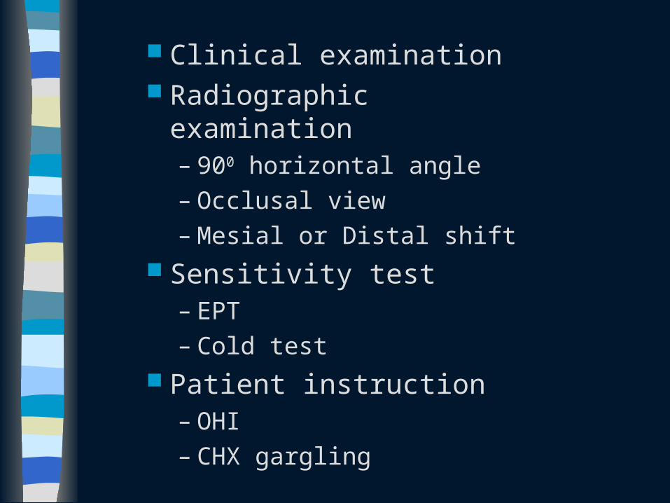
Clinical examination Radiographic examination
– 900 horizontal angle– Occlusal view– Mesial or Distal shift
Sensitivity test– EPT– Cold test
Patient instruction– OHI– CHX gargling
Treatment guidelines for fractures of teeth and alveolar bone
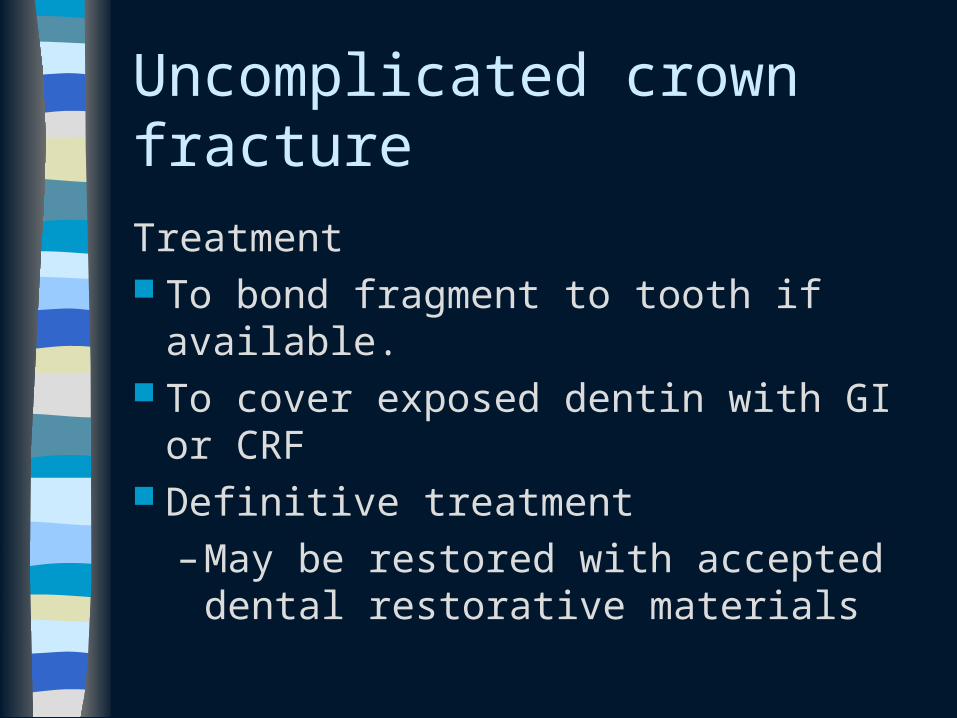
Uncomplicated crown fracture
Treatment To bond fragment to tooth if available. To cover exposed dentin with GI or
CRF Definitive treatment
– May be restored with accepted dental restorative materials
Complicated crown fracture
Open apex Preserve pulp vitality
– Pulp capping– Partial pulpotomy
– Ca(OH)2 or MTA
Complicated crown fracture
Closed apex with vital pulp Younger patient
– Pulp capping or partial pulpotomy Older patient
– RCT
Pulp necrosis RCT
Crown-root fracture
Closed apex with vital pulp Younger patient
– Pulp capping or partial pulpotomy Older patient
– RCT
Pulp necrosis RCT
Root fracture
Reposition – As soon as possible– Check position radiographically– Flexible splint, 4 weeks– Cervical fracture: up to 4 months
Follow-up– 1 yr at least– Pulp necrosis
• RCT for coronal fragment
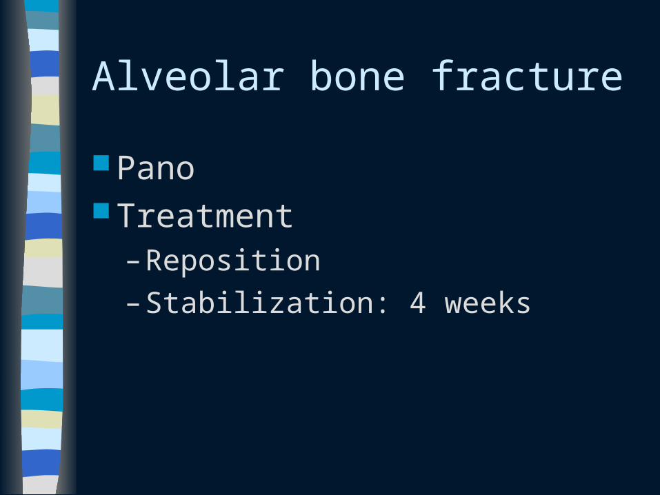
Alveolar bone fracture
Pano Treatment
– Reposition
– Stabilization: 4 weeks
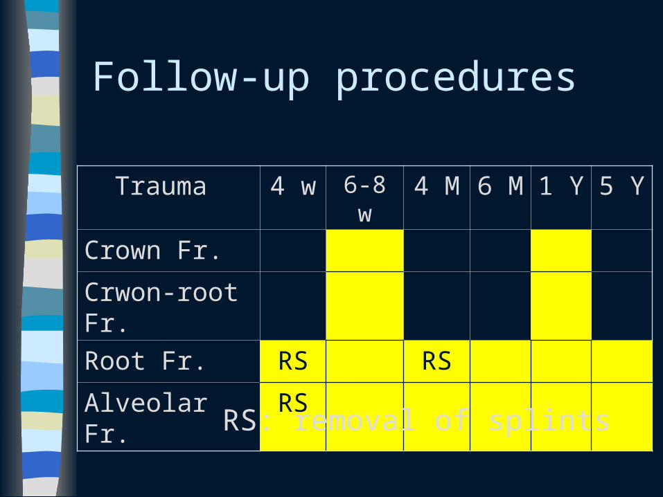
Follow-up procedures
Trauma 4 w 6-8 w 4 M 6 M 1 Y 5 Y
Crown Fr.
Crwon-root Fr.
Root Fr. RS RS
Alveolar Fr. RS
RS: removal of splints
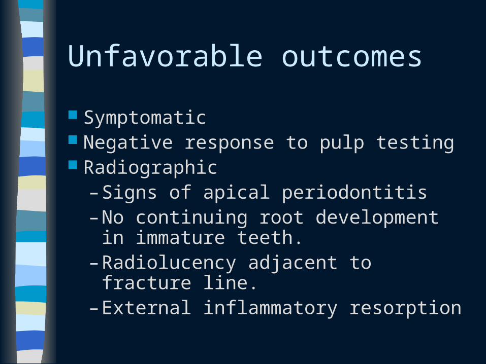
Unfavorable outcomes
Symptomatic Negative response to pulp testing Radiographic
– Signs of apical periodontitis– No continuing root development in
immature teeth.– Radiolucency adjacent to fracture line.– External inflammatory resorption
Treatment guidelines for luxation injuries
Concussion
No treatment is needed. Monitor pulpal condition for at least 1
year
Subluxation
Flexible splint, 2 weeks
Extrusive luxation
Reposition: gently re-inserting Stabilization
– flexible splint, 2 weeks Monitoring the pulpal condition
– Sensibility tests– Radiography
Lateral luxation
Reposition – Disengage tooth with forceps– Gently reposition into original location
Stabilization– Flexible splint, 4 weeks
Monitor the pulpal condition
Intrusive luxation
Incomplete root formation– Allow spontaneous repositioning to take
place within 3 weeks– Rapid orthodontic repositioning
Complete root formation– To be repositioned either orthodontically or
surgically as soon as possible.
– RCT with Ca(OH)2 dressing within 3 weeks
Follow-up procedures
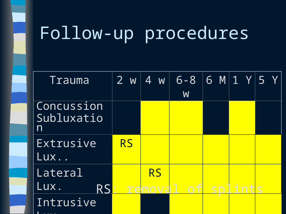
Trauma 2 w 4 w 6-8 w 6 M 1 Y 5 YConcussionSubluxation
Extrusive Lux..
RS
Lateral Lux. RS
Intrusive Lux
RS: removal of splints
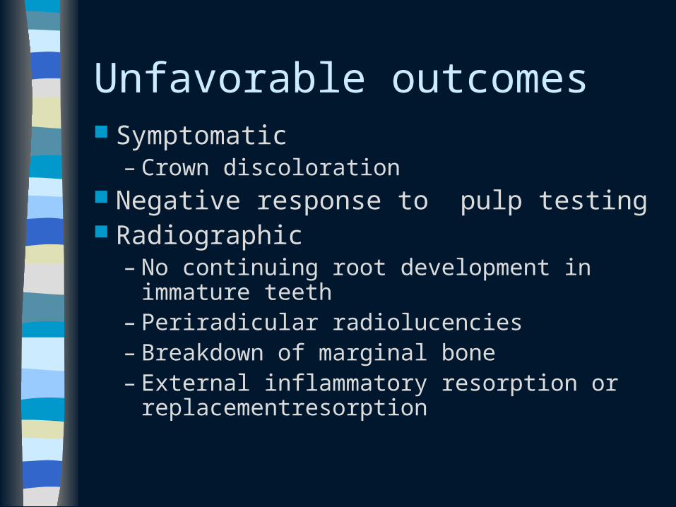
Unfavorable outcomes Symptomatic
– Crown discoloration Negative response to pulp testing Radiographic
– No continuing root development in immature teeth
– Periradicular radiolucencies– Breakdown of marginal bone– External inflammatory resorption or
replacementresorption
Treatment guidelines for avulsed permanent teeth
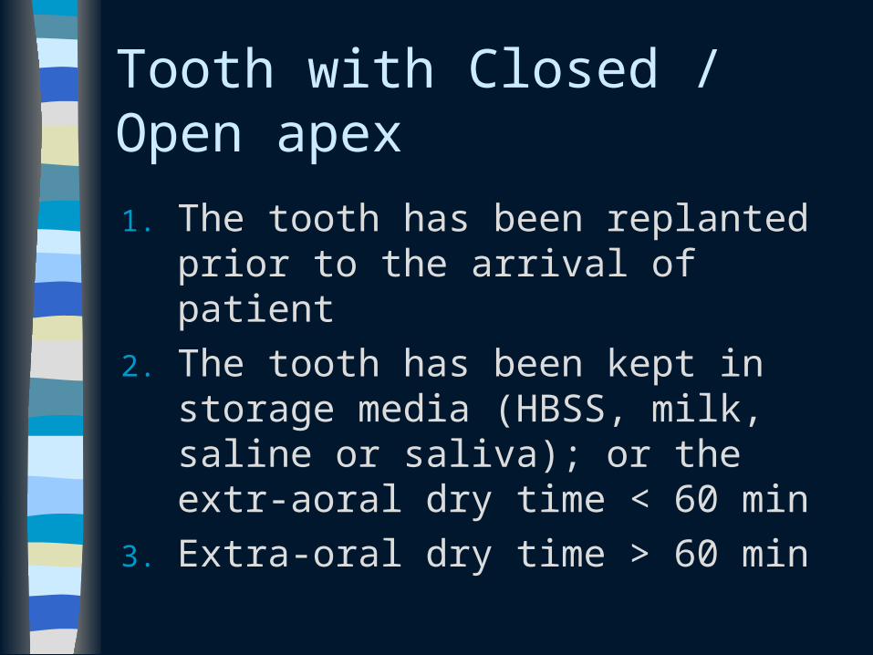
Tooth with Closed / Open apex
1. The tooth has been replanted prior to the arrival of patient
2. The tooth has been kept in storage media (HBSS, milk, saline or saliva); or the extr-aoral dry time < 60 min
3. Extra-oral dry time > 60 min
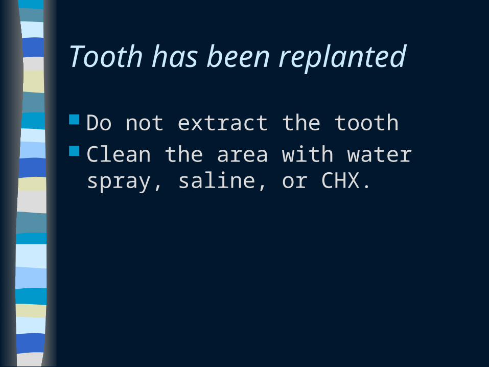
Tooth has been replanted
Do not extract the tooth Clean the area with water spray, saline,
or CHX.
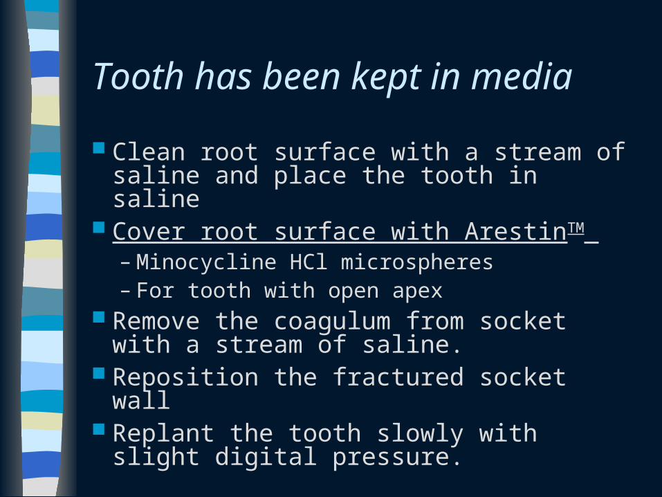
Tooth has been kept in media
Clean root surface with a stream of saline and place the tooth in saline
Cover root surface with ArestinTM – Minocycline HCl microspheres– For tooth with open apex
Remove the coagulum from socket with a stream of saline.
Reposition the fractured socket wall Replant the tooth slowly with slight
digital pressure.
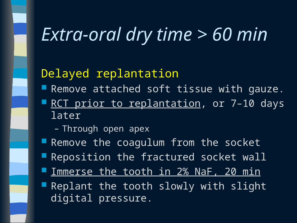
Extra-oral dry time > 60 min
Delayed replantation Remove attached soft tissue with gauze. RCT prior to replantation, or 7–10 days later
– Through open apex
Remove the coagulum from the socket Reposition the fractured socket wall Immerse the tooth in 2% NaF, 20 min Replant the tooth slowly with slight digital
pressure.
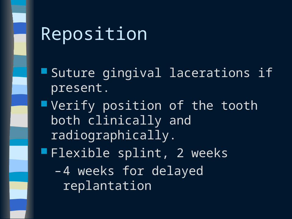
Reposition
Suture gingival lacerations if present. Verify position of the tooth both clinically
and radiographically. Flexible splint, 2 weeks
– 4 weeks for delayed replantation
Systemic antibiotics
Tetracycline (Doxycycline) for 7 days– Risk of discoloration– Not recommended for age < 12 y/o
Phenoxymethyl Penicillin (Pen V)
If the tooth has contacted soil, and if tetanus coverage is uncertain, refer to physician for evaluation and need for a tetanus booster.
Patient instruction
For all patients with dental trauma Soft diet, 2 weeks. Brush teeth with a soft toothbrush after
each meal. 0.12% CHX mouth rinse, bid, 1 week
Root canal treatment
RCT 7–10 days after replantation and before splint removal. – RCT prior to delayed replantation
Place Ca(OH)2 dressing until RCF
– 1 month Open apex: only when pulp necrosis
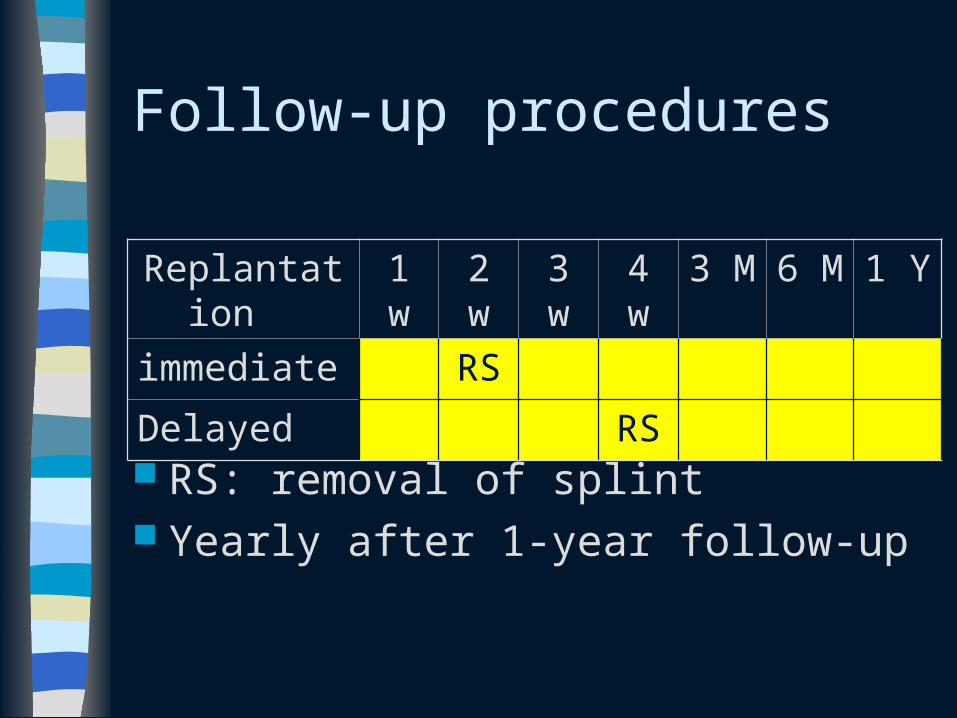
Follow-up procedures
RS: removal of splint Yearly after 1-year follow-up
Replantation
1 w 2 w 3 w 4 w 3 M 6 M 1 Y
immediate RS
Delayed RS
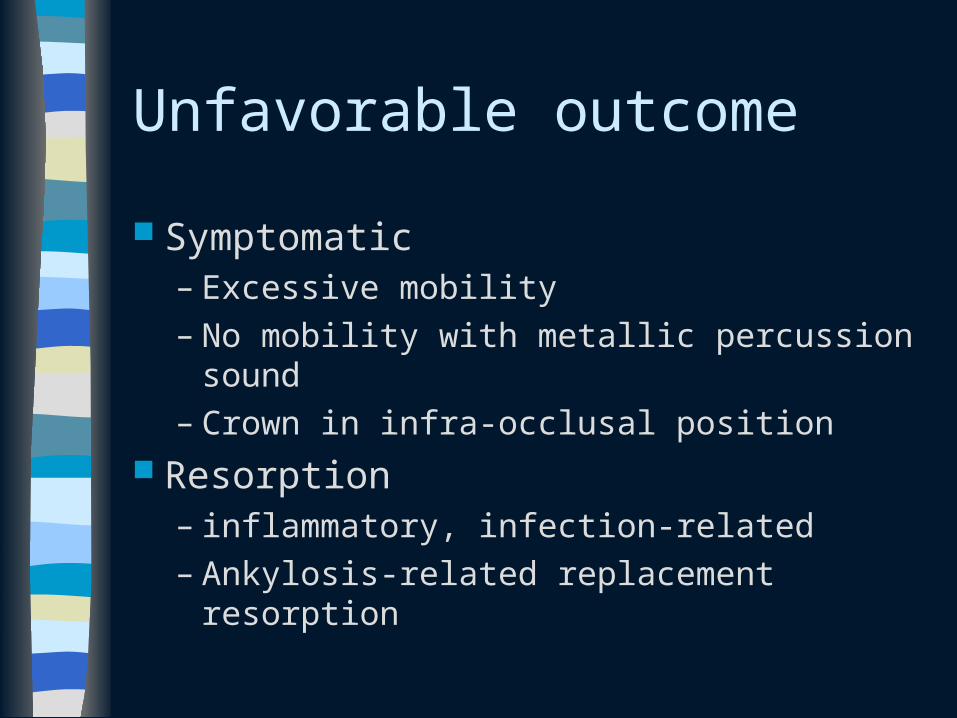
Unfavorable outcome
Symptomatic– Excessive mobility– No mobility with metallic percussion sound– Crown in infra-occlusal position
Resorption– inflammatory, infection-related– Ankylosis-related replacement resorption
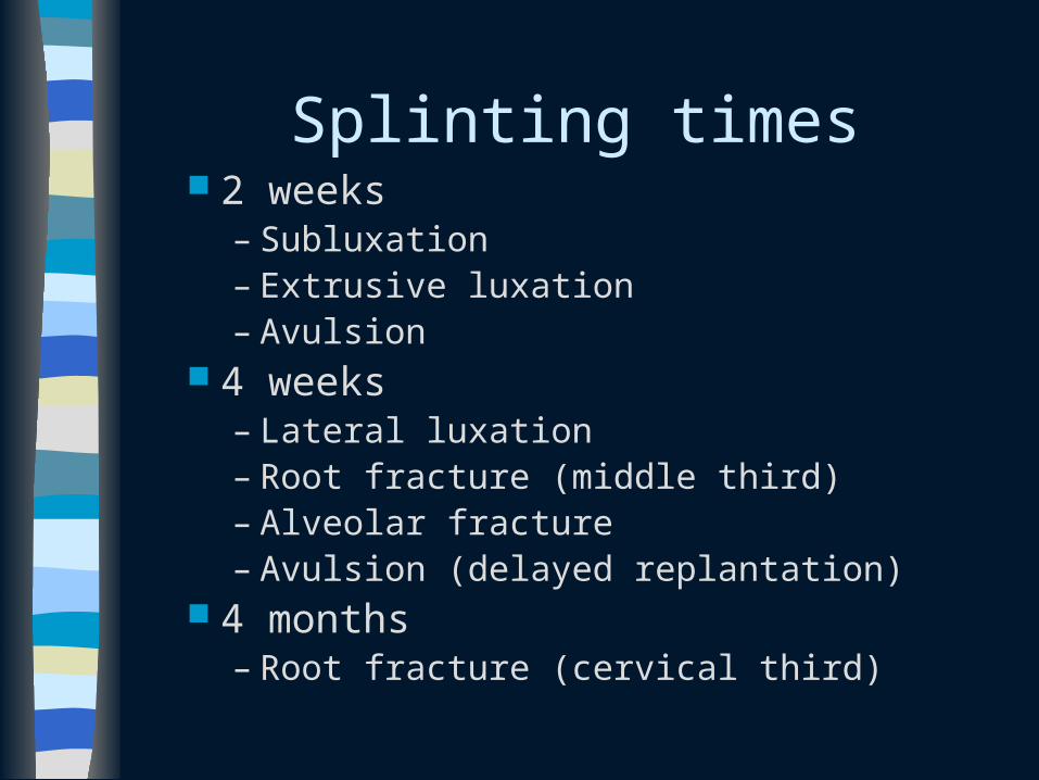
Splinting times 2 weeks
– Subluxation – Extrusive luxation – Avulsion
4 weeks– Lateral luxation– Root fracture (middle third) – Alveolar fracture– Avulsion (delayed replantation)
4 months– Root fracture (cervical third)


