
Cévy mozku a míchy
Veronika NěmcováRastislav Druga
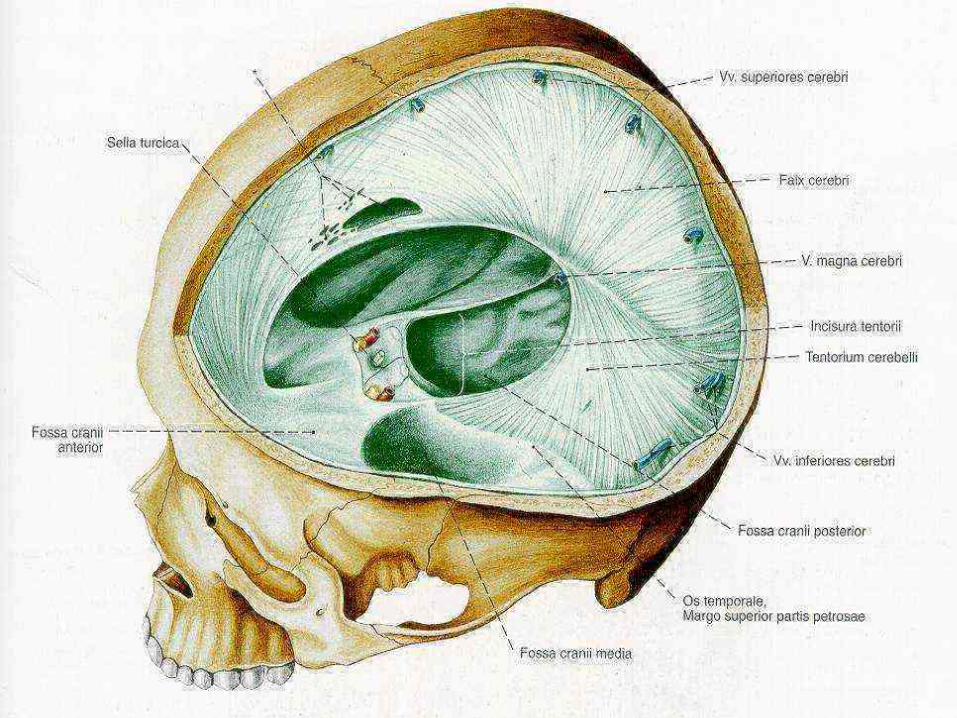
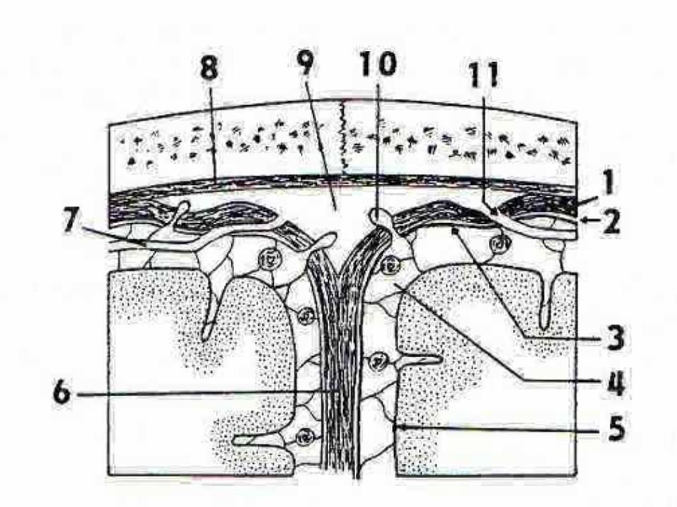
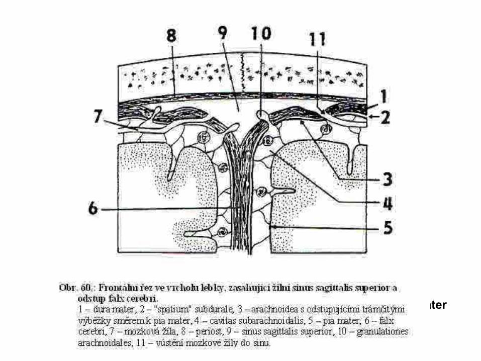
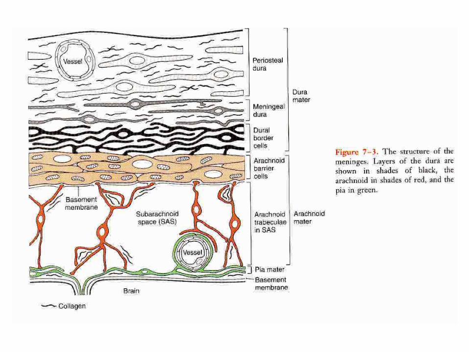
pia mater
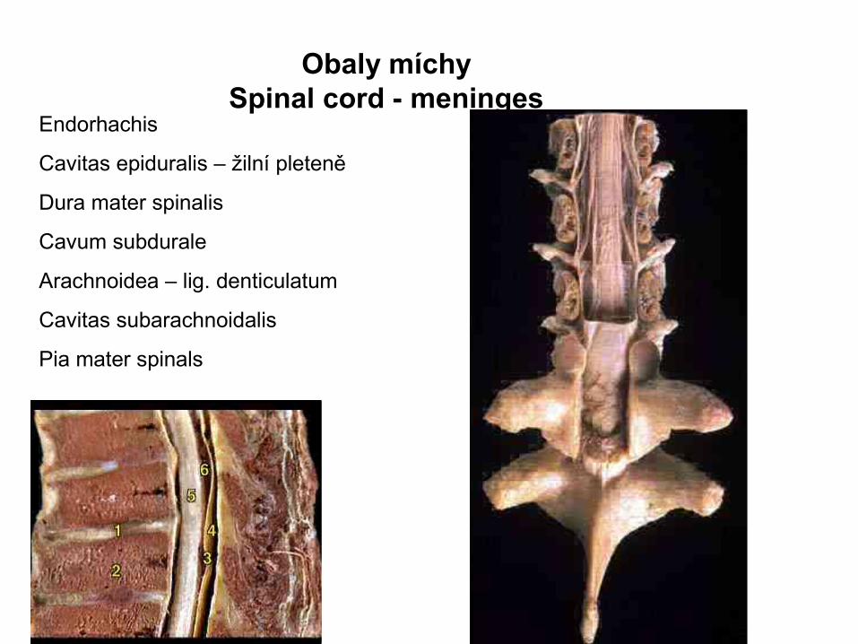
Obaly míchySpinal cord - meninges
Endorhachis
Cavitas epiduralis – žilní pleteně
Dura mater spinalis
Cavum subdurale
Arachnoidea – lig. denticulatum
Cavitas subarachnoidalis
Pia mater spinals
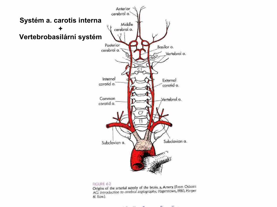
Systém a. carotis interna+
Vertebrobasilární systém
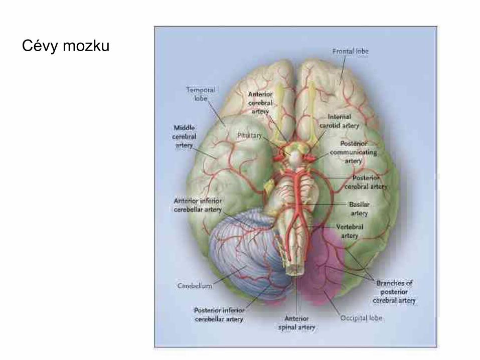
Cévy cieculusCévy mozku
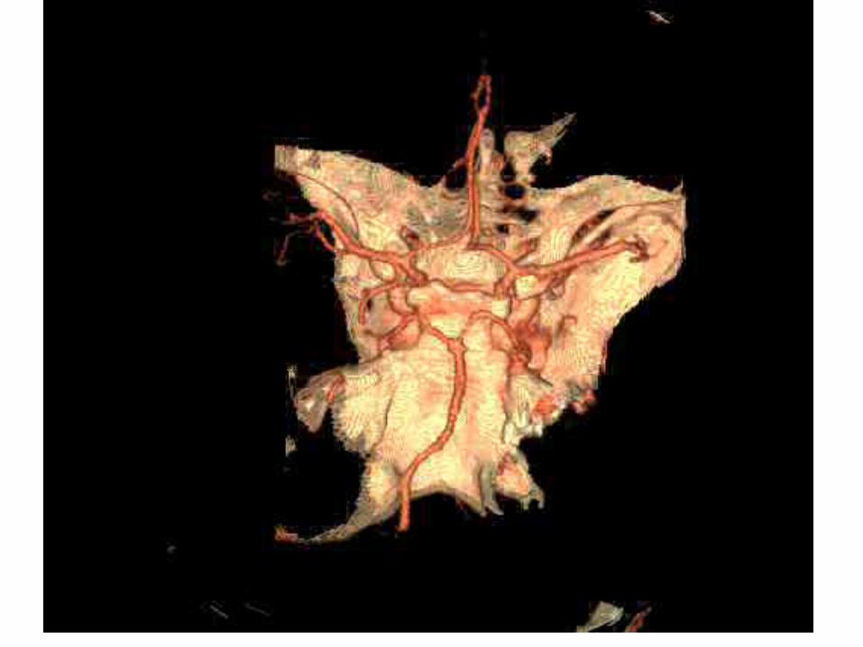
CT – AG, 3-D
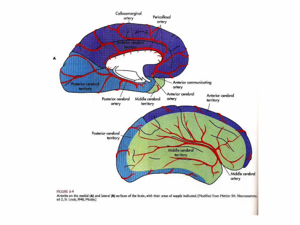
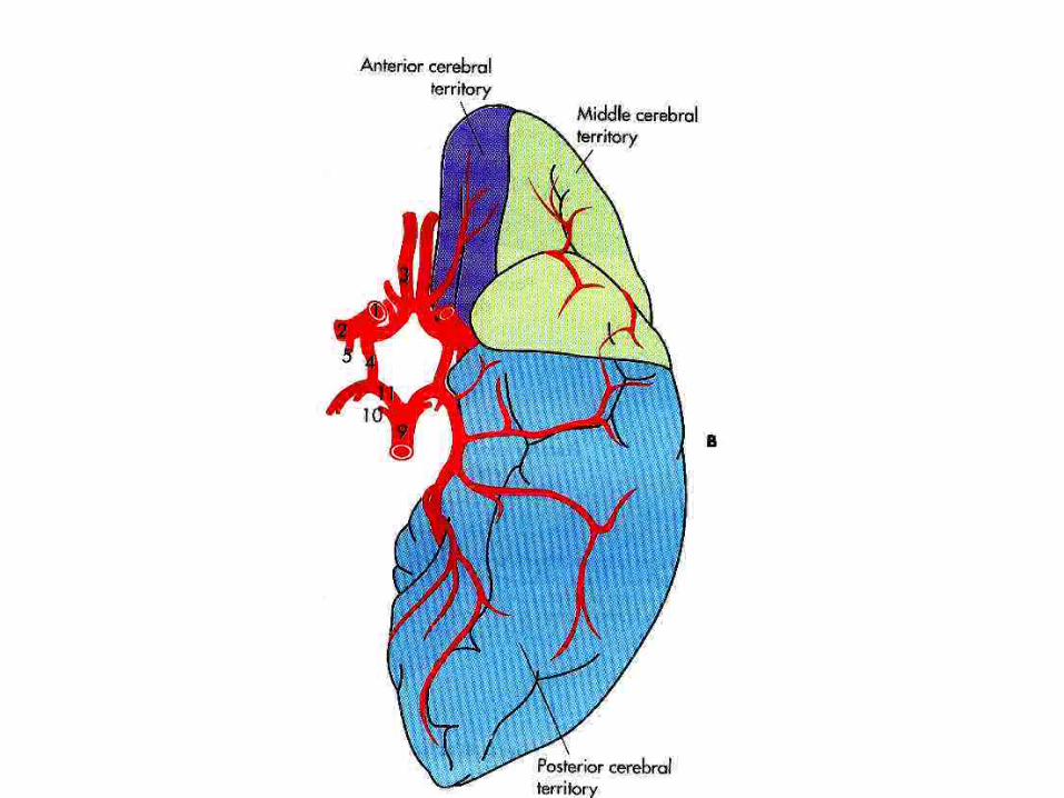

Teritoria mozkových tepen
a.cerebri anterior
a.cerebri media
a. cerebri posterior
a. choroidea anterior
a.cerebellaris superior
a.cerebellaris inferior posterior
a.cerebellaris inferior anterior
aa. lenticulostriaticae
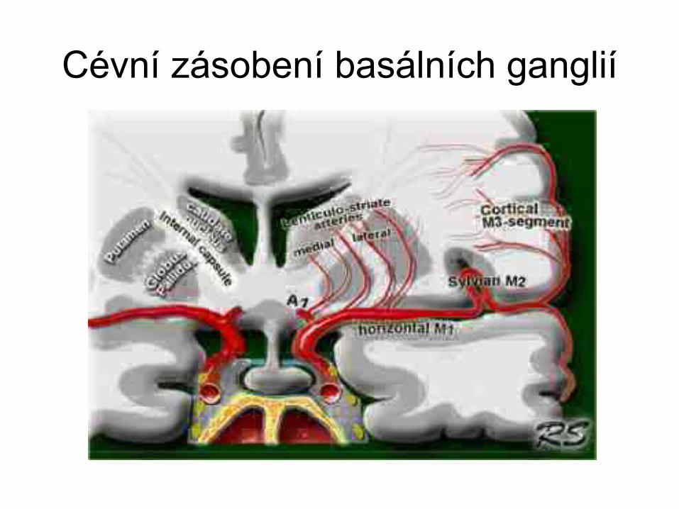
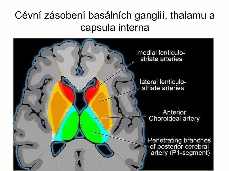
Cévní zásobení basálních ganglií, thalamu a capsula interna
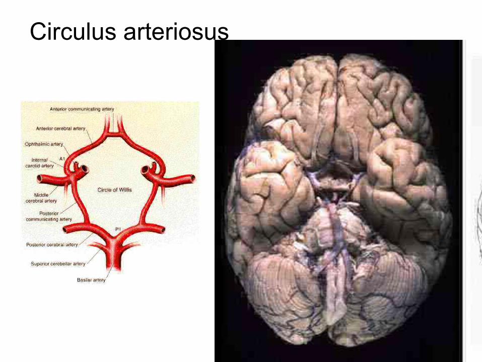
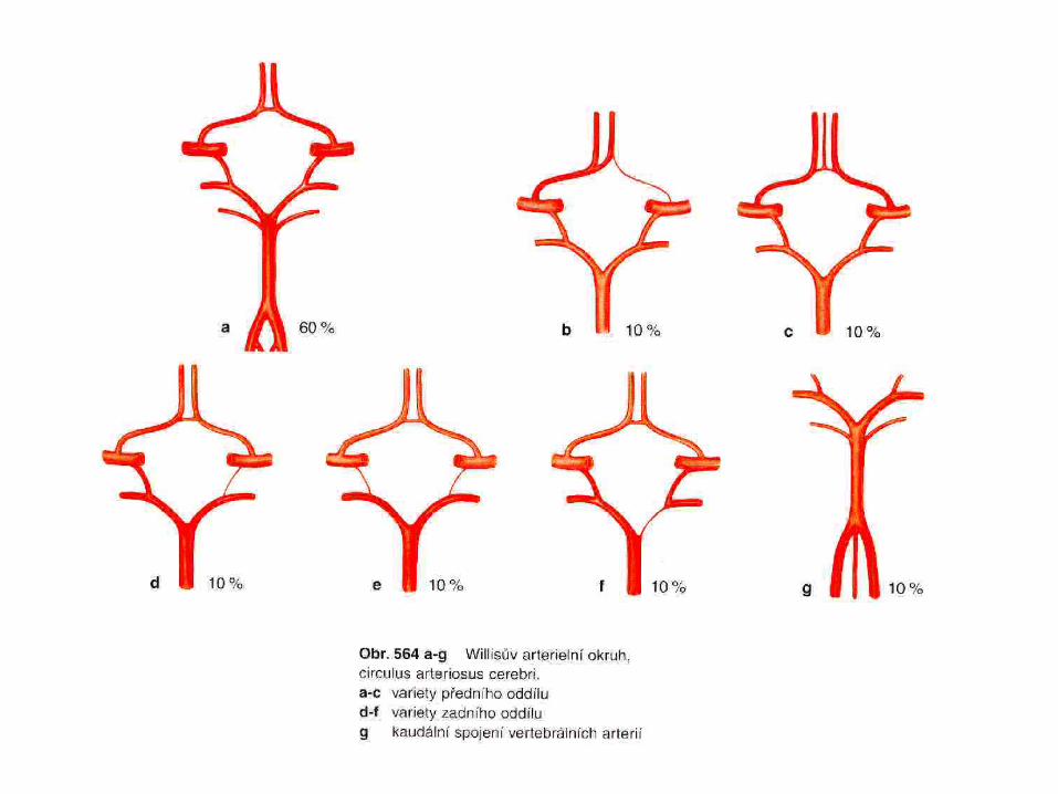
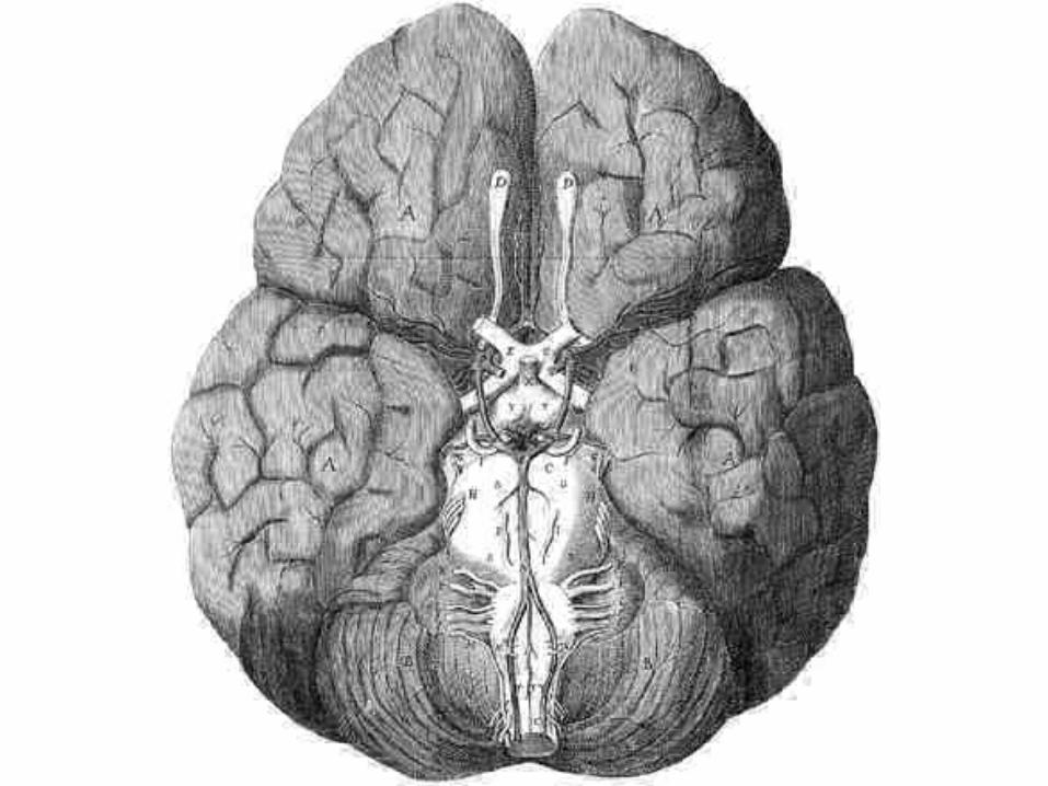
Circulus arteriosus
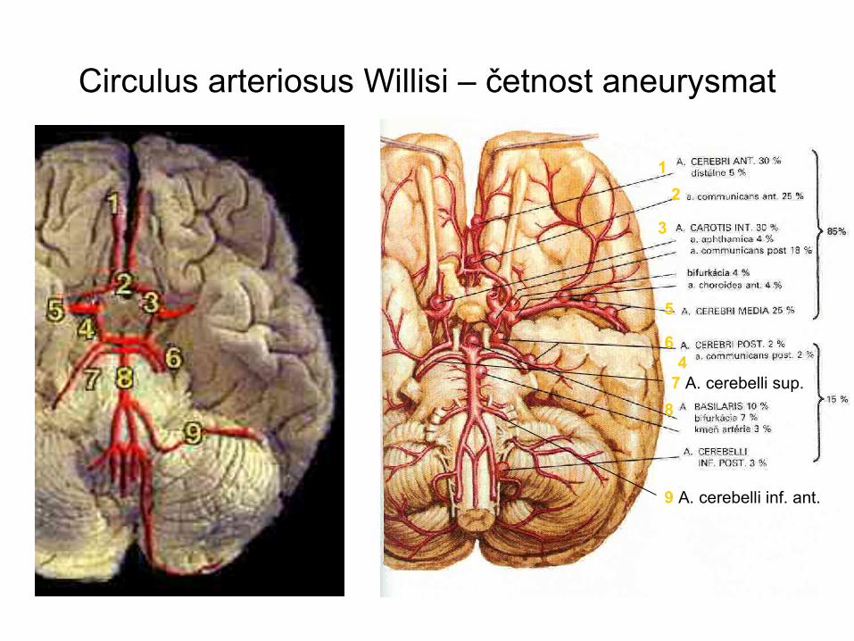
Circulus arteriosus Willisi – četnost aneurysmat
1
3
5
6
2
4
87 A. cerebelli sup.
9 A. cerebelli inf. ant.
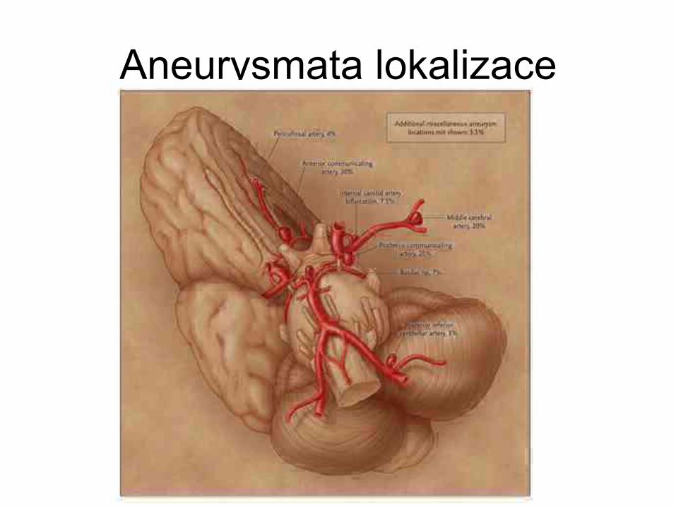
Aneurysmata lokalizace
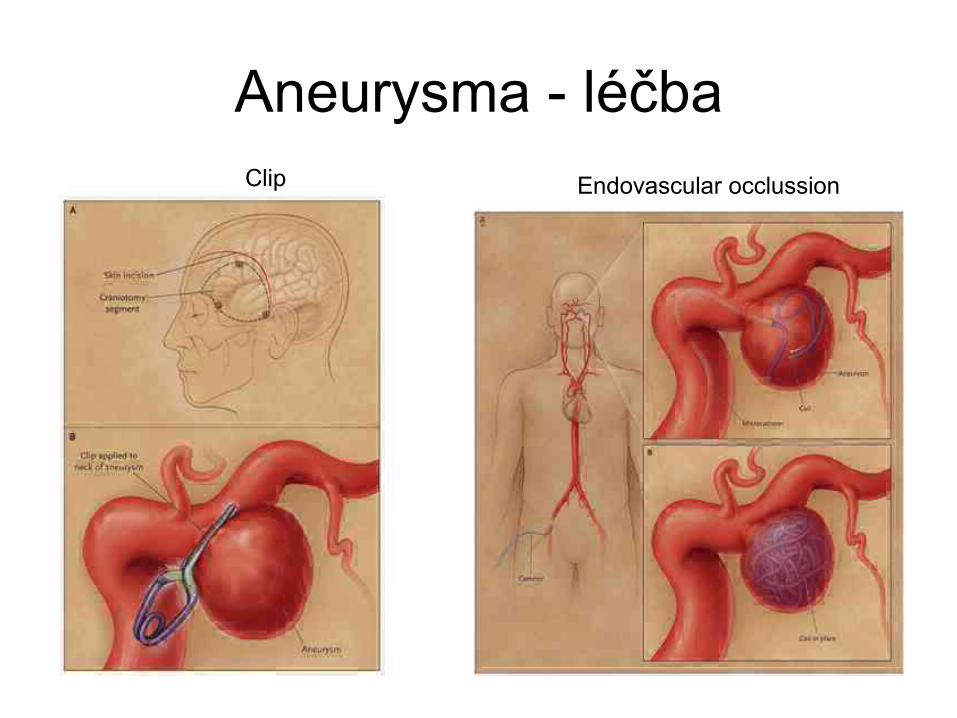
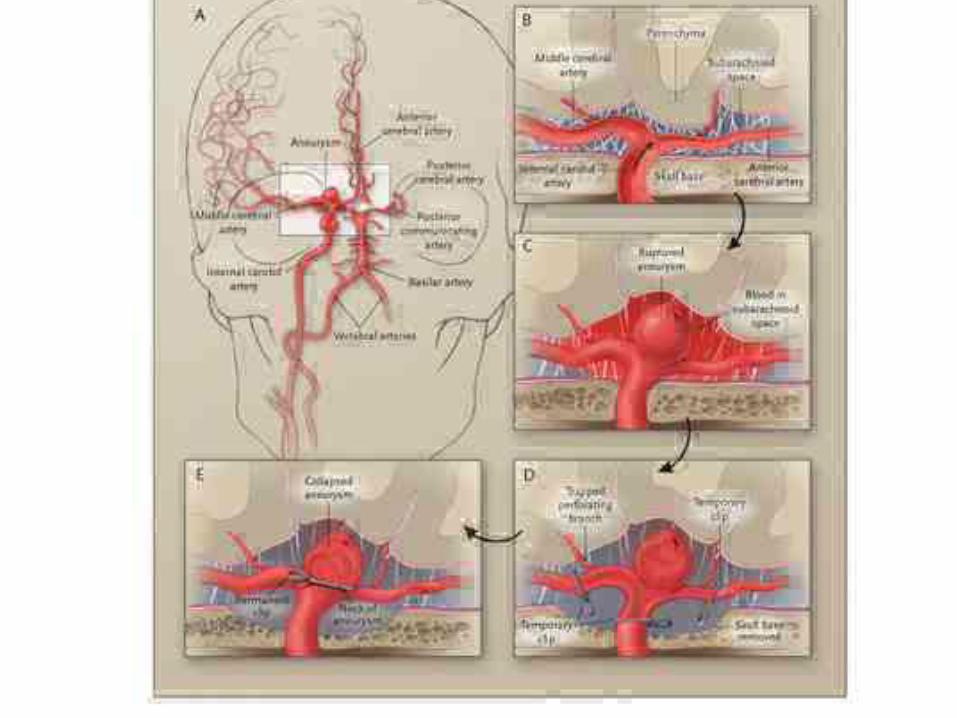
Aneurysma - léčbaEndovascular occlussionClip
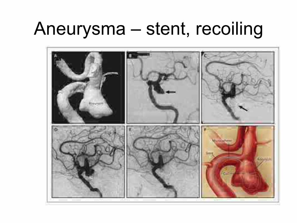
Aneurysma – stent, recoiling
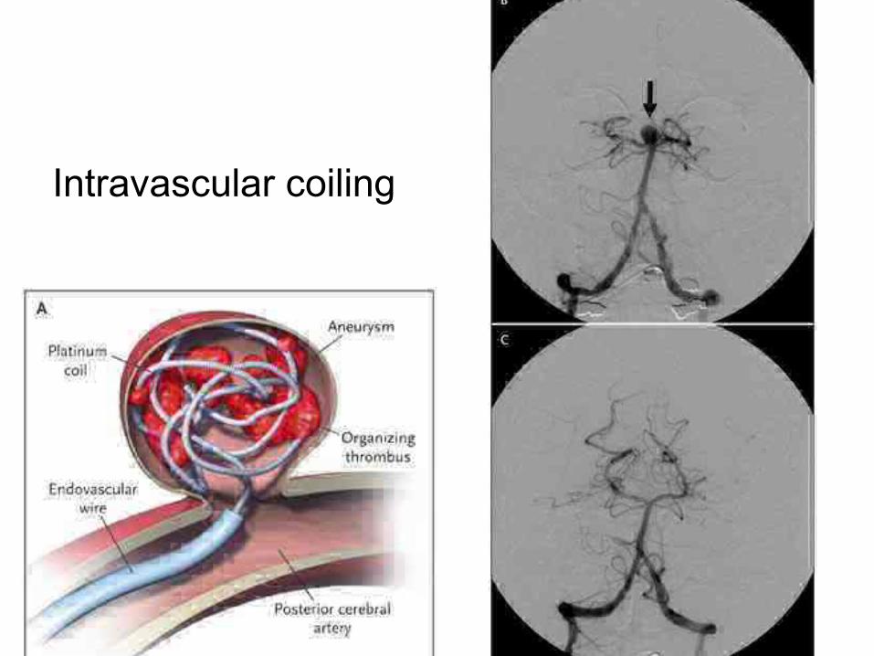
Intravascular coiling
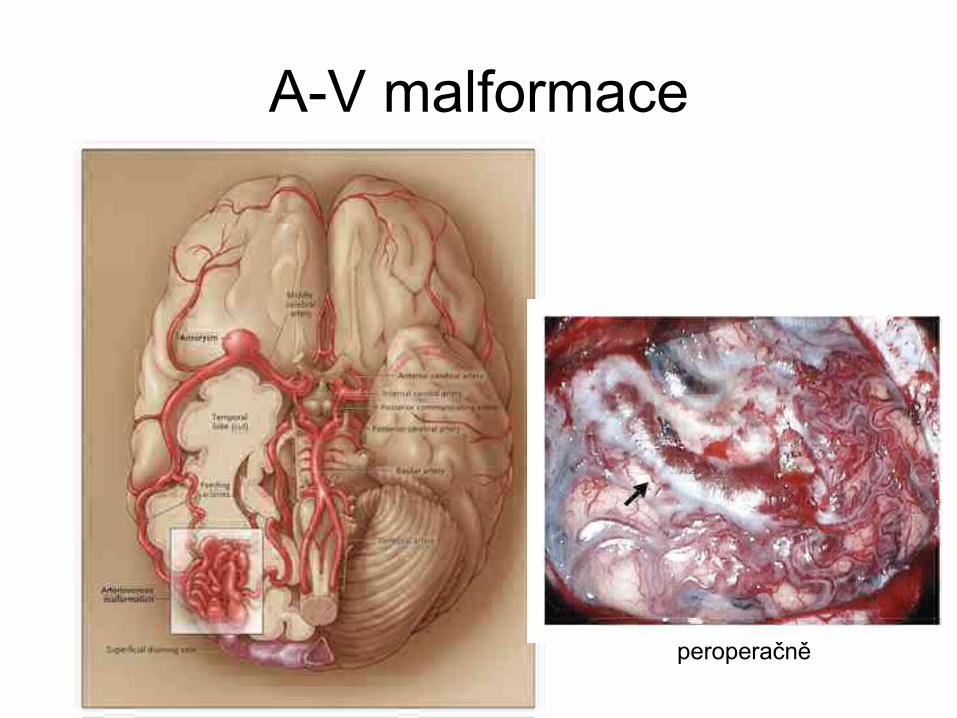
A-V malformace
peroperačně
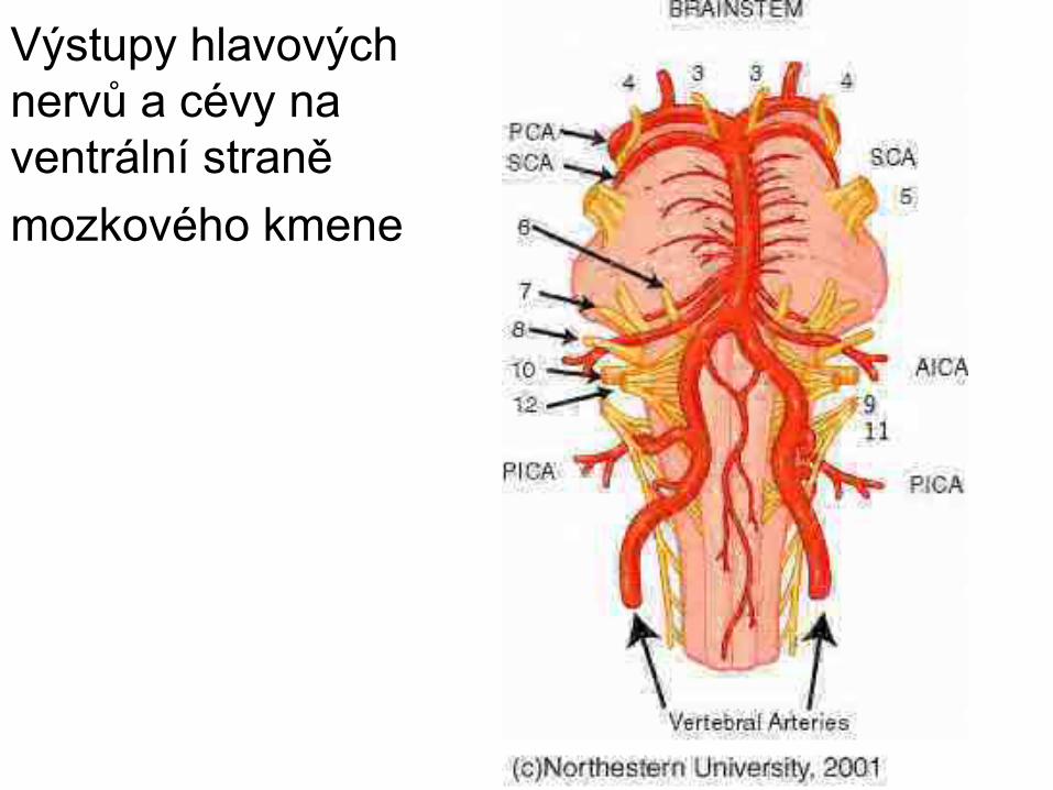
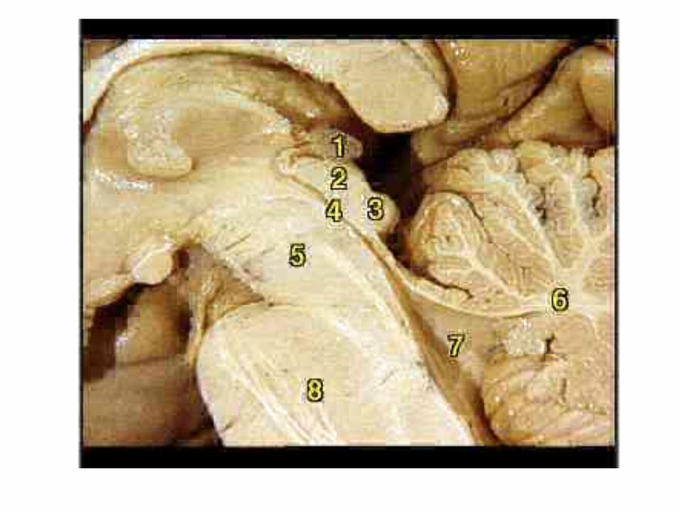
Výstupy hlavových nervů a cévy na ventrální straně mozkového kmene
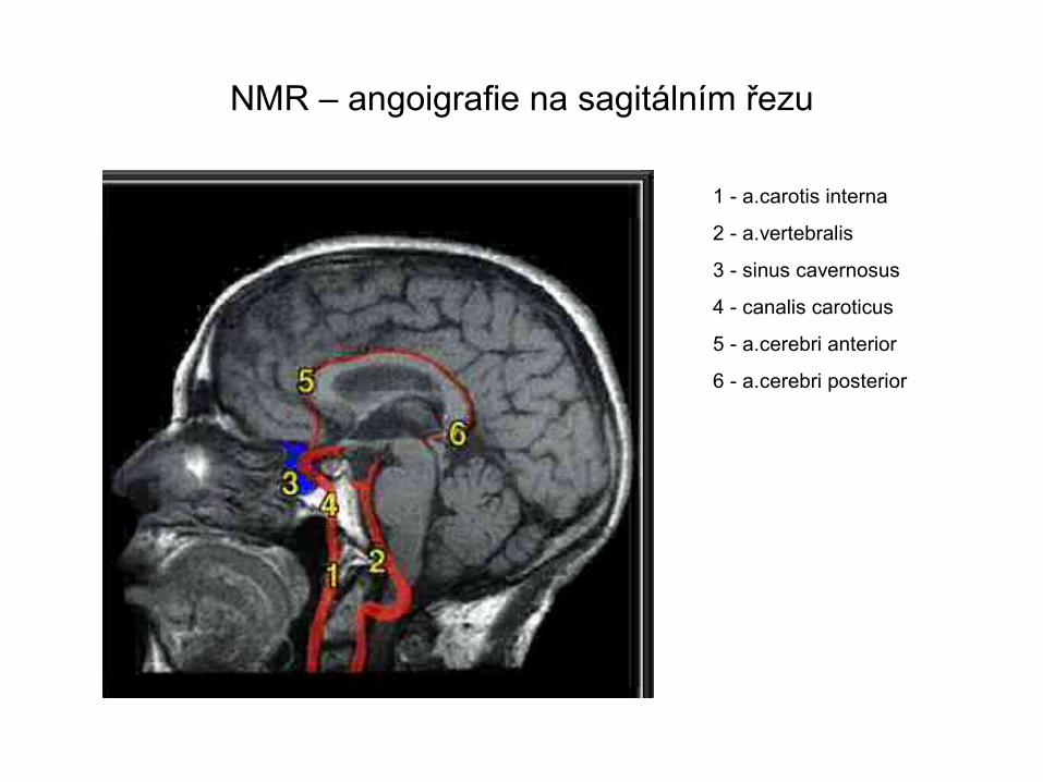
NMR – angoigrafie na sagitálním řezu
1 - a.carotis interna
2 - a.vertebralis
3 - sinus cavernosus
4 - canalis caroticus
5 - a.cerebri anterior
6 - a.cerebri posterior
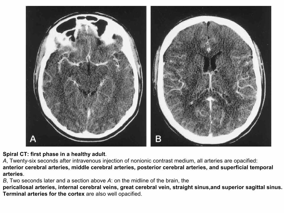
Spiral CT: first phase in a healthy adult.A, Twenty-six seconds after intravenous injection of nonionic contrast medium, all arteries are opacified: anterior cerebral arteries, middle cerebral arteries, posterior cerebral arteries, and superficial temporalarteries.B, Two seconds later and a section above A: on the midline of the brain, thepericallosal arteries, internal cerebral veins, great cerebral vein, straight sinus,and superior sagittal sinus.Terminal arteries for the cortex are also well opacified.
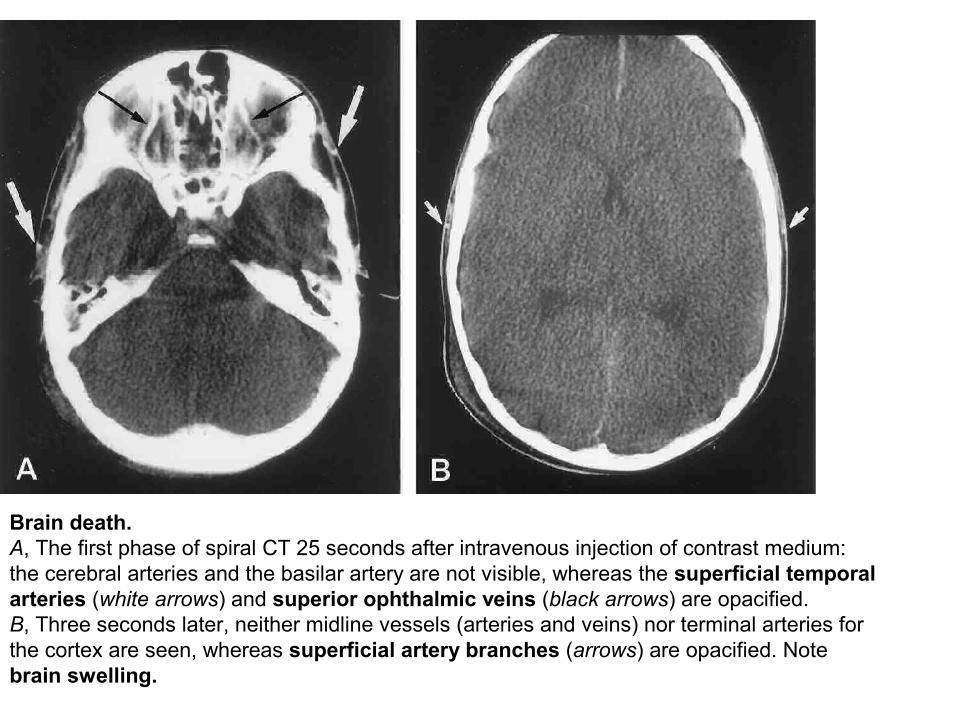
Brain death.A, The first phase of spiral CT 25 seconds after intravenous injection of contrast medium: the cerebral arteries and the basilar artery are not visible, whereas the superficial temporal arteries (white arrows) and superior ophthalmic veins (black arrows) are opacified.B, Three seconds later, neither midline vessels (arteries and veins) nor terminal arteries for the cortex are seen, whereas superficial artery branches (arrows) are opacified. Note brain swelling.

Thomas Willis (1621–1675)

The home of Thomas Willis from 1657 to 1667
.
Oxford, Beam Hall
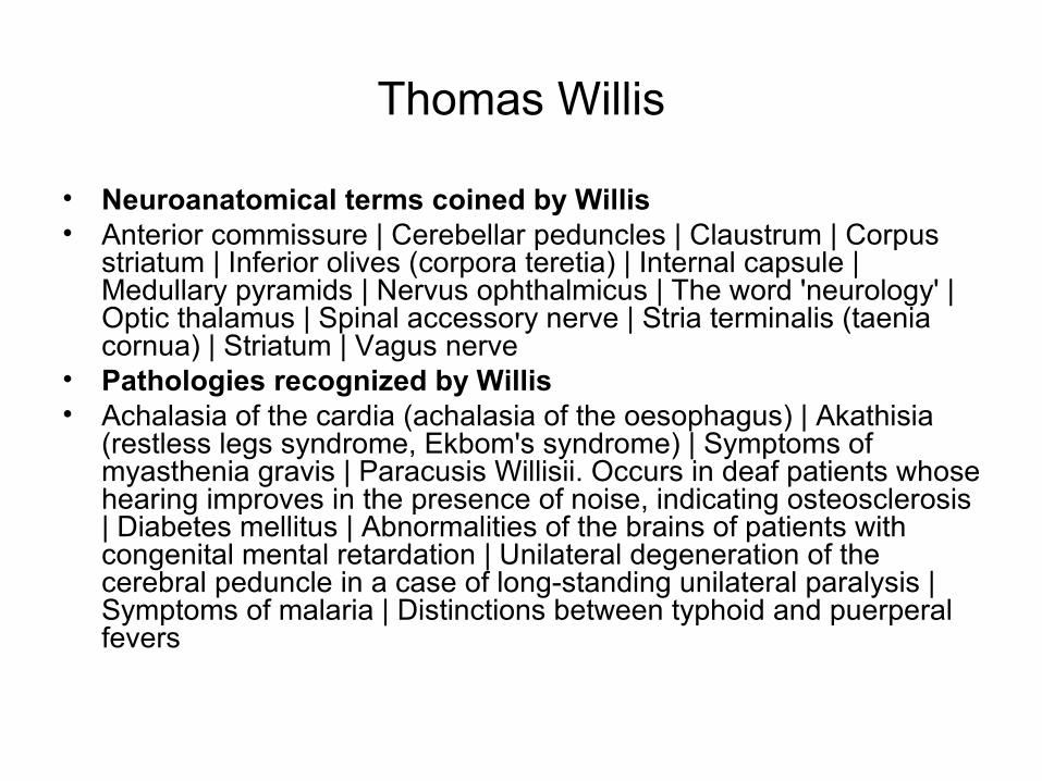
Thomas Willis
• Neuroanatomical terms coined by Willis• Anterior commissure | Cerebellar peduncles | Claustrum | Corpus
striatum | Inferior olives (corpora teretia) | Internal capsule | Medullary pyramids | Nervus ophthalmicus | The word 'neurology' | Optic thalamus | Spinal accessory nerve | Stria terminalis (taenia cornua) | Striatum | Vagus nerve
• Pathologies recognized by Willis• Achalasia of the cardia (achalasia of the oesophagus) | Akathisia
(restless legs syndrome, Ekbom's syndrome) | Symptoms of myasthenia gravis | Paracusis Willisii. Occurs in deaf patients whose hearing improves in the presence of noise, indicating osteosclerosis | Diabetes mellitus | Abnormalities of the brains of patients with congenital mental retardation | Unilateral degeneration of the cerebral peduncle in a case of long-standing unilateral paralysis | Symptoms of malaria | Distinctions between typhoid and puerperal fevers
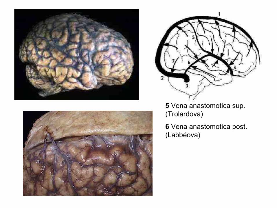
5 Vena anastomotica sup. (Trolardova)
6 Vena anastomotica post. (Labbéova)

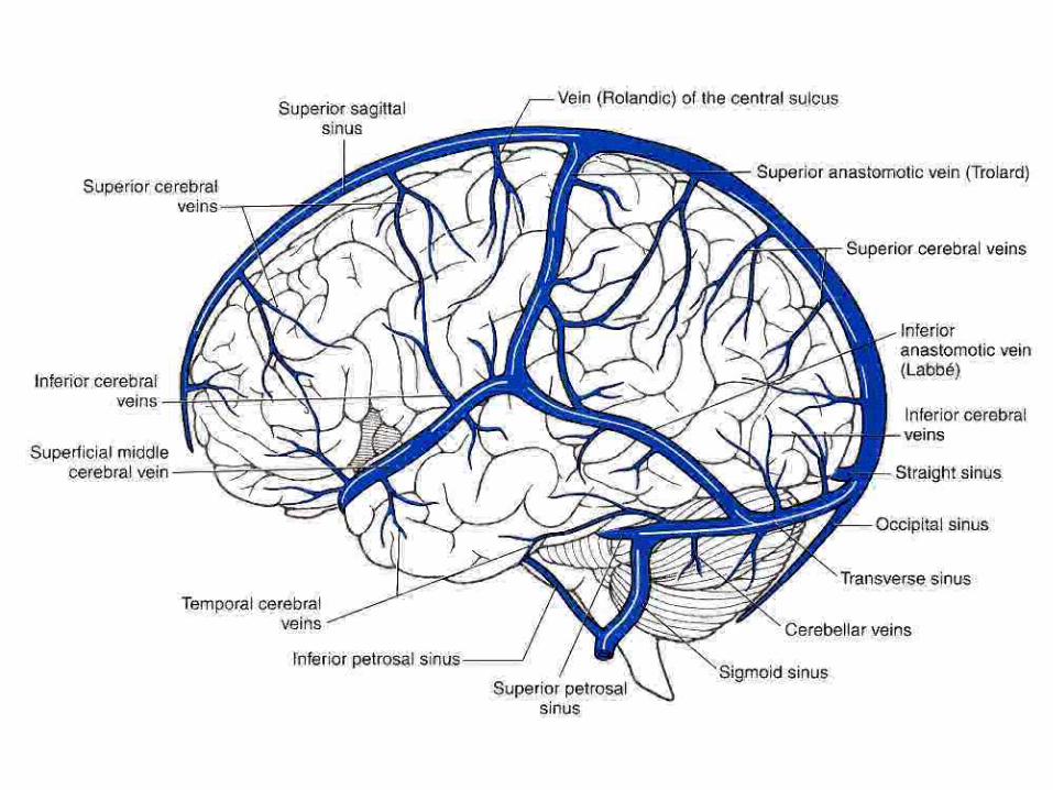
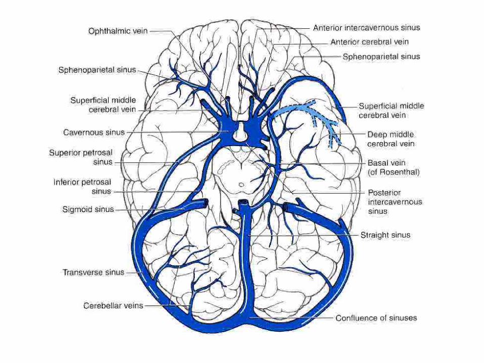
Žíly mozku, četnost trombos
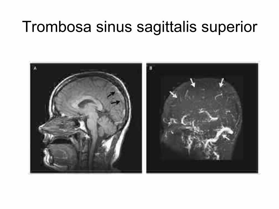
Trombosa sinus sagittalis superior
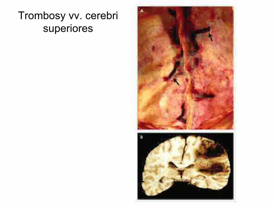
Trombosy vv. cerebri superiores
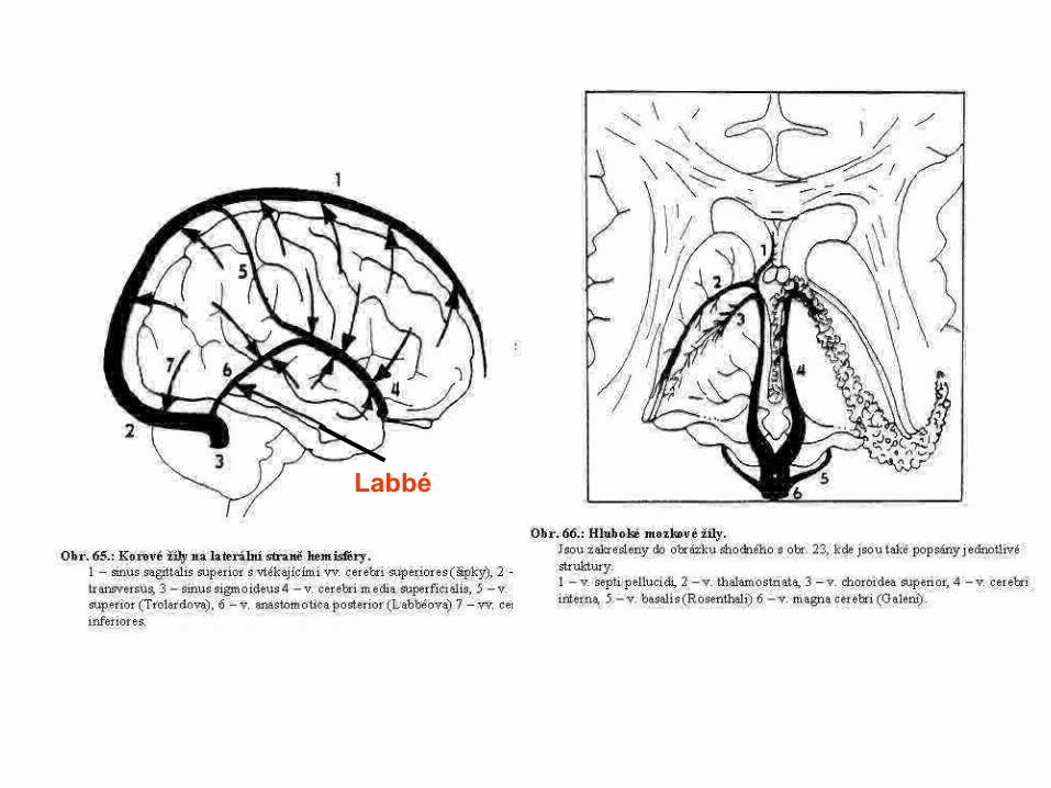
Labbé
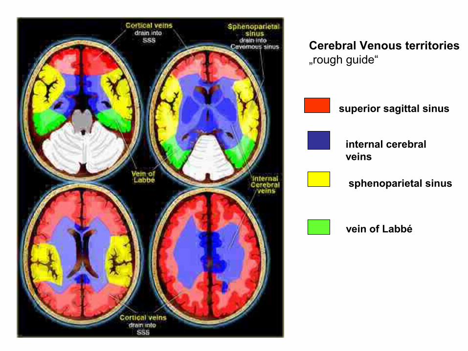
superior sagittal sinus
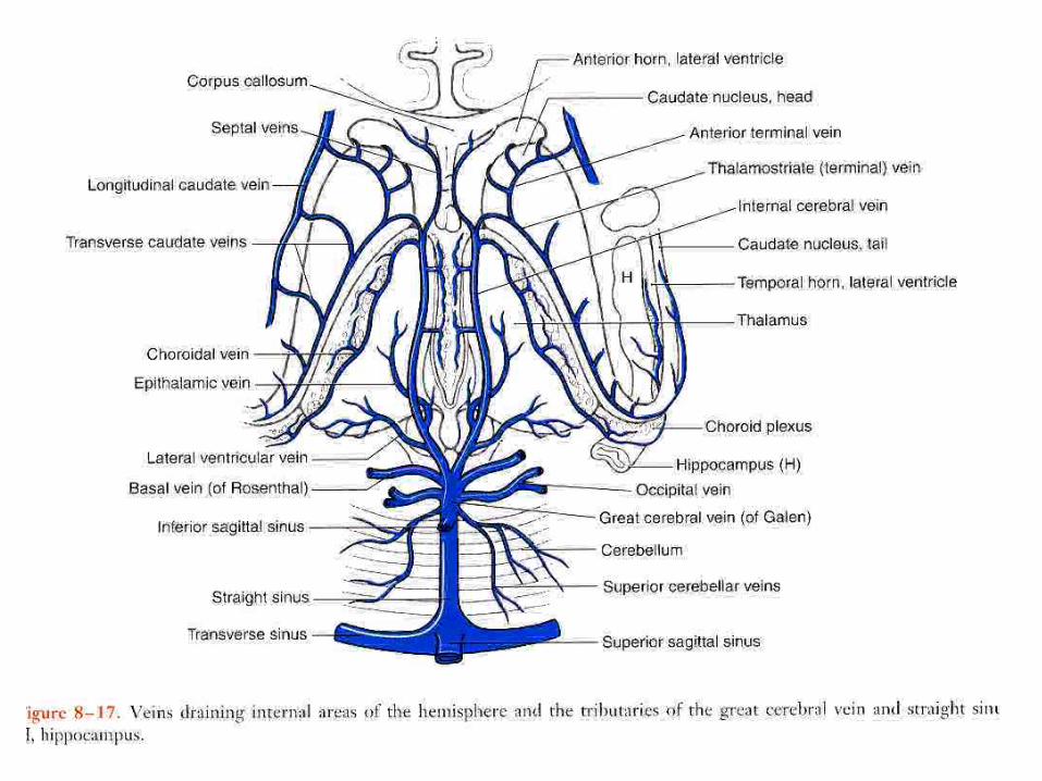
internal cerebral veins
vein of Labbé
sphenoparietal sinus
Cerebral Venous territories„rough guide“
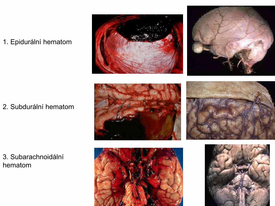
1. Epidurální hematom
2. Subdurální hematom
3. Subarachnoidální hematom

Epi
Subd
Subar
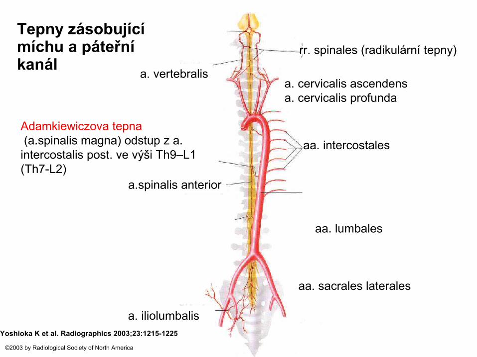
Tepny zásobující míchu a páteřní kanál
Yoshioka K et al. Radiographics 2003;23:1215-1225
©2003 by Radiological Society of North America
a.spinalis anterior
Adamkiewiczova tepna (a.spinalis magna) odstup z a. intercostalis post. ve výši Th9–L1 (Th7-L2)
aa. intercostales
a. iliolumbalis
aa. lumbales
aa. sacrales laterales
rr. spinales (radikulární tepny)
a. vertebralisa. cervicalis ascendensa. cervicalis profunda
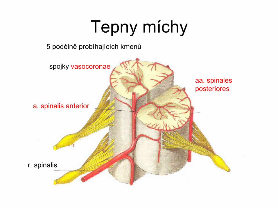
Tepny míchy
spojky vasocoronae
5 podélně probíhajících kmenů
r. spinalis
a. spinalis anterior
aa. spinales posteriores
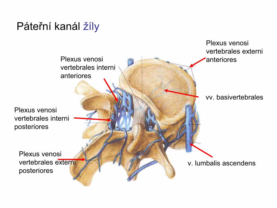
Páteřní kanál žílyPlexus venosi vertebrales externi anterioresPlexus venosi
vertebrales interni anteriores
Plexus venosi vertebrales interni posteriores
v. lumbalis ascendensPlexus venosi vertebrales externi posteriores
vv. basivertebrales
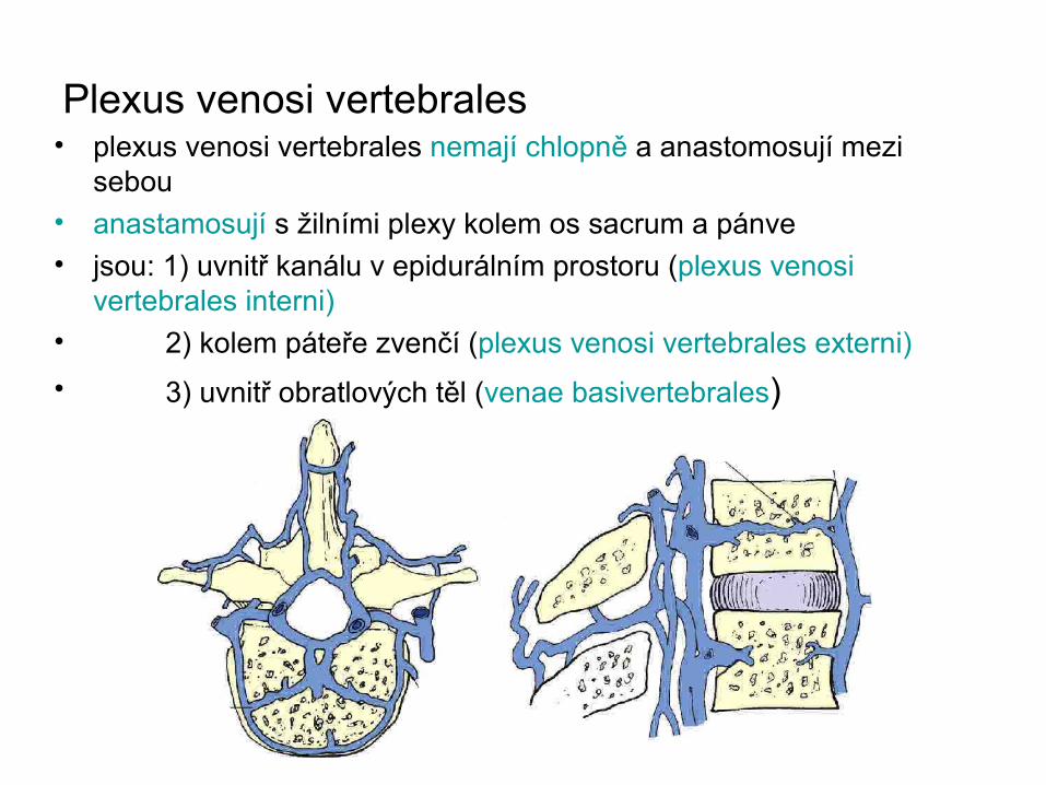
Plexus venosi vertebrales• plexus venosi vertebrales nemají chlopně a anastomosují mezi
sebou• anastamosují s žilními plexy kolem os sacrum a pánve• jsou: 1) uvnitř kanálu v epidurálním prostoru (plexus venosi
vertebrales interni)• 2) kolem páteře zvenčí (plexus venosi vertebrales externi)• 3) uvnitř obratlových těl (venae basivertebrales)
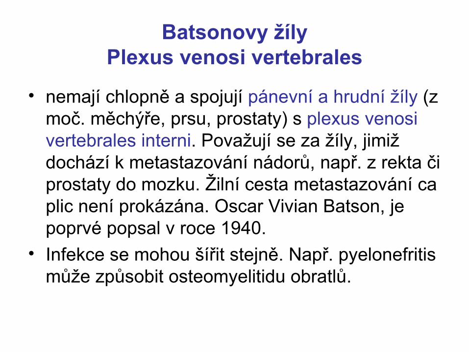
Batsonovy žílyPlexus venosi vertebrales
• nemají chlopně a spojují pánevní a hrudní žíly (z moč. měchýře, prsu, prostaty) s plexus venosi vertebrales interni. Považují se za žíly, jimiž dochází k metastazování nádorů, např. z rekta či prostaty do mozku. Žilní cesta metastazování ca plic není prokázána. Oscar Vivian Batson, je poprvé popsal v roce 1940.
• Infekce se mohou šířit stejně. Např. pyelonefritis může způsobit osteomyelitidu obratlů.


Mozkomíšní mokMozkomíšní mokTvorba v plexus choroideusTvorba v plexus choroideus
Komory a spatium subarachnoidale 140 mlKomory a spatium subarachnoidale 140 mlMechanická opora mozku („plave“)Mechanická opora mozku („plave“)
Chemická komunikace v CNS (neurony- mok-stěna Chemická komunikace v CNS (neurony- mok-stěna komor– neurony)komor– neurony)
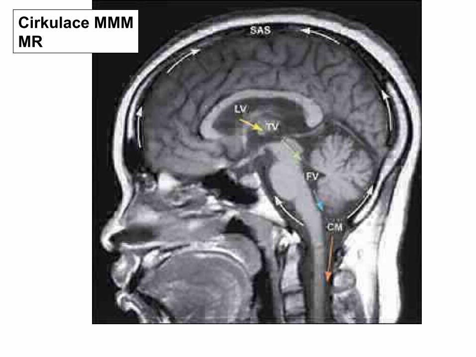
Cirkulace MMMMR
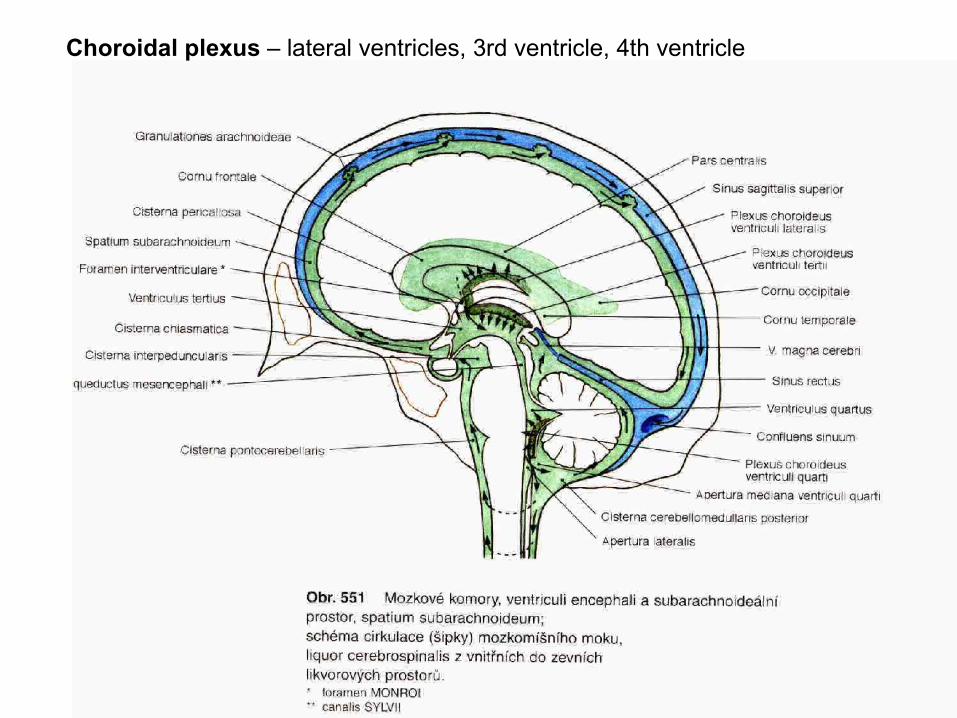
Choroidal plexus – lateral ventricles, 3rd ventricle, 4th ventricle

Granulationes arachnoidales
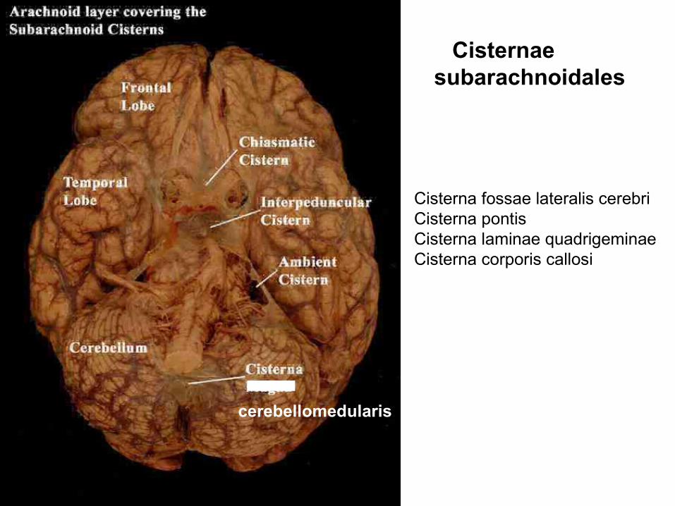
Cisternae subarachnoidales
cerebellomedularis
Cisterna fossae lateralis cerebriCisterna pontisCisterna laminae quadrigeminaeCisterna corporis callosi
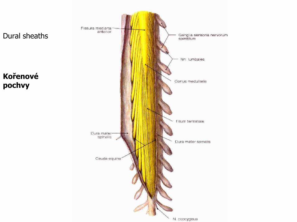
Dural sheaths
Kořenové pochvy
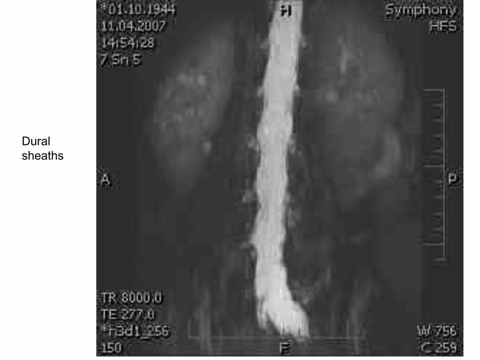
Dural sheaths
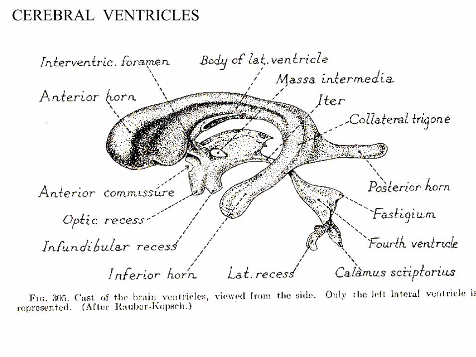
CEREBRAL VENTRICLES

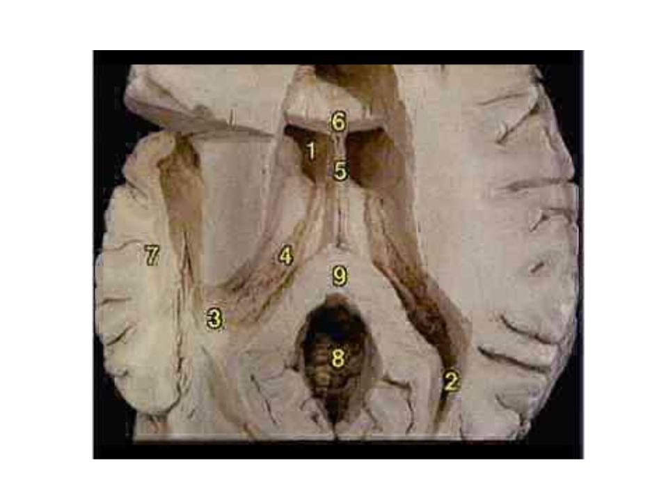
Cornu frontale ventriculi lateralis
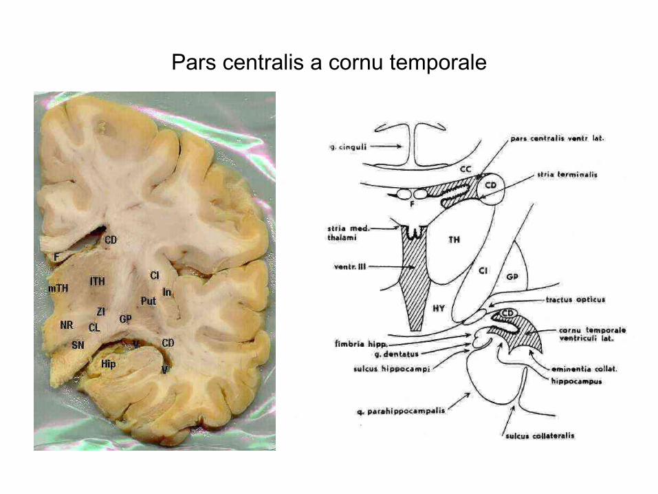
Pars centralis a cornu temporale
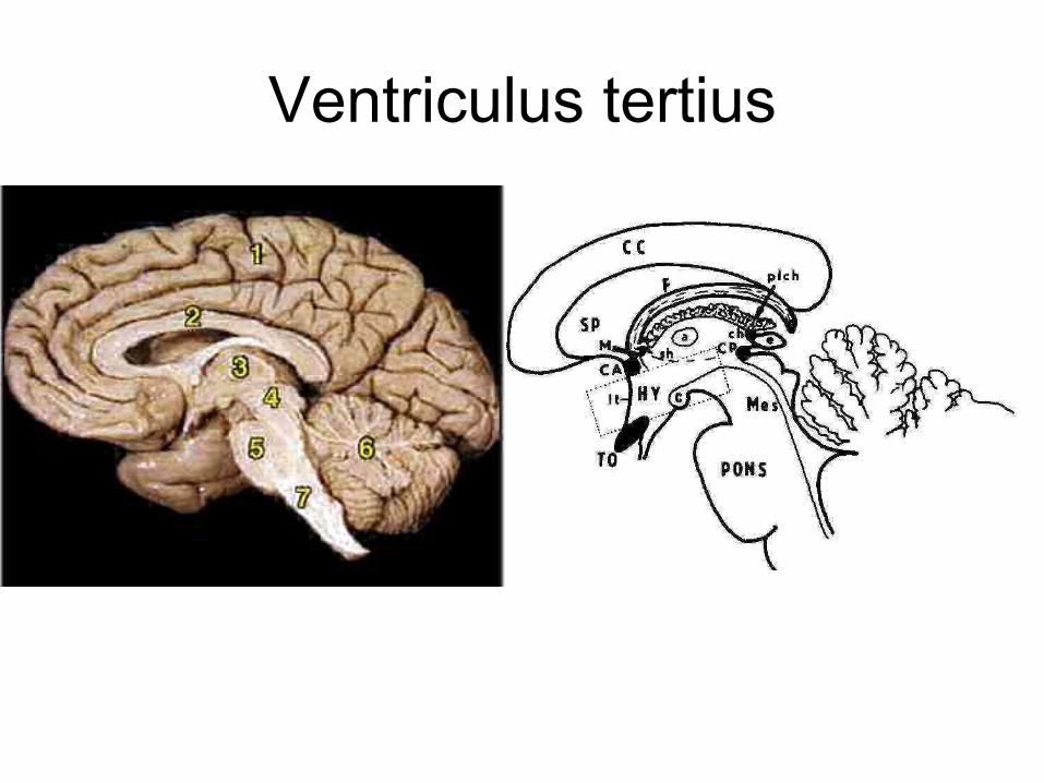
Ventriculus tertius
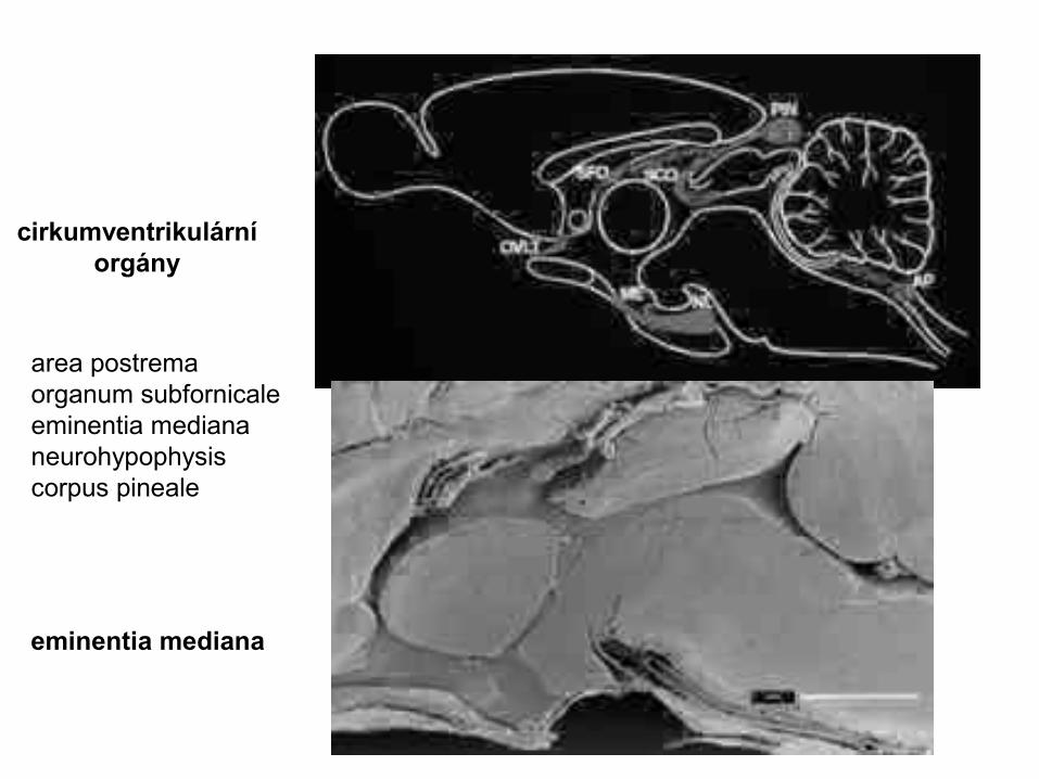
cirkumventrikulární orgány
eminentia mediana
area postremaorganum subfornicaleeminentia mediananeurohypophysiscorpus pineale

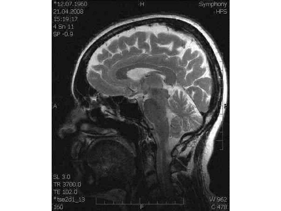
MRI – T2

MRI – T2
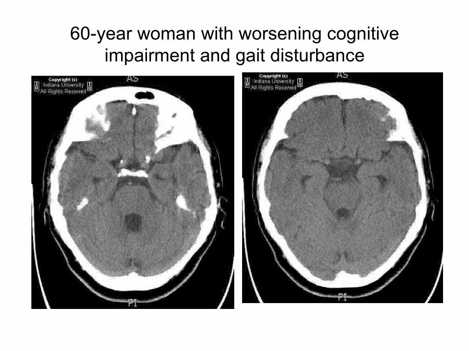
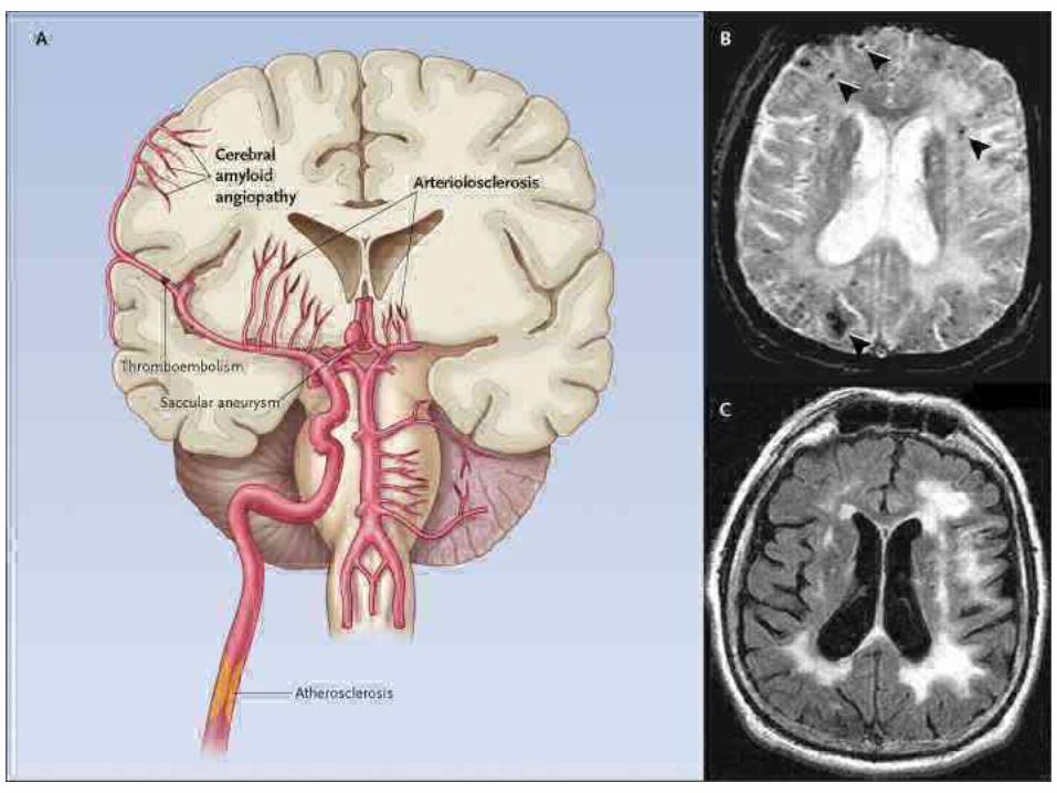
60-year woman with worsening cognitive impairment and gait disturbance
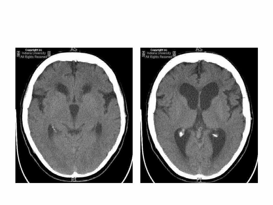
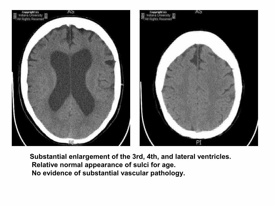
Substantial enlargement of the 3rd, 4th, and lateral ventricles. Relative normal appearance of sulci for age. No evidence of substantial vascular pathology.

• Classical clinical triad of dementia, gait disturbance, and urinary incontinence is seen with normal pressure hydrocephalus.
• Symptoms result from distortion of white matter by distended ventricles. • Patients commonly have a history of prior SAH or meningeal infection. • Gradient between ventricular system and subarachnoid space due to
incomplete subarachnoid block. • Radiographic key: Diffuse ventriculomegaly out of proportion to sulcal
prominence. • Not a radiographic diagnosis. Diagnosis made by improvement of
symptoms after shunting. • Radioisotope cisternogram shows early entry into the lateral
ventricles with persistence at 24-48 hours and delayed ascent to parasagittal regions.
• Flow void can be seen through the aqueduct of Sylvius on MR due to increased flow velocity
Normotensní hydrocephalusNormal pressure hydrocephalus
Použité zdroje sources
• Petrovický, Anatomie III• http://www.radiologyassistant.nl/en/484b8328cb6b2• http://www.nejm• http://www.auntminnie.com/index.asp
?Sec=edu• jiné webové zdroje a soukromý archiv


