effects of arterial ischemia and venous congestion on the lumbar nerve root in dogs
TRANSCRIPT
Effects of Arterial Ischemia and Venous Congestion on The Lumbar NerveRoot in Dogs
Shigeru Kobayashi,1 Kenichi Takeno,1 Tsuyoshi Miyazaki,1 Masafumi Kubota,2 Seichior Shimada,2 Takafumi Yayama,1
Kenzo Uchida,1 Eiki Normura,3 Erisa Mwaka,1 Hisatoshi Baba1
1Faculty of Medical Sciences, Department of Orthopaedics and Rebhailitation Medicine, The University of Fukui, 23-3 Shimozizuki, Matsuoka,Eiheiji, Fukui, Japan, 2Faculty of Medical Sciences, Department of Rehabilitation Medicine, The University of Fukui, Fukui, Japan, 3Department ofOrthopedic Surgery, Kawasaki Municipal Hospital, Kawasaki, Japan
Received 2 November 2007; accepted 3 April 2008
Published online 5 June 2008 in Wiley InterScience (www.interscience.wiley.com). DOI 10.1002/jor.20696
ABSTRACT: The development of radiculopathy in patients with lumbar canal stenosis is thought to be closely related to intraradicularedema resulting from compression. However, there is little agreement as to question which is more essential for intermittent claudication:ischemia or congestion. The aim of the present experimental investigation was to examine the effect of ischemia and congestion on the nerveroot using dogs. Theaorta was clampedas an ischemiamodel of the nerve root and the inferior vena cava was clamped as a congestion modelatthe sixth costal level for 30 min using forceps transpleurally. Measurements of blood flow, partial oxygen pressure, and conduction velocity inthe nerve root were repeated over a period of 1 h after release of clamping. Finally, we examined the status of intraradicular blood–nervebarrier under fluorescence and transmission electron microscope. Immediately after clamping of the inferior vena cava, the central venouspressure increased by about four times and marked extravasation of protein tracers was induced in the lumbar nerve root. Blood flow, partialoxygen pressure, and conduction velocity of the nerve root were more severely affected by aorta clamp, but this ischemia model did not showany intraradicular edema. The blood–nerve barrier in the nerve root was more easily broken by venous congestion than by arterial ishemia.In conclusion, venous congestion may be an essential factor precipitating circulatory disturbance in compressed nerve roots and inducingneurogenic intermittent claudication. � 2008 Orthopaedic Research Society. Published by Wiley Periodicals, Inc. J Orthop Res 26:1533–
1540, 2008
Keywords: nerve root; lumbar canal stenosis; intermittent claudication; blood flow; venous congestion
It is well known that symptom of lumbar canalstenosis, neurogenic intermittent claudication, occurswhen patients are standing or walking.1–4 Althoughthere have been many reports about neurogenic inter-mittent claudication associated with lumbar canalstenosis, the pathogenesis of claudication has not yetbeen completely clarified. Blau and Logue5 postulatedthat neurogenic intermittent claudication might beevoked with ischemic neuritis of the cauda equina.Evans6 advocated exercise-induced ischemia as thecause of intermittent claudication, which is the charac-teristic syndrome of this disease. They supposed thatreduced blood flow in the spinal nerve roots has beendemonstrated during exercise, and this might contributeto the pathogenesis of neurogenic claudication. Ehniet al.7 stressed the postural changes in the spinal canalon standing. He demonstrated a myelographic block inthe lordotic position, but flexion permitted the contrastmedium to pass. Yamada et al.8 reported the importanceof intermittent constriction of the cauda equina associ-ated with postural change. They thought that theligamentous fluvum had a significant role in dynamicnarrowing of the canal. Kavanaugh et al.9 reported thatthe increase of cerebrospinal fluid pressure below theblocked area might obstruct venous return and be causeof anoxia of the cauda equina. Verbiest10 thought thatthis theory deserved further consideration, because hecommonly found enlargement of the epidural venousplexus during decompression of spinal canal stenosis.
Basic research investigations in this aspect, however,are few, and details of circulatory problems in the nerveroots are not as well understood as in the peripheralnerve. The development of neurogenic intermittentclaudication in patients with lumbar canal stenosis isthought to be closely related to intraradicular edemaresulting from compression.11–13 However, the basicpathophysiology of circulatory disturbance induced byischemia and congestion is not fully understood. The aimof the present experimental investigation was to exam-ine the effect of ischemia and congestion on the nerveroot.
MATERIALS AND METHODSThe experiment was carried out under the control of the localanimal ethics committee in accordance with the guidelineson animal experiments in our university, Japanese govern-ment animal protection and management law, and Japanesegovernment notification on feeding and safekeeping of animals.Forty adult dogs, weighing 7 to 15 kg, were anesthetizedwith intramuscular injection of 3 mL of Ketalar (Ketamine50 mg/mL; Warner-Lumbert, Morris Plains, NJ) and venti-lated on a respirator under general anesthesia (O2: 3 mL/min,N2O: 3 mL/min, Halothane: 1.5 mL/min). The femoral arteryand vein were canulated, and arterial blood pressure andcentral venous pressure (CVP) were monitored in all animalsthroughout the experiment. A tip of the arterial catheter wasplaced in the femoral artery and a tip of the venous catheterwas inserted into the abdominal vena cava through thefemoral vein. Animals were maintained at constant physio-logic levels during experiment. Each animal was placed in theprone position on a flame. At first, the sixth and seventhlumbar laminae were removed, and the seventh lumbar nerveroot was exposed widely. Next, the sixth rib was removed andthe transpleural approach was used to access the aorta andinferior vena cava. The aorta was clamped as an ischemia
JOURNAL OF ORTHOPAEDIC RESEARCH NOVEMBER 2008 1533
Correspondence to: Shigeru Kobayashi (T: þ81-776-61-8383;F: þ81-776-61-8125; E-mail: [email protected])
� 2008 Orthopaedic Research Society. Published by Wiley Periodicals, Inc.

model of the nerve root (Fig. 1A) and the inferior vena cava wasclamped as a congestion model (Fig. 1B) at the sixth costallevel for 30 min using forceps transpleurally. Measurements ofblood flow, partial oxygen pressure (PO2), and conductionvelocity in the nerve root were repeated over a period of 30 minafter release of clamping.
Measurement of Intraradicular Blood FlowTo measure the intraradicular blood flow in 10 animals, atissue blood flow meter (DHM-3001, M.T. Giken Co., Tokyo,Japan) was employed. A small electrode with a diameter of200 mm (MHD-60: M.T. Giken Co.) was inserted into the dorsalroot at midpoint between the dural sac and the dorsal rootganglion.14,15 We used a polarized voltage of 600 mV and adirect current of 20 A for 25 s to electrochemically generatehydrogen in the dorsal root. We measured the blood flow twicebefore clamping the aorta (n¼ 5) or vena cava (n¼ 5), toestablish the average intraradicular blood flow in animals.
Measurement of Intraradicular PO2
PO2 in the nerve root was measured by the polarographicalmethod (Model POG-5000S PO2 Meter: M.T. Giken Co.).16,17
After calibration, a PO2 needle-type electrode with a diameterof 10 mm (POE-10N: M.T. Giken Co.) was inserted into thedorsal root at midpoint between the dural sac and the dorsalroot ganglion. After intraradicular PO2 stabilized, we recordedit twice and calculated the average value of the normal statebefore clamping the aorta (n¼ 5) or vena cava (n¼ 5).
Electrophysiological StudyNerve root conduction studies were carried out using anelectromyometer (Neuromatic 2000 DISA electromyographyunit, Dantec Electronics, Bristol, UK) using 10 animals. Thesciatic nerve was exposed at the thigh and stimulated with0.1-ms square-wave voltage pulses at a rate of 2/s using abipolar electrode. The stimulus intensity was adjusted to1.5–2 times of the motor threshold and 20 responses weresummated. The evoked potentials were recorded directly fromthe dorsal root at the entry site of the dural sac and proximal tothe dorsal root ganglion after clamping the aorta (n¼ 5) or venacava (n¼ 5), and then the sensory nerve conduction velocity wascalculated by ‘‘the distance between 2 site (m)/the time lagbetween 2 site (S).
Preparation for Fluorescence and TransmissionElectron Microscopic StudyFinally, we examine the status of the intraradicular blood–nerve barrier under fluorescence and transmission electronmicroscope after injection of Evans blue albumin (EBA) andHorseradish peroxidase (HRP) into the cephalic vein to findout what sort of circulatory disturbance occurred in the nerveroot.11 After clamping of the aorta (n¼ 5) or the vena cava(n¼ 5) for 30 min, EBA (10 mL/kg, molecular weightapproximately 59,000) and HRP (type-II, molecular weightapproximately 43,000; Sigma Co., St. Louis, MO) were injectedintravenously and allowed to circulate for 30 min. EBA wasprepared by mixing 5% bovine albumin (Wako Chemical Co.,Osaka, Japan) with 1% Evans blue (Sigma Chemical Co.). Theanimals were fixed by intraaortal perfusion with 4% paraf-ormaldehyde and 1% glutaraldehyde in 0.15 M cacodylatebuffer, pH 7.2 at 208C. The nerve root sections were resected atthe midpoint between the dural sac and the dorsal rootganglion, and at the outlet of dural sleeve of seventh lumbarnerve root bilaterally. These sections were divided into twogroups. After the nerve root sections fixed with 4% parafor-maldehyde for 24 h, 20 mm-thick sections were mounted with50% aqueous glycerin to be examined under the fluorescencemicroscope at 380 mmW (BX-51, Olympus, Tokyo, Japan). Theother sections were rinsed in 0.05 M Tris–HCl buffer,postfixed at room temperature for 3 h in 2% OsO4 in 0.1 Msodium cacodylate buffer, impregnated with 2% uranylacetate, dehydrated in graded ethanols, and embedded inepoxy resin. For light microscopy, 1–3 mm-thick toluidin blue-stained sections were used. For electron microscopy, ultrathinsections contrasted with uranyl acetate and lead citrate wereexamined a under electron microscope (JSM2000, NipponDenshi, Tokyo, Japan).
Statistical AnalysisFrom the record of each animal in both groups, arterial bloodflow, CVP, intraradicular blood flow, PO2, and conductionvelocity during experiment were determined. Unless other-wise stated, data are presented as the mean� the standarderror of the mean (SEM) of at least five separate experiments.Also, the averaged data were expressed as percentage of theaverage value before clamping of aorta or inferior venacava. Comparison values were performed using a repeated-measures analysis of variance and post hoc (Scheffe) comparedbefore and after clamping of aorta or inferior vena cava. Datewere entered into a database and analyzed by using SPSSstatistical soft-ware, version 14.0.J (SPSS Inc, Chicago, IL). Aprobability of 5% was considered statistically significant.
Figure 1. The aorta was clamped as an ischemic model of thenerve root (A), and the inferior vena cava was clamped as acongestion model (B) at the sixth costal level for 30 minutes usingforceps transplurally. [Color scheme can be viewed in the onlineissue, which is available at http://www.interscience.wiley.com]
1534 KOBAYASHI ET AL.
JOURNAL OF ORTHOPAEDIC RESEARCH NOVEMBER 2008

RESULTSImmediately after aorta clamping, blood pressure in thefemoral artery dropped to 26–40 mmHg and meantime(p<0.05) (Fig. 2A), central venous pressure was slightlyelevated (Fig. 2B). When the vena cava was clamped,central venous pressure increased to about four times ofthe pressure before clamping (p< 0.05) (Fig. 2B) andblood pressure in the femoral artery was reduced by half(p<0.05) (Fig. 2A). After release of clamping, botharterial and venous pressures quickly returned to thepressure before clamping.
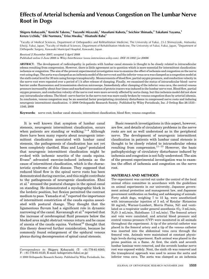
The blood flow in the seventh posterior nerve root dueto aorta and vena cava clamping fall to 50–60% of theblood flow before clamping in the ischemic model(p<0.05) and to about 20% in the congestion model(p<0.05). When the clamp was released, the intra-radicular blood flow in the congestion model was restoredwith in 1 h. The intraradicular blood flow in the ischemicmodel, however, did not recover and stayed at thereduced level (p<0.05) (Fig. 3A and Table 1). Thechanges of PO2 in the nerve root indicated a similartendency to blood flow, 50–60% drop in the ischemicmodel (p< 0.05) and 20–40% drop in the congestionmodel. After release of clamping, intraradicular PO2
recovered completely in both models (Fig. 3B andTable 1). Conduction velocity of the nerve root dimin-ished by 40–50% in the ischemia model (p< 0.05) and10–20% in the congestion model. This drop of conductionvelocity returned almost completely within 1 h afterrelease of clamping (Fig. 3C and Table 1).
After intravenous injection of protein tracers, markedextravasation of the tracers in the seventh lumbar nerveroot bilaterally was induced after 30-min clamping of thevena cava (Figs. 4B and 5C and D) but not by aortaclamping (Figs. 4A and 5A and B). There was noextravasation of EBA, and the blood–nerve barrier wasmaintained in the nerve root after clamping of the aorta(Fig. 4A). After clamping of the vena cava, however,the red fluorescence of EBA was seen outside theendoneurial microvessels and diffusely through out theendoneurial space of the nerve root (Fig. 4B). HRP hadthe same distribution as the EBA tracer. By electronmicroscopy, capillaries of the nerve root were shown tohave an inner diameter of approximately 5 to 10 mm.
After clamping of the aorta, HRP product did not appearin the endoneurial space when it was injected intra-venously (Fig. 5A), and their endotherial cells are linkedby tight junctions (zonulae occludens) showing theexistence of a blood–nerve barrier (Fig. 5B). SomeHRP-labeled vesicles related to paracellular transportwere detected in the endotherial cells. However, thesevesicles did not appear to carry the product away fromcapillary to the endoneurial space. After clamping of thevena cava, the extravasation of the HRP tracer wasobserved in endoneurial spaces between the nerve fibers,especially the perivascular spaces (Fig. 5C). The tightjunction between the endothelial cells appeared to intact.However, the pinocytotic vesicles in the endotheliumappeared to carry the dark-stained HRP product awayfrom the capillay lumen and the product extravasatedinto the endoneurial space (Fig. 5D). This increasedtrancellular transport of the tracer indicates breakdownof the blood–nerve barrier, leading to edema formationin the nerve root.
DISCUSSIONIschemic nerve root injury is a ubiquitous insult thatcan lead to a wide range of neuropathologic consequen-ces, depending on the severity and duration of theischemic event. Ischemic injury to nerve roots predom-inantly causes demyelination, although prolonged ische-mia can also interfere with axonal transport, leadingto axonal damage and Wallerian degeneration of thenerve fiber.18–21 Vascular damage and fibrosis arecommon findings within the spinal canal and interver-tebral foramina, and such vascular damage is signifi-cantly related to the severity of degenerative discdisease. Disc protrusion may lead to compression ofepidural veins and dilation of noncompressed veins.Cooper et al.22 noted a significant relationship betweenevidence of venous obstruction, intraneurial and peri-neurial fibrosis, and neural atrophy. Fibrosis mayfurther impede nutrient transfer to endoneurial fibers,as well as predisposing to nerve stretch injury. Thepresent study assessed the influence of arterial ische-mia and venous congestion resulting from obstruction ofblood flow without nerve root compression on intra-radicular blood flow and radicular function. As a result,
Figure 2. Changes in blood pressureafter aorta or inferior vena cava clamp.(A) Changes in blood pressure ofthe femoral artery. (B) Changes incentral venous pressure (CVP) (Sheffe;*#<0.05).
ARTERIAL ISCHEMIA AND VENOUS CONGESTION EFFECTS ON THE LUMBAR NERVE ROOT IN DOGS 1535
JOURNAL OF ORTHOPAEDIC RESEARCH NOVEMBER 2008
it was confirmed that nerve root ischemia had a moreserious influence on blood flow, PO2, and conductionvelocity than nerve root congestion. After 30 min ofnerve root ischemia, recovery occurred with reperfu-sion, but longer ischemic periods will cause a permanenteffect on radicular function due to oxygen deficiency.When changes of the femoral arterial and centralvenous pressures were monitored after obstruction ofblood flow, both the arterial and venous pressuresdecreased after aortic blockade and the arterial pres-sure increased slightly after obstruction of the inferiorvena cava. However, the central venous pressureshowed an approximately fourfold increase immediatelyafter obstruction of the inferior vena cava, and thissudden increase in venous pressure could have amarked influence on the capillary pressure in the nerveroots. Usubiaga et al.23 demonstrated that clamping ofthe vena cava can be used experimentally to increasethe systemic venous pressure. The same maneuver alsoproduces congestion of the epidural veins and increasesthe epidural pressure,24 but they did not describe thechanges of nerve root circulation.
The arachnoid membrane acts as a diffusion barrierfor the nerve root, and the blood–nerve barrier is alsocreated by the vascular endothelial cells of the endoneu-rial microvessels. These nerve root barriers protect andmaintain the nerve fibers in a constant environment. Thecapillary vessels of the nerve roots are lined byendothelial cells that contain only a few pinocyticvesicles and are bound by tight junctions to form theblood–nerve barrier. Protein tracers that are injectedintravenously do not normally leak out of the vessels dueto this barrier.11,13 When arterial ischemia was induced,protein tracers remained in the blood vessels, indicatingmaintenance of the integrity of the blood–nerve barrier
(Figs. 4A and 5A and B). On the other hand, venouscongestion disrupted the blood–nerve barrier, and therewas extravasation and edema in the nerve roots (Figs. 4Band 5C and D). Thus, the blood–nerve barrier thatregulates vascular permeability in the nerve root seemsto be susceptible to congestion, which raises the intra-vascular pressure rather than to ischemia, whichdecreases the pressure.
An experiment performed by Olmarker25 demon-strated that the capillaries and venules of the nerve rootcould be occluded by mild compression of around 30–40 mmHg. Takahashi et al.26 found that the epiduralpressure is only 15 to 18 mmHg during lumbar flexion inLSCS patients, but reaches 80–100 mmHg duringlumbar extension. The epidural pressure increases withwalking, and the patient then stops walking because ofleg pain and/or claudication. The pressure decreasesimmediately after walking is stopped, and leg pain thensubsides. There is a repeated pattern of increasing anddecreasing pressure during walking. Although, thesepressure changes are not great enough to disturb arterialblood flow, the epidural venous system may becomecongested if the pressure is higher than 10–30 mmHg.Accordingly, in patients with symptomatic stenosis, thesubarachnoid space is obstructed and the cauda equinanerve roots undergo strangulation by the diffusionbarrier in the arachnoid membrane, whereas the nutri-tional pathway to the nerve roots from the CSF is blockedand intermittent increases of compression may damagethe nerve tissue.
It is known that sites of nerve root compression byspinal canal stenosis frequently show gadoliniumenhancement on MR images, suggesting that there isbreakdown of the blood–nerve barrier and edema of thenerve root.11–13,27,28 In lumbar spinal stenosis associated
Figure 3. Changes in intraradicularblood flow (A), PO2 (B), and conductionvelocity (C) after aorta or inferiorvena cava clamp. The avereaged datawere expressed as percentage of theaverage value before clamping. (Sheffe;*#<0.05).
1536 KOBAYASHI ET AL.
JOURNAL OF ORTHOPAEDIC RESEARCH NOVEMBER 2008
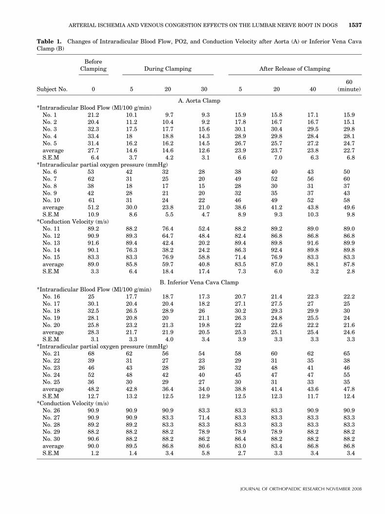
Table 1. Changes of Intraradicular Blood Flow, PO2, and Conduction Velocity after Aorta (A) or Inferior Vena CavaClamp (B)
Subject No.
BeforeClamping During Clamping After Release of Clamping
0 5 20 30 5 20 4060
(minute)
A. Aorta Clamp*Intraradicular Blood Flow (Ml/100 g/min)
No. 1 21.2 10.1 9.7 9.3 15.9 15.8 17.1 15.9No. 2 20.4 11.2 10.4 9.2 17.8 16.7 16.7 15.1No. 3 32.3 17.5 17.7 15.6 30.1 30.4 29.5 29.8No. 4 33.4 18 18.8 14.3 28.9 29.8 28.4 28.1No. 5 31.4 16.2 16.2 14.5 26.7 25.7 27.2 24.7average 27.7 14.6 14.6 12.6 23.9 23.7 23.8 22.7S.E.M 6.4 3.7 4.2 3.1 6.6 7.0 6.3 6.8
*Intraradicular partial oxygen pressure (mmHg)No. 6 53 42 32 28 38 40 43 50No. 7 62 31 25 20 49 52 56 60No. 8 38 18 17 15 28 30 31 37No. 9 42 28 21 20 32 35 37 43No. 10 61 31 24 22 46 49 52 58average 51.2 30.0 23.8 21.0 38.6 41.2 43.8 49.6S.E.M 10.9 8.6 5.5 4.7 8.9 9.3 10.3 9.8
*Conduction Velocity (m/s)No. 11 89.2 88.2 76.4 52.4 88.2 89.2 89.0 89.0No. 12 90.9 89.3 64.7 48.4 82.4 86.8 86.8 86.8No. 13 91.6 89.4 42.4 20.2 89.4 89.8 91.6 89.9No. 14 90.1 76.3 38.2 24.2 86.3 92.4 89.8 89.8No. 15 83.3 83.3 76.9 58.8 71.4 76.9 83.3 83.3average 89.0 85.8 59.7 40.8 83.5 87.0 88.1 87.8S.E.M 3.3 6.4 18.4 17.4 7.3 6.0 3.2 2.8
B. Inferior Vena Cava Clamp*Intraradicular Blood Flow (Ml/100 g/min)
No. 16 25 17.7 18.7 17.3 20.7 21.4 22.3 22.2No. 17 30.1 20.4 20.4 18.2 27.1 27.5 27 25No. 18 32.5 26.5 28.9 26 30.2 29.3 29.9 30No. 19 28.1 20.8 20 21.1 26.3 24.8 25.5 24No. 20 25.8 23.2 21.3 19.8 22 22.6 22.2 21.6average 28.3 21.7 21.9 20.5 25.3 25.1 25.4 24.6S.E.M 3.1 3.3 4.0 3.4 3.9 3.3 3.3 3.3
*Intraradicular partial oxygen pressure (mmHg)No. 21 68 62 56 54 58 60 62 65No. 22 39 31 27 23 29 31 35 38No. 23 46 43 28 26 32 48 41 46No. 24 52 48 42 40 45 47 47 55No. 25 36 30 29 27 30 31 33 35average 48.2 42.8 36.4 34.0 38.8 41.4 43.6 47.8S.E.M 12.7 13.2 12.5 12.9 12.5 12.3 11.7 12.4
*Conduction Velocity (m/s)No. 26 90.9 90.9 90.9 83.3 83.3 83.3 90.9 90.9No. 27 90.9 90.9 83.3 71.4 83.3 83.3 83.3 83.3No. 28 89.2 89.2 83.3 83.3 83.3 83.3 83.3 83.3No. 29 88.2 88.2 88.2 78.9 78.9 78.9 88.2 88.2No. 30 90.6 88.2 88.2 86.2 86.4 88.2 88.2 88.2average 90.0 89.5 86.8 80.6 83.0 83.4 86.8 86.8S.E.M 1.2 1.4 3.4 5.8 2.7 3.3 3.4 3.4
ARTERIAL ISCHEMIA AND VENOUS CONGESTION EFFECTS ON THE LUMBAR NERVE ROOT IN DOGS 1537
JOURNAL OF ORTHOPAEDIC RESEARCH NOVEMBER 2008

with intermittent claudication, Jinkins et al.12 firstreported gadolinium enhancement of the caudia equinaabove the level of stenosis. Our previous experimentalstudy showed that the blood–nerve barrier of the nerveroot is disrupted and intraradicular edema is producedby acute compression with a microsurgical clip at morethan 15 g of force for 1 h11 or by chronic compression dueto wrapping the nerve root for at least 1 month with asilastic tube slightly larger than the nerve root diame-ter.29 In patients with compression radiculopathy, totalcircumferential compression of the cauda equina asso-ciated with closure of the subarachnoid space is assumedto block all routes for the supply of nourishment andremoval of waste via the CSF, thereby triggering variousdisorders in combination with chemical factors releasedby inflammatory cells.30 Elevation of the capillary pres-sure induced by venous stasis is thought to cause intra-radicular edema and the inflammatory responseproduced by compression, as well as mechanical damageto the blood–nerve barrier, because venous blood flow isstopped by compression at a very low pressure. As aresult, the subarachnoid space is occluded, and conges-tion as well as nerve fiber degeneration occurs in thecauda equina. Efflux of excess fluid into the subarach-noid space is impaired by breakdown of the blood–nervebarrier, leading to an increase of endoneurial pres-sure.31,32 Although this pressure increase is reversible, acompartment syndrome may occur in the cauda equinaat the site of stenosis that disturbs blood flow33,34 andaxonal flow,19,20 provoking ectopic discharges or con-duction disturbance,35,36 which is essentially responsiblefor neurogenic intermittent claudication in nerve fiberswith chronic damage. Ikawa et al.37 demonstrated thatectopic firing was elicited by venous stasis in a rat model
Figure 4. Transverse sections of the nerve root seen under afluorescence microscope. (A) Ischemia model, EBA emits a brightred fluorescence, in clear contrast to the green fluroescence of thenerve tissue. After intravenous injection of EBA, EBA was limitedinside the blood vessels, and the blood–nerve barrier was main-tained. (B) Congestion model. EBA emits a bright red fluorescence,which leaked outside the blood vessles, and intraradicular edemawas seen under a fluorescent miscrscope.
Figure 5. Transverse sections of thecapillary in the neve root seen under anelectron microscope. (A,B) Ischemiamodel. After intravenous injection ofHRP, the tracer was blocked at the tightjunction to extravasate. The pinocytoticvesicles contain the reaction productseen in the endothelial cells. However,HRP is not observed in the endoneurialspace. (C,D) Congestion model. Afterintravenus injection of HRP, the elec-tron-dense reaction product of HRPpassed through endothelial cells andentered into the endoneurial space. Inthe endothelial cell of the capillary,pinocytotic vesicles containing the reac-tion product, about 50–70 nm in diam-eter, are thought to move toward theperivascular (endoneurial) space byvesicular transport. However, HRPwas blocked at the tight junction toestravasate. Abbreviations: A, arach-noid membrane; D, dura matter; E,endothelial cell; En, endoneurial space;L, capillary lumen; P, pinocytoticvesicle; T, tight junction.
1538 KOBAYASHI ET AL.
JOURNAL OF ORTHOPAEDIC RESEARCH NOVEMBER 2008

of lumbar canal stenosis. Thus, venous congestion maybe an essential factor precipitating circulatory disturb-ance in compressed nerve roots and inducing neurogenicintermittent claudication.
ACKNOWLEDGMENTSThe submitted manuscript does not contain information aboutmedical devices or drugs. No benefits in any form have beenreceived or will be received from a commercial party relateddirectly or indirectly to the subject of this article. Mr. NaruoYamashita provided expert help with the photography. Theauthors would like to thank Mrs. Kyoko Shimokawa, Ms. MikaOsaki, and Ms.Yukiko Horiuchi for their dedicated assistancein this study.
REFERENCES1. Verbiest H. 1954. A radicular syndrome from developmental
narrowing of the lumbar vertebral canal. J Bone Joint Surg Br36:230–237.
2. Verbiest H. 1975. Pathomorphological aspects of develop-mental lumbar stenosis. Orthop Clin North Am 6:177–196.
3. Schatzker J, Pennel GF. 1969. Spinal stenosis, a cause ofcauda equina compression. J Bone Joint Surg Br 50:606–618.
4. Kirkaldy-Willis WH, Paine KWE, et al. 1974. Lumbar canalstenosis. Clin Orthop 99:30–50.
5. Blau JN, Logue V. 1961. The natural history of intermittentclaudication of the cauda equina: an unusual syndromeresulting from central protrusion of a lumbar intervertebraldisc. Lancet 20:1081–1086.
6. Evans JG. 1964. Neurogenic intermittent claudication. BMJ17:985–987.
7. Ehni G. 1969. Significance of the small lumbar canal: Caudaequina compression syndrome due to spondylosis. J Neuro-surg 31:490–494.
8. Yamada H, Oya M, Okada T, et al. 1972. Intermittent caudaequina compression due to narrow spinal canal. J Neurosurg37:83–88.
9. Kavanaugh GJ, Svien HJ, Holman CB. 1968. ‘‘Pseudoclaudi-cation’’ syndrome produced by compression of cauda equina.JAMA 206:2477–2481.
10. Verbiest H. 1976. Neurogenic intermittent claudication.Amsterdam: North Holland Publishing Co; New York:American Elsevier; p 749–753.
11. Kobayashi S, Yoshizawa H, Hachiya Y, et al. 1993. Volvoaward winner in basic science studies: vasogenic edemainduced by compression injury to the spinal nerve root:distribution of intravenously injected protein tracers andgadolinium-enhanced magnetic resonance imaging. Spine18:1410–1424.
12. Jinkins JP. 1993. Gd-DTPA enhanced MR of the lumbarspinal canal in patients with claudication. J Comput AssistTomogr 17:555–562.
13. Kobayashi S, Uchida K, Takeno K, et al. 2006. Imaging of thecauda equina edema in lumbar canal stenosis using gadolinium-enhanced MR imaging: experimental constriction injury.AJNR Am J Neuroradiol 27:346–353.
14. Yoshizawa H, Kobayashi S, Kubota K. 1989. Effect ofcompression on intraradicular blood flow in dogs. Spine14:1220–1225.
15. Yoshizawa H, Kobayashi S, Hachiya Y. 1991. Blood supply ofnerve roots and dorsal root ganglia. Orthop Clin North Am22:195–211.
16. Connelly CM. 1957. Methods for measuring tissue oxygentension; theory and evaluation: the oxygen electrode. Fed Proc16:681–694.
17. Yagi S, Mikami G. 1961. On the applicability of the oxygenelectrode to the beating dog heart. J Exp Med 74:58–64.
18. Kobayashi S, Yoshizawa H, Yamada S. 2004. Pathology oflumbar nerve root compression. Part 1: Intraradicularinflammatory changes induced by mechanical compression.J Orthop Res 22:170–179.
19. Kobayashi S, Kokubo Y, Uchida K, et al. 2005. Effect of lumbarnerve root compression on primary sensory neurons and theircentral branches: Changes in the nociceptive neuropeptidessubstance P and somatostatin. Spine 30:276–282.
20. Kobayashi S, Sasaki S, Shimada S, et al. 2005. Changes ofcalcitonin gene-related peptide in primarily sensory neuronand their central branch after nerve root compression of thedog. Arch Phys Med Rehab 86:527–533.
21. Kobayashi S, Uchida K, Yayama T, et al. 2007. Motor neuroninvolvement in experimental lumbar nerve root compression. A.light and electron microscopic study. Spine 32:627–634.
22. Cooper RG, Freemont AJ, Hoyland JA, et al. 1995. Herniatedintervertebral disc-associated periradicular fibrosis and vas-cular abnormalities occur without inflammatory cell infiltra-tion. Spine 20:591–598.
23. Usubiaga JE, Moya F, Usubiaga LE. 1967. Effect of thoracicand abdominal pressure changes in the epidural spacepressure. Br J Anaeth 39:612–618.
24. Heddle RWL, Guiler ER. 1971. Epidural space manometryand multiple ligation in the investigation of posterior venacava occlusion in the rat. J Anat 110–491.
25. Olmarker K, Rydevik B, Holm S. 1989. Effect of experimental,graded compression on blood flow in spinal nerve roots; a vitalmicroscopic study on the porcine cauda equina. J Orthop Res7:817–823.
26. Takahashi K, Miyazaki T, Takino T. 1995. Changes inepidural pressure during walking in patients with lumbarspinal stenosis. Spine 20:2746–2749.
27. Jinkins R. 1993. MR of enhancing nerve roots in theunoperated lumbosacral spine. AJNR Am J Neuroradiol14:193–202.
28. Jinkins R. 1993. Magnetic resonance imaging of benign nerveroot enhancement in the unoperated and postoperativelumbosacral spine. Neuroimag Clin N Am 3:525–541.
29. Yoshizawa H, Kobayashi S, Morita T. 1995. Chronic nerveroot compression; Pathophysiologic mechanism of nerve rootdysfunction. Spine 20:397–407.
30. Kobayashi S, Baba H, Uchida K, et al. 2005. Effect ofmechanical compression on the lumbar nerve root: Local-ization and changes of intraradicular inflammatory cytokines,nitric oxide, and cyclooxygenase. Spine 30:1699–1705.
31. Rydevik B, Myers RR, Powell HC. 1989. Pressure increase indorsal root ganglion following mechanical compression;Closed compartment syndrome in nerve roots. Spine 14:574– 576.
32. Myers RR. 1997. The neuropathology of nerve injury and pain.In: Weinstein J, editor. Low back pain. A. scientific andclinical overview. Rosemont, IL: American Academy ofOrthpaedic Surgerons; p 247–264.
33. Kobayashi S, Shizu N, Suzuki Y, et al. 2003. Changes of nerveroot motion and intraradicular blood flow during an intra-operative SLR test. Spine 28:1427–1434.
34. Kobayashi S, Suzuki Y, Asai T, et al. 2003. Changes of nerveroot motion and intraradicular blood flow during an intra-
ARTERIAL ISCHEMIA AND VENOUS CONGESTION EFFECTS ON THE LUMBAR NERVE ROOT IN DOGS 1539
JOURNAL OF ORTHOPAEDIC RESEARCH NOVEMBER 2008

operative femoral nerve stretch test. J Neurosurg (Spine 3)99:298–305.
35. Howe JF, Loser JD, Calvin WH. 1977. Mechanosensitivity ofdorsal root ganglia and chronically injured axons: a physio-logical basis for the radicular pain of nerve root compression.Pain 3:25–41.
36. Calvin WH. 1979. Some design features of axons and howneuralgias may defeat them; advances in pain research andtherapy. Pain 3:297–309.
37. Ikawa M, Atsuta Y, Tsunekawa H. 2005. Ectopic firing due toartificial venous stasis in rat lumbar spinal canal stenosismodel. Spine 30:2393–2397.
1540 KOBAYASHI ET AL.
JOURNAL OF ORTHOPAEDIC RESEARCH NOVEMBER 2008