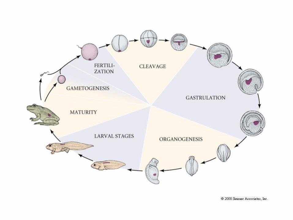
gastrulation - drosophila
DESCRIPTION
Figure 23.14 Homologous Pathways Specifying Neural Ectoderm in Protostomes ( Drosophila ) and Deuterostomes ( Xenopus ) D/V. Gastrulation - Drosophila. http://www.flybase.org/data/images/Animation/ AND Course Site (Movies). 4 STAGES OF ESTABLISHING DORSAL/VENTRAL – 4 SEQUENTIAL PATHWAYS +. - PowerPoint PPT PresentationTRANSCRIPT

Drosophila timescales (at 25°C): 3 hrs: cellular blastoderm with 5000 cells
with all cells’ identities specified24 hrs: embryo hatches as feeding larva
•Movies:–QT Drosophila Development -different views: embryogenesis –SEM morph
–QT Drosophila embryogenesis –QT Drosophila Cleavage: following Histone-GFP
–QT Drosophila Gastrulation: following Histone-GFP

Drosophila – 2 lectures (½ – 1- ½ )
• Cleavage
• View -gastrulation, organogen. frame metamorph.
• Once we know the embryo, meet the molecules• Because this is a largely ‘solved’ system
• Because these genes have key roles in all metazoans
• EVERY one of 5000 cleavage state cell has a D/V and A/P ‘molecular address’, and is therefore specified.
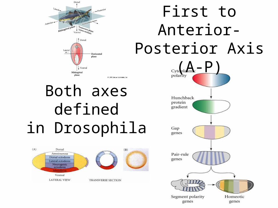
Both axes definedin Drosophila
First to Anterior-Posterior Axis (A-P)
A-P:-Termini
-Segmented body

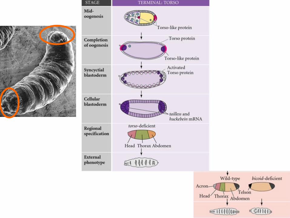
Acron and Telson are ‘TERMINAL’ structures


To overviewMovie:
“Embryo genesis”
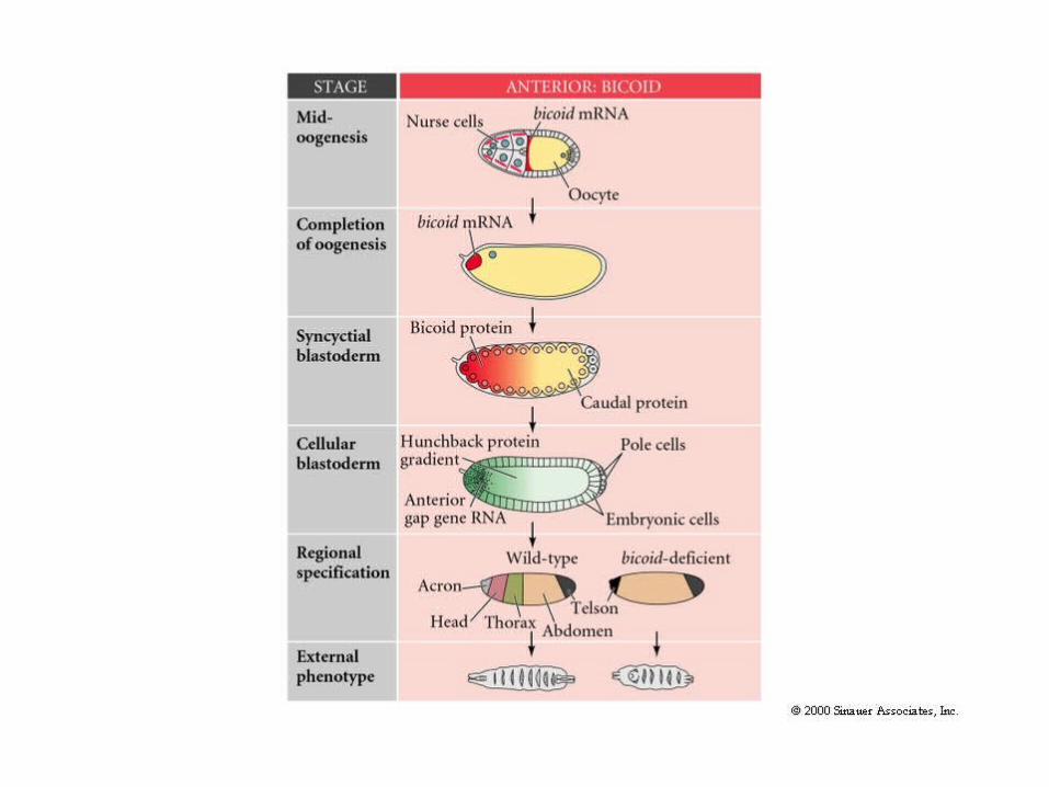


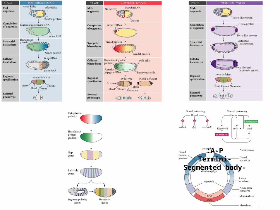
Bicoid mRNA 1. Bicoid RNA ‘caught’ at the ‘entrance’
2. Unanchored Bicoid RNA returned to the anterior side by dynein on MTs
Bicoid mRNA-binding protein

Show:Bcd-gastrulation
Gastrulation-dorsal

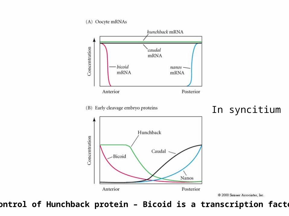
In syncitium
For control of Hunchback protein – Bicoid is a transcription factor, but Nanos . . .

Nanos is an RNA binding protein that PREVENTS Hunchback Translation

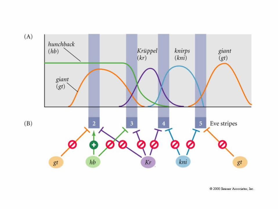
Gap genes are turned on in broad stripes by maternal genes, each other. ALL TFs.
Hunchback
Gt
Kr
kn
hb (later)


Gt
Kr
kn
hb (later)

Gap genes are turned on in broad stripes by maternal genes, each other
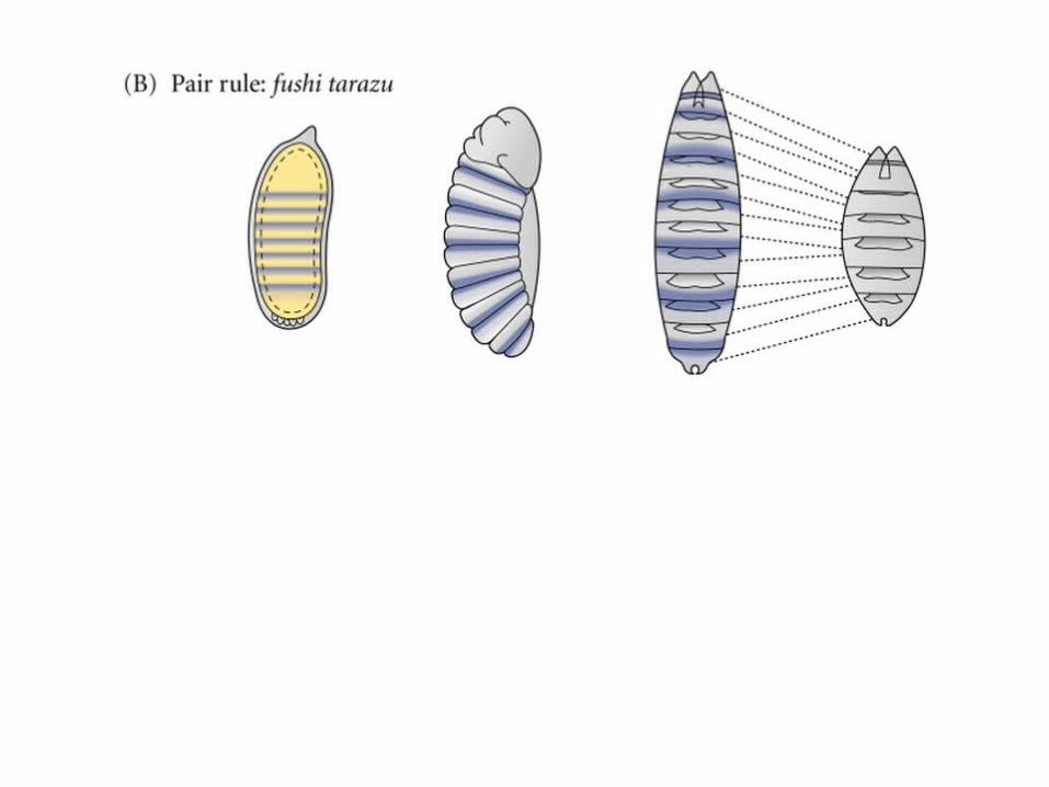
Pair rule genes are turned on in 7 stripes each, harder to conceptualize

Each stripe of theP-R gene has its Own enhancer.
Even-skippedgene – 7 stripes.

Each stripe has its own enhancer, responding to a different combinatorial of Gap and Maternal proteins
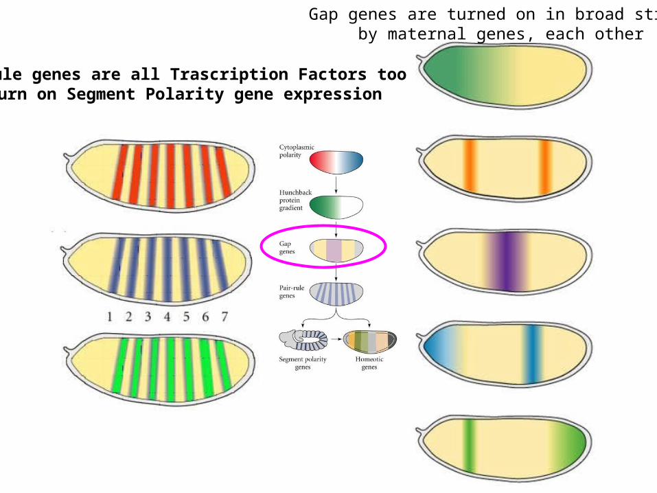
Gap genes are turned on in broad stripes by maternal genes, each other
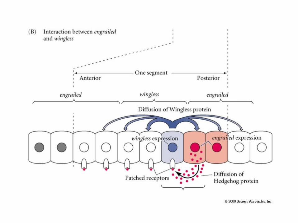
Pair rule genes are all Trascription Factors too – turn on Segment Polarity gene expression


hh hh hh hh Two morphogens/ligands/organizers in adjacent cells

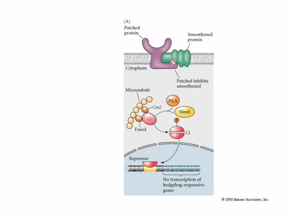
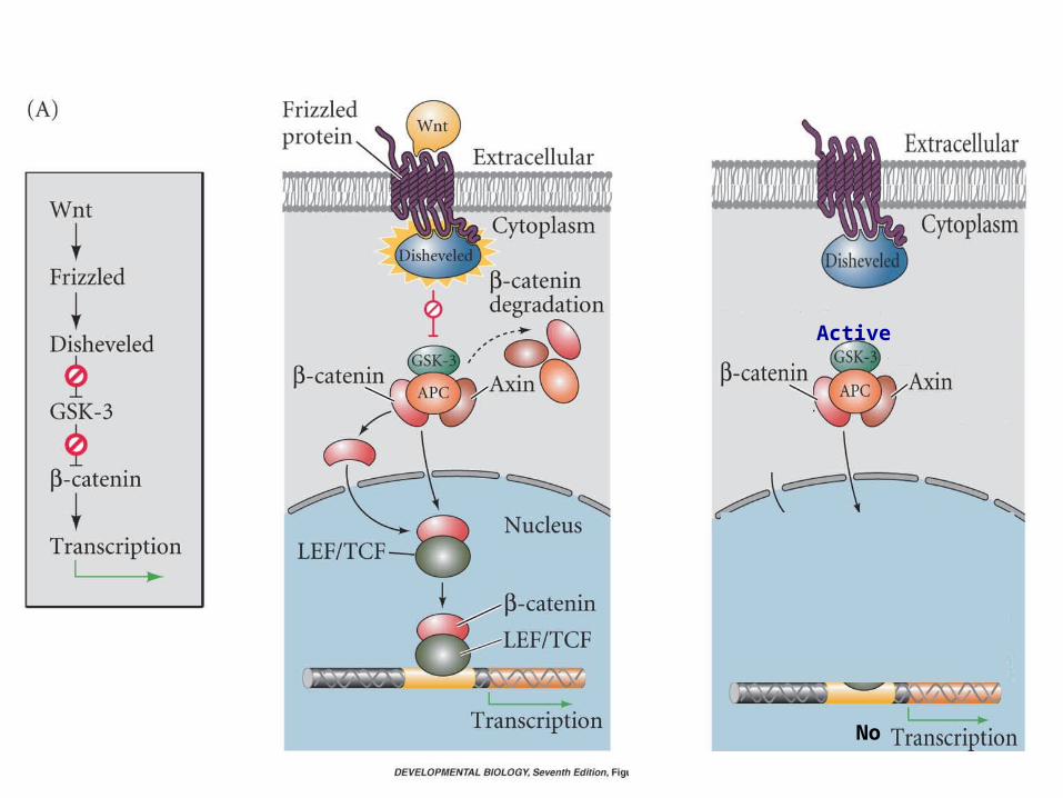
No
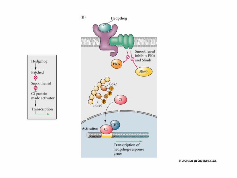
Active
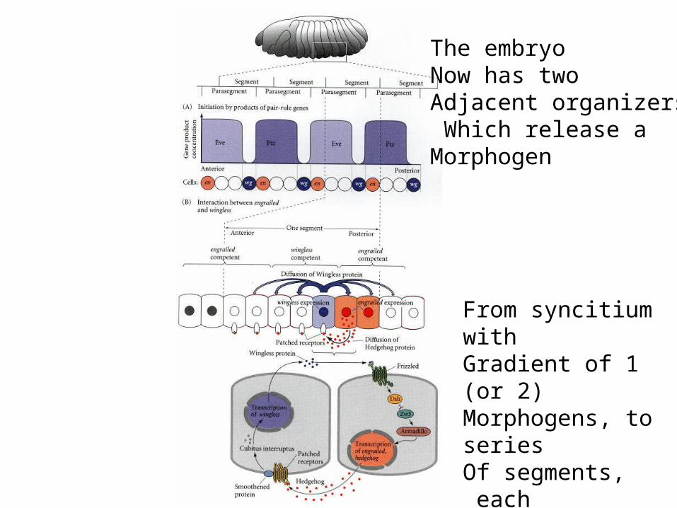
The embryoNow has twoAdjacent organizersWhich release a
Morphogen
From syncitium withGradient of 1 (or 2)Morphogens, to seriesOf segments, each
With 2 morphogens
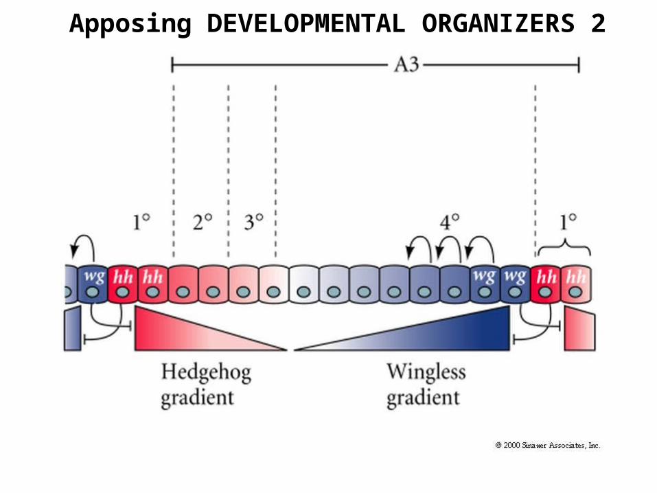
2 Apposing DEVELOPMENTAL ORGANIZERS

http://bcs.whfreeman.com/thelifewire/content/chp19/1902003.html


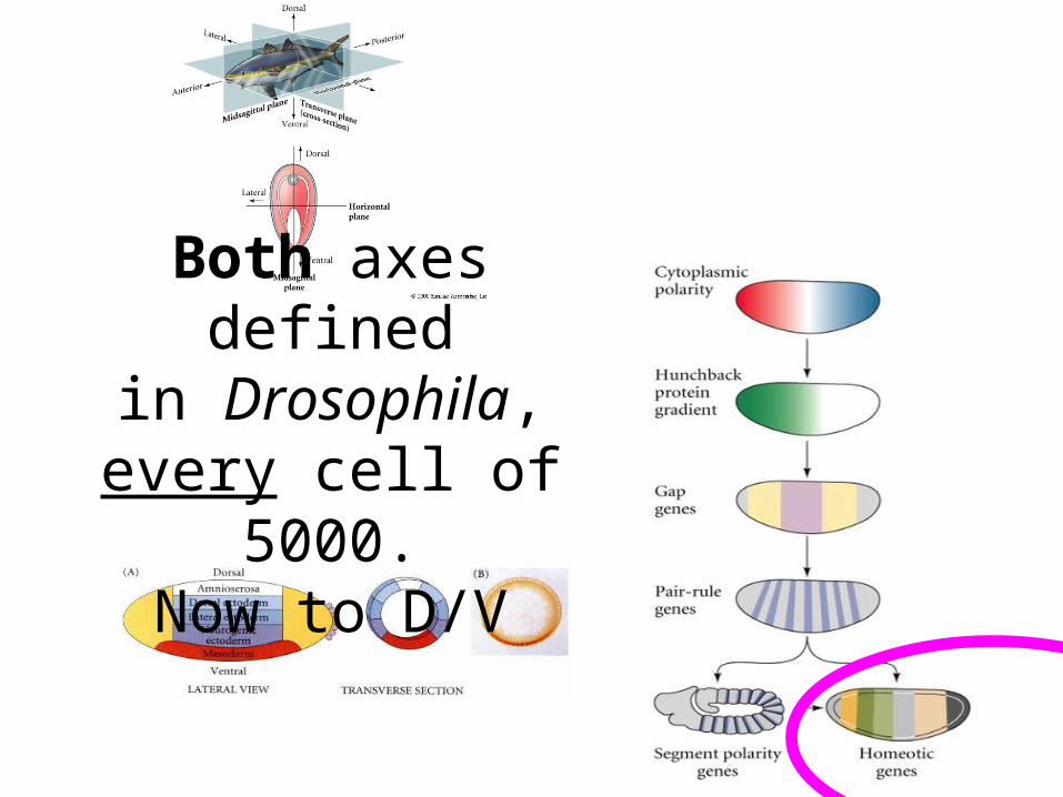
Both axes definedin Drosophila,
every cell of 5000.Now to D/V


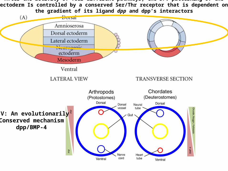
After the activity of four different pathways, the D/V patterning of the ectoderm Is controlled by a conserved Ser/Thr receptor that is dependent on the gradient of its ligand dpp and dpp’s interactors
D/V: An evolutionarilyConserved mechanism
dpp/BMP-4

Figure 23.14 Homologous Pathways Specifying Neural Ectoderm in Protostomes (Drosophila) and Deuterostomes (Xenopus) D/V
Gastrulation - Drosophila
http://www.flybase.org/data/images/Animation/
AND Course Site (Movies)


I. RTK pathway Sets follicle cell D/V state
II. Proteolytic cascade Sets embryos’ cell D/V state
III. Toll/Cactus/Doral Sets nuclear D/V state
IV. Dorsal TF thresholds Diff. pathway per D/V address
4 STAGES OF ESTABLISHING DORSAL/VENTRAL – 4 SEQUENTIAL PATHWAYS +
STAGE PATHWAY PATHWAY OUTCOME

I. RTK pathway Sets follicle cell D/V state
II. Proteolytic cascade Sets embryos’ cell D/V state
III. Toll/Cactus/Doral Sets nuclear D/V state
IV. Dorsal TF thresholds Diff. pathway per D/V address
4 STAGES OF ESTABLISHING DORSAL/VENTRAL – 4 SEQUENTIAL PATHWAYS +
STAGE PATHWAY PATHWAY OUTCOME
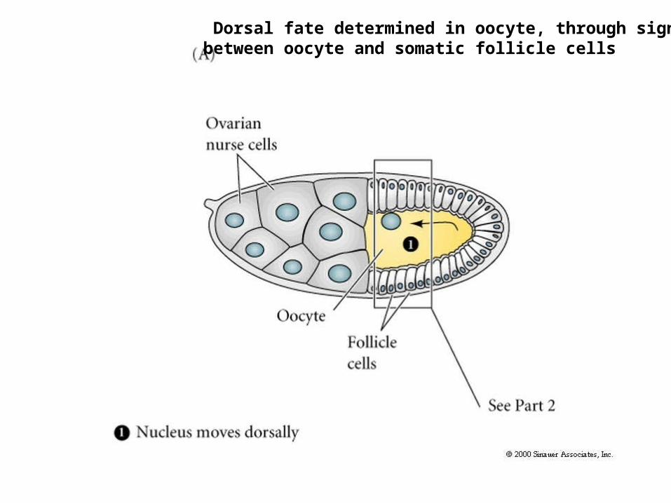
Dorsal fate determined in oocyte, through signaling between oocyte and somatic follicle cells
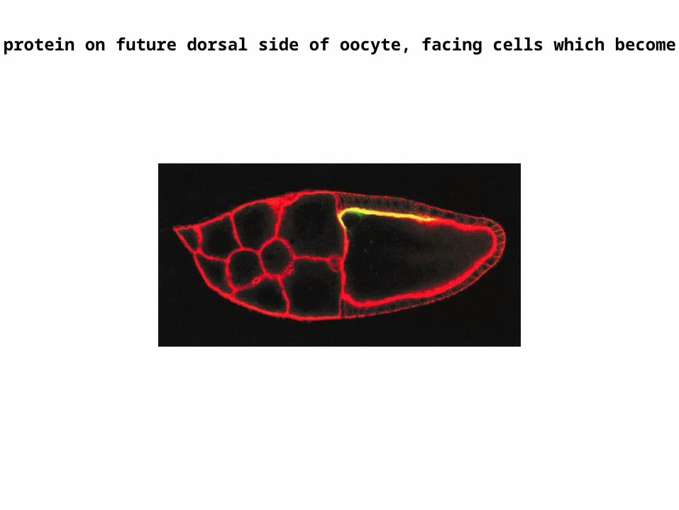
Gurken protein on future dorsal side of oocyte, facing cells which become dorsal


Human blood clotting cascade– Also a series of
(extracellular )proteolytic cleavages


Dorsalized Ventralized
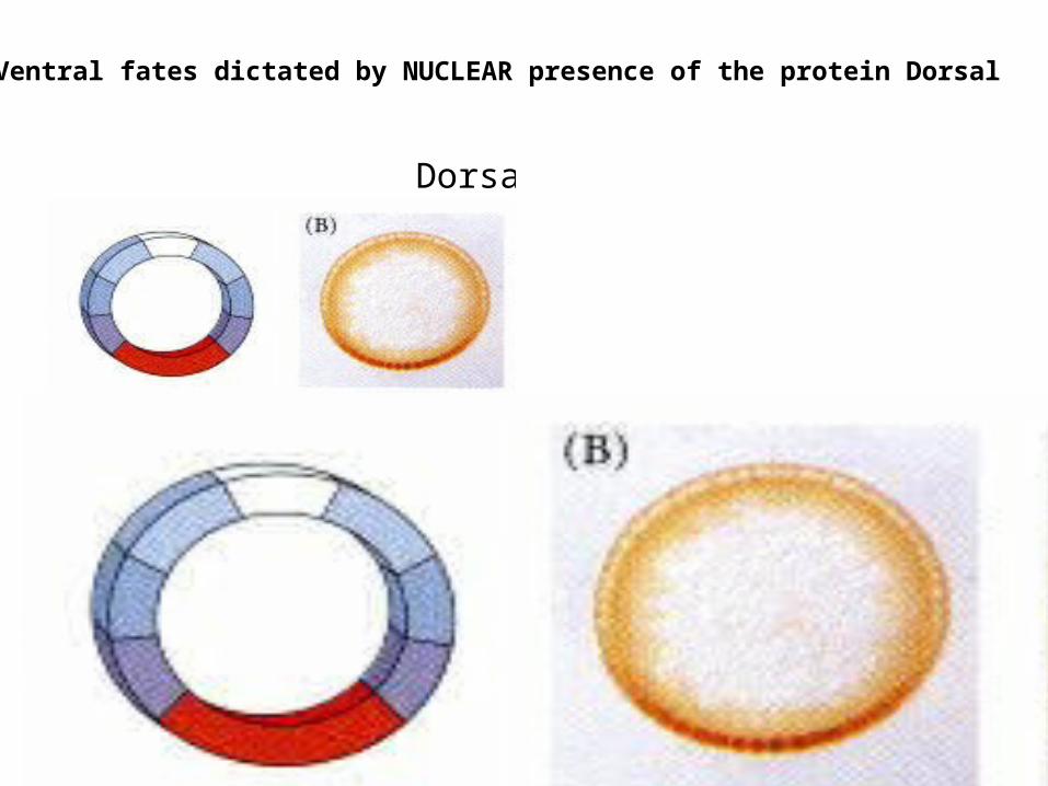
Ventral fates dictated by NUCLEAR presence of the protein Dorsal

Gradient of Nuclear Dorsal protein imparts D-V IDs to cells

Twist Protein specifies mesoderm

Lateral inhibition in neurectoderm to specify neruogenesis: Notch mediated
All Rhomboid expressing cells express Notch, then undergo a stochastic process for ¼ cells to become neuronal
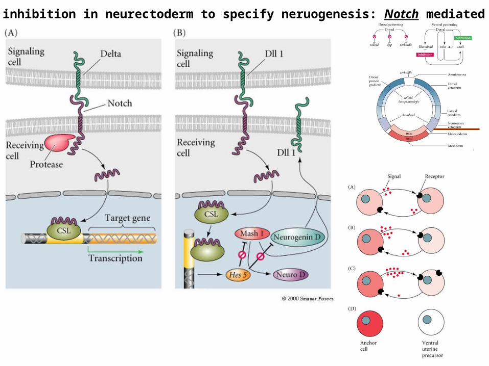
Lateral inhibition in neurectoderm to specify neruogenesis: Notch mediated

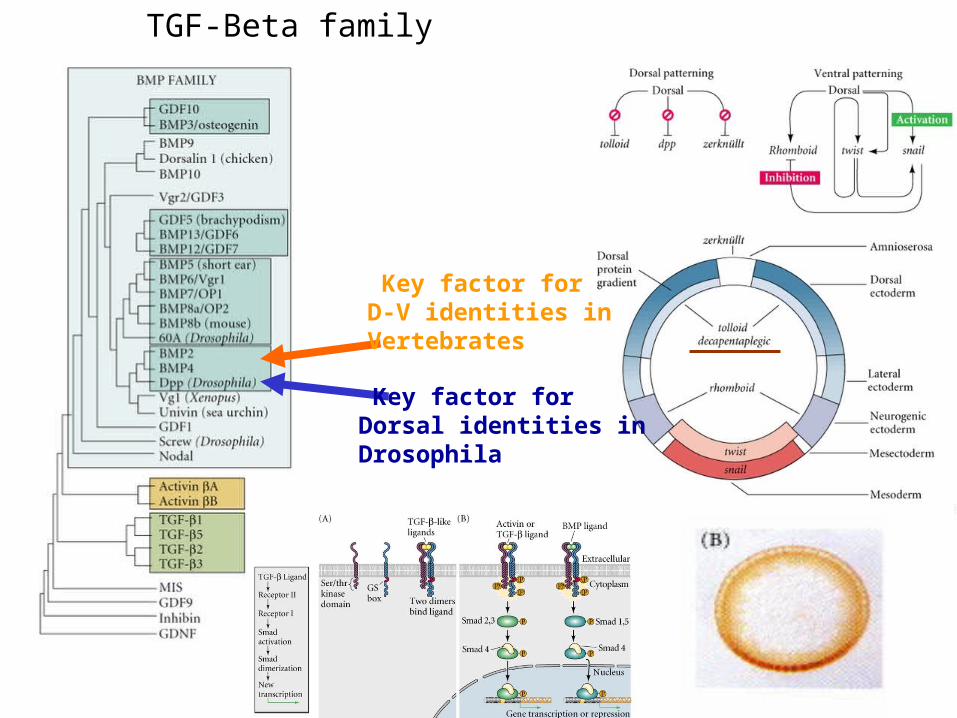
Key factor for Dorsal identities inDrosophila
Key factor for D-V identities inVertebrates
TGF-Beta family

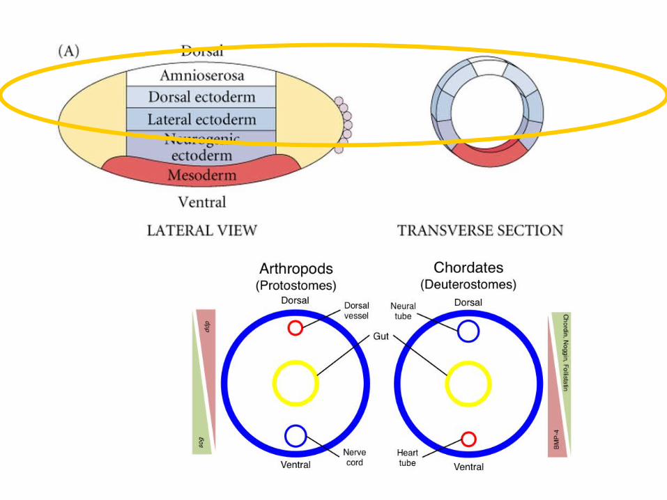
After the activity of four different pathways, the D/V patterning of the ectoderm Is controlled by a conserved Ser/Thr receptor that is dependent on the gradient of its ligand dpp and dpp’s interactors
D/V: An evolutionarilyConserved mechanism
dpp/BMP-4
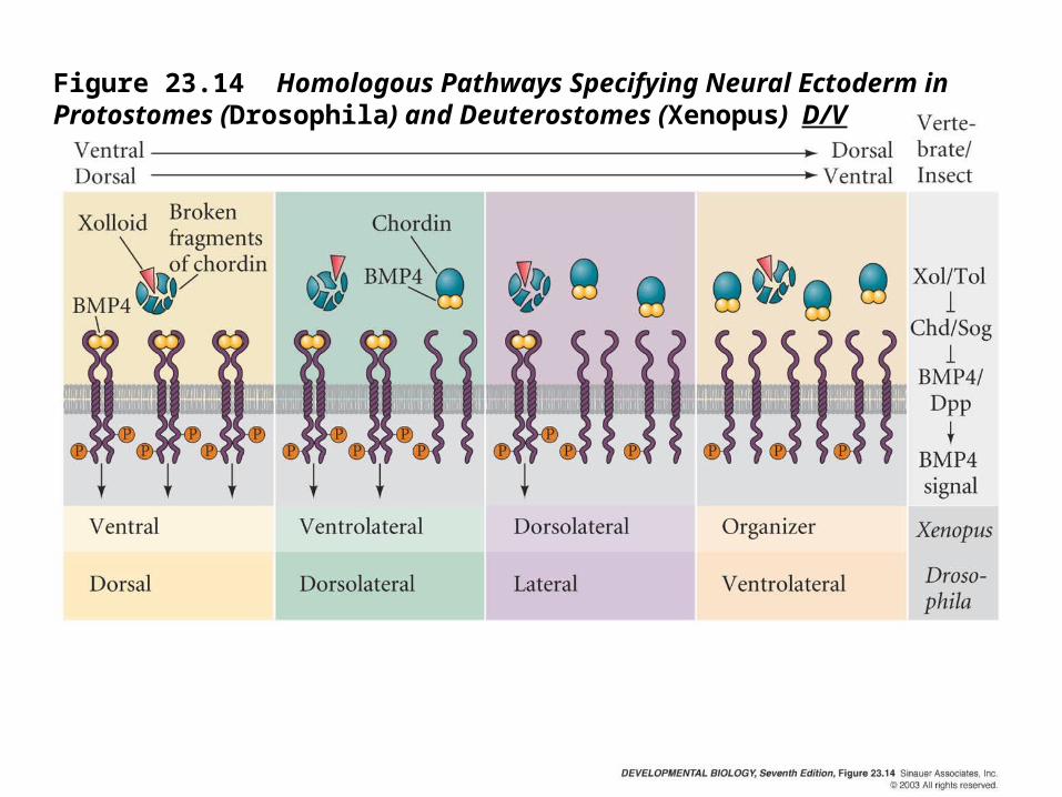
Figure 23.14 Homologous Pathways Specifying Neural Ectoderm in Protostomes (Drosophila) and Deuterostomes (Xenopus) D/V

The course primarily addresses development of urbilaterian descendents. All reflect on the ancestor-differences are details.


So now we’ve defined both axes
in Drosophila, includingevery cell of 5000.
msh, ind, and sogmark specific D/V cells/coordinates
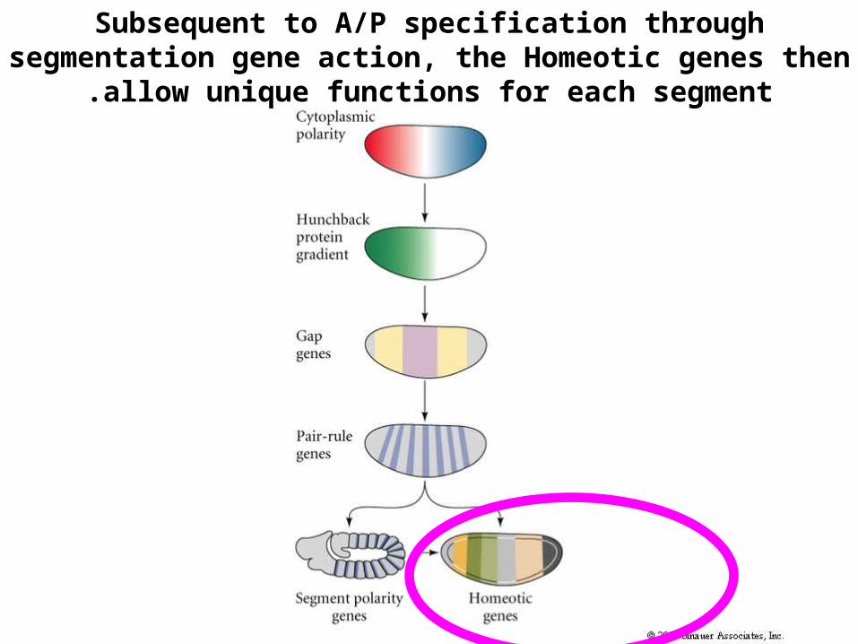
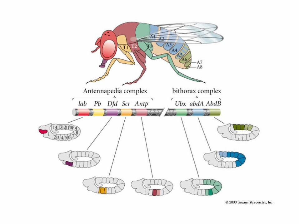
Subsequent to A/P specification through segmentation gene action, the Homeotic genes then allow unique functions for each segment.

Stopped here


All animals have related developmental histories


out inevert


Figure 6.16 Scanning Electron Micrograph of a Compound Eye in Drosophila
Eye disc patterning controlled by ‘reuse’ of the pathways seen in general axis specification

Figure 6.17 Differentiation of Photoreceptors in the Drosophila Compound Eye

Figure 6.18 Major Genes Known to be Involved in the Induction of Drosophila Photoreceptors