glavni i odgovorni urednik: prof. dr gordana ......izdavačka delatnost društva lekara vojvodine...
TRANSCRIPT

Izdavačka delatnost Društva lekara Vojvodine Srpskog lekarskog društva, Novi Sad, Vase Stajića 9Glavni i odgovorni urednik: prof. dr GORDANA DEVEČERSKI
MEDICINSKI PREGLEDČASOPIS DRUŠTVA LEKARA VOJVODINE SRPSKOG LEKARSKOG DRUŠTVA
PRVI BROJ JE ŠTAMPAN 1948. GODINE.
Glavni i odgovorni urednikProf. dr LJILJA MIJATOV UKROPINA
Pomoćnici urednikaAsist. dr sc. med. BOJANA KRSTONOŠIĆ
Asist. dr sc. med. BOJAN ZARIĆ
REDAKCIJSKI ODBORPredsednik: prof. dr PETAR SLANKAMENAC
Sekretar: prof. dr VIKTOR TILL
Prof. dr STOJANKA ALEKSIĆ, HamburgProf. dr KAREN BELKIĆ, StockholmProf. dr JEAN-PAUL BEREGI, Lille CedexProf. dr JELA BOROTA, Novi SadProf. dr MILAN BREBERINA. Novi SadProf. dr RADOVAN CVIJANOVIĆ, Novi SadProf. dr GROZDANA ČANAK, Novi SadProf. dr IVAN DAMJANOV, Kansas CityProf. dr DRAGAN DANKUC, Novi SadProf. dr GORDANA DEVEČERSKI, Novi SadProf. dr RAJKO DOLEČEK, OstravaProf. dr MIRJANA ĐERIĆ, Novi SadProf. dr SRĐAN ĐURĐEVIĆ, Novi SadProf. dr VERA GRUJIĆ, Novi SadProf. dr TATJANA IVKOVIĆ LAZAR, Novi Sad Prof. dr JÁNOS JAKÓ, BudapestProf. dr MARINA JOVANOVIĆ, Novi SadProf. dr DRAGAN KATANIĆ, Novi SadProf. dr ALEKSANDAR KIRALJ. Novi SadProf. dr ALEKSANDAR KNEŽEVIĆ, Novi Sad Prof. dr DRAGAN KOVAČEVIĆ, Novi Sad
Prof. dr SMILJANA MARINKOVIĆ, Novi SadProf. dr MARIOS MARSELOS, IoanninaProf. dr LJILJA MIJATOV UKROPINA, Novi SadProf. dr MIROSLAV MILANKOV, Novi SadProf. dr IGOR MITIĆ, Novi SadProf. dr NADA NAUMOVIĆ. Novi SadProf. dr ANA OROS, Novi SadProf. dr VERA JERANT PATIĆ, Novi SadProf. dr LJUBOMIR PETROVIĆ, Novi SadProf. dr MIODRAG RADULOVAČKI, ChicagoProf. dr JOVAN RAJS, DanderydProf. dr PETAR E. SCHWARTZ, New HavenProf. dr PETAR SLANKAMENAC, Novi SadProf. dr VIKTOR TILL, Novi SadProf. dr TAKASHI TOYONAGA. KobeProf. dr KONSTANTIN VIKTOROVIĆ SUDAKOV, MoskvaProf. dr NADA VUČKOVIĆ, Novi SadProf. dr ZORAN VUJKOVIĆ, Banja LukaProf. dr PETAR VULEKOVIĆ, Novi SadProf. dr RELJA ŽIVOJNOVIĆ, Antwerpen
Lektor za srpski jezik: Dragica PantićLektor za engleski jezik: Jasminka Anojčić
Tehnički sekretar: Vesna ŠaranovićTehnička podrška: „Grafit”, Novi Sad
Izrada UDK i deskriptora: Biblioteka Medicinskog fakulteta, Novi Sad
MEDICINSKI PREGLED izlazi dvomesečno (šest dvobroja godišnje), u tiražu od 1000 primeraka. Pretplata za pojedince sa teritorije Srbije za 2014. godinu iznosi 3.000,00 dinara (sa uračunatim PDV-om), a 4.000,00 dinara za pojedince van teritorije Srbije, a za ustanove 8.000,00 dinara (uz dodavanje PDV-a). Uplate se vrše na račun broj 340-1861-70 ili 115-13858-06, s naznakom „Dodatna članarina za Medicinski pregled”.
Copyright ® Društvo lekara Vojvodine Srpskog lekarskog društva Novi Sad 1998.
Prijem rukopisa vrši se u elektronskoj formi na stranici: aseestant.ceon.rs/index.php/medpreg/. Adresa Redakcije: Društvo lekara Vojvodine Srpskog lekarskog društva,
21000 Novi Sad, Vase Stajića 9, Tel. 021/521-096; 063/81 33 875
E-mail: [email protected]; Web: www.dlv.org.rs
Štamparija: Uprava za zajedničke poslove pokrajinskih organa - Odsek za poslove štamparije

Publishing Sector of the Society of Physicians of Vojvodina of the Medical Society of Serbia, Novi Sad, Vase Stajica 9, Editor-in-Chief Prof. GORDANA DEVECERSKI, MD, PhD
MEDICAL REVIEWJOURNAL OF THE SOCIETY OF PHYSICIANS OF VOJVODINA OF THE
MEDICAL SOCIETY OF SERBIATHE FIRST ISSUE WAS PUBLISHED IN 1948
Editor-in-Chief Prof. LJILJA MIJATOV UKROPINA, MD, PhD
Assistants to the Editor-in-Chief Assist. Asist. BOJANA KRSTONOŠIĆ, MD, PhD
Asist. BOJAN ZARIĆ, MD, PhD
EDITORIAL BOARD
President: Prof. PETAR SLANKAMENAC, MD, PhD Secretary: Prof. VIKTOR TILL, MD, PhD
Prof. STOJANKA ALEKSIĆ, MD, PhD, HamburgProf. KAREN BELKIĆ, MD, PhD, StockholmProf. JEAN-PAUL BEREGI, MD, PhD, Lille CedexProf. JELA BOROTA, MD, PhD, Novi SadProf. MILAN BREBERINA, MD, PhD. Novi SadProf. RADOVAN CVIJANOVIĆ, MD, PhD, Novi SadProf. GROZDANA ČANAK, MD, PhD, Novi SadProf. IVAN DAMJANOV, MD, PhD, Kansas CityProf. DRAGAN DANKUC, MD, PhD, Novi SadProf. GORDANA DEVEČERSKI, MD, PhD, Novi SadProf. RAJKO DOLEČEK, MD, PhD, OstravaProf. MIRJANA ĐERIĆ, MD, PhD, Novi SadProf. SRĐAN ĐURĐEVIĆ, MD, PhD, Novi SadProf. VERA GRUJIĆ, MD, PhD, Novi SadProf. TATJANA IVKOVIĆ LAZAR, MD, PhD, Novi Sad Prof. JÁNOS JAKÓ, MD, PhD, BudapestProf. MARINA JOVANOVIĆ, MD, PhD, Novi SadProf. DRAGAN KATANIĆ, MD, PhD, Novi SadProf. ALEKSANDAR KIRALJ, MD, PhD, Novi SadProf. ALEKSANDAR KNEŽEVIĆ, MD, PhD, Novi Sad Prof. DRAGAN KOVAČEVIĆ, MD, PhD, Novi Sad
Prof. SMILJANA MARINKOVIĆ, MD, PhD, Novi SadProf. MARIOS MARSELOS, MD, PhD, loanninaProf. LJILJA MIJATOV UKROPINA, MD, PhD, Novi SadProf. MIROSLAV MILANKOV, MD, PhD, Novi SadProf. IGOR MITIĆ, MD, PhD, Novi SadProf. NADA NAUMOVIĆ, MD, PhD, Novi SadProf. ANA OROS, MD, PhD, Novi SadProf. VERA JERANT PATIĆ, MD, PhD, Novi SadProf. LJUBOMIR PETROVIĆ, MD, PhD, Novi SadProf. MIODRAG RADULOVAČKI, MD, PhD, ChicagoProf. JOVAN RAJS, MD, PhD, DanderydProf. PETAR E. SCHWARTZ, MD, PhD, New HavenProf. PETAR SLANKAMENAC, MD, PhD, Novi SadProf. VIKTOR TILL, MD, PhD, Novi SadProf. TAKASHI TOYONAGA, MD, PhD, KobeProf. KONSTANTIN VIKTOROVIĆ SUDAKOV, MD, PhD, MoscowProf. NADA VUČKOVIĆ, MD, PhD, Novi SadProf. ZORAN VUJKOVIĆ, MD, PhD, Banja LukaProf. PETAR VULEKOVIĆ, MD, PhD, Novi SadProf. RELJA ŽIVOJNOVIĆ, MD, PhD, Antwerpen
Proof-reading for Serbian Language: Dragica PantićProof-reading for English Language: Jasminka Anojčić
Technical Secretary: Vesna ŠaranovićTechnical Support: ”Grafit” Novi Sad
UDC and descriptors prepared by: the Library of the Faculty of Medicine, Novi Sad
MEDICAL REVIEW is published two-monthly (six double issues per a year) in the circulation of 1000 co-pies. Payment for individuals from the territory of Serbia for the year 2014 is 3,000.00 dinars (the VAT being calculated in) and 4,000.00 dinars for the individuals outside the territory of Serbia, and 8,000.00 dinars (+ the VAT) for institutions. The payments are to be made to the account number 340-1861-70 or 115-13858-06, with the remark ”Additional membership fee for the Medical Review”.
Copyright ® Društvo lekara Vojvodine Srpskog lekarskog društva Novi Sad 1998.
The manuscripts can be submited on the web-page: aseestant.ceon.rs/index.php/medpreg/.Address Editorial: Društvo lekara Vojvodine Srpskog lekarskog društva, 21000 Novi Sad, Vase Stajića 9,
Tel. 021/521-096; 063/81 33 875E-mail: [email protected]; Web: www.dlv.org.rs
Printed by: Department for Joint Affairs of Provincial Authorities - Sector for Printing
M E D I C I N S K I P R E G L E DČASOPIS DRUŠTVA LEKARA VOJVODINE SRPSKOG LEKARSKOG DRUŠTVA
Novi Sad Vase Stajića 9 Srbija
Med Pregl 2014; LXVII (5-6): 135-192. Novi Sad: maj-juni.
SADRŽAJ
UVODNIK
Karen Belkić i Olesja NedićMEDICINA RADA NEKAD I SAD: U KOM PRAVCU DALJE .................................................................................................
ORIGINALNI NAUČNI RADOVI
Ljiljana Kesić, Radojka Delić, Dragan Mihailović, Milica S. Petrović i Tijana Đ. DelićMORFOLOŠKA I MORFOMETRIJSKA ANALIZA PROMENA U USNOJ DUPLJI KOJE JE IZAZVALA CANDIDA ALBICANS – EKSPERIMENTALNI RAD .....................................................................................................................................
Đuka Ninković Baroš, Vesna S. Gajanin, Radoslav B. Gajanin i Bogdan ZrnićKOMPARATIVNA ANALIZA USPEHA LEČENJA PSORIJAZE STANDARDNIM TERAPIJSKIM MODALITETIMA I BALNEOTERAPIJOM ....................................................................................................................................................................
PREGLEDNI ČLANCI
Mihailo I. Stjepanović, Violeta Mihailović Vučinić, Dragana Jovanović, Milija Mijajlović, Vesna Škodrić Trifunović i Mirjana M. StjepanovićTERAPIJA NEUROSARKOIDOZE - NOVINE I IZAZOVI .......................................................................................................
STRUČNI ČLANCI
Olgica Milankov, Milena Bjelica i Radojica SavićKOJOM VRSTOM MLEKA JE MOGUĆE PREVENIRATI NASTANAK SIDEROPENIJSKE ANEMIJE KOD ODOJ-ČADI – KOMPARATIVNA STUDIJA ............................................................................................................................................
Ivana Bajkin, Artur Bjelica, Tijana Ičin, Vesna Dobrić, Branka Kovačev Zavišić i Milica Medić StojanoskaUTICAJ ESTARA FTALNE KISELINE NA FETALNO ZDRAVLJE ..........................................................................................
PRIKAZI SLUČAJEVA
Ana Tadić, Tatjana Puškar i Branislava PetronijevićUPOTREBA FIBRINSKIH BLOKOVA BOGATIH KONCENTROVANIM FAKTORIMA RASTA U PREIMPLANTO-LOŠKIM AUGMENTACIONIM PROCEDURAMA ...................................................................................................................
Danijela Mandić, Lana Nežić i Ranko ŠkrbićTEŠKA HIPERKALIJEMIJA IZAZVANA PROPRANOLOLOM ...............................................................................................
Marija Trenkić Božinović, Predrag Jovanović, Gordana Zlatanović, Dragan Veselinović, Aleksandra Aracki Trenkić i Milan TrenkićRETINALNA HEMORAGIJA KAO KOMPLIKACIJA DRUZA OPTIČKOG DISKA U TRUDNOĆI ................................
139-147
149-153
154-160
161-166
167-171
172-175
177-180
181-184
185-189

M E D I C A L R E V I E WJOURNAL OF THE SOCIETY OF PHYSICIANS OF VOJVODINA OF THE MEDICAL SOCIETY OF SERBIA
Novi Sad Vase Stajića 9 Serbia
Med Pregl 2014; LXVII (5-6): 135-192. Novi Sad: May-June.
CONTENTS
EDITORIAL
Karen Belkić and Olesja NedićOCCUPATIONAL MEDICINE -THEN AND NOW: WHERE WE COULD GO FROM HERE....................................................................
ORIGINAL STUDIES
Ljiljana Kesić, Radojka Delić, Dragan Mihailović, Milica S. Petrović and Tijana Đ. Delić MORPHOLOGIC AND MORPHOMETRIC ANALYSIS OF ALTERNATIONS IN THE ORAL CAVITY CAUSED BY CANDIDA ALBICANS – EXPERIMENTAL WORK ..........................................................................................................................................................
Đuka Ninković Baroš, Vesna S. Gajanin, Radoslav B. Gajanin and Bogdan ZrnićCOMPARATIVE ANALYSIS OF SUCCESS OF PSORIASIS TREATMENT WITH STANDARD THERAPEUTIC MODALITIES AND BALNEOTHERAPY ..................................................................................................................................................................................
REVIEW ARTICLES
Mihailo I. Stjepanović, Violeta Mihailović Vučinić, Dragana Jovanović, Milija Mijajlović, Vesna Škodrić Trifunović and Mirjana M. StjepanovićTREATMENT OF NEUROSARCOIDOSIS - INNOVATIONS AND CHALLENGES ..................................................................................
PROFESSIONAL ARTICLES
Olgica Milankov, Milena Bjelica and Radojica SavićWHAT KIND OF MILK CAN PREVENT INFANT’S SIDEROPENIC ANEMIA – COMPARATIVE STUDY ........................................
Ivana Bajkin, Artur Bjelica, Tijana Ičin, Vesna Dobrić, Branka Kovačev Zavišić and Milica Medić StojanoskaEFFECTS OF PHTHALIC ACID ESTERS ON FETAL HEALTH ...................................................................................................................
CASE REPORTS
Ana Tadić, Tatjana Puškar and Branislava PetronijevićAPPLICATION OF FIBRIN RICH BLOCKS WITH CONCENTRATED GROWTH FACTORS IN PRE-IMPLANT AUGMENTATI-ON PROCEDURES .............................................................................................................................................................................................
Danijela Mandić, Lana Nežić i Ranko ŠkrbićSEVERE HYPERKALEMIA INDUCED BY PROPRANOLOL .......................................................................................................................
Marija Trenkić Božinović, Predrag Jovanović, Gordana Zlatanović, Dragan Veselinović, Aleksandra Aracki Trenkić and Milan TrenkićRETINAL HEMORRHAGES AS ONE OF COMPLICATIONS OF OPTIC DISC DRUSEN DURING PREGNANCY ..............................
139-147
149-153
154-160
161-166
167-171
172-175
177-180
181-184
185-189


Med Pregl 2014; LXVII (5-6): 139-147. Novi Sad: maj-juni. 139
Summary Occupational medicine has a long-standing history in the regi-on of the former Yugoslavia with seminal contributions to the theory and practice of this discipline. This tradition should be expanded to incorporate psychosocial stressors. We review the sociological work stress models and empirical evidence glea-ned thereby, and then the occupational stressor index, an additi-ve burden model developed from a cognitive ergonomics per-spective. In numerous studies, the occupational stressor index is significantly associated with risk behaviors: smoking, obesi-ty and sedentariness and clinical outcomes: hypertension, is-chemic heart disease, dyslipidemia and type 2 diabetes. The occupational stressor index characterizes the work conditions of physicians including surgeons and anesthesiologists; profe-ssional drivers and other groups at elevated risk for stress-rela-ted disorders. Much of these empirical data are from this regi-on. Work-stress related health disorders are a major public heal-th problem, with enormous human and economic costs. A more proactive role for physicians is needed vis-à-vis our working environment and that of patients. We physicians face a heavy job stressor burden strongly implicated with adverse health out-comes. The challenge is to identify effective strategies to lower the risk of work-stressor related illness. The critical gap is the lack of evidence-based guidelines. Intervention studies are nee-ded in which job stressors are ameliorated as a therapeutic/pre-ventive modality; the logical starting point is within our own profession. We also suggest how the relevant clinical competen-ce could be enhanced. Alongside clinical enhancement should be the full restoration of physician empowerment to implement work-related recommendations. A participatory action research perspective by physicians for physicians and for our patients is needed.Key words: Occupational Medicine; Workload; Stress, Psyc-hological; Occupational Diseases; Health Promotion; Risk Factors
SažetakMedicina rada ima dugogodišnju tradiciju u regionu bivše Jugo-slavije sa originalnim doprinosima u teoriji i praksi, koje bi tre-balo proširivati uključivanjem psihosocijalnih stresora. Prikazani su sociološki modeli utvrđivanja postojanja profesionalnog stresa i dokazi iz empirijskih istraživanja. Opisan je i novi model in-deksa profesionalnog stresa (Occupational Stressor Index), pote-kao iz perspektive kognitivne ergonomije. U brojnim studijama indeksom profesionalnog stresa utvrđena je značajna povezanost profesionalnog stresa sa rizičnim ponašanjima: pušenje, goja-znost, sedenternost i kliničkim ishodima: hipertenzija, ishemij-ska bolest srca, dislipidemija i dijabetes tipa 2. Indeks profesio-nalnog stresa sveobuhvatno opisuje sve uslove rada lekara, uklju-čujući hirurge i anesteziologe, profesionalnih vozača i drugih profesija sa povišenim rizikom za pojavu zdravstvenih poreme-ćaja povezanih sa stresom na radu. To je empirijski utvrđeno i u ovom regionu. Zdravstveni poremećaji u vezi sa stresom na radu su veliki javnozdravstveni problem, sa ogromnim ljudskim i eko-nomskim posledicama. Ukazano je na potrebu proaktivnije uloge lekara naspram našeg radnog okruženja i pacijenata. Lekari se su-očavaju sa teškim zdravstvenim ishodima kao posledicom posto-janja velikog profesionalnog opterećenja. Izazov je identifikovati efektivne strategije kojima bi se smanjili rizici od pojave bolesti povezanih sa stresom na radu. Kritični raskorak je u nedostatku smernica zasnovanih na dokazima. Potrebne su interventne studi-je u kojima bi se na utvrđene profesionalne stresore terapijski/pre-ventivno delovalo; logično polazište je naša vlastita profesija. Ta-kođe sugerišemo kako se relevantne kliničke kompetencije mogu pojačati. Zajedno sa poboljšanjem kliničkog znanja trebalo bi po-boljšati osposobljenost lekara u sprovođenju preporuka u vezi sa radom i radnim okruženjem. Potrebna je perspektiva aktivnog učešća u istraživanju lekara za lekare i lekara za pacijente.Ključne reči: Medicina rada; Izgaranje na poslu; Psihološki stres; Profesionalna oboljenja; Promocija zdravlja; Faktori ri-zika
EDITORIAL UVODNIKDepartment of Oncology/Pathology, Karolinska Institute, Sweden1 EditorialClaremont Graduate University UvodnikSchool of Community and Global Health, Claremont, California, USA2 UDK 616-057Institute for Health Promotion and Disease Prevention Research DOI: DOI: 10.2298/MPNS1406139BUniversity of Southern California School of Medicine, Los Angeles, California3
Ambulatory Health Care Center, Novi Sad, SerbiaDivision for Occupational Health Protection4
OCCUPATIONAL MEDICINE - THEN AND NOW: WHERE WE COULD GO FROM HERE
MEDICINA RADA NEKAD I SAD: U KOM PRAVCU DALJE
Karen BELKIĆ1-3 and Olesja NEDIĆ4
Corresponding Author: Karen Belkić, M.D., PhD, Department of Oncology-Pathology, Karolinska Institutet, Building P9 2nd floor P.O. Box 260, Stockholm SE 17176 Sweden: E-mail: [email protected]
Belkić K, et al. Occupational medicine - then and now140
Rich History of Occupational Medicine in the Region of the Former Yugoslavia
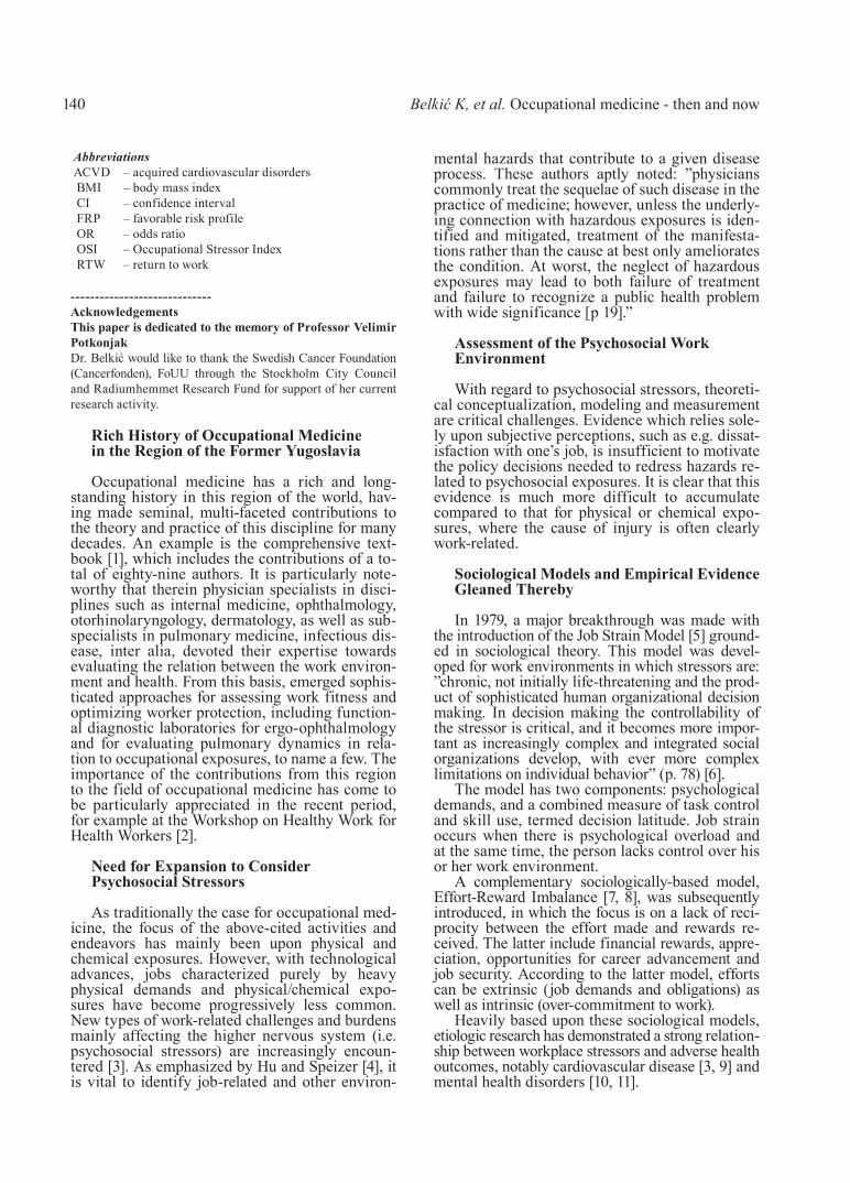
Occupational medicine has a rich and long-standing history in this region of the world, hav-ing made seminal, multi-faceted contributions to the theory and practice of this discipline for many decades. An example is the comprehensive text-book [1], which includes the contributions of a to-tal of eighty-nine authors. It is particularly note-worthy that therein physician specialists in disci-plines such as internal medicine, ophthalmology, otorhinolaryngology, dermatology, as well as sub-specialists in pulmonary medicine, infectious dis-ease, inter alia, devoted their expertise towards evaluating the relation between the work environ-ment and health. From this basis, emerged sophis-ticated approaches for assessing work fitness and optimizing worker protection, including function-al diagnostic laboratories for ergo-ophthalmology and for evaluating pulmonary dynamics in rela-tion to occupational exposures, to name a few. The importance of the contributions from this region to the field of occupational medicine has come to be particularly appreciated in the recent period, for example at the Workshop on Healthy Work for Health Workers [2].
Need for Expansion to Consider Psychosocial Stressors
As traditionally the case for occupational med-icine, the focus of the above-cited activities and endeavors has mainly been upon physical and chemical exposures. However, with technological advances, jobs characterized purely by heavy physical demands and physical/chemical expo-sures have become progressively less common. New types of work-related challenges and burdens mainly affecting the higher nervous system (i.e. psychosocial stressors) are increasingly encoun-tered [3]. As emphasized by Hu and Speizer [4], it is vital to identify job-related and other environ-
mental hazards that contribute to a given disease process. These authors aptly noted: ”physicians commonly treat the sequelae of such disease in the practice of medicine; however, unless the underly-ing connection with hazardous exposures is iden-tified and mitigated, treatment of the manifesta-tions rather than the cause at best only ameliorates the condition. At worst, the neglect of hazardous exposures may lead to both failure of treatment and failure to recognize a public health problem with wide significance [p 19].”
Assessment of the Psychosocial Work Environment
With regard to psychosocial stressors, theoreti-cal conceptualization, modeling and measurement are critical challenges. Evidence which relies sole-ly upon subjective perceptions, such as e.g. dissat-isfaction with one’s job, is insufficient to motivate the policy decisions needed to redress hazards re-lated to psychosocial exposures. It is clear that this evidence is much more difficult to accumulate compared to that for physical or chemical expo-sures, where the cause of injury is often clearly work-related.
Sociological Models and Empirical Evidence Gleaned Thereby
In 1979, a major breakthrough was made with the introduction of the Job Strain Model [5] ground-ed in sociological theory. This model was devel-oped for work environments in which stressors are: ”chronic, not initially life-threatening and the prod-uct of sophisticated human organizational decision making. In decision making the controllability of the stressor is critical, and it becomes more impor-tant as increasingly complex and integrated social organizations develop, with ever more complex limitations on individual behavior” (p. 78) [6].
The model has two components: psychological demands, and a combined measure of task control and skill use, termed decision latitude. Job strain occurs when there is psychological overload and at the same time, the person lacks control over his or her work environment.
A complementary sociologically-based model, Effort-Reward Imbalance [7, 8], was subsequently introduced, in which the focus is on a lack of reci-procity between the effort made and rewards re-ceived. The latter include financial rewards, appre-ciation, opportunities for career advancement and job security. According to the latter model, efforts can be extrinsic (job demands and obligations) as well as intrinsic (over-commitment to work).
Heavily based upon these sociological models, etiologic research has demonstrated a strong relation-ship between workplace stressors and adverse health outcomes, notably cardiovascular disease [3, 9] and mental health disorders [10, 11].
AbbreviationsACVD – acquired cardiovascular disorders BMI – body mass index CI – confidence interval FRP – favorable risk profile OR – odds ratio OSI – Occupational Stressor Index RTW – return to work
-----------------------------AcknowledgementsThis paper is dedicated to the memory of Professor Velimir PotkonjakDr. Belkić would like to thank the Swedish Cancer Foundation (Cancerfonden), FoUU through the Stockholm City Council and Radiumhemmet Research Fund for support of her current research activity.

Med Pregl 2014; LXVII (5-6): 139-147. Novi Sad: maj-juni. 141
Occupational Stressor Index: Comprehensive Model based on Cognitive Ergonomics
Considering the success of this line of research, together with the worsening of work conditions, which is occurring worldwide, it becomes incum-bent upon us to sharpen our tools, so that efforts to create more flexible and healthier work envi-ronments become maximally effective [12]. Vital to these efforts is to incorporate a cognitive ergo-nomics perspective, one which addresses how hu-man be ings actually process information, make decisions, and carry out actions [13]. The Occupa-tional Stressor Index (OSI) [11, 12, 14] is an addi-tive burden model developed from this cognitive ergonomics perspective and which also incorpo-rates key aspects of the Job Strain and Effort–Re-ward Imbalance models. The OSI analyzes work in relation to demands on mental resources and how these demands are controlled by the individ-ual, consistent with the Energy Regulation Theory [15]. This theory shows that the two job-strain di-mensions are closely coupled, such that with suf-ficient decision latitude, a person can modulate even fairly onerous, although not overwhelming, psychological workload to meet his or her needs and capacities. At the same time, it becomes criti-cal to rigorously define and guard against expo-sure to overwhelming job demands. With the help of cognitive ergonomics, the burden of work proc-esses upon the central nervous system can be de-scribed in a relatively objective way [12, 14].
Within the OSI, the work environment is viewed as a whole, including task-level issues, work schedule, physical and chemical exposures as well as broader organizational factors that can all contribute to the total stressor burden. In other words, the OSI provides a comprehensive assess-ment of an individual’s job conditions, akin to and compatible with the clinical approach of taking a complete occupational history, with the added benefit of quantitative information and normative data. Of particular note is the inclusion of key stressor dimensions such as threat avoidant vigi-lance [16] that are missing from the sociological models. Without consideration of these relatively ”silent” factors such as the need to maintain high levels of vigilance to avoid potentially disastrous consequences, the stressor burden of our own pro-fession, that of nurses and other health profession-als, airline pilots, bus drivers, inter alia, is sub-stantially underestimated [11, 12]. A version of the updated 2014 OSI model is presented in Table 1.
Empirical Studies using the OSI
In a substantial number of published studies [17–27] the total OSI, its aspects and many of the elements were found to be significantly associated with risk behaviors such as smoking, obesity and sedentariness and with clinical outcomes, includ-
ing arterial hypertension, ischemic heart disease, as well as dyslipidemia, type 2 diabetes, inter alia. Moreover, the total OSI scores and OSI profiles help identify and characterize the work conditions of occupational groups such as surgeons, anesthe-siologists, other physician categories, as well as urban mass transit operators, long-route truck drivers and other professional driver groups at el-evated risk for stress-related disorders. The cited empirical data were obtained in large measure, though by no means exclusively, within this re-gion. The OSI questionnaires, including the most recent 2014 versions, have been validated for use herein, having been prepared via the translation-back-translation method. Permission to use any of the OSI instruments should be obtained from the 1st author. We provide permission free-of-charge for all clinical and research endeavors aimed at improving job conditions and health.
Starting with our Own Profession: Why Physicians?
Physicians who complete the rigorous train-ing and enter the workplace are a highly selected group. Consequently, among our profession, there is a very strong ”healthy worker effect”, such that it is expected that disease occurrence will be sub-stantially lower than in the general population or even in other occupations. This is a particularly important consideration for stress-related illnesses such as the acquired cardiovascular disorders, as well as mental health disorders [3, 28, 29]. In other words, in training, hiring and retention into most of these highly stressful professions, there is a marked selection of mentally and physically very healthy persons, since such persons are more likely to be productive and adaptable to difficult work situa-tions [29]. Furthermore, physicians are well aware of lifestyle-related and other factors that contribute to or protect against these illnesses.
Risk for Stress-Related Disorders among Physicians despite a ”Super-Healthy Worker Effect”
In this light, the strong and consistent evidence that physicians are at increased risk of suicide [30–32] and burnout [33–36] strongly implicates a work-related etiology. Stressors such as harassment/de-grading experiences, night shift work, violence from patients and patient suicide, inter alia, have been identified as precipitating factors [34, 37–39].
Although evidence is still lacking that physi-cians are at increased risk for the stress-related or the so-called acquired cardiovascular disorders (ACVD) [40] compared to other occupational groups [41], once hypertension develops, physi-cians appear to be at high risk for complications. This statement is based upon a 7-year follow-up study of 160 physicians and nurses in Vojvodina

142
[42]. In comparison to 122 hospital employees without clinical duties, the health professionals had a relative risk = 3.7 (95% confidence interval (CI) = 1.6 - 8.6) for developing cardiovascular or
cerebrovascular complications. These findings corroborate the special etiological importance of occupational stressors in the progression from hy-pertension to ischemic heart disease [43, 44].
Belkić K, et al. Occupational medicine - then and now
Table 1. Bilingual Version (English-Serbian) of the Occupational Stressor Index version 2014Tabela 1. Indeks profesionalnih stresora verzija 2014Aspects-LevelsAspekti-Nivoi
UnderloadPodoptere ćenje
High DemandVisoki zahtevi
StrictnessStrogost-tačnost
Time pressureSpoljašnji vre-menski pritisak
Exposure to noxinsIzloženost noksama
Avoidance/Symbo-lic Aversiveness
Averzivnost/Izbe-gavanje opasnosti
Conflict/UncertaintyKonflikti/neizve-
snost
InputPrimanje informacija
• Homogeneous signals
Istovrsne informacije
• Low frequency of in-coming signals
Retko pristizanje novih signala
• Works alone-without need for communication
Radi sam bez po-trebe za komuni-
kacijom
• Several infor-mation sourcesViše izvora in-
formacija• Heterogeneous
informationRaznorodne in-
formacije• Heavy burden on visual systemPrimarno vizuel-
no opažanje • High frequency of incoming si-
gnalsVisok tok novih
informacija• 3 sensory mo-
dalities3 čulna nadraža-ja istovremeno• Communicati-
on essential Neophodnost
komunikacije pri radu
• Strict require-ments for signal
detectionStrogi zahtevi za tačnost u detek-
ciji signala
• No control over speed of
incoming signalsNe kontroliše
brzinu pristižućih infor-
macija
• Glare Bljesak
• Noise Buka
• High level of attention (Serious consequences of
momentary lapse)Visok nivo trajne
pažnje/nesagledive posledice
momentalnog pada nivoa
pažnje• Visually-distur-
bing scenes Izloženost vizuelno
uznemirujućim scenama
• Listens to emoti-onally-disturbing
occurrencesIzloženost emocio-nalno uznemiruju-ćim događajima
• Signal/noise con-flict
Nejasna razlika između šuma i signala
• Signal/signal con-flict
Nejasna razlika između različitih
signala
Central Decisi-on-MakingDonošenje odluka
• Decisions auto-matic from inputOdluke slede au-
tomatski na osnovu priml-
jenih informacija
• Complex deci-sions/Složene
odluke • Complicated
decisionsKomplikovane
odluke• Decisions af-fect work of ot-hers/Odluke uti-ču na rad drugih• Rapid decision-
makingDonošenje brzih
odluka
• Strict problem-solving strategyOgraničenja u
strategiji rešava-nja problema• Strictly-defi-
ned correct deci-sion/Strogo
ograničen broj tačnih odluka
• Decisions cannot be postponed
Odluke se ne mogu odložiti
• Serious (potenti-ally fatal) con-sequences of a
wrong decision
Teške (eventualno smrtonosne) posle-
dice pogrešnih odluka
• Missing informati-on needed for deci-sion/Nedostatak in-formacija za dono-
šenje odluka• Contradictory in-
formationProtivrečne infor-
macije• Unexpected events change work plan/
Novi plan rada zbog nepredviđenih do-
gađaja
Output/Task PerformanceIzvršavanje za-dataka
• Homogenous tasks
Istovrsni zadaci• Simple Tasks
Jednostavni za-daci
• Nothing to do (includes waiting
time) Nedovoljan po-sao - nema ništa
da radi(uključujući vre-
me čekanja)
• Heterogeneous tasks
Raznorodni za-daci
• Simultaneous task performanceIstovremeno iz-vršavanje zada-
taka• Complex tasksSloženi zadaci
• Rapid task per-formance Brzo
izvršavanje zadataka
• Work must meet a strictly-
defined standardStroga
kontrola rada po pravilima
• No control over rate of task
performanceNema uticaja na
tempo rada
• Isometric lifting
Dizanje tereta
• Vibration Vibracije
• Hazardous task performance
Akutne opasnosti pri radu
• Conflicting de-mands
Protivrečni zadaciTask performance
hampered by:Ometanje rada
zbog:• Extrinsic pro-
blems/Spoljašnjih problema
• Interruptions from people
Prekida od strane saradnika (ljudi)
GeneralOpšti
• Fixed pay Fiksna plata
• Inadequate pay Neadekvatna
plata• No chances for
upgrade Nemogućnost napredovanja u
karijeri • Lack of reco-gnition of work
Nedostatak pri-znanja za rad
• Piece rate workPlata po učinku• Long work ho-urs/Dugo radno
vreme• Holds 2+ jobsHonorarni rad• Lack of rest
breaksNedostatak pau-ze u toku rada• Night shift work/Noćni/smenski rad
• Lack of paid vacations (inclu-ding being obli-ged to work du-ring that time)
Nedostatak pla-ćenog odmora
(uključujući ako radi za vreme
plaćenog odmo-ra)
• Fixed body po-sition/Fiksiran telesni položaj
• Confined wor-kspace/Sužen radni prostor
• Lack of autono-mous workspace Nema sopstve-
nog radnog pro-stora
• Limited in ta-king time off from work/
Ograničene mo-gućnosti uzima-
nja slobodnih dana/sati
Low influence over/Ograničen
uticaj na: • Schedule/Radni
razpored• Tasks/Zadatke• Policy/Politiku
ustanove • With whom
one works/Izbor saradnika
• Deadlinepressure
Rad vezan za vremenski rok
• Speed-upUbrzavanje rada
• HeatVisoka tem-
peratura • ColdNiska
temperatura • Gases, fu-mes, dusts
Gasovi, pare, praši-
ne
• Work AccidentDoživljene povre-
de na radu• Witnessed work
accidentSvedok povrede na
radu• Work-related liti-gation/Testifying in court/Parniče-
nje na sudu• Suicide occu-
rrenceSamoubistvo u
okviru rada• Lack of functio-ning emergency
systemNedostatak siste-ma za slučaj opa-
snosti
• Emotionally-char-ged work atmosphe-re/Emocionalno op-
terećena radna atmosfera-konflikti• Lack of help with work-related diffi-culties/Nedostatak pomoći od kolega• Opposition to ca-reer advancement
Protivljenje unapre-đenju karijere
• Violations of beha-vior norms/abuses of power Kršenje
normi ponašnja/zlo-upotreba vlasti
• No grievance re-dress/Nema načina
žalbe• Threat of job loss
Pretnja otpuštanjem• Job lacks coheren-ce/Posao bez smisla

Med Pregl 2014; LXVII (5-6): 139-147. Novi Sad: maj-juni. 143
In a case-control study [24, 45] applying the OSI among 208 physicians employed at the Novi Sad Clinical Center, the total OSI score was significantly higher for the cases (those with one or more of the ACVD) compared to the control group of physicians. Two dimensions: high demands and threat avoidant vigilance were dominant in showing higher exposure among the cases. The stressors that most consistently and significantly distinguished physicians with ACVD from referents were long work hours, speed-up, and threat of job loss. It was concluded that phy-sicians are a heavily burdened oc cupational group, and several occupational stressors are significantly associated with case status [24, 45].
Several other studies [17, 18, 46] from the same cohort examined the relationship be tween work stressors assessed via the OSI and lifestyle-related risk factors for cancer and heart disease (smoking, obesity, sedentariness, and alcohol consumption). It is noteworthy that Novi Sad is a region with a high prevalence of lifestyle-related risk factors for cancer and heart disease. In the first of these studies, focus-ing upon the 112 participating female physicians [17], the total OSI score and several aspects of oc-cupational stress, notably threat avoidance alone or in combination, showed significant multivariate as-sociations with the lifestyle-related risk factors for cancer and heart disease, as did individual stressors identified by the OSI. The latter included long work hours, restricted problem-solving strategies, insuffi-cient help with clinical difficulties, supervisory re-sponsibility (significant for obesity or sedentari-ness), and problems hampering patient care (signifi-cant for smoking). More recently, a comparison of the participating male and female surgeons/anesthe-siologists and the other physicians revealed a signif-icantly higher total OSI score in the former, with night shift work identified as a significant correlate of lifestyle-related risk factors, whereas the total OSI was implicated for the male and female physi-cians working in non-surgical specialties [18, 46].
A comparative study from the Novi Sad physician cohort was also performed among 35 female physi-cians with and 74 without clinically-diagnosed hyper-tension [26]. Adjusting for covariates including body mass index (BMI), having an OSI high demand score above the mean yielded an odds ratio (OR) of 3.14 (95% CI=1.05–9.43) for hypertension. However, over-weight physicians without diagnosed hypertension were more often current and heavier smokers. The to-tal OSI score was significantly lower among the phy-sicians with the favorable risk profile, defined in that study as not a current smoker and without diagnosed hypertension. The most powerful multivariate model for favorable risk profile (FRP) included having a hob-by and lower BMI, with total threat avoidant vigilance score below the mean showing a highly significant ad-justed association (OR=0.30, CI=0.12–0.78, p=0.01). Disturbances from other people and listening to emo-tionally disturbing occurrences had a significant in-verse multivariate relation with FRP.
Stress-Related Disorders as Occupational Sentinel Health Events among Physicians
Taken together, these findings suggest that the occurrence of stress-related disorders among physi-cians warrants particular attention. The concept of ”occupational sentinel health events” is helpful in this context [47]. Thereby, the health problem of the individual is viewed as a potential health problem of the wider group, such that others who are also ill at the workplace are actively sought out, and the oc-cupational hazards are identified and ameliorated [48, 49]. With regard to physicians, important warn-ing signs would be that at a given workplace there has been a physician suicide occurrence or attempt, one or more physicians with severe burnout, or a physician, especially if young, who has had a myo-cardial infarction or other serious cardiovascular or cerebrovascular event. Insofar as the total OSI score is also very high, chances are that this is not an iso-lated occurrence. Rather, it is likely that dangerous conditions are present for other physicians at the same workplace. These considerations have been pivotal in spurring debate about whether, e.g., myo-cardial infarction among physicians should be rec-ognized as a work-related disease [50].
A More Proactive Role for Physicians vis-à-vis our Work Environment and for Patients
Clearly, then, a more proactive role for physi-cians can be envisioned with response to our own working environment, as well as that of our patients. Specifically, we are the ones called upon to decide about our patients’ work fitness. Within that frame-work, we are obliged to make recommendations to improve the working conditions of our patients. This is especially important since work-stress related health disorders are increasingly recognized as a major public health problem, affecting millions of people, with enormous human and economic costs [51–55]. At the same time, as illustrated in the above-reviewed empirical data, we physicians face a heavy job stressor burden which is strongly implicated with adverse health outcomes. The key challenge is to identify the most effective strategies to lower the risk of work-stressor related illness, both for our pa-tients and for ourselves. The critical gap is the lack of evidence-based guidelines.
Intervention Studies for Etiologic Research and Prevention
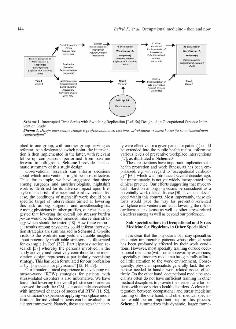
Randomized controlled intervention studies rep-resent the strongest line of evidence in etiologic re-search. Moreover, they provide a very practical means of testing the efficacy of prevention strate-gies [3]. With regard to ameliorating occupational stressors, one of the most robust intervention study designs is the ”Interrupted time-series with switch-ing replication” [56]. The intervention is first ap-
144
plied to one group, with another group serving as referent. At a designated switch point, the interven-tion is then implemented in the latter, with relevant follow-up comparisons performed from baseline forward in both groups. Scheme 1 provides a sche-matic summary of this study design.
Observational research can inform decisions about which interventions might be most effective. Thus, for example, we have suggested that since among surgeons and anesthesiologists, nightshift work is identified for its adverse im pact upon life-style-related risk of cancer and cardio vascular dis-ease, the conditions of nightshift work should be a specific target of interventions aimed at lowering this risk among surgeons and an esthesiologists. Among physicians in other profiles, our results sug-gested that lowering the overall job stressor burden per se would be the recommended intervention strat-egy which should be tested [18]. How these empiri-cal results among physicians could inform interven-tion strategies are summarized in Scheme 2. On-site visits to the worksite can yield invaluable insights about potentially modifiable stressors, as illustrated for example in Ref. [57]. Participatory action re-search [58] whereby the persons involved in the study actively and iteratively contribute to the inter-vention design represents a particularly promising strategy. This has been formulated for our profession as by ”physicians for physicians” [12, 14, 59].
Our broader clinical experience in developing re-turn-to-work (RTW) strategies for patients with stress-related disorders is also informative. We have found that lowering the overall job stressor burden as assessed through the OSI, is consistently associated with improved chances of successful RTW [11, 52]. The clinician’s experience applying workplace modi-fications for individual patients can be invaluable in a larger framework. Namely, those changes that clear-
ly were effective for a given patient or patient(s) could be extended into the public health realm, informing various levels of preventive workplace interventions [47], as illustrated in Scheme 3.
These realizations have important implications for health protection and work fitness, as has been em-phasized, e.g. with regard to ”occupational cardiolo-gy” [60], which was introduced several decades ago, but unfortunately, is not yet widely incorporated into clinical practice. Our efforts suggesting that myocar-dial infarction among physicians be considered as a potentially work-related disease [50] have been devel-oped within this context. Most importantly, these ef-forts would pave the way for prevention-oriented workplace interventions aimed at lowering the risk of cardiovascular disease as well as other stress-related disorders among as well as beyond our profession.
Sub-specializations in Occupational and Stress Medicine for Physicians in Other Specialties?
It is clear that the physicians of many specialties encounter innumerable patients whose clinical state has been profoundly affected by their work condi-tions. However, most specialty training outside occu-pational medicine (with some noteworthy exceptions, especially pulmonary medicine) has generally afford-ed little attention to the work environment. Conse-quently, physician specialists generally lack the ex-pertise needed to handle work-related issues effec-tively. On the other hand, occupational medicine spe-cialists often do not have sufficient training in other medical disciplines to provide the needed care for pa-tients with more serious health disorders. A closer in-tegration between occupational and stress medicine training on the one hand, and other medical special-ties would be an important step in this process. Scheme 3 summarizes this dynamic, larger frame-
Belkić K, et al. Occupational medicine - then and now
Scheme 1. Interrupted Time Series with Switching Replication [Ref. 56] Design of an Occupational Stressor Inter-vention StudyShema 1. Dizajn interventne studije o profesionalnim stresorima: ,,Prekidana vremenska serija sa naizmeničnom replikacijom”

Med Pregl 2014; LXVII (5-6): 139-147. Novi Sad: maj-juni. 145
work in which such clinicians could best contribute to develop and implement evidence-based guidelines for creating healthier work places for our profession as well as for our patients.
Where are We Now and where do We Go from Here?
Much has changed since the earlier days when occupational medicine was a leading discipline
Scheme 2. Occupational Intervention Strategies for Physicians Informed by Observational Findings from the OSIShema 2. Intervencije na uslovima rada lekara bazirane na empirijskim istraživanjima primenom OSI
Scheme 3. The Role of the Clinician/Physician Specialist and the OSI in the Larger Framework of Creating Healthy Workplaces, as adapted from Refs. [11, 47] Shema 3. Uloga lekara/kliničara specijaliste i OSI u kreiranju zdravog radnog mesta, prilagođena iz Ref. [11, 47].

146
with broad influence and extensive empowerment to implement recommendations in the working lives of millions of people in this region. Not only has this empowerment been severely eroded, but, as discussed, the nature of work-related factors that impact upon health has also changed pro-foundly, not only here, but globally. We have out-lined some strategic directions based upon empiri-cal evidence garnered in large measure from this region. Intervention studies in which job stressors are ameliorated as a therapeutic/preventive modal-
ity are urgently needed and the logical starting point is with our own profession. We have also suggested how the relevant clinical competence should be enhanced. Alongside this clinical en-hancement should be the full restoration of the empowerment of these physician specialists to im-plement work-related recommendations. A partici-patory action research perspective [58] by physi-cians and for physicians and for our patients would be the recommended approach.
References1. Stanković D, Beritić T, Cvetanov V, Killibarda M, Mar-
kičević A, Mikov M, et al, eds. Medicina rada. Beograd, Za-greb: Medicinska Knjiga; 1984.
2. South East Europe Workplace Academy - SEEWA 2011 Healthy workplaces for health workers - June 27 to July 2, 2011 – Zagreb: SEEWA; 2011.
3. Belkić K, Landsbergis P, Schnall P, Baker D. Is job strain a major source of cardiovascular disease risk? Scand J Work Environ Health 2004;30:85-128.
4. Hu H, Speizer F. Influence of environmental and occupa-tional hazards on disease. In: Braunwald E, Fauci A, Kasper D, Hauser D, Longo D, Jameson J, eds. Harrison’s principles of in-ternal medicine. 15th ed. New York (NY): McGraw-Hill, Inc; 2001. p 19–21.
5. Karasek RA. Job demands, job decision latitude and mental strain: Implications for job redesign. Adm Sci Q 1979;24:285-307.
6. Karasek RA, Theorell T. The demand-control-support model and CVD. Occup Med 2000;15:78-83.
7. Siegrist J. Adverse health effects of high-effort/low-re-ward conditions. J Occup Health Psych 1996;1:27-41.
8. Siegrist J, Peter R. Measuring effort-reward imbalance. Düsseldorf: University of Düsseldorf; 1999.
9. Kivimäki M, Virtanen M, Elovainio M, Kouvonen A, Vään änen A, Vahtera J. Work stress in the etiology of coro-nary heart disease: a meta-analysis. Scand J Work Environ Health 2006;32:431-42.
10. Stansfeld S, Candy B. Psychosocial work environment and mental health: a meta-analytic review. Scand J Work En-viron Health. 2006;32:443-62.
11. Belkić K, Savić Č. Job stressors and mental health: a proactive clinical perspective. London: World Scientific Pu-blishers; 2013.
12. Belkić K, Savić Č. The occupational stress index: an ap-proach derived from cognitive ergonomics applicable to clinical practice. Scand J Work Environ 2008;32(Suppl 6):169-75.
13. Welford AT. The measurement of sensory-motor per-formance: survey and reappraisal of twelve years’ progress. Ergonomics 1960;3:189-230.
14. Belkić K. The occupational stress index: an approach derived from cognitive ergonomics and brain research for cli-nical practice. Cambridge: Cambridge Interna tional Science Publishing; 2003.
15. Gaillard AWK. Comparing the concepts of mental load and stress. Ergonomics 1993;36:991-1005.
16. Fuller R. A conceptualization of driving behaviour as threat avoidance. Ergonomics 1984;27:1139-55.
17. Belkić K, Nedić O. Workplace stressors and lifestyle-relat-ed cancer risk factors among female physicians: assessment using the occupational stress index. J Occup Health 2007;49:61-71.
18. Belkić K, Nedić O. Night work, total occupational bur-den and cancer/cardiovascular risk factors in physicians. Med Pregl 2012;65(11-12):461-9.
19. Belkić K, Pavlović S, Djordjević M, Uglješić M, Micković Lj. Determinants of cardiac risk in professional drivers. Kardiologija 1992;13:145-9.
20. Belkić K, Savić Č, Theorell T, Rakić Lj, Ercegovac D, Djordjević M. Mechanisms of cardiac risk among professional drivers. Scand J Work Environ Health 1994;20:73-86.
21. Djindjić N, Jovanović J, Djindjić B, Jovanović M, Jova-nović J. Associations between the occupational stress index and hypertension, type 2 diabetes mellitus, and lipid disorders in middle-aged men and women. Ann Occup Hyg 2012;56:1051-62.
22. Emdad R, Belkić K, Theorell T, Cizinsky S, Savić Č, Olsson K. Work environment, neurophysiologic and psychop-hysiologic models among professional drivers with and witho-ut cardiovascular disease. Stress Med 1997;13:7-21.
23. Emdad R, Belkić K, Theorell T, Cizinsky S. What prevents professional drivers from following physicians’ car-diologic advice? Psychother Psychosom 1998;67:226-40.
24. Nedić O, Belkić K, Filipović D, Jocić N. Work stre-ssors among physicians with the acquired cardiovascular dis-orders: assessment using the occupational stress index. Med Pregl 2008;61:22-34.
25. Nedić O, Belkić K, Filipović D, Jocić N. Gender as an important effect modifier between exposure to work stressors among physicians and the occurrence of cardiovascular disea-se. Med Pregl 2008;61:343-9.
26. Nedić O, Belkić K, Filipović D, Jocić N. Job stressors among female physicians: relation to having a clinical diagnosis of hypertension. Int J Occup Environ Health 2010;16:330-40.
27. Uglješić M, Belkić K, Simeunovic-Micković Lj, Vu-kajlović M. Implementation of a plan for cardiac prevention among professional drivers as a high-risk group. Srp Arh Ce-lok Lek 1992;120(Suppl 1):49-51.
28. McMichael A. Standardized mortality ratios and the ”healthy worker effect”. Scratching the surface. J Occup Med 1976;18:165-8.
29. Van Dijk F. Work-related musculoskeletal and mental disorders. Central Eur J Occup Environ Med 1995;1:292-305.
30. Aasland OG, Ekeberg O, Schweder T. Suicide rates from 1960 to 1989 in Norwegian physicians compared with other educational groups. Soc Sci Med 2001;52:259-65.
31. Hawton K, Agerbo E, Simkin S, Platt B, Mellanby R. Risk of suicide in medical and related occupational groups: a national study based on Danish case population-based regis-ters. J Affective Disord 2011;134:320-6.
32. Schernhammer E, Colditz G. Suicide rates among physicians: a quantitative and gender assessment (meta-analy-sis). Am J Psychiatry 2004;161:2295-302.
Belkić K, et al. Occupational medicine - then and now

Med Pregl 2014; LXVII (5-6): 139-147. Novi Sad: maj-juni. 147
33. Arigoni F, Bovier P, Sappino A. Trend of burnout among Swiss doctors. Swiss Med Wkly 2010; 140:w13070.
34. Kumar S, Hatcher S, Huggard P. Burnout in psychia-trists. Int J Psychiatry Med 2005;35:405-16.
35. Balch C, Shanafelt T. Combating stress and burnout in surgical practice. Adv Surg 2010;44:29-47.
36. Shanafelt T, Boone S, Tan L, Dyrbye L, Sotile W, Satele D, et al. Burnout and satisfaction with work-life bal-ance among US physicians relative to the general US popula-tion. Arch Intern Med 2012;172:1377-85.
37. Frank E, Dingle A. Self-reported depression and sui-cide attempts among U.S. women physicians. Am J Psychiatry 1999;156:1887-94.
38. Fridner A, Belkić K, Marini M, Minucci D, Pavan L, Schenck-Gustafsson K. Recent suicidal ideation among fe-male university hospital physicians in Sweden and Italy: cross-sectional associations with work stressors. Gender Med. 2009;6:314-28.
39. Fridner A, Belkić K, Minucci D, Pavan L, Marini M, Pingel B, et al. The work environment and recent suicidal thoughts among male university hospital physicians in Swe-den and Italy. Gender Med 2011;8:269-79.
40. Eliot RS. Stress and the heart. Mt. Kisco: Futura Pu-blishing; 1974.
41. Carpenter L, Swerdlow A, Fear N. Mortality of doctors in different specialities: findings from a cohort of 20 000 NHS hospital consultants. Occup Environ Med 1997;54:388-95.
42. Nedić O, Filipović D, Solak Z. Job stress and cardiovascu-lar diseases among health workers. Med Pregl 2001;54:423-31.
43. Schwartz J, Pickering T, Landsbergis P. Work-related stress and blood pressure: current theoretical models and con-siderations from a behavioral medicine perspective. J Occup Health Psychol 1996;1:287-310.
44. Uchiyama S, Kurasawa, T, Sekizawa T, Nakatsuka H. Job strain and risk of cardiovascular events in treated hyper-tensive Japanese workers: hypertension follow-up group study. J Occup Health 2005;47:102-11.
45. Nedić O. Occupational stressors and physician health (dissertation). Novi Sad, University of Novi Sad; 2006.
46. Nedić O, Belkić K. Workplace health promotion among surgeons and anesthesiologists, 12th Congress of occupational medicine. Serbia, Zlatibor, October 2013. Belgrade: SLD; 2013.
47. Fisher J, Belkić K. A public health approach in clinical practice.Occup Med 2000;15:245-53.
48. Markowitz S. The role of surveillance in occupational health. In: Rom W, ed. Environmental and occupational medi-cine. Philadelphia: Lippincott-Raven Publishers; 1998. p. 19-29.
49. Mullan R, Murthy L. Occupational sentinel health events: an updated list for physician recognition and public health surveillance. Am J Ind Med 1991;19:775-99.
50. Nedić O. Akutni infarkt miokarda kao povreda na radu ili profesionalno oboljenje. Pravni Život 2006;1(9):497-510.
51. Ahola K, Virtanen M, Honkonen T, Isometsä E, Aro-maa A, Lönnqvist J. Common mental disorders and subse-quent work disability: a population-based health 2000 study. J Affect Disord 2011;134:365-72.
52. Belkić K. Return-to-work in Scandinavia: experience and insights with a focus on Sweden. In: Talmage JB, Melhorn JM, Hyman M, eds. A physician’s guide to return to work. Chi-cago: American Medical Association Press; 2011. p. 465-72.
53. Guthrie R, Ciccarelli M, Babic A. Work-related stress in Australia: the effects of legislative interventions and the cost of treatment. Int J Law Psychiatry 2010;33:10-5.
54. Pro J. Working with common psychiatric problems. In: Talmage JB, Melhorn JM, Hyman M. Physician’s guide to return to work. Chicago: American Medical Association Press; 2005. p. 305-20.
55. Sultan-Taieb H. Modeling the economic burden of di-seases imputable to stress at work. Eur J Health Econom 2005; 50:16-23.
56. Beehr T, O’Hara K. Methodological designs for the evaluation of occupational stress interventions. In: Kasl S, Cooper C, eds. Research methods in stress and health psycho-logy. New York: John Wiley; 1987. p. 79-112.
57. Belkić K, Schnall P. On a San Francisco public tran-sport line: burden and consequences upon the human opera-tor. The Job Stress Network Website: Center for Social Epide-miology (www.workhealth.org), 2000.
58. Israel B, Goldenhar L, Baker E, Heaney C. Occupatio-nal stress, safety and health: conceptual framework and prin-ciples for effective prevention interventions. J Occup Health Psychol 1996;1:261-86.
59. Savić Č. The physician and stress.17th Meeting in Sombor Medical Society of Vojvodina. Sombor: DLV; 2002. str. 52-4.
60. Maisano G, Gobbato F, Julian D, Mulcahy R. Workshop on occupational cardiology. Eur Heart J 1988;9(Suppl L):1-131.
Rad je primljen 13. III 2014.Prihvaćen za štampu 13. III 2014.BIBLID.0025-8105:(2014):LXVII:5-6:139-148.

Štampu ovog časopisa ”Medicinski pregled” pomogla je Vlada AP Vojvodine
Printing of this journal ”Medical Review”has been supported bythe Government of the AP of Vojvodina
Med Pregl 2014; LXVII (5-6): 149-153. Novi Sad: maj-juni. 149
Introduction
Candida species fungi are commonly present in healthy individuals, and Candida albicans is the
most prevalent species [1, 2]. The leading cause of candidiasis, Candida albicans, is a dimorphic fun-gus that resides as a commensal of the oral mucosa and the gastrointestinal tract mucosa. Changes in the oral ecosystem or in the immunological system of the host can lead to candidiasis development [2–4]. Candidiasis has become a human disease of increas-
Corresponding Author: Prof. dr Ljiljana Kesić, Klinika za stomatologiju, 18000 Niš, Bulevar Zorana Đinđića 52, E-mail: [email protected]
ORIGINAL STUDIESORIGINALNI NAUČNI RADOVIUniversity of Niš, Faculty of Medicine, Dental Clinic Original study Department of Oral medicine and Periodontology1 Originalni naučni radPrivate dental practice, Novi Sad2 UDK 616.31-022.7-094:612.08University of Niš, Faculty of Medicine, Institute of Patological Anatomy3 DOI: 10.2298/MPNS1406149KUniversity of Novi Sad, Faculty of Medicine, Dental Division, Doctoral Academic Studies Student4
MORPHOLOGIC AND MORPHOMETRIC ANALYSIS OF ALTERNATIONS IN THE ORAL CAVITY CAUSED BY CANDIDA ALBICANS – EXPERIMENTAL WORK
MORFOLOŠKA I MORFOMETRIJSKA ANALIZA PROMENA U USNOJ DUPLJI KOJE JE IZAZVALA CANDIDA ALBICANS – EKSPERIMENTALNI RAD
Ljiljana KESIĆ1, Radojka DELIĆ2, Dragan MIHAILOVIĆ3, Milica S. PETROVIĆ1 and Tijana Đ. DELIĆ4
SummaryIntroduction. Candidiasis has become a human disease of increas-ing importance in the last decades. The aim of the study is to estab-lish pathomorphological alterations caused by the blastospores of the Candida albicans as well as morphometric alterations. Materi-al and Methods. The experiment was carried out on 2.5-month-old rats, weighting 110–130 g. The study sample was divided into the animals infected by a submucous inoculation in the periodontal re-gion and the controls. The gingival specimens were taken, prepara-tions were done and stained by the hematoxylin-eosin and Periodic acid Schiff methods. Results. The following alterations were found out by the stereological analysis: an average volume of nuclei of the gingival epithelial cells was 111.82 µm3 (SD=25.34) on the first day. A statistically significant increase in the volume of nuclei in the ex-perimental group began to occur from the fourth day (202.97 µm3; SD=31.16, p<0.05) and the highest value of the nuclei volume was found out on the eight day of the experiment (316.83 µm3; SD=40.15). Conclusion. Blastospores of Candida albicans are pathogenic for the gingival tissue where they cause degenerative necrotic alterations of the granulomatous character and after the fourth day from the inoculation, the development of the pseudohy-phae was observed. The obtained values of stereologic measure-ment show the acute increase in the volume of nuclei.Key words: Mouth; Candida albicans; Candidiasis; Rats; Gingiva; Spores, Fungal
SažetakPoslednjih godina kandidoza predstavlja oboljenje od posebnog značaja. Cilj ovog istraživanja bio je da se utvrde i ispitaju pato-morfološke promene uzrokovane blastosporama kandide (Candi-da albicans) kao i morfometrijske promene. Materijal i metode. Eksperimentalna studija je urađena na pacovima starosti 2,5 mese-ci, težine 110−130 g. Životinje su inficirane submukoznom inoku-lacijom u region parodonta. Takođe, formirana je kontrolna grupa. Uzimani su isečci sa gingive, napravljeni preparati su bojeni sa he-matoksilin-eozin i perjodna kiselina-Schiff metodom. Rezultati. Stereološkom analizom utvrđene su sledeće promene: u prvom danu srednja vrednost zapremine nukleusa epitelnih ćelija gingi-ve iznosila je 111,82 µm3 (SD = 25,34). Zapaženo je statistički značajno povećanje zapremine jedra od četvrtog dana (202,97 µm3; SD = 31,16, p < 0,05), a najveća vrednost zapremine jedra iznosila je osmog dana eksperimenta (316,83 µm3; SD = 40,15). Zaključak. Blastospore kandide (Candida albicans) patogene su za tkivo gingive, gde uzrokuju degenerativne nekrotične alteracije granulomatoznog karaktera. Posle četvrtog dana od inokulacije, pronađene su pseudohife. Dobijene vrednosti stereometrijske ana-lize pokazale su postojanje akutnog uvećanja zapremine jedara.Ključne reči: Usna šupljina; Candida albicans; Kandidijaza; Pacovi; Gingiva; Spore gljivica
-----------------------------In memory of my father Prof. dr. Georgi Penev (1933-2012), Medi-cal faculty University of Niš, Institute for Pathological Anatomy

Kesić Lj, et al. Analysis of changes at oral experimental candidiasis150
ing importance in the last decades due to the increas-ing number of patients with immunological involve-ment associated with the infection by the human im-munodeficiency virus (HIV) and the use of immu-nosuppressive agents after organ transplantation or antineoplastic therapy [1]. In immunocompromised hosts, however, saprophytic colonization often leads to opportunistic mucosal or life-threatening deep or-gan infection. Invasion of the human gastrointestinal mucosa by Candida albicans and its passage across the bowel wall into the bloodstream is an important portal of entry for this opportunistic pathogen in the neutropenic host, leading to systemic or disseminat-ed candidiasis [5]. In addition, hematogenous candi-diasis is a frequent complication in treatment of pa-tients with acute leukemia [6]. Many researchers de-veloped several experimental models in rats in order to understand the mechanisms related to the patho-genesis of oral candidiasis. The oral cavity of these animals is easily colonized by Candida and develops similar lesion in relation to those observed among human beings [1, 3]. Many predisposing factors for oral candidiasis have been studied in experimental models, such as: broad-spectrum antibiotics therapy [7, 8], the use of acrylic prosthesis [9], diabetes mel-litus [10], topical use of corticosteroids [11], xerosto-mia [12, 13] and immunosuppressive therapy [4, 14]. An important cofactor associated with the pathogen-esis of oral candidiasis appears to be the virulence of the infecting organism [15, 16]. The specific features of the fungus that contribute to the development of oral candidiasis include its ability to adhere to and colonize the oral mucosa [17], its ability to form cy-lindrical appendages termed germ tubes [18], and its cell surface hydrophobicity [19]. In addition, pheno-typic and genotypic switching [20, 21], extracellular aspartyl proteinase secretion [22, 23], and phospholi-pase production [24] appear to play a subsidiary role in the pathogenicity.
Animal models represent powerful tools in eluci-dating the molecular and cellular pathogenesis of can-didiasis (previously reviewed in [1, 3]). The principal advantage in studying animals instead of human be-ings is that the animal and its environment can be controlled [4], allowing a precise cause-and-effect longitudinal analysis of host-pathogen interactions. In addition, these models obviate the procurement of tis-sue samples from human patients, which can often be problematic. The usefulness of animal models of can-didiasis includes not only the study of pathogenesis but also the in vivo assessment of novel antifungals, immunomodulators and potential Candida vaccines [25, 26]. The aim of thix study was to establish patho-morphological alterations caused by the blastospores of Candida albicans as well as morphometric altera-tions in the rats.
Material and Methods
The experiment was carried out on 2.5-month-old rats, weighting 110–130 g. A group of ten rats was infected with blastospores of Candida albi-cans in the dosage of 400.000 in 0.5 ml of physio-logical solution for an animal (determined in the Spenser chamber). The animals were infected by a submucous inoculation in the periodontal region. The control group consisted of three animals which were kept under the same conditions as those for the experimental group. The rats were sacrificed after 24 hours, after the second, fourth, sixth and eighth days from the moment of infection. The cuts of gin-gival tissue were taken, preparations were done and stained by the hematoxylin-eosin (HE) and Periodic acid Schiff (PAS) methods.
Stereologic analysis of the average volume of the nuclei of the gingival epithelial cells was also carried out according to the Gunderssen ”nuclea-tor principle” (1988).
The sinus-dependent test system was used to measure the intercept length according to the for-mula Vv = (l x π ∕3). The x100 objective was used.
The Student t-test was used for the statistical analysis of the obtained results.
Results
The alteration took place only at the gingival level in the gingival tissue in the acute phase up to the fourth day from the infection (Figure 1).
Inflammatory-edematous alterations were ascer-tained by the presence of the budding blastospores and pseudomicellar fibers in the gingival tissue in the acute phase and from the fourth day onwards granulomatous alterations occurred with abscess for-mation in addition to numerous predominantly eosi-nophile elements and pseudohyphae. Giant cells of the Langhans type and the foreign body type were present in complexes. The alterations appeared due to the effects of blastospores and candidine, their metabolic product (figures 2 and 3).
AbbreviationsHIV – human immunodeficiency virusAIDS – acquired immunodeficiency syndromeHE – hematoxylin-eosin
Figure 1. Gingival tissue with edema and blastospores. HE, X 400. Slika 1. Tkivo gingive sa edemom i blastosporama HE, X 400

Med Pregl 2014; LXVII (5-6): 149-153. Novi Sad: maj-juni. 151
The development of excess fibrous connective tissue, that is fibrosis and sclerosis, occurred in the chronic phase (Figure 4).
The control sample (Figure 5) showed intact epithelial lamina with subepithelial muscle tissue.
The following alterations were determined by the stereological analysis:
– an average volume of nuclei of the gingival epithelial cells on the first day was 111.82 µm3, (SD=25.34).
– a statistically significant increase in the vol-ume of nuclei in the experimental group began to occur from the fourth day (202.97 µm3; SD=31.16, p<0.05);
– the highest value of the nuclei volume was found out on the eight day of the experiment (316.83 µm3; SD=40.15).
Discussion
The animals that inoculated by Candida albicans in the oral cavity developed clinical and microscopy lesions of candidiasis in the tongue dorsum even without presenting predisposing factors, such as the administration of antibiotics, immunosuppression, carbohydrate-rich diet or xerostomia. These data confirm that the experimental candidiasis can be in-duced by a simple inoculation of a pathogenic strain of Candida albicans [28]. The recognition that Can-dida is an important pathogen, particularly in the immunocompromised host, has resulted in a vast body of in vitro investigations evaluating its virulent attributes in an attempt to elucidate the pathogenesis of the disease. The progress made in understanding some of these features, such as the mechanisms that result in adherence to host tissues [29], cell surface hydrophobicity [30], switching phenomena of the yeast [22, 31], secretion of aspartyl proteinases [24], and phospholipase production [25], is very impres-sive. Nonetheless, in vivo studies either in humans or in animals are essential to elucidate and fully com-prehend the mechanisms leading to candidal infec-tion. The host oral defenses against Candida essen-tially fall into two categories: nonspecific immune
Figure 2. Edematous gingival tissue, diffuse infiltrati-on with inflammatory elements and necrosis in the central part. HE, X 400.Slika 2. Edematozno tkivo gingive, difuzna infiltracija sa inflamatornim elementima i nekrozom u centralnom delu HE, X 400
Figure 3. Subepithelial soft tissue with Candida albi-cans plaque. HE, X 400. Slika 3. Subepitelijalno meko tkivo sa plakom Kandide albikans HE, X 400
Figure 4. Gingival tissue with granulomatous process, ne-crotic fields and Candida albicans blastospores. HE, X 400.Slika 4. Tkivo gingive sa granulomatoznim procesom, nekrotičnim poljima i blatosporama Kandide albikans HE, X 400
Figure 5. Control sample without pathohistologic alte-rations: intact epithelial lamina and subepithelial musc-le tissue. HE, X 400.Slika 5. Kontrolni uzorak bez patohistoloških prome-na: neoštećena epitelijalna lamina i subepitelijalno mi-šićno tkivo HE, X 400

152
mechanisms (e.g., integrity of the mucosae, commen-sal bacteria, polymorphonuclear leukocytes, macro-phages, and salivary factors) and specific immune mechanisms (e.g., serum antibodies, secretory anti-bodies, and cell-mediated immunity) [32]. The strati-fied squamous epithelium of the oral mucosa forms a continuous surface that protects the underlying tis-sues and functions as an impervious, mechanical barrier. The protection so provided is dependent on the degree of keratinization and the continuous desq-uamation or shedding of epithelial cells. Indeed, the latter mechanism is considered to play a pivotal role in maintaining a healthy oral mucosa and in limiting candidal colonization and infection. The interaction between Candida species and the commensal micro-bial flora is perhaps the next critical mechanism modulating oral candidal colonization [33]. The com-mensal flora regulates yeast numbers by inhibiting the adherence of yeasts to oral surfaces by competing for sites of adherence as well as for the available nu-trients. A number of studies have also shown, both in vivo in gnotobiotic mice and in vitro, that candidal colonization of epithelia could be suppressed by streptococci, which are the predominant resident commensals of oral mucosal surfaces [33–35]. Con-sequently, the process of infection can be viewed as a competition between the ability of fungal cells to multiply and the host antimicrobial response. Obvi-ously, for an infected host to survive and recover, it is crucial to impede the ability of pathogens to multiply [36]. Although different species of Candida, such as Candida glabrata, Candida tropicalis, Candida kru-sei and Candida dubliniensis, are at present recog-
nized as increasing opportunistic pathogens specially in HIV infected individuals and acquired immuno-deficiency syndrome (AIDS) patients, Candida albi-cans still remains the most common yeast isolated in humans [37]. In some ways, it is surprising that Can-dida albicans is uniquely associated with animals and human, as it has no specific nutrient require-ments that would prevent it from surviving in the outside environment [38]. The use of mouse models appears appropriate since progression of both sys-temic and oral candidiasis closely resembles that ob-served in humans [39].
Conclusion
Based on the results obtained from the experi-mental testing of effects of blastospores of Candi-da albicans the following conclusions can be drawn:
1. Blastospores of Candida albicans given in dosage of 400.000 in 0.5 ml of the physiological solution are pathogenic for the gingival tissue where they cause degenerative necrotic alterations of the granulomatous character. We suppose that candidine, as a metabolic product of Candida al-bicans, play a great role in the pathohistological alterations of the gingiva.
2. Development of the pseudohyphae was found after the fourth day from the inoculation
3. The obtained values of stereologic measure-ment in the acute increase of the nuclei volume is probably a consequence of the nearby focus of in-fection or toxic effects of Candida albicans.
Kesić Lj, et al. Analysis of changes at oral experimental candidiasis
References1. Samaranayake YU, Samaranayake LP. Experimental oral
candidiasis in animal models. Clin Microbiol Rev 2001;14:398-429.2. Webb BC, Thomas CJ, Wilcox MD, Harty DW, Knox
KW. Candida-associated denture stomatitis. Aetiology and management: a review. Aust Dent J 1998;43:45-50.
3. Alenn CM. Animal models of oral candidiasis: a revi-ew. Oral Surg Oral Med Oral Pathol 1994;78:216-21.
4. de Repentigny L. Animal models in the analysis of Candida host-pathogen interactions. Curr Opin Microbiol 2004;7:324-9.
5. Walsh TJ, Merz WG. Pathologic features in the human alimentary tract associated with invasiveness of Candida tro-picalis. Am J Clin Pathol 1986;85:498-502.
6. Abi-Said D, et al. The epidemiology of hematogenous candidiasis caused by different Candida species. Clin Infect Dis 1997;24:1122-8.
7. Allen CM, Beck FM, Lurie FA, Pinsky HM. Role of te-tracycline in pathogenesis of chronic can didiasis of rat tongu-es. Infect Immun 1985;47:480-3.
8. Fisker AV, Rindon-Schiott C, Philipsen HP. Short-term oral candidiasis in rats, with special reference to the site of in-fection. Acta Pathol Microbiol Immunol Scand 1982;90:49-57.
9. Shakir BS, Smith CJ, Martin MV. Epithelial mitotic ac-tivity during the induction of palatal candidiasis in the Wistar rat. J Oral Pathol 1986;15:375-80.
10. Wasan KM, Conklin JS. Evaluation of renal toxicity and antifungal activity of free and liposomal amphotericin B fo-
llowing a single intravenous dose to diabetic rats with systemic candidiasis. Antimicrob Agents Chemother 1996;40(8):1806-10.
11. Deslauries N, Coulombe C, Carre B, Goulet JP. Topi-cal application of a corticosteroid destabilizes the host-parasi-te relationship in an experimental model of the oral carrier state of Candida albicans. FEMS Immunol Med Microbiol 1995;11:45-55.
12. Jorge AOC, Totti MAG, Almeida OP, Scully C. Oral candidiasis established in the sialoadenectomized rat. J Oral Pathol Med 1993;22:54-6.
13. Jorge AOC, Totti MAG, Almeida OP, Scully C. Effect of sialoadenectomy on the carriage of Candida albicans in the mouths of rats. J Oral Pathol Med 1993;22:138-40.
14. Takakura N, et al. A novel murine model of oral can-didiasis with local symptoms characteristic of oral thrush. Microbiol Immunol 2003;47:321-6.
15. McCullough MJ, Ross BC, Reade PC. Candida albi-cans: a review of its history, taxanomy, epidemiology, viru-lence attributes and methods of strain differentiation. Int J Oral Maxillofac Surg 1996;25:36-144.
16. Ruechel R. Virulence factors of Candida species. In: Samaranayake LP, MacFarlane TW, eds. Oral Candidiasis. London: Wright; 1990.
17. Kennedy MJ. Adhesion and association mechanisms of Candida albicans. Curr Top Med. Mycol 1998;2:73-169.

Med Pregl 2014; LXVII (5-6): 149-153. Novi Sad: maj-juni. 153
18. Casanova M, Cervera AM, Gozalbo D, Martinez, JP. Hemin induces germ tube formation in Candida albicans. In-fect Immun 1997;65:4360-4.
19. Hazen KC. Participation of yeast cell surface hydrop-hobicity in adherence of Candida albicans to human epithelial cells. Infect Immun 1989;57:1894-900.
20. Scherer S, Magee PT. Genetics of Candida albicans. Microbiol Rev 1990;54:226-41.
21. Soll DR. High frequency switching in Candida albi-cans. Clin Microbiol Rev 1992;5:183-203.
22. De Bernardis FP, et al. Elevated aspartic proteinase secretion and experimental pathogenicity of Candida albicans isolates from oral cavities of subjects infected with human immunodeficiency virus. Infect Immun 1996;64:466-71.
23. Wu T, Samaranayake LP, Cao BY, Wang J. In vitro proteinase production by oral Candida albicans isolates from individuals with and without HIV infection and its attenuati-on by antimycotic agents. J Med Microbiol 1996;44:311-6.
24. Lane T, Garcia JR. Phospholipase production in morpho-logical variants of Candida albicans. Mycose 1991;34:217-20.
25. Bromuro C, et al. Interplay between protective and in-hibitory antibodies dictates the outcome of experimentally disseminated candidiasis in recipients of a Candida albicans vaccine. Infect Immun 2002;70:5462-70.
26. Roilides E, Walsh T. Recombinant cytokines in au-gmentation and immunomodulation of host defenses against Candida spp. Med Mycol 2004;42:1-13.
27. Allen CM, Paulson R, Duncan R. Clinical, histologic and scanning electron microscopic study of the development of chronic candidiasis of the rat tongue. J Oral Pathol Med 1989;18:352-9.
28. Kennedy MJ, et al. Molecular basis of Candida albi-cans adhesion. Med Mycol 1992;30:95-122.
29. Hazen KC, Brawner DL, Riesselman MH, Cutler JE, Jutila MA. Differential adherence of hydrophobic and hydrop-hilic Candida albicans yeast cells to mouse tissues. Infect Im-mun 1991;59:907-12.
30. Soll DR, Langtimm CJ, McDowell J, Hicks J, Galask R. High-frequency switching in Candida strains isolated from vaginitis patients. J Clin Microbiol 1987;25:1611-22.
31. Challacombe SJ. Immunology of oral candidiasis. In: Samaranayake LP, MacFarlane TW, eds. Oral candidiasis. London: Wright; 1990. p. 104-23.
32. Samaranayake LP, MacFarlane TW, editors. Oral can-didiasis. London: Wright; 1990.
33. Liljemark WF, Gibbons RJ. Suppresion of C. albicans by human oral streptococci in gnotobiotic mice. Infect Immun 1973;8:846-9.
34. Nair R, Samaranayake LP. The effect of oral commen-sal bacteria on candidal adhesion to human buccal epithelial cells. J Med Microbiol 1996;45:179-85.
35. Samaranayake LP, Hamilton D, MacFarlane TW. The effect of indigenous bacterial populations on buccal epithelial cells on subsequent microbial adhesion in vitro. Oral Microbi-ol Immunol 1994;9:236-40.
36. Mims CA, Nash A, Stephen J. In MIMS’ Pathogenesis of infectious disease. London: Academic press; 2001. p. 67-82.
37. Giammanco GM, et al. Value of morphotyping for the characterization of Candida albicans clinical isolates. Mem Inst Oswaldo Cruz 2005;5:483-90.
38. Hube B. From commensal to pathogen: stage- and ti-ssue-specific gene expression of Candida albicans. Curr Opin Microbiol 2004;7:336-41.
39. Rozell B, Ljungdahl PO, Martínez P. Host-pathogen interactions and the pathological consequences of acute syste-mic Candida albicans infections in mice. Curr Drug Targets 2006;7:483-94.
Rad je primljen 14. III 2014.Recenziran 21. III 2014.Prihvaćen za štampu 25. III 2014.BIBLID.0025-8105:(2014):LXVII:5-6:149-153.
154
Introduction
Psoriasis is a chronic, inflammatory, immune-mediated skin disease characterized by increased epidermal proliferation and differentiation, and ac-celerated angiogenesis with dilated blood vessels
and inflammation in the skin [1]. The prevalence of psoriasis is the same in both sexes, and in all socio-economic groups in the society [2]. Epidemiologic and immunogenetic studies have indicated that pre-disposition for psoriasis is hereditary. Belić et al. have shown the correlation among hereditary com-
Corresponding Author: Doc. dr Vesna Gajanin, Univerzitet u Banjoj Luci, Medicinski fakultet Banja Luka,78000 Banja Luka, Save Mrkalja 14, E-mail: [email protected]
Clinical Center of Banja Luka1 Original study University of Banja Luka Originalni naučni radFaculty of Medicine2 UDK 616.517-08-036.8 DOI: 10.2298/MPNS1406154N
COMPARATIVE ANALYSIS OF SUCCESS OF PSORIASIS TREATMENT WITH STANDARD THERAPEUTIC MODALITIES AND BALNEOTHERAPY
KOMPARATIVNA ANALIZA USPEHA LEČENJA PSORIJAZE STANDARDNIM TERAPIJSKIM MODALITETIMA I BALNEOTERAPIJOM
Đuka NINKOVIĆ BAROŠ1, Vesna S. GAJANIN2, Radoslav B. GAJANIN1 and Bogdan ZRNIĆ2
SummaryIntroduction. Psoriasis is a chronic, inflammatory, immune-me-diated skin disease. In addition to standard therapeutic modalities (antibiotics, cytostatics, phototherapy, photochemotherapy and re-tinoids), nonstandard methods can be used in the treatment of psoriasis. This includes balneotherapy which is most commonly used in combination with therapeutic resources. The aim of this research was to determine the length of remission of psoriasis in patients treated with standard therapeutic modalities, balneothe-rapy, and combined treatment (standard therapeutic modalities and balneotherapy). Material and Methods. The study analyzed 60 adult patients, of both sexes, with different clinical forms of psoriasis, who were divided into three groups according to the applied therapeutic modalities: the first group (treated with stan-dard therapeutic modalities), the second group (treated with bal-neotherapy) and the third group (treated with combined therapy-standard methods therapy and balneotherapy). The Psoriasis Area and Severity Index was determined in first, third and sixth week of treatment for all patients. The following laboratory analysis were performed and monitored: C reactive protein, iron with total iron binding capacity, unsaturated iron binding capacity and ferri-tin, uric acid, rheumatoid factors and antibodies to streptolysin O in the first and sixth week of treatment. Results. The average len-gth of remission in patients treated with standard therapeutic mo-dalities and in those treated with balneotherapy was 1.77 ± 0.951 months and 1.79 ± 0.918 months, respectively. There was a stati-stically significant difference in the duration of remission between the patients treated with combination therapy and patients treated with standard therapeutic modalities (p=0.019) and balneotherapy (p=0.032). Conclusion. The best results have been achieved when the combination therapy was administered.Key words: Psoriasis; Drug Therapy; Balneology; Combined Modality Therapy; Remission Induction; Body Surface Area; Severity of Illness Index; Diagnostic Tests, Routine
SažetakUvod. Psorijaza je hronična, inflamatorna, imunoposredova-na kožna bolest. Pored standardnih terapijskih modaliteta (anti-biotici, citostatici, fototerapija, fotohemoterapija i retinoidi), u terapiji se primenjuju i nestandardne terapijske metode kao bal-neoterapija, ali najčešće kombinacija više terapijskih sredstava. Cilj rada bio je da se utvrdi dužina remisije psorijaze kod paci-jenata lečenih standardnim terapijskim modalitetima, balneo-terapijom i kombinovanim lečenjem (standardnim terapijskim modalitetima i balneoterapijom). Materijal i metode. Analizi-rano je 60 odraslih pacijenata oba pola, obolelih od različitih kliničkih oblika psorijaze, podeljenih u tri grupe prema prime-njenim terapijskim modalitetima: grupa I (lečena standardnim terapijskim modalitetima), grupa II (lečena balneoterapijom) i grupa III (lečena kombinovanim terapijskim modalitetima − standardnim metodama i balneoterapijom). Svim pacijentima smo određivali indeks procene težine psorijaze u 1, 3. i 6. nede-lji lečenja. Pratili smo laboratorijske analize: C-reaktivni pro-tein, gvožđe, ukupni kapacitet vezanog gvožđa, kapacitet vezi-vanja nezasićenog gvožđa, feritin, mokraćnu kiselinu, reuma-toidne faktore i antitela na streptolizin O u 1. i 6. nedelji lečenja. Rezultati. Prosečna dužina remisije kod pacijenata lečenih standardnim terapijskim modalitetima iznosi 1,77 ± 0,951 me-seci, a kod pacijenata lečenih balneoterapijom iznosi 1,79 ± 0,918 meseci. Kod pacijenata lečenih kombinovanom terapijom, period remisije je u proseku trajao 2,47 ± 0,743 meseci. Postoji statistički značajna razlika u dužini trajanja remisije između pacijenata lečenih kombinovanom terapijom i pacijenta lečenih standardnim terapijskim modalitetima (p = 0,019), odnosno balneoterapijom p = 0,032). Zaključak. Primena kombinovane terapije pokazala je najbolje rezultate u lečenju psorijaze.Ključne reči: Psorijaza; Terapija; Balneologija; Kombinovani terapijski modaliteti; Remisija; Površina tela; Indeks procene težine bolesti; Rutinski dijagnostički testovi
Ninković Baroš Đ, et al. Treatment of psoriasis
Med Pregl 2014; LXVII (5-6): 154-160. Novi Sad: maj-juni. 155
ponent, frequency and time of appearance of psoria-sis [3]. Psoriasis is associated with a certain human leukocyte antigen (HLA), and it is believed that the immune system plays an important role in the patho-genesis of psoriasis [4]. Poljački et al. have found as-sociation between psoriasis and other dermatologi-cal diseases (lichen planus, alopecia areata and vitil-igo) [5]. Psoriasis is characterized by clearly defined erythematous plaques and/or papules that are cov-ered by silver-white scaling. According to the degree of the skin involvement, psoriasis can be localized or generalized, which may turn into erythrodermia (involvement of skin greater than 90%) [6-8]. This disease may be acute, subacute, but it is usually chronic. The plaque form of psoriasis is the most common form of psoriasis [9]. The diagnosis of pso-riasis is usually set on the basis of medical history, clinical and diagnostic signs (”stearin candles”, ”last skins” and ”blood droplets”) [10]. The definitive di-agnosis is confirmed by pathological examination [11]. According to the percentage of the affected skin, which is determined by Evans formula for burn, calculating the Psoriasis Area and Severity In-dex (PASI score), which analyzes the degree of ery-thema, infiltration and desquamation on the affected skin. Reduction in PASI score of 75% is considered the ”gold standard” for measuring the response to therapy (PASI-75) [12–14]. In patients with psoriasis, we can see a variety of laboratory abnormalities, such as the increased concentrations of C-reactive protein (CRP), leukocytosis with a predominance of neutrophils, increased elastase, lactoferrin and α1-antitrypsin. Neutrophils play a key role in the clini-cal development of psoriasis. Monitoring these markers could predict relapse of the disease. In addi-tion, there is also anemia, thrombocytosis and hype-ruricemia [15–19]. Patients with psoriasis often have negative values of rheumatoid factor. Increased val-ue of rheumatoid factor appears among elderly pa-tients with psoriasis and in patients with psoriasis with rheumatic comorbidity [20]. Increased levels of antibodies to streptolysin O (ASO titre) were found in the serum of patients in response to infection with hemolytic streptococcus groups A, C or G. Strepto-coccal infection usually leads to eruptive guttate psoriasis, and deterioration of other clinical forms of psoriasis. Therefore, it makes sense to introduce an-tibiotic therapy in such patients [21,22].
Treatment of psoriasis requires a multidiscipli-nary approach. The combination of multiple thera-peutic agents is another approach to the treatment of psoriasis and is convenient because it reduces the cu-mulative toxic effects of the disease [23]. Standard therapeutic modalities include phototherapy (ultravi-olet B (UVB), combined ultraviolet A (UVA) and UVB wave), photochemotherapy (UVA - with sys-temic photosensitizer - PUVA), combination of retin-oids and PUVA (Re-PUVA), cytotoxic therapy, retin-oids, antibiotic and biological therapy [24–27]. Local treatment of psoriasis includes local corticosteroid therapy, vitamin D analogues, anthralin prepara-tions, tar and topical retinoids, calcineurin inhibitors, keratinolytics and emollients [28, 29]. In addition to standard therapeutic modality in the treatment of psoriasis, we can also use non-therapeutic procedure, such as balneotherapy [30–33]. Application of ther-mal mineral water containing sulfur can have a good therapeutic effect on psoriasis and other skin diseas-es. Thermal water also has anti-inflammatory, kerat-oplastic and antipruritic effect. It is used as a single modality or in combination with other therapeutic modalities [34–37].
The aim of this study was to determine the du-ration of remission of psoriasis in patients treated with standard therapeutic modalities, balneothera-py and combination treatment (standard therapeu-tic modalities and balneotherapy). It is also neces-sary to determine whether there is a statistically significant difference in the duration of remission of psoriasis when compared to the applied treat-ments of psoriasis.
Material and Methods
This prospective study included 60 adult pa-tients, of both sexes, with different clinical forms of psoriasis. Graph 1 shows the structure of patients by sex and age. The study sample included 60 pa-tients, 62% men and 38% women. Most of the re-spondents (25.0%) were older than 60 years of age, and 11.7% younger than 29 years of age. The most common form of psoriasis in the total number of respondents was a generalized form of plaque pso-riasis, which was diagnosed in 21 cases (35%). Oth-er forms were less present (Graph 2).
The patients were randomly selected from those who had consulted the doctor at the Department of Dermatology and Venereal Diseases of the Clinical Center of Banja Luka. All the respondents gave their written consent to be included in the study. Their data were entered in the questionnaire de-signed according to the data analyzed in the study. The questionnaire contained basic information about the patients, their diagnosis, therapeutic modality, PASI score in the first, third and sixth week, and the value of laboratory tests in the first and sixth week. Psoriasis was diagnosed according to the clinical parameters and histological analysis of the modified skin in all patients. PASI score was deter-
AbbreviationsHLA – human leukocyte antigenPASI score – Psoriasis Area and Severity IndexCRP – C-reactive proteinASTO – antistreptolysin O titerUV – ultravioletPUVA – psoralen + UVARe-PUVA – retinoids and PUVARf – rheumatoid factorTIBC – total iron binding capacityUIBC – unsaturated iron binding capacityLSD – least significant difference test

156
mined according to the severity of illness and the total area of the affected skin.
The patients were divided into three groups ac-cording to the applied therapeutic modalities:
The patients at the Department of Skin and Ve-nereal Diseases, Clinical Center of Banja Luka, treated with standard therapeutic modalities (26 patients). The standard therapeutic modality in-volved systemic antibiotic therapy, methotrexate, phototherapy and photochemotherapy. Local ke-ratinolytic therapy, corticosteroid preparations, di-tranol and emollients were also applied.
The patients with different clinical forms of psoriasis treated with balneotherapy at the Kulaši Spa, Republic of Srpska, for three weeks (19 pa-tients). Water of Kulaši Spa has characteristics of healing thermal mineral water, its temperature be-ing 30.6 degrees Celsius. Water is sterile, highly alkaline (pH 11.75) with low mineralization (168 mg/l). The spa water was used twice a day in the form of baths, hot tubs and swimming in the pool for half an hour. The patients with less severe forms of psoriasis used bath tubs, while the pa-tients with moderate and severe forms of psoriasis swam in the pool twice a day for half an hour. Af-ter the end of treatment, the neutral local therapy (Galsanae cream) was prescribed.
The patients with psoriasis who were treated with standard therapeutic modalities combined with balneotherapy (15 patients). After hospitali-zation or outpatient treatment at the Department of Skin and Venereal Diseases, Clinical Center of Banja Luka, the patients were sent to the Kulaši Spa, for a period of 21 days. In cooperation with the dermatovenerologist, the spa treatment proto-col was determined according to the severity of the clinical picture measured on PASI score.
Remission was followed over the period of three months. The clinical examination and PASI scores were measured in the first, third and sixth week of treatment to determine the period of re-mission. At the beginning and at the end of the study, all patients underwent the following labora-tory tests: ASTO, Rheumatoid factor (Rf), CRP, uric acid, iron with ferritin (determined only at the end of the treatment) and total iron binding capac-ity (TIBC), unsaturated iron binding capacity (UIBC).
The statistical analysis of the results used de-scriptive and analytical statistics. For statistical
analysis of data, the Statistical Package for the So-cial Sciences (SPSS) (version 17) was used.
Results
The average values of PASI score were deter-mined in the first, third and sixth week of the treat-ment (Table 1). Fisher’s Exact Test showed a statis-tically significant difference (p=0.049) in the mean valu- es of PASI score in the first week of treatment among different groups. In the first week, the pa-tients treated with standard therapeutic modalities had PASI score values significantly higher than in the group of patients treated with balneotherapy, but in comparison with the group of patients treated with combined therapy, there were no statistically significant differences in the values of PASI scores. During the second measurement (the third week), the mean PASI scores were the highest in the group of patients treated with standard therapeutic modal-ities (11.05±9.550).
The factor analysis showed no statistically sig-nificant difference between the values of PASI scores among the tested groups of patients (p = 0.145). The third measurement (in the sixth week of treatment) showed that the group of patients treated with balneotherapy had the lowest mean values of PASI score (2.85 ± 2.556); whereas the patients treated with standard therapy had the highest value (5.68 ± 4.423).
There was no statistically significant difference in PASI scores between different groups of all pa-tients (p = 0.54). The use of Robust test showed a statistically significant difference in the values of PASI score (in sixth week) between different groups of patients (p = 0.037). In order to determine which groups of patients differed significantly among themselves, Post-hoc tests (Tukey post-hoc test) were used. These statistical tests showed that there were statistically significant differences in the values of PASI scores measured in the sixth week of treatment among the patients treated with standard therapeutic modalities and balneotherapy (p = 0.043). However,
Graph 1. Subjects by gender and ageGrafikon 1. Ispitanici po polu i starosti
Graph 2. Distribution of patients by diagnosed form of psoriasisGrafikon 2. Distribucija pacijenata prema dijagnosti-kovanom obliku psorijaze
156 Ninković Baroš Đ, et al. Treatment of psoriasis

Med Pregl 2014; LXVII (5-6): 154-160. Novi Sad: maj-juni. 157
there was no difference between the group treated with combination therapy and the one treated with standard therapy (p = 0.443), and the group treated with combination therapy and balneotherapy (p = 0.577). The results of Friedman’s test showed that there were statistically significant differences be-tween the values of PASI scores determined in the first, third and sixth week of treatment in all patients (60 patients) (p = 0.000). The values of PASI score in the sixth week of measurements were statistically significantly lower than the values of PASI score in the first week (Wilcoxon’s test: Z = -6.031, p = 0.000) and the values of PASI score in the third week (Wilcoxon’s test: Z = - 5.626, p = 0.000). Also, the values of PASI scores determined in the third week of treatment were statistically significantly lower than the values in the first week (Wilcoxon’s test: Z = - 6.031 p = 0.000). The value of PASI score during the study period of six weeks in all treated patients statistically significantly decreased because of ap-plied therapeutic modalities (Graph 3).
The average values of CRP are shown in Table 2. The difference between the average value of CRP at the beginning and at the end of treatment was not statistically significant in any of the groups, but after the end of treatment, there was a decrease in the val-ue of CRP as a result of therapy. The factor analysis of the average value of CRP in the first and second measurements showed that there was no statistically
significant difference in CRP levels among the study groups (p = 0.326 and p = 0.605).
The therapy applied for six weeks led to an in-crease in the value of iron in most patients, no matter whether the patient had had a reduced iron value or the values of iron had been within the reference val-ues. The average value of iron in all patients in the first week of treatment was 18.95±7,834 µmol/L and in the sixth week 19.10±5.996 µmol/L. The recorded increase in the value of iron in the sixth week by 0.15 µmol/L was not statistically significant (Table 2). The factor analysis of the average values of TIBC in relation to therapeutic modality showed that there was no statistically significant difference in both measurements among all tested patients treated with different modalities (TIBC in the first week of p = 0.062, and in the sixth week of p = 0.352). The factor analysis of the average values of UIBC in relation to therapeutic modality showed that there was no sta-tistically significant difference in both measure-ments among all tested patients treated with differ-ent therapeutic modalities (UIBC in the first week of p = 0.659, UIBC in the sixth week of p = 0.698).
The average value of ferritin in all patients was 80.56 ng/mL. The highest value was in the pa-tients treated with balneotherapy (90.17 ng/mL), and the lowest in the patients treated with stand-ard therapeutic modalities (71.91 ng/mL). Differ-ences in ferritin values were not statistically sig-nificant (p = 0.829).
Uric acid levels in the psoriasis patients treated with different therapeutic modalities are shown in Table 2. The factor analysis did not establish that these differences of mean values uric acid were sta-tistically significant in the first and sixth week among different groups of patients (p = 0.929 and p = 0.599).
The ASTO values were tested by Kruskal-Wal-lis test, and the results showed that there were no statistically significant differences in the values of ASTO titer in the first week (H = 3.082, p = 0.214) and at the end of the sixth week (H = 2.525, p = 0.283) among different groups of patients.
Rf-values of the test latex and Waaler-Rose test were determined in patients during the first and at the end of the sixth week in the patients treated with different therapeutic modalities. In our study there were only few respondents with positive rheumatoid factor value.
Graph 3. PASI score determined in the first, third and sixth week of treatment with different therapeutic met-hodsGrafikon 3. PASI skor određen u prvoj, trećoj i šestoj nedelji lečenja različitim terapeutskim metodama PASI – indeks proširenosti i težine psorijaze
Table 1. Average PASI score determined in the first, third and sixth week of treatment with different therapeutic methodsTabela 1. Prosečni PASI skor određen u prvoj, trećoj i šestoj nedelji lečenja različitim terapeutskim metodamaAverage PASI score±SDPASI Prosečni skor
Standard therapyStandardna terapija
BalneotherapyBalneoterapija
Combination therapyKombinovana terapija
First week/Prva nedelja 24.65±20.452 12.16±11.070 21.48±15.091Third week/Treća nedeljna 11.05±9.550 6.36±6.410 10.60±7.528Sixth week/Šesta nedelja 5.68±4.423 2.85±2.556 4.17±3.950N (number of patient)/N (broj pacijenata) 26 19 15PASI - indeks proširenosti i težine psorijaze

Ninković Baroš Đ, et al. Treatment of psoriasis158
The success rate of psoriasis treatment is defined by the duration of remission. The analysis of the ob-tained results showed that the remission lasted for 1.77 ± 0.951 months on average in the group of pa-tients treated with standard therapeutic modalities. In the patients treated with balneotherapy the remission lasted for 1.79 ± 0.918 months on average, whereas in the patients treated with combination therapy of re-mission it lasted for 2.47 ± 0.743 months on average.
Discussion
The prevalence of psoriasis is the same in both sexes and in all socio-economic groups in society [2]. There were 62% of men and 38% of women in our study sample. The average age of the partici-pants was 48.6 (the youngest respondent was 20 and the oldest 83 years of age). The highest number of patients had stable, localized or generalized form of plaque psoriasis (51.7%), which is consistent with the literature data [9].
The PASI score is determined at the beginning, during and at the end of the treatment in order to evaluate the success of a particular treatment in pso-riasis [13]. The greatest values in the PASI score in the first week were found in the group of patients treated with standard therapy (24.65 ± 20.452) and the smallest group of patients treated with balneo-therapy (12.16 ± 11.070). During the measuring of PASI score, the values in the sixth week of treatment showed that the patients treated with balneotherapy had the lowest average value of 2.85 ± 2.85, and the patients treated with standard therapy had the high-est value (5.68 ± 4.423). The use of Robust test showed a statistically significant difference in the values of PASI score in the sixth week among differ-ent groups of patients (p = 0.037). The comparative analyzes and the Tukey and least significant differ-ence (LSD) tests showed that a statistically signifi-cant difference occured in the values of PASI score in the group of patients treated with standard therapy and balneotherapy (p = 0.043 and p = 0.017). The differences between the combination therapy and the standard therapy were not statistically significant,
but the values of both groups were significantly low-er than in the baseline PASI score (p = 0.443 and p = 0.226). In addition, the difference between the com-bination therapy and balneotherapy was not statisti-cally significant, but the values of PASI score in the combined therapy were significantly lower com-pared to the PASI score values at the beginning (p = 0.319 and p = 0.577).
The results of our study indicate a positive effect of the combined treatment of psoriasis using standard therapy modalities and balneotherapy. The study on the treatment of chronic plaque psoriasis conducted in the Comano Spa in Italy in 2008 had also con-firmed this result. There was a reduction of PASI score within two weeks after the implementation of combination therapy (photobalneotherapy) (p <0.001) [31]. Kazandijeva et al. note that climate therapy may be an alternative to conventional treatment of psoria-sis [35].
CRP has been proposed as a marker for psoriasis severity assessment and monitoring of the disease because it is not based on visual assessment, unlike the PASI score [15]. The factor analysis of the aver-age value of CRP in the first and sixth measurement showed that there was no statistically significant dif-ference in CRP levels among the study groups (p = 0.326 and p = 0.605). However, CRP remained slight-ly elevated at the end of therapy. The reduction of the CRP value in all groups of patients was not statisti-cally significant. The results are consistent with the results of Chodorowska et al. who found that the con-centration of CRP remained elevated during remis-sion, compared with healthy controls [16].
Hyperuricemia is frequently present in patients with psoriasis. This correlation was first observed by Eisen and Seegmiller in 1961. They found a cor-relation between concentrations of uric acid in the serum and skin changes in 30-40% patients with psoriasis [18]. The patients treated with balneother-apy had the highest average values (324.58 ± 70.049), and the patients treated with combination therapy had the lowest average values of uric acid (294.44 ± 24.491). The application of combined therapy had the best effect in reducing serum uric
Table 2. Average laboratory parameters by different therapeutic methodsTabela 2. Prosečni laboratorijski parametric po različitim terapeutskim metodamaLaboratory parameters ±SDLaboratorijski parametri±SD
Standard therapyStandardna terapija
BalneotherapyBalneoterapija
Combination therapyKombinovana terapija
C-reactive protein (mg/L) first week/prva nedelja 6,19±8,498 2,86±3,190 7,30±14,204C-reactive protein (mg/L) sixth week/šesta nedelja 4,50±8,509 2,33±3,243 3,57±7,975Iron/gvožđe(µmol/L) first week/prva nedelja 17,90±8,021 20,76±2,019 18,45±6,120
Iron/gvožđe(µmol/L) sixth week/šesta nedelja 18,56±5,292 19,70±1,706 19,29±5,421
Uric acid/mokraćna kiselina (µmol/L) first week/prva nedelja 339,61±97,647 344,26±75,990 332,07±26,210
Uric acid/mokraćna kiselina (µmol/L)sixth week/šesta nedelja 317,00±96,324 324,58±70,049 294,44±24,491

Med Pregl 2014; LXVII (5-6): 154-160. Novi Sad: maj-juni. 159
acid. The average value of uric acid was 313.76 ± 87.753 in all patients. These differences in mean values of serum uric acid were not statistically sig-nificant in the first and sixth measurement in all patients (p = 0.929 and p = 0.599).
There were very few respondents in all thera-peutic groups with ASTO positive values (> 200 IU/ml). ASTO values in respondents tested by Kruskal-Wallis test showed that there was no statis-tically significant difference in ASTO values in the first week (H = 3.082, p = 0.214) and at the end of the sixth week (H = 2.525, p = 0.283) among the different groups of patients. In order to determine the effect of different therapeutic modalities in the treatment of psoriasis, we followed our patients over a period of three months. The obtained results showed that there was no statistically significant difference in the duration of remission between the patients treated with the combination therapy and the patients treated with standard therapeutic mo-dalities (p = 0.019) balneotherapy (p = 0.032).
It can be noted that the combination therapy showed the best results in the treatment of psoria-sis. The study of synchronous application of nar-
rowband UVB therapy and swimming in the Dead Sea in 60 ambulatory patients with psoriasis con-ducted by Schiffner et al. showed good results of the combination therapy in psoriasis [36]. The study of Brockow et al, done in 160 adult patients with psoriasis and PASI score exceeding 10, who were treated with balneotherapy and UVB phototherapy, showed that the combination therapy was more ef-fective than UVB therapy at the end of sixth week [37]. Bathing in geothermal seawater combined with NB-UVB therapy in psoriasis results in faster clinical and histological improvement, produces longer remission time and allows lower NB-UVB doses than UVB therapy alone [38].
Conclusion
It has been found that there is a statistically signifi-cant difference in the duration of remission between the patients treated with the combination therapy and the patients treated with standard therapy or balneo-therapy. The application of combined treatment shows the best results in the treatment of psoriasis.
References1. Smith CH, Barker JN. Psoriasis and its management. BMJ
2006;333(7564):380-4.2. Islam MT, Paul HK, Zakaria SM, Islam MM, Shafiquzza-
man M. Epidemiologica determinants of psorisis. Mymensingh Med J 2011;20(1):9-15.
3. Belić D, Ristić-Nikolić S, Damjan S, Ratkov I, Četke M. Naslednost psorijaze. Med Pregl 2004;57(3-4):171-4.
4. Sabat R, Philipp S, Hoflich C, Kreutzer S, Wallace E, Asadullah K, et al. Immunopathogenesis of psoriasis. Exp Der-matol 2007;16(10):779-98.
5. Poljački MN, Begenešić M, Đuran VD, Matović Lj, Matić M, Jovanović M, i dr. Psorijaza i autoimuna obolenja. Med Pregl 2002;55(7-8):325-8.
6. Karadaglić Đ, izd. Dermatologija. Beograd: Vojnoizda-vački zavod; 2000 (Serbian).
7. Lebwohl M. Psoriasis. Lancet 2003;361(9364):1197-204. 8. Griffiths CE, Barker JN. Pathogenesis and clinical featu-
res of psoriasis. Lancet 2007;370(9583):263-71. 9. Meier M, Sheth PB. Clinical spectrum and severity of
psoriasis. Curr Probl Dermatol 2009;38:1-20. 10. Pariser DM, Bagel J, Gelfand JM, Korman NJ, Ritchlin
CT, Strober BE, et al. National psoriasis foundation clinical con-sensus on disease severity. Arch Dermatol 2007;143(2):239-42.
11. Weedon D, editor. David Weedon skin pathology. 2nd ed. London: Churchill Livingstone; 2002.
12. Louden BA, Pearce DJ, Lang W, Feldman SR. A simpli-field psoriasis area severity index (SPASI) for rating psoriasis severity in clinick patients. Dermatol Online J 2004;10(2):7.
13. Katz KA. Psoriasis Area and Severity Index 50 as an en-dpoint in psoriasis trials: an uncovincing proposal. J Am Acad Dermatol 2005;53(3):547-51.
14. Carlin CS, Feldman SR, Krueger JG, Menter A, Krueger CG. A 50% reduction in the psoriasis area severity index (PASI 50) is a clinically significant endpoint in the assessment of psori-asis. J Am Acad Dermatol 2004;50(6):859-66.
15. Coimbra S, Oliveira H, Reis F, Belo L, Rocha S, Quinta-nilha A, et al. C-reactive protein and leucocyte activation in pso-riasis vulgaris according to severity and therapy. J Eur Acad Dermatol Venereol 2010;24(7):789-96.
16. Chodorowska G, Wojnowska D, Juszkiewicz-Borowiec M. C-reactive protein and α2-macroglobulin plasma activity in medium–severe and severe psoriasis. J Eur Acad Dermatol Ve-nereol 2004;18(2):180-3.
17. Ghosh A, Mukhopadhyay S, Kar M. Role of free reactive iron in psoriasis. Indian J Dermatol Venereol Leprol 2008;74:277-8.
18. Eisen AZ, Seegmiller JE. Uric acid metabolism in psori-asis. J Clin Invest 1961;40:1486-96.
19. Gonzales-Gay MA, Gonzales-Juanatey C, Vazquez-Ro-driguez TR, Gomez-Acebo I, Miranda-Filloy JA, Paz-Carreira J, et al. Asymptomatic hyperuricemia and serum uric acid concen-tration correlate with subclinical atherosclerosis in psoriatic ar-thritis patients without clinicaly evident cardiovascular disease. Semin Arthritis Rheum 2009;39(3):157-62.
20. Sutton B, Corper A, Bonagura V, et al. The structure and origin rheumatoid factors. Immunol Today 2000;21(4):177-83.
21. Gudjonsson JE, Thorarinsson AM, Sigurgeirsson B, Kristinsson KG, Valdimarsson H. Streptococcal throat infec-tions and exacerbation of chronic plaque psoriasis: a prospec-tive study. Br J Dermatol 2003;149(3):530-4.
22. Prinz JC. The role of streptococci in psoriasis. Hautar-zt. 2009;60(2):109-15.
23. Van de Kerkof PC. Therapeutic strategies: rotational thera-py and combinations. Clin Exp Dermatol 2001;26(4):356-61.
24. Lapolla W, Yentzer BA, Bagel J, Halvorson CR, Fel-dman SR. A review of phototherapy protocols for psoriasis treatment. J Am Acad Dermatol 2011;64(5):936-49.
25. Kalb RE, Strober B, Weinstein G, Lebwohl M. Metho-trexate and psoriasis: 2009 National psoriasis foundation con-sensus conference. J Am Acad Dermatol 2009;60(5):824-37.
Ninković Baroš Đ, et al. Treatment of psoriasis160
Rad je primljen 2. I 2014.Recenziran 14. I 2014.Prihvaćen za štampu 17. III 2014.BIBLID.0025-8105:(2014):LXVII:5-6:154-160.
26. Colombo D, Cassano N, Altomare G, Giannetti A, Vena GA. Psoriasis relapse evalution with week-end cyclosporine a tre-atment: results of a randdomized, double-blind, multicentric study. Int J Immunopathol Pharmacol. 2010;23(4):1143-52.
27. Blasco AJ, Lazaro P, Ferrandiz C, Garcia-Diez A, Liso J. Efficiency of biologic agents in the treatment of moderate to se-vere psoriasis. Actas Dermosifiliogr 2009;100(9):792-803.
28. Halverstam CP, Lebwohl M. Nonstandard and off-la-bel therapies for psoriasis. Clin Dermatol 2008;26(5):546-53.
29. Feldman SR, Yentzer BA. Topical clobetasol propio-nate in the treatment of psoriasis: a review of newer formula-tions. Am J Clin Dermatol 2009;10(6):397-9.
30. Nasermoaddeli A, Kagamimori S. Balneotherapy in Me-dicine: A Review. Environ Health Prev Med 2005;10(4):171-9.
31. Peroni A, Gisondi P, Zanoni M, Girolomoni G. Balne-otherapy for chronic plaque psoriasis at Comano spa in Tren-tino, Italy. Dermatol Ther. 2008;21(Suppl 1):S31-8.
32. Falagas ME, Zarkadoulia E, Rafailidis P.The therapeutic effect of balneotherapy: Evaluation of the evidence from rando-mized controlled trials. Int J Clin Pract 2009;63(7):1068-84.
33. Roos S, Hammes S, Ockenfels HM. Psoriasis. Natural versus artificial balneotherapy. Hautarzt 2010;61(8):683-90.
34. Leslie KS, Millington GW, Levell NJ. Sulphur and skin. J Cosmet Dermatol 2004;3(2):94-8.
35. Kazandijeva J, Grozdev I, Darlenski R, Tzankov N. Cli-matotherapy of psoriasis. Clin Dermatol 2008;26(5):477-85.
36. Schiffner R, Schiffner RJ, Wolfl G, Landthaler M, Glassl A, Walther T, et al. Evaluation of a multicentre study of synchronous application of narrowband ultraviolet B photot-herapy (TL-01) and bathing in Dead Sea salt solution for pso-riasis vulgaris. Br J Dermatol 2000;142(4):740-7.
37. Brockow T, Schiener R, Franke A, Resch KL, Peter RU. A pragmatic randomized controlled trial on the effective-ness of highly concentrated saline spa water baths followed by UVB compared to UVB only in moderate to severe psoria-sis. J Altern Complement Med 2007;13(7):725-32.
38. Eysteinsdóttir JH, Ólafsson JH, Agnarsson BA, Lúðvíksson BR, Sigurgeirsson B. Psoriasis treatment: faster and long-standing results after bathing in geothermal seawater. A randomized trial of three UVB phototherapy regimens. Photo-dermatol Photoimmunol Photomed 2014;30(1):25-34.
Med Pregl 2014; LXVII (5-6): 161-166. Novi Sad: maj-juni. 161
REVIEW ARTICLES PREGLEDNI ČLANCIClinical Center of Serbia, Belgrade, Serbia, Department of Pulmonary Diseases1 Review articleUniversity of Belgrade, Serbia, Faculty of Medicine2 Pregledni članakClinical Center of Serbia, Belgrade, Serbia, Department of Neurology3 UDK 616.83-002.7-08Department of Psychiatric Disorders ”Dr Laza Lazarević”, Belgrade, Serbia4 DOI: 10.2298/MPNS1406161S
TREATMENT OF NEUROSARCOIDOSIS - INNOVATIONS AND CHALLENGES
TERAPIJA NEUROSARKOIDOZE – NOVINE I IZAZOVI
Mihailo I. STJEPANOVIĆ1, Violeta MIHAILOVIĆ VUČINIĆ1,2, Dragana JOVANOVIĆ1,2, Milija MIJAJLOVIĆ 3, Vesna ŠKODRIĆ TRIFUNOVIĆ1,2 and Mirjana M. STJEPANOVIĆ4
Introduction
Sarcoidosis is a systemic granulomatous dis-ease most frequently affecting the lungs and hilar lymph nodes. The lungs are affected in 90-95% of cases, while peripheral lymph nodes are affected
Corresponding Author: Dr Mihailo Stjepanović, Klinika za pulmologiju, Klinički centar Srbije, 11000 Beograd, Koste Todorovića 26, E-mail: [email protected]
SummaryIntroduction: Sarcoidosis affects the central nervous system more frequently than it used to be believed. While the cranial nerves are most frequently affected, neurosarcoidosis can in-volve other nervous system tissues as well. Treatment of Neu-rosarcoidosis: Although a lot of drugs have proved useful in treating neurosarcoidosis, corticosteroids are still the gold stand-ard in treatment of these patients. Therapeutic protocols differ regarding the dose of these drugs. Symptomatic neurosarcoido-sis should always be treated with pulse corticosteroid therapy. People with diabetes, high blood pressure, osteoporosis and tu-berculosis should be carefully monitored, as they are prone to complications associated with treatment with corticosteroids. In cases when treatment with corticosteroids does not show the de-sired results or therapy is discontinued due to the development of side effects, there are other pharmacologic options, such as methotrexate, mycophenolate mofetil, cyclophosphamide, chlo-roquine, azathioprine, thalidomide, and infliximab. It should be noted that the treatment response to the above mentioned regi-mens, except for infliximab, is relatively slow compared to cor-ticosteroids; therefore, corticosteroids should be taken into ac-count in all states and particularly in the acute phase of the dis-ease. Conclusion: It is the existence of different forms of the disease, lack of local diagnostic criteria and different and non standardized therapy that makes the treatment of this disease difficult. Despite advances in pharmacotherapy and radiological diagnosis, it is necessary to develop better diagnostic strategies in order to set the optimal therapeutic approach.Key words: Sarcoidosis; Drug Therapy; Central Nervous Sy-stem Diseases; Diagnosis; Immunosuppressive Agents; Gluco-corticoids + therapeutic use
SažetakUvod. Sarkoidoza zahvata centralni nervi sistem češće nego što se ranije smatralo. Dok su kranijalni nervi najčešće pogođeni, neurosarkoidoza može zahvatiti i druga tkiva nervnog sistema. Terapija neurosarkoidoze. Iako se dosta lekova pokazalo kori-snim u lečenju neurosarkoidoze, kortikosteroidi i dalje predstav-ljaju zlatni standard u lečenju ovih bolesnika. Terapijski režimi se razlikuju u pogledu doziranja lekova. Simptomatska neuro-sarkoidoza uvek se leči pulsnim dozama kortikosteroidne terapi-je. Osobe sa šećernom bolesti, povišenim krvnim pritiskom, tu-berkulozom i osteoporozom treba pažljivo pratiti, pošto su sklo-ne razvoju komplikacija u vezi sa terapijom kortikosteroidima. U slučajevima kada tretman kortikosteroidima ne pokazuje že-ljene rezultate ili je terapija prekinuta zbog razvoja neželjenih efekata, postoje i druge farmakološke opcije, poput metotreksa-ta, mikofenolat-mofetila, ciklofosfamida, hlorokina, azatioprina, talidomida i infliksimaba. Treba napomenuti da je na navedene terapijske režime, izuzev infliksimaba, terapijski odgovor rela-tivno spor u odnosu na kortikosteroide – dakle kortikosteroidi treba da se uzmu o obzir u svim stanjima, naročito u akutnoj fazi bolest. Zaključak. Upravo postojanje različitih oblika ovog obo-ljenja, odsustvo dijagnostičkih kriterijuma i različita i nestandar-dizovana terapija čine lečenje ove bolesti težim. Uprkos napre-dovanjima u farmakoterapiji i radiološkoj dijagnostici, potrebno je razviti bolje dijagnostičke strategije kako bi se postavio što optimalniji terapijski pristup.Ključne reči: Sarkoidoza; Terapija; Oboljenja centralnog ner-vnog sistema; Dijagnoza; Imunosupresivna terapija; Gluko-kortikoidi + terapija
---------------------------Acknowledgement The present work was supported by the Ministry of Education and Science of the Republic of Serbia, Projects No. 175046 and 175081, 2011-2014.

Stjepanović M, et al. Neurosarcoidosis162
in 50-70% of cases. Nowadays it is known that sarcoidosis can affect any organ. Although the in-volvement of the nervous system is rather rare, it can lead to serious complications.
According to the literature data, clinically mani-fested neurosarcoidosis is presented in about 5-15% of patients with systemic sarcoidosis. However, it is difficult to state the exact number because of a large number of subclinical cases, i.e. cases without clearly evident symptoms. Autopsy studies have shown that only half neurosarcoidosis cases are di-agnosed during lifetime. Sarcoidosis can affect any part of the nervous system, most frequently dis-playing symptoms by cranial nerves. Chronic form of neurosarcoidosis is particularly resistant to the applied medicament therapy [1,2,3]. Corticosteroid drugs are the first line therapy, but due to their side-effects and sometimes inadequate therapeutic re-sponse, other immunosuppresive drugs have fre-quently been used lately. This article has been aimed at explaining the choice of drugs, their effi-ciency and administration at our department.
Neurosarcoidosis Treatment
Both neurosarcoidosis and sarcoidosis are di-agnosed according to the clinical and radiological findings (magnetic resonance of endocranium with contrast), laboratory findings (angiotensin-converting enzyme-ACE in liquor), histopatholog-ical confirmation, provided that all other possible causes of granulomatous inflammation have been previously excluded. The most reliable diagnostic procedure is certainly the nervous tissue biopsy, showing the presence of non caseating granuloma, although it is rarely applied in clinical practice. Basically there are three clinical forms of neuro-sarcoidosis where appropriate therapy should be concerned:
1) Patients with neurosarcoidosis confirmed by biopsy should be treated immediately in order to avoid permanent damage of the central nervous system (CNS).
2) Initiation of therapy must be taken into con-sideration when it is impossible to perform biopsy but the patients have magnetic resonance (MR) and positron emission tomography (PET) scan findings indicating neurosarcoidosis and con-firmed systemic sarcoidosis, and particularly in cases when infection, neoplastic and other disor-ders can be excluded. In these cases the therapy can be ‘diagnostic’ as well if there is no remission
of symptoms under the applied therapy, thus sug-gesting that neurosarcoidosis is rather improbable.
Immunosuppresive drugs such as corticoster-oids can give temporary improvement even though it is not sarcoidosis but tuberculosis or lymphoma, for example. Therapy must be administered with caution and all its risk must be taken into account.
3) An asymptomatic patient with neurosar-coidosis proved by biopsy or some other noninva-sive method presents a therapy dilemma. The deci-sion to treat these patients is individual and it is based on the localization of lesion and its evolution during time and the risk from the therapy [3, 4].
Although many drugs have been proved to be useful in neurosarcoidosis treatment, corticoster-oids still present the golden standard in treatment of these patients. Therapy regimens differ regard-ing dosing of these drugs. Symptomatic neurosar-coidosis is always treated with pulse corticosteroid therapy. The patients with diabetes, hypertension, tuberculosis and osteoporosis should be carefully followed since they are prone to complications re-sulting from corticosteroid therapy. In cases when corticosteroid therapy does not give desired re-sults or the therapy has been discontinued due to the development of side–effects, there are other pharmacological options, such as methotrexate, mycophenolate mofetil, cyclophosphamide, chlo-roquine, azathioprine, thalidomide and inflixi-mab. It should be noted that the therapeutic re-sponse to the above mentioned regimens, except for infliximab, is relatively slow compared to cor-ticosteroids; therefore, corticosteroids should be taken into account in all states and particularly in the acute phase of the disease [5].
Methotrexate (MTH), analogous to folic acid, is most frequently used in neurosarcoidosis thera-py. To treat sarcoidosis, MTH can be used inde-pendently or as a therapy agent ”saving”, i.e. re-ducing the required doses of pronison in treatment of some of its clinical forms. The precise mecha-nism of MTH effect in sarcoidosis treatment has not been clarified so far. MTH acts as an inhibitor of growth and functions of different cell popula-tions, as well as a specific modulator of cytokines and their production and proliferation of fibrob-last. In this way methotrexate shows its anti-in-flammatory effect. Lower et al. [6] found that 61% of patients treated by MTH as a replacement for corticosteroid therapy responded adequately to the treatment. When combined with corticosteroids to treat neurosarcoidosis, MTH reduces the neces-sary dose of prednisone by half, thus reducing the long-term side-effects of corticosteroids. MTH is a well tolerated drug, but it requires regular check-up of complete blood count and liver function be-fore the initiation of therapy and during the treat-ment in order to avoid blood dyscrasia and hepato-toxicity. It is necessary to point out that the posi-tive effects of MTH can be noticed only with cu-mulative dose, i.e. after at least six months of
AbbreviationsCNS – central nervous systemMR – magnetic resonancePET – positron emission tomographyMTH – methotrexateMMF – mycophenolate mofetilTNF – tumor necrosis factorRT – radiotherapy
Med Pregl 2014; LXVII (5-6): 161-166. Novi Sad: maj-juni. 163
treatment. The positive response to MTH therapy has been described in numerous studies and it var-ies from 60 to 80% of patients. The most serious complication of MTH therapy is its hepatotoxicity. This complication can sometimes be irreversible. The risk certainly increases with the existence of previous liver damage that a patient may have be-fore MTH therapy. The risk of development of hepatotoxicity effects is increased with the exist-ence of diabetes mellitus, alcohol use and obesity. Hepatotoxic effects of MTH are increased with a cumulative dose exceeding 5 grams or in the pres-ence of a kidney disorder, i.e. renal insufficiency. Serum transaminases (aspartate transaminase – AST and alanine transaminase – ALT) must be checked every 6-8 weeks during the therapy. A moderate increase in serum transaminases can be noticed in 30% of the patients on average.
It is usually not necessary to discontinue MTH therapy in these patients because the increased transaminases get normalized spontaneously. How-ever, some patients still require a MTH dose reduc-tion. In case of elevated serum transaminase values in patients with sarcoidosis, liver sarcoidosis must be excluded as a possible cause of transaminase el-avations prior to the initiation of therapy.
It is known that sarcoidosis, being a multisys-tem disease, can affect liver as well, which is manifested in increase of liver enzymes in serum. Simultaneously, 1 mg dose of folic acid a day is recommended in order to prevent macrocytic ane-mia and possible abnormalities in the liver func-tion, gastrointestinal intolerance and cardiovascu-lar disorders due to the increase in homocysteine concentration in plasma [7,8].
Mycophenolate mofetil (MMF) is an inhibitor of monophosphate dehydrogenase, the enzyme necessary in purine synthesis and weakening of T and B proliferation. It was used before in cutane-ous and gastrointestinal sarcoidosis treatment. Re-cently MMF has proved to be an efficient and well tolerated drug in neurosarcoidosis treatment [9].
Cyclophosphamide is mainly used in severe forms of neurosarcoidosis. In the study performed in 7 patients with neurosarcoidosis treated by cy-clophosphamide, the clinical response was record-ed in four patients, which was documented by the improvement on images made by MR or by exam-ination of cerebrospinal liquor [10].
Azathioprine is another cytotoxic agent used in sarciodosis treatment. Although its toxicity is sim-ilar to MTH toxicity, azathioprine is considered to be significantly potent immunosuppressive agent in treatment of many disorders caused by immune response disorders. The biggest restriction in aza-thioprine therapy is its potential carcinogenicity. Azathioprine has strong immunosuppressive ef-fect on inflammatory cells (lymphocytes, neu-trophils, macrophages,). Muller- Quernheim et al. [11] have demonstrated supreme effects of azathi-oprine on the increased values of tumor necrosis
factor (TNF) from alveolar macrophages in pa-tients with active sarcoidosis. The recommended dose of azathioprine is 2-3 mg/kg. Patients with methyltransferase deficiency have a significantly increased risk for developing neutropenia during azathioprine therapy. Neutropenia is also the most serious toxic effect of azathioprine and is certainly dependant on the total dose of the applied therapy. Regular checking of complete blood count is rec-ommended, i.e. white blood cells, every two to three months in the patients on regular azathio-prine therapy. Feeling of nausea and fatigue fre-quently appear during azathioprine therapy. In a controlled study aimed at giving comparative ef-fects of azathioprine therapy and MTH in the pa-tients with sarcoidosis, azathioprine was signifi-cantly more frequently associated with the side-effects displayed by gastrointestinal system [12]. It is not surprising that the feeling of nausea is de-pendent on the applied dose of azathioprine. Al-though the toxic effects related to liver or pancreas are rare in azathioprine therapy, regular check-ups of the liver function are recommended. Since re-gular blood analyses are necessary in this group of patients, simultaneous checking of liver en-zymes is also recommended. Carcinogenic effects of azathioprine therapy remain its biggest prob-lem. Studies dealing with organ transplantation is-sues showed a significantly increased risk of ma-lignancy in patients on azathioprine therapy. The risk is particularly expressed in patients on triple immunosuppressive therapy. On the other hand, studies performed in order to follow the patients on this therapy, but without previous organ trans-plantation, showed no increased risk of developing malignancy although these patients had been on azathioprine therapy for several years. The effi-ciency of azathioprine is different in treatment of sarcoidosis patients. In their study, Müller-Quern-heim et al. described 11 patients with sarcoidosis who had positive response to therapy, and only three of them had relapse of disease after discon-tinuation of therapy [12]. On the other hand, Lewis et al. reported positive response in only two out of nine treated patients [13]. Literature data on the ef-ficiency of azathioprine usually state that two thirds of patients have positive response to this therapy. There are no controlled studies on azathioprine ef-ficiency in relation to methotrexate in patients with chronic sarcoidosis. In one study on uveitis associ-ated with sarcoidosis, MTH was the first cytostatic drug used in therapy. The efficiency of this therapy was observed in 36 out of 53 patients; the patients who did not respond to MTH therapy continued azathioprine treatment (21 patients). Only six pa-tients (29%) from the group without the positive therapy response to MTH, responded favorably to mono azathioprine therapy [14, 15].
Chloroquine and hydroxychlorine, which be-long to a group of anti-inflammatory drugs, have also shown the efficiency in sarcoidosis treatment.

Stjepanović M, et al. Neurosarcoidosis164
In a study [16] performed on 12 treated patients, 10 out of 12 of those who had not responded to corticosteroid therapy showed either improvement or stabilization of symptoms. The biggest problem in using this therapy is its ocular toxicity. The tox-icity is cumulative and usually reversible. Routine ophthalmologic check-ups are recommended to all patients on this therapy. Less frequent toxic ef-fects of this therapy are skin changes (rash) as well as hepatotoxicity. The inhibition of cytokine re-lease from the alveolar macrophages underlies the anti-inflammatory effect of this therapy. This in-cludes the release of TNF, although other cytokines can be inhibited by the effect of these drugs. The inhibition effect of cytokine release depends on the dose and, accordingly, the described therapy is more efficient where the accumulation of the drug is more intense, e.g. in the skin. In a randomized study on chronic lung sarcoidosis [17], all patients were initially treated with high doses of chloro-quine (750 mg a day), and the resulting clinical re-sponse was favorable. After these high doses, the patients were divided into two groups, so the first group received low doses of 250 mg a day or they had no therapy. All the patients with sarcoidosis from the group which had been treated by mor-bostatic doses of chloroquine, had significantly lower number of clinical relapses of disease, i.e. a low percentage of aggravation at the end of this study. This drug has also proved to be very success-ful in neurosarcoidosis treatment. Here the percent-age of improvement is certainly not so dramatically high. One of the possible explanations is low drug concentration in the brain tissue and the lungs in relation to the skin [18].
The aforementioned statements on sarcoidosis therapy suggest that TNF suppression may have a significant role in sarcoidosis treatment. Three known anti-TNF agents used in treatment of immu-nologically caused diseases in the United States are: antagonist of TNF receptors–etanercept and mono-clonal antibodies such as infliximab and adalimu-mab. The effect mechanism of anti-TNF agents has been studied in other diseases, not exactly in sar-coidosis. Etanercept, being a soluble TNF receptor, binds from the circulation with free TNF, thus pre-venting TNF from binding with the cell receptors and their activation as well. Infliximab and adali-mumab are monoclonal antibodies binding to free TNF in the circulation. Infliximab and adalimubab have the ability of binding even to the surface of cells releasing TNF. Binding is done via immu-noglobulin G (IgG) antibodies and may lead to cell lysa. Infliximab was first used in the therapy of re-fractory lung and skin sarcoidosis in 2001 [19]. Since then there have been numerous published studies reporting the positive effect of infliximab applied to treat not only lung and skin sarcoidosis, but also chronic eye sarcoidosis, upper respiratory airways and muscle sarcoidosis. Etanercept has not proved so efficient in sarcoidosis treatment.
There are more studies dealing with this subject which show modest results in sarcoidosis treatment achieved by this agent. These studies only confirm the equality of placebo response and this therapy agent [20]. Experience is similar in the therapy of Crohn’s disease and other inflammatory diseases. The real reason for achieving high concentrations of this drug in circulation may be its intravenous administration, as is the case with infliximab. Tox-ic effects of anti-TNF therapy are caused by the re-action on the protein component of therapeutics. Etanercept and adalimumab are applied subcutane-ously, so here the reaction is local on the application site. Infliximab is applied intravenously so the re-actions such as anaphylaxis may be expected. All the above mentioned therapy agents are associated with the increased risk of infection development. The highest risk is the development of granuloma-tous infections, especially tuberculosis and histo-plasmosis. The risk of developing tuberculosis in-fection is significantly higher in patients treated by infliximab than in those treated by etanercept [21]. In the last ten years, several studies have been per-formed on infliximab application in neurosarcoido-sis treatment and its efficiency in patients who do not respond to corticosteroid therapy [22, 23]. In the sample of ten patients with sarcoidosis, inflixi-mab alleviated the symptoms in 9, including two patients with neurosarcoidosis [24]. Vital capacities improved significantly in 138 patients with chronic lung sarcoidosis [25]. Other studies have shown clinical efficiency of infliximab in patients with cy-clophosphamide refractory neurosarcoidosis [26]. Seven patients with refractory neurosarcoidosis treated by combination of MMF and infliximab had clinical improvement without complications [27]. In addition, infliximab can also be used for urgent stabilization of patients with severe condi-tion or in patients with neurosarcoidosis aggravated due to unsuccessful treatment with high doses of corticosteroids [28]. When pharmacotherapy fails in treatment and/or side-effects cannot be tolerated, the alternative treatment is radiotherapy (RT) of CNS. RT functions on the principle of locating and destroying the cells, such as macrophages and lym-phocytes which are directly involved in granuloma forming and which are metabolically active. Recent studies have shown minimal and moderate efficien-cy of RT in persistent sarcoidosis of CNS. In the biggest study performed so far, researchers treated four patients with neurosarcoidosis resistant to pharmacotherapy by radiotherapy. The complete remission of most of the symptoms was achieved in one patient, and the partial and minimal remission was achieved in two patients and one patient, re-spectively [29]. In another study, two out of three patients treated by RT had almost complete remis-sion of symptoms [30]. Since the patients in all previous treatments received different doses of gray (Gy) during radiation, the comparison of ra-diation doses was not possible. Further research is
Med Pregl 2014; LXVII (5-6): 161-166. Novi Sad: maj-juni. 165
necessary which would be oriented towards the optimal RT strategy. As mentioned before, the his-topathological finding of the involved tissue re-mains the gold standard in neurosarcoidosis diag-nostics. From the therapeutic standpoint, neuro-surgery is indicated only when pharmacology is without effect, or in patients with urgent or life threatening conditions. Complications, such as se-vere hydrocephalus and increased intracranial pressure, can be treated by ventriculoperitoneal shunt. Afterwards, surgical resection of the white mass may be taken into consideration in life threatened patients [31].
Conclusion
It is the variety of forms of this disease, the absen-ce of diagnostic criteria and different and non stan-dardized therapy that make this treatment difficult. In our country, there is a lack of controlled studies on the treatment of these patients, particularly on the application of new immunosuppressive drugs. In spi-te of advances in pharmacology and radiology, it is necessary to develop better diagnostic strategies in order to set optimal therapeutic approach.
References1. Škodrić-Trifunović V, Vučinić V, Simić-Ogrizović S,
Stević R, Stjepanović M, Ilić K, et al. Mystery called sarcoi-dosis: forty four years follow up of chronic systemic disease. Srp Arh Celok Lek. 2012;140(11-12):768-71.
2. Savić M, Stjepanović M, Mihailović-Vučinić V, Gvoz-denović B, Filipović S, Mujović N. Sarkoidoza: novine u dija-gnostici. Med Pregl 2013;66(Suppl 1):22-5.
3. Stjepanović M. Vanplućne komplikacije plućne sarkoi-doze-neurosarkoidoza: evaluacija, terapija i komplikacije za-hvaćenosti nervnog sistema (specijalistički rad). Beograd: Medicinski fakultet, Univerzitet u Beogradu; 2013.
4. Stjepanović M, Mihailović-Vučinić V, Dudvarski-Ilić A, Videnović-Ivanov J, Škodrić-Trifunović V, Popević S. Kliničke manifestacije neurosarkoidoze. Med Pregl 2013;66(Suppl 1):54-9.
5. Filipović S, Mihailović-Vučinić V, Omčikus M, Videno-vić-Ivanov J, Stjepanović M, Šumarac Z. Hitotriozidaza: poten-cijalni biomarker u sarkoidozi. Med Pregl 2013;66(Suppl 1):26-8.
6. Lower EE, Broderick JP, Brott TG, Baughman RP. Dia-gnosis and management of neurological sarcoidosis. Arch In-tern Med 1997;157(16):1864-8.
7. Vucinic-Mihailovic V. What is the future of Metho-trexate in sarcoidosis? A study and review. Curr Opin Pulm Med 2002;8:470-6.
8. Baughman RP, Winget DB, Lower EE. Methotrexate is steroid sparing in acute sarcoidosis: results of a double blind, ran-domized trial. Sarcoidosis Vasc Diffuse Lung Dis 2000;17(1):60-6.
9. Chaussenot A, Bourg V, Chanalet S, Fornari JM, Lebrun C. Neurosarcoidosis treated with mycophenolate mo-fetil: two cases. Rev Neurol (Paris). 2007;163(4):471-5.
10. Doty JD, Mazur JE, Judson MA. Treatment of corti-costeroid-resistant neurosarcoidosis with a short-course cyc-lophosphamide regimen. Chest 2003;124(5):2023-6.
11. Müller-Quernheim J, Kienast K, Held M, Pfeifer S, Co-stabel U. Treatment of chronic sarcoidosis with an azathiopri-ne/prednisolone regimen. Eur Respir J 1999;14(5):1117-22.
12. McKendry RJ, Cyr M. Toxicity of methotrexate compared with azathioprine in the treatment of rheumatoid arthritis: a case-control study of 131 patients. Arch Intern Med 1989;149(3):685-9.
13. Jewis SJ, Ainslie GM, Bateman ED. Efficacy of azat-hioprine as second-line treatment in pulmonary sarcoidosis.Sarcoidosis Vasc Diffuse Lung Dis. 1999;16(1):87-92.
14. Baughman RP, Lower EE, Bradley DA, et al. Use of cytotoxic therapy for chronic ophthalmic sarcoidosis. Sarcoi-dosis Vasc Diffuse Lung Dis 1999;16:S17.
15. Hoitsma E, Drent M, Sharma OP. A pragmatic approa-ch to diagnosing and treating neurosarcoidosis in the 21 st century. Curr Opin Pulm Med 2010;16:472-9.
16. Sharma OP. Effectiveness of chloroquine and hydroxyc-hloroquine in treating selected patients with sarcoidosis with ne-urological involvement. Arch Neurol. 1998;55(9):1248-54.
17. Kalish RS, Koujak S. Minocycline inhibits antigen processing for presentation to human T cells: additive inhibi-tion with chloroquine at therapeutic concentrations. Clin Im-munol 2004;113(3):270-7.
18. Mihailović-Vučinić V, Stjepanović M, Videnović-Iva-nov J, Gvozdenović B, Filipović S, Dudvarski-Ilić A. Nestero-idna terapija sarkoidoze. Med Pregl 2013;66(Suppl 1):60-6.
19. Baughman RP, Lower EE. Infliximab for refractory sarco-idosis. Sarcoidosis Vasc Diffuse Lung Dis 2001;18(1):70-4.
20. Baughman RP, Lower EE, Bradley DA, Raymond LA, Ka-ufman A. Etanercept for refractory ocular sarcoidosis: results of a double-blind randomized trial. Chest 2005;128(2):1062-47.
21. Banse C, Bisson-Vaivre A, Kozyreff-Meurice M, Vitte-coq O, Goëb V. No impact of tumor necrosis-factor antagonists on the joint manifestations of sarcoidosis. Int J Gen Med 2013; 6:605-11.
22. Vorselaars AD, Verwoerd A, van Moorsel CH, Keij-sers RG, Rijkers GT, Grutters JC. Prediction of relapse after discontinuation of infliximab therapy in severe sarcoidosis. Eur Respir J 2014;43:602-9.
23. Sollberger M, Fluri F, Baumann T, Sonnet S, Tamm M, Steck AJ, et al. Successful treatment of steroid-refractory ne-urosarcoidosis with infliximab. J Neurol 2004;251(6):760-1.
24. Doty JD, Mazur JE, Judson MA. Treatment of sarcoi-dosis with infliximab. Chest 2005;127:1064-71.
25. Baughman RP, Drent M, Kavuru M, Judson MA, Co-stabel U, du Bois R, et al. Infliximab therapy in patients with chronic sarcoidosis and pulmonary involvement. Am J Respir Crit Care Med 2006;174(7):795-802.
26. Sodhi M, Pearson K, White ES, Culver DA. Infliximab therapy rescues cyclophosphamide failure in severe central ner-vous system sarcoidosis. Respir Med 2009;103(2):268-73.
27. Moravan M, Segal BM. Treatment of CNS sarcoido-sis with infliximab and mycophenolate mofetil. Neurology 2009;72:337-40.
28. Stern BJ, Aksamit A, Clifford D, Scott TF. Neurosarco-idosis study group. Neurologic presentations of sarcoidosis. Ne-urol Clin 2010;28(1):185-98.

Stjepanović M, et al. Neurosarcoidosis166
29. Menninger MD, Amdur RJ, Marcur RB Jr. Role of ra-diotherapy in the treatment of neurosarcoidosis. Am J Clin Oncol 2003;26:e115-e118.
30. Kang S, Suh JH. Radiation therapy for neurosarcoido-sis: report of three cases from a single institution. Radiat On-col Invest. 1999;7:309-12.
31. Stjepanović M, Mihailović-Vučinić V, Jovanović D, Mijaj-lović M, Škodrić-Trifunović V, Videnović-Ivanov J. Radiological presentation of neurosarcoidosis. Med Pregl 2014;67(1-2):24-7.
Rad je primljen 24. X 2013.Recenziran 10. III 2014.Prihvaćen za štampu 17. III 2014.BIBLID.0025-8105:(2014):LXVII:5-6:161-166.
Med Pregl 2014; LXVII (5-6): 167-171. Novi Sad: maj-juni. 167
PROFESSIONAL ARTICLESSTRUČNI ČLANCIUniversity of Novi Sad, Faculty of Medicine, Novi Sad Professional articleInstitute for Child and Youth Healthcare of Vojvodina Stručni članakDepartment of Pediatrics UDK 616.155.194:613.287]-053.31 DOI: 10.2298/MPNS1406167M
WHAT KIND OF MILK CAN PREVENT INFANT’S SIDEROPENIC ANEMIA – COMPARATIVE STUDY
KOJOM VRSTOM MLEKA JE MOGUĆE PREVENIRATI NASTANAK SIDEROPENIJSKE ANEMIJE KOD ODOJČADI – KOMPARATIVNA STUDIJA
Olgica MILANKOV, Milena BJELICA and Radojica SAVIĆ
Introduction
The problem of iron deficiency anemia, although being a multi-interesting topic, is still not enough
talked about. According to the reports of the World Health Organization (WHO), anemia is ”... one of the most common diseases of undernou-rishment in the world...”. Thirty percent of the
Corresponding Author: Prof. dr Olgica Milankov, Institut za zdravstvenu zaštitu dece i omladine Vojvodine, 21000 Novi Sad, Hajduk Veljkova 10, E-mail: [email protected]
SummaryIntroduction. The most common cause of sideropenic anemia in infants, during the period of their fast growth and development, is inadequate nutrition or insufficient intake of food rich in iron. The aim of this paper is to provide the insight into the problem of anemia and to emphasize nutrition as an important etiologic factor in the onset and prevention of anemia in infants. Material and Methods. Two retrospective studies were conducted at the Institute for Child and Youth Healthcare of Vojvodina, Department for Infant and Small Children’s Pathology. The first study covered the period of eight years (1988-1995), and it included a total of 507 children, aged 1-24 months. The second study covered the period of two years (2010-2011) and a total of 290 children aged 1-12 months were in-cluded. The diagnosis of anemia was made according to clinical ex-amination or after taking routine laboratory tests. According to the criteria of the World Health Organization, all children were divided into those with severe, moderate or mild anemia. Results. Out of 507 children examined in the first study, 333 (65.68%) were breastfed, while 174 (34.32%) had never been breastfed. In the second study, 206 (71.03%) out of 290 children were breastfed, while 56 (19.31%) had never been breastfed. In both studies the highest percentage of children breastfed for the longest period was among children with mild form of anemia, while the children who were breastfed for the shortest period had severe anemia. In addition, the highest percent-age of anemic children was supplementary fed with cow’s milk in both studies. Conclusion. Short natural diet, early introduction of supplementation and choice of milk could be determining factors in the development and manifestation of anemia.Key words: Milk; Infant; Anemia, Iron-Deficiency; Breast Fee-ding; Infant Nutritional Physiological Phenomena; Risk Factors; Hemoglobins; Iron
SažetakUvod. Najčešći uzrok nastanka sideropenijske anemije kod odojča-di, tj. u periodu ubrzanog rasta i razvoja, jeste nepravilna ishrana ili nedovoljan unos gvožđa hranom. Cilj rada je da pruži uvid u pro-blem pojave anemije i da potencira ishranu kao važan etiološki fak-tor za nastanak, odnosno prevenciju anemije u odojčadskom uzra-stu. Materijal i metode. Retrospektivnim studijama koje su spro-vedene u Institutu za zdravstvenu zaštitu dece i omladine Vojvodi-ne, na Odeljenju za patologiju odojčeta i malog deteta, obuhvaćeno je 507 dece uzrasta 1−24 meseca u periodu od 1988. do 1995. godi-ne i 290 dece uzrasta 1−12 meseci tokom 2010. i 2011. godine. Dija-gnoza malokrvnosti postavljena je na osnovu kliničkog pregleda ili nakon uzimanja rutinskih laboratorijskih nalaza. Sva deca su pode-ljena, prema kriterijumima Svetske zdravstvene organizacije, na decu sa teškom, umerenom ili lakom anemijom. Rezultati. U prvoj studiji, od ukupnog broja dece, 333 (65,68%) dece bilo je dojeno, dok 174 (34,32%) nikada nije bilo na prirodnoj ishrani. U drugoj studiji, od ukupnog broja dece, 206 (71,03%) bilo je dojeno, dok 56 (19,31%) nikada nije sisalo. U obe studije, najveći procenat najduže dojene dece bio je među decom sa lakim oblikom anemije, dok su najkraće dojena deca bila sa teškim oblikom anemije. Isto tako je u obe studije najveći procenat teško anemične dece dohranjivan krav-ljim mlekom. Zaključak. Kratkotrajna prirodna ishrana, rano za-početa dohrana i izbor mleka mogli bi biti determinišući faktori u nastanku i ispoljavanju anemije. Ključne reči: Mleko; Odojče; Sideropenijska anemija; Dojenje; Fiziologija ishrane odojčeta; Faktori rizika; Hemoglobina; Gvožđe

Milankov O, et al. Prevention of infant’s sideropenic anemia168
world’s population, that is 1.3 billion people have iron deficiency; the percentage being 43%, 51% and 37% in the children of pre-school population, wo-men and children of school-age, respectively [1].
Iron deficiency anemia is the most common he-matological disorder among infants encountered in everyday practice. Although many studies have tac-kled this problem, it has not lost any of its relevance over time, since the factors causing iron deficiency anemia are still present in our environment [2]. Three quarters of cases of anemia in the first two years of life are caused by iron deficiency. During this period of life there is an increased need for iron, combined with iron loss and undernourishment, which is why this age is known as ”hematologically vulnerable age”. According to some authors, anemia affects children from poor social, hygienic and cul-tural backgrounds. Studies indicate that iron defici-ency anemia has a growing trend, especially in eco-nomically deprived conditions, in the countries where iron is almost completely absent in the diet of population, or in countries where the diet is reduced because of poverty, war, inadequate agricultural po-licy or wrong social doctrine [2-4]. However, this type of anemia is also present in rich, industrialized countries, where it is caused by the consumption of refined and technologically processed food, which is rich in energy but less valuable in quality. A cer-tain amount of iron and other elements involved in the synthesis of hemoglobin is lost in the process of food production. Therefore, many countries started iron supplementation of different types of food in order to compensate for inadequate food intake of iron. Flour, salt, cereals, rice and various spices are enriched with iron to prevent the development of anemia [5]. The most often cause of sideropenic anemia in infants during the period of their fast growth and development is incorrect nutrition or in-sufficient intake of food rich in iron. This is the rea-son why this anemia falls into the group of nutritio-nal anemias. The prevalence of anemia in this vul-nerable period is more than 30% [2, 6]. It is well known that both human and cow’s milk contain insu-fficient amount of iron (1.5 mg/L and 0.5 mg/L, res-pectively). Keeping in mind the average absorption of iron of about 10%, it is to be expected that infant’s nutrition will be deficient in iron, because milk is the main and basic food for an infant. Due to the increa-sed need for iron intakes at this age, and because of the relatively low value of iron in foods, even in its most perfect form, with a proper diet, the quantity of iron consumed is not enough for most children to pre-vent the development of nutritional anemia. Therefo-re, iron deficiency in infants is often caused by the use of milk diet without added iron or with inadequ-ate addition of iron [2, 7, 8]. During the first year of life, infants born at term require 160 mg, and pre-
mature infants about 240 mg of iron to meet the needs of erythropoiesis. Approximately 50 mg of iron is provided from the disintegrated erythrocytes that are physiologically present in the first week of life. The rest of iron must be provided through food. Human and cow’s milk contain small amounts of iron (about 1 mg Fe/L). However, iron in human milk is absorbed better (49% vs. 10% in cow’s milk). This is the reason why infants who are breastfed for the first six months of life have higher levels of se-rum ferritin and saturation of transferrin greater than children fed with cow’s milk. In addition, in children who were fed with unmodified cow’s milk, iron deficiency can occur not only because of scarce iron content of milk and reduced absorption of iron from milk, but because of possible bleeding from gastrointestinal organs [2, 8].
The aim of this paper is to provide at least a partial insight into the problem of anemia and to emphasize nutrition as an important etiologic fac-tor for the development of anemia in infants.
Material and Methods
We compared two retrospective studies conduc-ted at the Institute of Child and Youth Healthcare of Vojvodina, Department for Infant and Small Children’s Pathology in different periods of time. The first one was conducted from 1988 to 1995 and included 507 hospitalized children aged from 1 to 24 months. The second study was conducted from 2010 to 2011 and included 290 hospitalized children aged from 1 to 12 months. The children were re-ferred to the Institute with different diagnoses, and diagnosis of anemia was based on the typical history, physical examination and the results of routine labo-ratory tests. All laboratory data were obtained in the laboratory of the Institute for Child and Youth Heal-thcare of Vojvodina in Novi Sad and the following instruments were used: automatic biochemical sy-stem Hitachi 704, hematological counter MS9 (Me-let Schlosing), ISE analyzer AVL 983 Scales Sartori-us 6000, centrifuge Hermle 9 and mixer Hanna 4. Hematological parameters were obtained by using the hematological instrument MS9. This instrument determines the number of erythrocytes with the help of volumetric impedance (Baker’s principle). The principle of counting and determining the size of ce-lls is based on the difference in the conductivity of pure cells and diluents in which the cells are suspen-ded. The level of hemoglobin (HGB) was determi-ned by the standard cyanmethemoglobin method.
According to the criteria of the WHO, the entire population of children was divided into three groups by the values of hemoglobin, biochemical and hema-tological status. These groups were as follows: 1) children with severe anemia (hemoglobin level be-low 70 g/L), 2) children with moderate anemia (he-moglobin level between 70 and 90 g/L), and 3) chil-dren with a mild form of anemia (hemoglobin 90 to 110 g/L). All children were analyzed according to
AbbreviationsHGB – hemoglobin WHO – World Health Organization
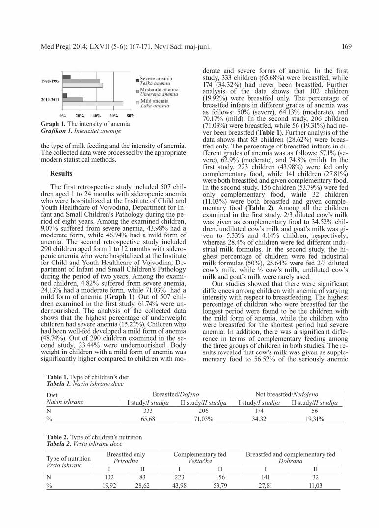
Med Pregl 2014; LXVII (5-6): 167-171. Novi Sad: maj-juni. 169
the type of milk feeding and the intensity of anemia. The collected data were processed by the appropriate modern statistical methods.
Results
The first retrospective study included 507 chil-dren aged 1 to 24 months with sideropenic anemia who were hospitalized at the Institute of Child and Youth Healthcare of Vojvodina, Department for In-fant and Small Children’s Pathology during the pe-riod of eight years. Among the examined children, 9.07% suffered from severe anemia, 43.98% had a moderate form, while 46.94% had a mild form of anemia. The second retrospective study included 290 children aged form 1 to 12 months with sidero-penic anemia who were hospitalized at the Institute for Child and Youth Healthcare of Vojvodina, De-partment of Infant and Small Children’s Pathology during the period of two years. Among the exami-ned children, 4.82% suffered from severe anemia, 24.13% had a moderate form, while 71.03% had a mild form of anemia (Graph 1). Out of 507 chil-dren examined in the first study, 61.74% were un-dernourished. The analysis of the collected data shows that the highest percentage of underweight children had severe anemia (15.22%). Children who had been well-fed developed a mild form of anemia (48.74%). Out of 290 children examined in the se-cond study, 23.44% were undernourished. Body weight in children with a mild form of anemia was significantly higher compared to children with mo-
derate and severe forms of anemia. In the first study, 333 children (65.68%) were breastfed, while 174 (34.32%) had never been breastfed. Further analysis of the data shows that 102 children (19.92%) were breastfed only. The percentage of breastfed infants in different grades of anemia was as follows: 50% (severe), 64.13% (moderate), and 70.17% (mild). In the second study, 206 children (71.03%) were breastfed, while 56 (19.31%) had ne-ver been breastfed (Table 1). Further analysis of the data shows that 83 children (28.62%) were breas-tfed only. The percentage of breastfed infants in di-fferent grades of anemia was as follows: 57.1% (se-vere), 62.9% (moderate), and 74.8% (mild). In the first study, 223 children (43.98%) were fed only complementary food, while 141 children (27.81%) were both breastfed and given complementary food. In the second study, 156 children (53.79%) were fed only complementary food, while 32 children (11.03%) were both breastfed and given comple-mentary food (Table 2). Among all the children examined in the first study, 2/3 diluted cow’s milk was given as complementary food to 34.52% chil-dren, undiluted cow’s milk and goat’s milk was gi-ven to 5.33% and 4.14% children, respectively; whereas 28.4% of children were fed different indu-strial milk formulas. In the second study, the hi-ghest percentage of children were fed industrial milk formulas (50%), 25.64% were fed 2/3 diluted cow’s milk, while ½ cow’s milk, undiluted cow’s milk and goat’s milk were rarely used.
Our studies showed that there were significant differences among children with anemia of varying intensity with respect to breastfeeding. The highest percentage of children who were breastfed for the longest period were found to be the children with the mild form of anemia, while the children who were breastfed for the shortest period had severe anemia. In addition, there was a significant diffe-rence in terms of complementary feeding among the three groups of children in both studies. The re-sults revealed that cow’s milk was given as supple-mentary food to 56.52% of the seriously anemic
Table 1. Type of children’s dietTabela 1. Način ishrane dece
Diet Način ishrane
Breastfed/Dojeno Not breastfed/NedojenoI study/I studija II study/II studija I study/I studija II study/II studija
N 333 206 174 56% 65,68 71,03% 34.32 19,31%
Graph 1. The intensity of anemiaGrafikon 1. Intenzitet anemije
Table 2. Type of children’s nutritionTabela 2. Vrsta ishrane dece
Type of nutrition Vrsta ishrane
Breastfed onlyPrirodna
Complementary fedVeštačka
Breastfed and complementary fedDohrana
I II I II I IIN 102 83 223 156 141 32% 19,92 28,62 43,98 53,79 27,81 11,03

170
children in the first study and 64.3% in the second study, 43.05% of moderately anemic children in the first study and 51.4% in the second study and 42.44% of the children with a mild form of anemia in the first study and 53.9% in the second study.
Discussion
Anemia in children under two years of age is a common health problem due to the fact that their growth requires a high intake of iron which is usu-ally not provided by their diet [9]. By comparing these two studies it was observed that the percenta-ges of the children with severe and moderate form of anemia were twice lower in the second study, while the percentage of the children with a mild form of anemia almost doubled compared to the first study. Anemia is usually caused by several associated factors, rather than individual ones. The important factors are the impact of environment, socio-economic factors, habits in the family, espe-cially the ones related to the mother, certain indivi-dual characteristics (gender, time and way of deli-very, birth weight and associated diseases) as well as the child’s diet. The level of nutrition is certainly correlated with different quality of alimentary defi-cit. In infancy, the overall mental and physical de-velopment depends primarily on the diet, and the diet relies almost entirely on the attitude, percepti-on, cultural and health education of the mother [2]. Most of the authors think that children suffering from anemia due to iron deficiency tend to have lower body weight [10, 11]. Other authors, however, believe that better nutritional status does not neces-sarily mean higher hemoglobin values, that these two factors are not directly related [12–14]. Accor-ding to the data from both studies, the body weight of children with a mild form of anemia was signifi-cantly higher compared to the children with mode-rate and severe forms of anemia.
It is well known that milk is a prototype of food poor in iron. In the first six months of life, milk is the basic food. It has been shown that the absorption of iron from breast milk is much higher compared to cow’s milk. Fortified milk formula contains the highest amount of iron, and its use is recommended in the diet of children under one year of age. Literature indicates that anemia has the highest prevalence in children fed with cow’s milk, while it is less common in breastfed chil-dren. Prevalence of anemia is the lowest in chil-dren fed with adapted milk formulas. These are the reasons why the WHO recommends that in-fants who are not breastfed should not get diluted or unmodified cow’s milk until the age of one. Iron rich baby formulas are therefore highly reco-mmended [2, 15–19]. The comparison of these two studies shows that the children from the second study sample were breastfed in a slightly higher percentage (71.03%) compared to the first study (65.68%). In both studies, the children with a mild
form of anemia were breastfed in the highest per-centage.
In 1928, Helen McKay concluded that anemia was common in infants on complementary diet, but did not know the exact reason for this phenomenon. Eight decades later, studies suggested that the re-sults of her research about the dependence of infant feeding and the development of anemia could be accepted with only minor adjustments. Therefore, she is considered to be the founder of modern study of anemia caused by iron deficiency. At that time, breastfeeding lasted much longer than today, and the results of her study showed that the hemo-globin concentration was higher in breastfed chil-dren. Complementary feeding at that time was ac-tually cow’s milk powder, without the addition of iron and other minerals or vitamins. She identifi-ed the real cause of this phenomenon to be better absorption of iron from human milk (50%) than from cow’s milk (10%). Approximately one third of the infants who were fed with cow’s milk showed blood loss through stool, which could be another factor in developing anemia due to iron deficiency. In addition, she suggested a definition of anemia, which is very similar to the one given by the WHO 40 years later [2, 15, 20].
As for the kind of milk introduced during the complementary feeding, 2/3 diluted cow’s milk was mainly used in the first study, whereas in the second study, the use of industrial milk formulas was domi-nant, which is in accordance with the recommenda-tions of the WHO. Frequent use of industrial milk formulas in nutrition of children, that was observed in the second study, may indicate a higher level of health education of mothers and may suggest the in-crease in socio-economic standards of the populati-on compared to the previous periods [2, 5].
Inadequate nutrition is a significant problem, es-pecially in this vulnerable population. Since defici-ent diet has a negative impact on general health of population, anemia is considered to be not only a health problem, but a social problem as well [2]. The analysis of the results of our studies shows that chil-dren who used cow’s milk as complementary food had severe form of anemia. Short-termed natural nutrition, early supplementation, and choice of milk could be determining factors in the development and manifestation of anemia [2, 8, 16, 19, 20]. Brea-stfeeding for more than six months of life without the addition of iron, early feeding with unmodified cow’s milk or solid foods can be the causes of side-ropenic anemia. It can also be caused if non-fortifi-ed baby formulas are used longer than four months without introducing other mixed foods. Children who are born at term and breastfed for the first six months of life are not at risk in terms of iron defici-ency. After this age, if the child is not breastfed, su-pplementary diet should include industrial milk in-fant formula enriched with iron. At the same time, it is recommended to introduce mixed foods. If so-lid foods are introduced to children who are still
Milankov O, et al. Prevention of infant’s sideropenic anemia

Med Pregl 2014; LXVII (5-6): 167-171. Novi Sad: maj-juni. 171
being breastfed, it can improve the bioavailability of iron from human milk [21].
Conclusion
Anemia due to iron deficiency effects people of all ages and from all economic groups, although it is more common among the younger population groups, particularly infants and young children aged from 1 to 24 months. It is particularly com-mon among people with insufficient nutrition. From what has been said, one can only imagine the extent to which iron deficiency anemia is wi-despread in our country and worldwide. What is yet undiscovered is the percentage of patients who are not registered and are thus not treated.
The comparison of two retrospective studies shows that the percentage of children with severe and moderate forms of anemia had decreased
through the years and it was twice lower in the se-cond study, while the percentage of children with a mild form of anemia had increased, in fact it do-ubled in the second study. Both studies confirmed the significant role of breastfeeding in prevention of sideropenic anemia. Also, it has been observed that the diet of children changes through the years and that diluted 2/3 cow’s milk, which was previo-usly frequently used in complementary feeding of children, is now often replaced by adapted milk formulas, which is in accordance with the World Health Organization recommendations.
Given the high prevalence of anemia in infants (30% of children treated at the Department), all the necessary social, economic and educational measu-res muste be taken to correct the diet and to ensure optimal nutritional and energy needs, as well as the needs for iron, and thus prevent development of si-deropenic anemia in this age group.
References1. McLean E, Cogswell M, Egli I, Wojdyla D, de Benoist
B.Worldwide prevalence of anaemia. WHO vitamin and mi-neral nutrition information system, 1993-2005. Public Health Nutr 2009;12(4):444-54.
2. Milankov O. Faktori rizika kod sideropenijske anemije odojčadi (doktorska disertacija). Beograd: Univerzitet u Beo-gradu; 2003.
3. Diaz JR, Cagigas A, Rodriguez R. Micronutrient defi-ciencies in developing and affluent countries. Eur J Clin Nutr 2003;57(1):70-2.
4. Dos Reis MC, Nakano AM, Silva IA, Gomes FA, Perei-ra MJ. Prevalence of anemia in children three to 12 months old in a health service in Ribeirão Preto, SP, Brazil. Rev Lat Am Enfermagem 2010;18:792-9.
5. Marx JJM. Iron deficiency in developed countries: pre-valance, influence of lifestyle factors and hazards of preventi-on. Eur J Clinic Nutr 1997;51(8):491-4.
6. Duque X, Flores-Hernández S, Flores-Huerta S, Mén-dez-Ramírez I, Muñoz S, Turnbull B, et al. Prevalence of ane-mia and deficiency of iron, folic acid, and zinc in children yo-unger than 2 years of age who use the health services provided by the Mexican Social Security Institute. BMC Public Health 2007;30:345.
7. Perišić V, Borislav J. Pedijatrija. Beograd: Medicinski fakultet; 2010.
8. Milankov O. Ishrana odojčadi: iskustva, novi trendovi i preporuke. Med Pregl 2013;66:5-10.
9. World Health Organization. The world health report 2002: reducing risks, promoting healthy life. Geneva: WHO; 2002.
10. Wise J. Daily iron during pregnancy improves birth weight. BMJ 2013;346:f3997.
11. Amrutha Veena K, Kowsalya S, Kothandapani S. Mi-cronutrient malnutrition profile of infants in South India. J Hum Nutr Diet 2014;27(1):54-7.
12. Suskind DL. Nutritional deficiencies during normal growth. Pediatr Clin North Am 2009;56(5):1035-53.
13. Friel JK, Aziz K, Andrews WL, Harding SV, Courage ML, Adams RJ. A double-masked, randomized control trial of iron supplementation in early infancy in healthy term bre-ast-fed infants. J Pediatr 2003;143(5):582-6.
14. Long H, Yi JM, Hu PL, Li ZB, Qiu WY, Wang F, et al. Benefits of iron supplementation for low birth weight infants: a systematic review. BMC Pediatr 2012;12:99.
15. Elalfy MS, Hamdy AM, Abdel Maksoud SS, Abdel Megeed RI. Pattern of milk feeding and family size as risk factors for iron deficiency anemia among poor Egyptian in-fants 6 to 24 months old. Nutr Res 2012;32:93-9.
16. Dube K, Schwartz J, Mueller MJ, Kalhoff H, Kersting M. Iron intake and iron status in breastfed infants during the first year of life. Clin Nutr 2010;29:773-8.
17. Sawasdivorn S, Taeviriyakul S. Are infants exclusive-ly breastfed up to 6 months of age at risk of anemia? J Med Assoc Thai 2011;94:178-82.
18. Oliveira MA, Osório MM. Cow’s milk consumption and iron deficiency anemia in children. J Pediatr (Rio J) 2005; 81:361-7.
19. Ziegler EE. Consumption of cow’s milk as a cause of iron deficiency in infants and toddlers. Nutr Rev 2011;69:37-42.
20. Hopkins D, Emmett P, Steer C, Rogers I, Noble S, Emond A. Infant feeding in the second 6 months of life rela-ted to iron status: an observational study. Arch Dis Child 2007;92:850-4.
21. Baker R, Greer F. Diagnosis and prevention of iron de-ficiency and iron-deficiency anemia in infants and young children (0-3 years of age). Pediatrics 2010;126:1040.
Rad je primljen 17. IX 2013.Recenziran 18. X 2013.Prihvaćen za štampu 20. XII 2013.BIBLID.0025-8105:(2014):LXVII:5-6:167-171.
Introduction
Interest in chemical matters that disrupt the en-docrine system work - endocrine disrupting chemi-cals (EDCs) has been increased over the last several years. EDCs may affect the synthesis, secretion, mechanism of action, metabolism and elimination of hormones in humans and animals, with harmful health consequences.
Phthalates, esters of 1,2-dicarboxylic acid - phthalic acid, are synthetic industrial chemical compounds which were introduced in 1920. Ever since 1933 and the synthesis of di (2-ethylhexyl) phthalates (DEHP), the phthalates have been the most common chemical compounds with the possi-bility to disrupt the endocrine system [1]. The effect of phthalates depends on dosage, duration of action and stage of the development of the individual, thus
Corresponding Author: Dr Ivana Bajkin, Klinika za endokrinologiju, dijabetes i bolesti metabolizma, 21000 Novi Sad, Hajduk Veljkova 1-7, E-mail: [email protected]
SummaryIntroduction. Phthalates are synthetic industrial compounds ca-pable of disrupting endocrine system. Effects of phthalates de-pend on dosage, duration of action and stage of development of the individual, thus making the fetus, newborn, and children at puberty the most vulnerable groups. Metabolism of Phthalates: Metabolism of these compounds consists of at least two steps: hy-drolysis and conjugation. They are mainly excreted in urine, with a low percent being excreted through feces. Exposure to Phtha-lates. Exposure to the effects of phthalates begins at the intrauter-ine stage since the phthalates pass through the placental barrier. Phthalates may be found in plastic products, toys, medical equip-ment, industrial materials, food, and clothes. Determination of Phthalate Levels in Humans. Urine is the best sample for evalu-ating phthalate levels in humans because of rapid phthalate me-tabolism and high concentrations of metabolites in the urine. Fe-tal Testicular Dysgenesis Syndrome: Fetal testicular dysgenesis syndrome involves disorders of male genital tract such as short-ened anogenital distance, hypospadia, cryptorchidism, malforma-tions of seminal vesicles, prostate, epididymis and it results from the harmful effects of phthalates. Other Effects of Phthalates on Health. Negative effects of phthalates on female health are mostly reflected in anovulation, premature puberty, changes in duration of pregnancy. There is a possible effect on neurocognitive devel-opment, occurrence of allergies, asthma, testicular carcinoma, hepatic and renal damages, insulin resistance and obesity, thyroid dysfunction. Conclusion. Further studies are needed to establish the safe phthalate concentration in certain products and to deter-mine more negative consequences of exposure to phthalate.Key words: Phthalic Acids; Fetus; Endocrine Disruptors; Pla-stics; Gonadal Dysgenesis; Insulin Resistance
SažetakUvod. Ftalati su sintetska industrijska jedinjenja koja imaju sposobnost da remete funkciju endokrinog sistema. Njihovi efekti zavise od doze, dužine dejstva i razvojnog stadijuma je-dinke, te su fetus, novorođenče i deca u pubertetu najugrože-nije kategorije. Metabolizam ftalata. Metabolizam ftalata odvija se u najmanje dva koraka − hidroliza i konjugacija. Ek-skrecija najvećeg dela ftalata obavlja se urinom, međutim ma-nji procenat se izlučuje fecesom. Izloženost ftalatima. Izlože-nost ftalatima počinje još intrauterino, pošto oni slobodno pro-laze placentarnu barijeru. Prisutni su u plastičnim prozvodima, igračkama, medicinskim instrumentima, industrijskim materi-jalima, hrani, odeći. Određivanje nivoa ftalata u ljudskom organizmu. Urin je materijal izbora za određivanje nivoa ftala-ta, zbog njihovog brzog metabolizma i visokih koncentracija metabolita ftalata u urinu. Sindrom fetalne testikularne diz-genezije nastaje kao posledica štetnog dejstva ftalata; podrazu-meva anomalije genitalnog trakta: skraćena ano-genitalna dis-tanca, hipospadija, kriptorhizam, malformacije semenih vezi-kula, prostate, epididimisa. Drugi efekti ftalata na zdravlje. Kod osoba ženskog pola negativan uticaj ftalata ogleda se u anovulaciji, preranom pubertetu, promenama u dužini trajanja trudnoće. Smatra se da neželjeni efekti ftalata mogu da se ispo-lje i kroz neurokognitivne poremećaje, pojavu alergija, astmu, karcinom testisa, oštećenja jetre i bubrega, insulinsku rezisten-ciju i gojaznost, tiroidnu disfunkciju. Zaključak. Neophodna su dalja istraživanja kojima bi se odredile bezbedne koncentra-cije ftalata, ali i da bi se uočili i do sada neprepoznati neželjeni efekti ftalata na ljudsko zdravlje. Klučne reči: Ftalna kiselina; Fetus; Endokrina disrupcija; Plastika; Gonadna disgenezija; Insulinska rezistencija
Clinical Centre of Vojvodina, Novi Sad Professional articleDepartment of Endocrinology, Diabetes and Metabolic Diseases1 Stručni članakDepartment of Gynecology and Obstetrics2 UDK 616-008:547.584 i University of Novi Sad, Faculty of Medicine3 UDK 618.33:547.584Health Centre, Novi Sad4 DOI: 10.2298/MPNS1406172B
EFFECTS OF PHTHALIC ACID ESTERS ON FETAL HEALTH
UTICAJ ESTARA FTALNE KISELINE NA FETALNO ZDRAVLJE
Ivana BAJKIN1, Artur BJELICA2,3, Tijana IČIN1, Vesna DOBRIĆ4, Branka KOVAČEV ZAVIŠIĆ1,3 and Milica MEDIĆ STOJANOSKA1,3
Bajkin I, et al. Phthalates and fetal health172

Med Pregl 2014; LXVII (5-6): 172-175. Novi Sad: maj-juni. 173
making the fetus, newborn, and children at puberty the most vulnerable groups [2]. According to the study of Latini et al. from 2003, the exposure to the effects of phthalates begins at the intrauterine stage since the phthalates pass through the placental bar-rier; another important issue in relation to the phtha-lates, i.e. EDCs, is that intrauterine exposure may not be manifested until adolescence or even later [1]. It is also known that exposure to phthalates may be manifested only as disorders of the next generations (through modification of factors that regulate gene expression) [1]. Because of the known harmful ef-fects, the European Union countries as well as our country have limited the use of DEHP, butyl benzyl phthalate (BzBP) and di-n-butyl phthalate (DBP) in the production of children’s toys and items intended for the child care, while the restrictions on the use of di-isononyl phthalate (DINP), di-isodecyl phthalate (DIDP) and di-n-octyl phthalate (DNOP) apply only to the manufacturers of toys and items intended for child care that children can put in the mouth [3]. Ap-plication of DEHP and DBP and BzBP is not per-mitted in cosmetic production in the European Un-ion because of reproductive toxicity [4].
Metabolism of Phthalates
After they enter the body, phthalates are subject-ed to hydrolysis and conjugation [4]. Monoestar phthalates are created by hydrolysis. In vivo and in vitro studies proved monoester phthalates to be bio-logically more active than their diesters [5,6]. Short chain phthalates are excreted in the urine as mo-noester phthalates, and the long chain phthalates are subjected to further metabolism in terms of hydroxy-lation and oxidation after which they are excreted in urine and feces [7, 8]. Their biological half-life is short, more than 60% is excreted in 24 hours [1, 9].
Exposure to Phthalates
Low molecular weight phthalates, for example di-methyl phthalate (DMP), DBP, are present in cosmetic products (nail polish, perfumes, facial creams, shampoos, body lotions...), while high mo-lecular weight phthalates, for example DEHP, BBzP, DNOP, DINP, DIDP, are present in plastic
containers, adhesives, clothes of raincoat type and plastic products with polyvinyl chloride which is added in order to improve flexibility [4]. Phthalates are also found in medical instruments such as cen-tral venous and urinary catheters, as well as in packaging for total parenteral nutrition and intrave-nous infusion. They are also present in some medi-cations [11]. Foodstuffs, such as cereals, bread, bis-cuits, cakes, nuts, oils and fats, can be found in packaging made of plastics containing DEHP, DBP and DEHP and di-isobutil phthalate (DIBP), and thus they are in contact with phthalates [12].
Toys are another important source of exposure to phthalate (soothers, teethers, bath toys), and they can enter the body either orally, or by inhala-tion, through skin and parenterally [1,10].
Determination of Phthalate Levels in Humans
Phthalates do not tend to bioaccumulate and their half-life is less than 24 hours [1,10]. There have been attempts at determining phthalate levels in saliva, serum, seminal fluid, meconium and placenta but, the validation of these procedures have shown that phthalates are excreted in a very small percentage in this way [13, 14]. Urine, ma-ternal milk, serum and amniotic fluid are most frequently used nowadays as material to assess the presence of phthalates in the body [15]. Urine was proven to be the best sample in epidemiological studies in regards to the rapid metabolism of phthalates and high concentrations of the metabo-lites in the urine. Further advantages of urine as material for determining levels of phthalates is that it can be collected in a noninvasive way and may reflect exposure to phthalates in the last few days, even weeks [16, 17]. In all the above men-tioned samples, the level of monoesters, i.e. phtha-lates metabolites, are determined because the level of monoesters is higher than the level of diesters of phthalic acid, and the contamination of the sam-ple by ubiquitous diesters during the collection, storage and analysis itself is avoided [18].
Fetal Testicular Dysgenesis Syndrome
In the last fifteen years, a number of studies on experimental animals (rats) have proven that phtha-lates, especially DEHP, DBP, BBzP, when acting in a critical period of the development of genital tract, lead to disturbances in androgen-signaling pathway [18,19]. In almost all previous studies, the anti-an-drogen effect of phthalates in newborn males was examined, but it was also shown that the negative effects of phthalates on female health are reflected in anovulation, premature puberty, changes in the duration of pregnancy and other disorders [2, 10].
Fetal testicular dysgenesis syndrome (”phthalate syndrome” in rodents) involves disorders of male genital tract in terms of shortened anogenital dis-tance, hypospadia, cryptorchidism, malformations
AbbreviationsEDCs – endocrine disrupting chemicals DEHP – di (2-ethylhexyl) phthalatesMEHP – mono(ethyl-hexyl) phthalateBBzP – butyl benzyl phthalateDBP – di-n-butyl phthalateDINP – di-isononyl phthalateDIDP – di-isodecyl phthalateDMP – di-methyl phthalateDnOP – di-n-octyl phthalateMEP – mono-ethyl phthalate MBP – mono-butyl phthalateMMP – mono-methyl phthalate

Bajkin I, et al. Phthalates and fetal health174
of the seminal vesicles, prostate, and epididymis [18, 20]. According to contemporary literature, the stated syndrome is a consequence of reduced level of fetal testosterone, insulin-like growth factor-3 (IGF-3) and follicule stimulating hormone (FSH) [18, 21]. A negative correlation between levels in breast milk and free testosterone of babies was ob-served, while there was a positive correlation be-tween mono-ethyl phthalate (MEP) and mono-butyl (MBP) with sex hormone bingind globuline (SHBG) and mono-metyl phthalate (MMP) and MEP and MBP with the ratio of lutenzing hormone (LH) and free testosterone [22].
Other effects of phthalates on health
Exposure to phthalates is significantly associated with the duration of pregnancy [2]. According to some studies, the chemical structure of DEHP and prostaglandin/thromboxane, interleukin-1 connects the phthalates with induction of intrauterine inflam-matory processes as well as shortening of pregnan-cy [2, 23]. Some results suggested an association between levels of mono-(2-ethylhexyl) phthalate in preconceptional period and early pregnancy loss, while many authors pointed out the impact of phtha-lates on the low birth weight [9, 16, 18].
Recent research suggests a possible effect on neurocognitive development, as well as on the de-velopment of allergies, asthma, testicular carcino-ma, hepatic and renal damages, insulin resistance and obesity, thyroid dysfunction [18].
An interesting fact is that exposure to a certain type of phthalates varies among different socio-economic groups, which is probably the conse-quence of certain products whose use is signifi-cantly different among these groups [24].
Conclusion
Considering the widespread use of phthalates and exposure of large human population to phtha-lates in the environment, food or items for person-al use their harmful impact on health need to be tested. Numerous experimental, epidemiological and observational studies of human population have suggested their most common side effects, but there are still many uncertainties. It is charac-teristic that detrimental effect is not only dose de-pendent. The duration of exposure is rather impor-tant: exposure to low doses of phthalates over a long period of time can lead to endocrine and met-abolic disorders. Especially sensitive categories are the fetus and newborn, as well as pubertal
Table 1. Phthalate diesters and their metabolites (taken from Frederiksen H, Skakkebek NE, Andersson AM. Metabolism of phthalates in humans, Mol Nutr Food Res 2007;51:899-911.)Tabela 1. Diestri ftalata i njihovi metaboliti (preuzeto iz Frederiksen H, Skakkebek NE, Andersson AM. Meta-bolism of phthalates in humans. Mol Nutr Food Res 2007;51:899-911.)
Phthalates/Ftalati Metabolites/Metabolitidi-methyl-phthalate/di-metil-ftalat DMP mono-methyl-phthalate/mono-metil-ftalat MMP
di-ethyl-phthalate/di-etil-ftalat DEP mono-ethyl-phthalate/mono-etil-ftalat MEPdi-n-butyl phthalate/di-n-butil-ftalat DBP mono-butyl phthalate/mono-butil-ftalat MBPdi-n-butil-phthalate/di-n-butil-ftalat DBP mono-butil-ftalat/mono-izo-butil-ftalat MBP
di-iso-butyl phthalate/di-izo-butil-ftalat DiBP mono-iso-butyl phthalate/mono-butil-benzil ftalat MiBPbutyl benzyl phthalate/butil-benzil-ftalat BBzP mono-butyl benzyl phthalate/mono2-etil-heksil-ftalat MBzP
di-2-ethyl-hexyl phthalatedi-2-etil-heksilftalat DEHP mono2-ethyl -hexyl phthalate
mono-2-etil-5 hidroksiheksil ftalat MEHP
mono-2-ethyl-5 hydroxyhexyl phthalatemono-2-etil-5 oksoheksil ftalat 5OHMEHP
mono-2-ethyl-5 oxohexyl phthalatemono-2-etil-5 karboksipentil ftalat 5oxoMEHP
mono--2-ethyl-5 carboxy pentyl phthalatemono-2-etil-5 karboksipentil ftalat 5chMEHP
mono-2-carboxy-hexyl-phthalatemono-2-carboksi-heksil-ftalat 2chMMHP
di-iso-nonyl phthalate/di-izo-nonil-ftalat mono-iso-nonyl phthalate/mono-izo-nonil-ftalat MiNPmono-hydroxy-iso-nonyl phthalatet
mono-hidroksi-izo-nonil-ftalat OH-MiNP
mono-oxo-iso-nonyl phthalatemono-okso-izo-nonil-ftalat oxo-MiNP
mono-carboxy-iso-octyl phthalatemono-karboksi-izo-oktil-ftalat cx-MiNP

Med Pregl 2014; LXVII (5-6): 172-175. Novi Sad: maj-juni. 175
References1. Frederiksen H, Skakkebek NE, Andersson AM. Metaboli-
sm of phthalates in humans. Mol Nutr Food Res 2007;51:899-911.
2. Latini G, De Felice C, Presta G, Del Vecchio A, Paris I, Ruggieri F, et al. In utero exposure to di-(2-ethylhexyl)phtha-late and duration of human pregnancy. Environ Health Per-spect 2003;111:1783-5.
3. CDC’s Fourth national report on human exposure to environmental chemicals, updated tables, february 2012; Ava-ilable from: http://www.cdc.gov/exposurereport.
4. Commission directive 2004/93/EC of 21 September 2004 amending council directive 76/768/EEC for the purpose of adapting its Annexes II and III to technical progress. Brussels: European Parliament; 2004.
5. Heindel JJ, Powell CJ. phthalate ester effects on rat Ser-toli cell function in vitro: effects of phthalate side chain and age og animal. Toxicol Appl Pharmacol. 1992;115:116-23.
6. Blount BC, Silva MJ, Caudill SP, Needham LL, et al. Le-vels of seven urinary phthalate metabolites in a human referen-ce population. Environ Health Perspect 2000;108:979-82.
7. Koch HM, Bolt HM, Preuss R, Angerer J. New metabo-lites of di(2-ethylhexyl)phthalate (DEHP) in human urine and serum after single oral doses of deuterium labeled DEHP. Arch Toxicol 2005;79:367-76.
8. Silva MJ, Barr DB, Reidy JA, Kato K, Malek NA, Hodge CC, et al. Glucuronidation patterns of common urinary and serum monoester phthalates metabolites. Arch Toxicol 2003;77:561-7.
9. Braun JM, Sathyanarayana S, Hauser R. Phthalate expo-sure and children’s health. Curr Opin Pediatr 2013;25:247-54.
10. Rudel RA, Gray JM, Engel CL, Rawsthorne TW, Dod-son RE, Ackerman JM, et al. Food packaging and bisphenol A and bis(2-ethylhexyl)phthalate exposure: findings from a die-tary intervention. Environ Health Perspect 2011;119:914-20.
11. Hernandez-Diaz S, Mitchell AA, Kelly KE, Calafat AM, Hauser R. Medications as a potential source of exposure to phthalates in U.S. population. Environ Health Perspect 2009;117:185-9.
12. Wormuth M, Scheringer M, Vollenweider M, Hungerbuc-hler K. What are the sources of exposure to eight frequently used phtalic acid esters in Europeans? Risk Anal 2006;26:803-24.
13. Kato K, Silva M, Needham L, Calafat A. Quantifying phthalate metabolites in human meconium and semen using au-
tomated off-line solid-phase extraction coupled with on-line SPE and isotope-dilution high performance liquid chromato-graphy-tandem mass spectrometry. Anal Chem 2006;78:6651-5.
14. Silva M, Reidy J, Samandar E, Herbert A, Needham L, Calafat A. Detection of phthalate metabolites in human sa-liva. Arch Toxicol 2005;79:647-52.
15. Calafat AM, McKee RH. Integrating biomonitoring exposure data into the risk assasment process: phthalates (di-ethyl phthalate and di(2-ethylhexyl)phthalate) as a case study. Environ Health Perspect 2006;114:1783-9.
16. Braun JM. Smith KW, Williams PL. Variability of urine phthalate metabolite an bisphenol a concentrations before and during pregnancy. Environ Health Perspect 2012;120:739-45.
17. Hauser R, Meeker JD, Park S, Silva JM, Calafat AM. Temporal variability of urinary phthalate metabolit levels in men of reproductive age. Environ Health Perspect 2004;112:1734-40.
18. Swan SH. Environmental phthalate exposure in relati-on to reproductive outcomes and other health endpoints in hu-mans. Environ Res 2008;108:177-84.
19. Foster PM, Mylchreest E, Gaido KW, Sar M. Effects of phthalate esters on the developing reproductive tract of male rats. Hum Reprod Update 2001;7:231-5.
20. Welsh M, Saunders PT, Fisken M, Scott HM, Hutchin-son HM, Smith LB, et al. Identification in rats of a program-ming window for reproductive tract masculinisation, disrupti-on of which leads to hypospadias and cryptorchism. J Clin Invest 2008;118:1478-90.
21. Sharpe RM, Irvine DS. How strong is the evidence of a link between environmental chemicals and adverse effects on human reproductive health? Br Med J 2004;328:447-51.
22. Main KM, Mortensen GK, Kaleva MM, Boisen KA, Da-magaard IN, et al. Human breast milk contamination with phthala-tes alternations of endogenous reproductive hormones in infants three months of age. Environ Health Perspect 2006;114:270-6.
23. Maroziene L, Grazuleviciene R. Maternal exposure to low-level air pollution and pregnancy outcomes: a population based study. Environ Health Perspect 2002;9:6.
24. Kobrosly RW, Parlett LE, Stahlhut RW, Barret ES, Swan SH. Socioeconomic factors and phthalate metabolite concentratins among United States women of reproductive age. Environ Res 2012;115:11-7.
Rad je primljen 11. III 2014.Recenziran 20. III 2014.Prihvaćen za štampu 25. III 2014.BIBLID.0025-8105:(2014):LXVII:5-6:172-175.
children. Endocrine disrupting chemicals have the epigenetic influence and these disorders can be manifested in the next generations. Further re-search aimed at timely recognition of adverse ef-
fects and adjusting the concentrations of the chemical compounds in products is needed in or-der to avoid their adverse effects on human health.


Med Pregl 2014; LXVII (5-6): 177-180. Novi Sad: maj-juni. 177
CASE REPORTSPRIKAZI SLUČAJEVAUnivesity of Novi Sad, Faculty of Medicine, Novi Sad Case reportDental Clinic of Vojvodina Prikaz slučajaDepartment of Oral Surgery1 UDK 616.314-089.843Department of Dental Prosthetics2 DOI: 10.2298/MPNS1406177T
APPLICATION OF FIBRIN RICH BLOCKS WITH CONCENTRATED GROWTH FACTORS IN PRE-IMPLANT AUGMENTATION PROCEDURES
UPOTREBA FIBRINSKIH BLOKOVA BOGATIH KONCENTROVANIM FAKTORIMA RASTA U PREIMPLANTOLOŠKIM AUGMENTACIONIM PROCEDURAMA
Ana TADIĆ1, Tatjana PUŠKAR2 and Branislava PETRONIJEVIĆ2
Introduction
In our everyday clinical practice, we face a rela-tively large number of patients presenting with a substantial deficit of the residual alveolar ridge due to the loss of single or several teeth who require im-plant-prosthetic treatment. In such situations, the
augmentation of lost bony structures is indicated in order to provide optimal conditions for dental im-plant placement and subsequent prosthetic rehabili-tation [1]. Nowadays, a whole range of modern sur-gical procedures and a variety of dental materials for reconstruction of bony defects of the upper and lower jaws and for the augmentation of lost structu-
Corresponding Author: Dr Ana Tadić, Klinika za stomatologiju Vojvodine,21000 Novi Sad, Hajduk Veljkova 12, E-mail: [email protected]
SummaryIntroduction. Growth factors are mediators regulating the key processes of tissue regeneration, including cell proliferation and differentiation, extracellular matrix synthesis, chemotaxis and angiogenesis. In addition to the role they play in haemostasis and inflammatory processes, thrombocytes are of major importance in the reparation of mineralized and soft tissues. Application of fibrin rich blocks with concentrated growth factors is one of the latest approaches to guided bone regeneration and augmentation of lost bony structures of the alveolar ridge. Case report. This paper presents a case of a female patient who underwent recon-struction of the defect of residual alveolar ridge of the upper jaw by applying fibrin rich blocks with concentrated growth factors and subsequent placement of two titanium endosteal implants five months after wound healing. Conclusion. The loss of a sin-gle tooth or several teeth sometimes entails the augmentation of lost bony structures in order to provide optimal conditions for dental implant placement and subsequent prosthetic rehabilitati-on. A range of contemporary surgical procedures and a variety of dental materials for reconstruction of bony defects of the upper and lower jaws are available nowadays. The method described in this paper, i.e. the application of concentrated growth factors is one of the latest approaches which poses no risk of transmissible and allergic diseases and is at the same time cost effective.Key words: Alveolar Bone Loss; Alveolar Ridge Augmenta-tion; Intercellular Signaling Peptides and Proteins; Surgery, Oral; Female; Adult; Dental Implantation, Endosseous
SažetakUvod. Faktori rasta su biološki medijatori koji regulišu ključne procese u reparaciji tkiva, uključujući ćelijsku proliferaciju, dife-rencijaciju, sintezu ekstracelulanog matriksa, hemotaksu i angi-ogenezu. Osim učešća u hemostazi i inflamaciji, trombociti ima-ju veliku ulogu u reparaciji mineralizovanih i mekih tkiva. Upo-treba fibrinskih blokova bogatih koncentrovanim faktorima ra-sta predstavlja jednu od najsavremenijih metoda koja se koristi kod vođene koštane regeneracije pri nadoknadi izgubljenih ko-štanih struktura alveolarnog grebena viličnih kostiju. Prikaz slučaja. U radu je prikazan slučaj pacijentkinje kod koje je ura-đena rekonstrukcija defekata rezidualnog alveolarnog grebena gornje vilice fibrinskim blokovima bogatim koncentrovanim faktorima rasta, u koji su nakon perioda zarastanja od pet meseci ugrađena dva titanijumska endoosealna implantata. Zaključak. U slučajevima gubitka jednog ili više zuba, nekada je neophod-na nadoknada izgubljenih koštanih struktura radi obezbeđivanja optimalnih uslova za ugradnju dentalnih implantata i protetsku rehabilitaciju koja sledi. Savremenim kliničarima na raspolaga-nju stoje brojne operativne procedure kao i veliki broj različitih materijala koji se koriste u rekonstrukciji koštanih defekata gor-nje i donje vilice. Prikazana metoda rada sa preparatima koncen-trovanih faktora rasta spada u jednu od najsavremenijih, bez opasnosti od prenošenja transmisionih bolesti, pojave alergijskih reakcija. U ekonomskom smislu je u potpunosti isplativa.Ključne reči: Gubitak alveolarne kosti; Nadoknada alveolar-nog grebena; Faktori rasta; Oralna hirurgija; Žensko; Odrasli; Stomatološka endosealna implantacija

Tadić A, et al. Augmentation with concentrated growth factors178
res of the residual alveolar ridge are available. The application of fibrin rich blocks with concentrated growth factors (CGF) is one of the latest approa-ches to guided bone regeneration (GBR). Fibrin rich blocks are applicable either alone or mixed/combined with any of synthetic bone grafts [1, 2].
According to numerous studies, growth factors are mainly located in blood plasma and thrombo-cytes. Growth factors are biological mediators re-gulating the major processes of tissue restoration, including cell proliferation and differentiation, extracellular matrix synthesis, chemotaxis and an-giogenesis. In addition to the role they play in hae-mostasis and inflammatory processes, thrombo-cytes are of major importance in the reparation of mineralized and soft tissues [3].
The most important and most extensively inve-stigated growth factors are platelet-derived growth factor (PDGF), transforming growth factor (TGF), epidermal growth factor (EGF) and insulin-like growth factor 1 (IGF-1) [4]. The first generation of these preparations, platelet rich plasma (PRP), has been well known for more than 15 years. PRP was first introduced into clinical practice by Marx in 1998 [5], whereas platelets rich in growth factor (PRGF) are the second-generation platelet concen-trates. Platelet-rich fibrin (PRF) was first introdu-ced by Choukroun in 2000 as the platelet concen-trates of the third generation along with CGF in-troduced by Sacco in 2006. CGFs are characteri-zed by higher density, larger amount of growth factors, higher viscosity and better adhesion capa-city as compared to PRF. All preparations are pro-duced from the patient’s fresh venous blood [6].
The application of autologous fibrin rich blocks has no adverse effects; it is considered a safe and simple procedure for the surgeon. At the same time, it is highly effective and economically feasi-ble for the patient.
Case report
A 28-year-old female patient came to the Depar-tment of Oral Surgery and Implantology of the Den-tal Clinic of Vojvodina because of the loss of tooth 22 and pain in the region of tooth 23. The clinical examination revealed fresh extraction wound after the last extraction of tooth 22 and intraoral fistula in the root tip area of the tooth 23. The patient was re-ferred to cone beam computed tomography (CBCT) scan. The CBCT scan revealed a radicular cyst on tooth 23 associated with massive destruction of the surrounding bony tissue (Figure 1).
After completing the diagnostic procedure and consulting the specialist in dental prosthetics, the therapy options were considered. The definitive therapeutic strategy was established, encompassing three stages. Stage 1 included the extraction of tooth 23, cyst curettage and the reconstruction of bony de-fects in regions of teeth 22 and 23 by applying CGF blocks. The second stage, after bone healing, inclu-ded the placement of two endoosseous titanium im-plants at the position of teeth 22 and 23. The third stage, subsequent to the successfully completed osseointegration, included the placement of two im-plant-supported metal-ceramic crowns. After obtai-ning the patient’s written consent, the suggested the-rapy plan was carried out. After full-thickness mu-coperiosteal flap had been lifted, the extraction of tooth 23 along with complete curettage of the cystic process and surrounding pathologically altered tis- sue was performed under local anaesthesia. The re-sulting bony defects were entirely filled out with CGF blocks previously prepared from the patient’s venous blood and the flap was sutured back in place with synthetic non-absorbable monofilament suture (figures 2 and 3).
Five months after surgery, the control CBCT scan revealed the complete regeneration and repa-ration of the bone tissue and satisfactory dimensi-ons of the residual alveolar ridge, that being the prerequisite for the placement of endoosseous im-plants of 3.3 mm in diameter and 12 mm in length. (figures 4 and 5). After implantation, the entire surgical region was covered with pressed CGF barrier membrane and the flap was put back in place and sutured with synthetic non-absorbable monofilament suture in order to minimize the accumulation of soft deposits and to alleviate po-tential tissue reactions [7, 8].
Four months after implantation procedure, the patient underwent prosthetic restoration procedure at the Department of Dental Prosthetics. The pro-cedure included the placement of two implant-su-pported metal-ceramic crowns (Figure 6).
AbbreviationsCGF – concentrated growth factorsPRP – platelet rich plasma PRGF – platelets rich in growth factor PRF – platelet-rich fibrin CBCT – cone beam computed tomography GBR – guided bone regeneration
Figure 1. Preoperative CBCT scanSlika 1. Preoperativni CBCT snimak

Med Pregl 2014; LXVII (5-6): 177-180. Novi Sad: maj-juni. 179
Discussion
Application of CGF blocks is one of the most recent approaches to practical application of tissue engineering methods in oral surgery and implan-tology [4]. This is of particular importance in im-plantology, having in mind the growing need for
augmentation procedures in cases of unfavourable anatomic conditions (horizontal and vertical au-gmentation, sinus-lift, etc.) [2].
The comparison of protocols for CGF block preparation revealed different utilization rates of collected blood, being 10% in PRP and PRGF and 30–40% in PRF and CGF. Numerous authors have reported positive results of the application of CGF in the augmentation of bone architecture of the re-sidual alveolar ridge and establishment of adequate anatomical conditions for a successful implant-prosthetic rehabilitation of toothless patients. Blocks with CGF enhance the processes of bony structure reparation and regeneration thus shorte-ning the healing time from the initial surgical pro-cedure, i.e. alveolar ridge augmentation to the mo-ment of placing the endoosseous implant into the augmented region. As aforementioned, CGF blocks are applicable alone or combined with any of synt-hetic bone grafts. However, novel trends in GBR give preference to natural graft material, i.e. auto-transplants. In a broader sense, CGF blocks could be considered natural graft material [1, 2, 6].
Figure 2. Intraoperative bony defectSlika 2. Intraoperativni koštani defekt
Figure 3. Inserted CGF blocksSlika 3. Postavljeni CGF preparati
Figure 4. Control CBCT scanSlika 4. Kontrolni CBCT snimak
Figure 5. Inserted endosteal implants Slika 5. Postavljeni endoosealni implantati
Figure 6. Definitive appearance after treatmentSlika 6. Definitivni izgled nakon terapije

Tadić A, et al. Augmentation with concentrated growth factors180
Conclusion
In our everyday clinical practice, we face a rela-tively large number of patients indicated for im-plant-prosthetic treatment. The loss of a single to-oth or several teeth results in substantial deficit of the residual alveolar ridge in such patients. Such si-tuations require the augmentation of lost bony structures in order to provide optimal conditions for dental implant placement and subsequent pro-
sthetic rehabilitation. Oral surgeons have the who-le range of modern surgical procedures and a vari-ety of dental materials for reconstruction of bony defects of the upper and lower jaws at their dispo-sal. The method presented in this paper, i.e. the application of concentrated growth factors is one of the latest approaches, which carries no risk of either transmissible or allergic diseases, and is at the same time highly effective and economically feasible.
References1. Mirković S, Đurđević Mirković T, Puškar T. Applicati-
on of concetrated growth factors in reconstruction of bone de-fects after removal of large jaw cyst: report of two cases. Voj-nosanit Pregl 2014 in press.
2. Mirković S, Đurđević Mirković T, Petrović L, Božić D. The use of concentrate growth factors in gyded bone regene-ration after lateral sinus lift procedure: case report. Heal-thMED 2013;7(2):700-4.
3. Plachokova AS, Nikolidakis D, Mulder J, Jansen JA, Creugers NH. Effect of platelet-rich plasma on bone regenera-tion in dentistry: a systematic review. Clin Oral Implant Res 2008;19(6):539-45.
4. Lazić Z, Bubalo M, Petković-Ćurčin A, Duka M, Mi-hajlović B. Terapijska primena plazme bogate trombocitima u oralnoj hirurgiji. Vojnosanit Pregl 2009;66(10):821-5.
5. Marx RE, Carlson ER, Eichstaedt RM, Schimmele SR, Strauss JE, Georgeff KR. Platelet-rich plasma: growth factor enhancement for bone grafts. Oral Surg Oral Med Oral Pathol Oral Radiol Endod 1998;85(6):638-46.
6. Choukroun J, Diss A, Simonpieri A, Girard MO, Schoe-ffler C. Platelet-rich fibrin (PRF): a second generation platelet concentrate. Part IV: clinical effects on tissue healing. Oral Surg Oral Med Oral Pathol Oral Radiol Endod 2006;101(3):56-60.
7. Mirković S, Džambas LJ, Selaković S. Uticaj različitih materijala za šivenje na intenzitet tkivne reakcije usne šuplji-ne. Zb Matice Srpske Prir Nauke 2008;115:91-9.
8. Mirković S, Đurđević Mirković T, Bajkin B, Šarčev I. Izbor hirurškog šavnog materijala prilikom suturiranja u usnoj duplji: klinička studija. Med Pregl 2010;63(7-8):497-501.
Rad je primljen 18. III 2014.Recenziran 20. III 2014.Prihvaćen za štampu 25. III 2014.BIBLID.0025-8105:(2014):LXVII:5-6:177-180.
Med Pregl 2014; LXVII (5-6): 181-184. Novi Sad: maj-juni. 181
University Clinical Center, Banja Luka Case reportClinic of Internal Medicine1 Prikaz slučajaFaculty of Medicine, Banja Luka UDK 615.22.06:616-008.9Department of Clinical Pharmacology2 DOI: 10.2298/MPNS1406181M
SEVERE HYPERKALEMIA INDUCED BY PROPRANOLOL
TEŠKA HIPERKALIJEMIJA IZAZVANA PROPRANOLOLOM
Danijela MANDIĆ1, Lana NEŽIĆ2 and Ranko ŠKRBIĆ2
Introduction
Potassium is the principal intracellular cation, and maintenance of its distribution between the intracellular and the extracellular compartments relies on several homeostatic mechanisms. When these mechanisms are perturbed, hypokalemia or hyperkalemia may occur [1]. Severe hyperkalemia increases the risk of fatal dysrhythmias and there-fore early recognition of its electrocardiographic (ECG) manifestations is clinically very important. The ECG changes associated with high potassium level may include peaked T waves, prolongation of the PR interval, and QRS widening followed by loss of atrial activity, ventricular fibrillation, and asystole. The pathological ECG changes of hyper-kalemia are proportional to the serum potassium level, and can be detected when its level is greater than 6.0 mmol/L [1].
Case report
A female patient, aged 50, was referred to an in-ternal medical examination as she was experienc-ing a feeling of general weakness, tightness behind the sternum and numbness in the limbs, the symp-toms that had been occurring increasingly over the previous month. The medical history showed that a week prior to the onset of the symptoms, the patient had been diagnosed with angina pectoris and there-fore she had been receiving propranolol a 40 mg per day, divided into two doses. Additionally, the patient was fasting for two weeks over the previous month due to a dental intervention. The medical history also showed the presence of comorbid hy-pothyreosis and osteoporosis, treated with 100 μg levothyroxine daily and calcium supplement. The clinical examination established the general condi-tion to be relatively good with arterial blood pres-sure 120/70 mmHg and normal auscultatory find-
SummaryIntroduction. Hyperkalemia secondary to beta-adrenergic re-ceptor blockade occurs in 1-5% of patients and is likely to deve-lop with non-cardio-selective beta-blockers. Case Report. We have described hyperkalemia in a patient with angina pectoris receiving propranolol, clinically manifested as weakness, ti-ghtness behind the sternum and numbness in the limbs. Labora-tory tests showed hyperkalemia (6.6 mmol/L), peaked T wave and a corrected QT interval of 510 ms. After discontinuation of propranolol, decline in potassium level, normalisation of electro-cardiographic changes and clinical improvement were achieved. Causal relationship of drug related hyperkalemia has been con-firmed as probable/likely according to Naranjo Adverse Drug Reaction Probability Score of 7 and the World Health Organiza-tion Uppsala Monitoring Centre Probability Scale. Conclusion. Hyperkalemia can be unpredictable and life-threatening compli-cation of propranolol or a non-selective adrenergic beta blocker treatment, and requires timely identification of cause and imple-mentation of therapeutic measures.Key words: Hyperkalemia; Propranolol; Electrocardiography; Drug-Related Side Effects and Adverse Reactions; Signs and Symptoms; Female; Middle Aged; Angina Pectoris; Risk Factors
SažetakUvod. Hiperkalijemija uzrokovana blokadom beta adrenergičkih receptora javlja se kod 1−5% pacijenata, a najčešće je izazvana neselektivnim beta blokatorima. Prikaz slučaja. Prikazali smo pacijentkinju s hiperkalijemijom i kliničkim manifestacijama u vidu slabosti, bolova iza grudne kosti i trnjenja u ekstremitetima, koja je uzimala propranolol zbog angine pektoris. Laboratorijske analize pokazale su hiperkalijemiju (6,6 mmol/l), visoke T-talase i korigovani QT interval od 510 ms. Nakon prekida uzimanja propranolola, dolazi do pada nivoa kalijuma, normalizacije elek-trokardiografskih promena i poboljšanja kliničkog nalaza. Uzročna povezanost između leka i hiperkalijemije klasifikovana je kao verovatna prema Naranjo skali za procenu verovatnoće skorom 7 i World Health Organization Uppsala Monitoring Cen-tre skalom verovatnoće. Zaključak. Hiperkalijemija može biti nepredvidiva i po život opasna komplikacija primene proprano-lola ili drugih neselektivnih beta blokatora i zahteva pravovre-menu identifikaciju i sprovođenje terapijskih mera.Klučne reči: Hiperkalijemija; Propranolol; Elektrokardiogram; Nus efekti i neželjene reakcije izazvane lekovima; Znaci i simp-tomi; Žensko; Srednje godine; Angina pectoris; Faktori rizika
Corresponding Author: Dr Danijela Mandić, Univerzitetsko klinički centar, Banja Luka, Klinika za unutrašnje bolesti,78000 Banja Luka, Save Mrkalja 14, E-mail: [email protected]
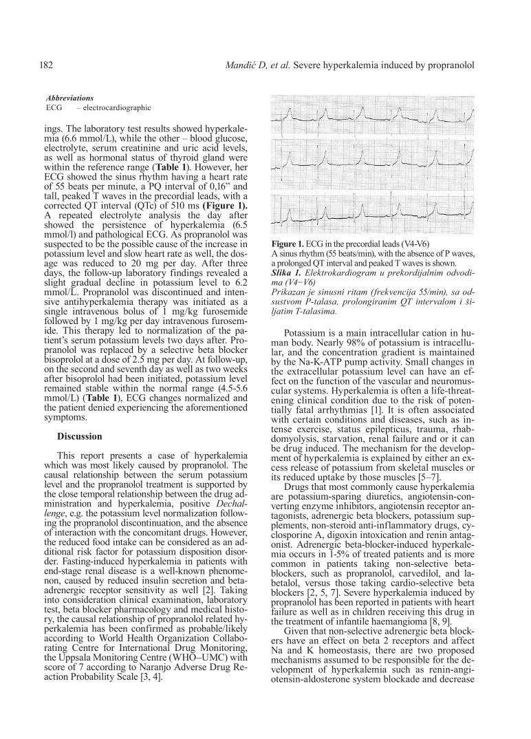
Mandić D, et al. Severe hyperkalemia induced by propranolol182
ings. The laboratory test results showed hyperkale-mia (6.6 mmol/L), while the other – blood glucose, electrolyte, serum creatinine and uric acid levels, as well as hormonal status of thyroid gland were within the reference range (Table 1). However, her ECG showed the sinus rhythm having a heart rate of 55 beats per minute, a PQ interval of 0,16” and tall, peaked T waves in the precordial leads, with a corrected QT interval (QTc) of 510 ms (Figure 1). A repeated electrolyte analysis the day after showed the persistence of hyperkalemia (6.5 mmol/l) and pathological ECG. As propranolol was suspected to be the possible cause of the increase in potassium level and slow heart rate as well, the dos-age was reduced to 20 mg per day. After three days, the follow-up laboratory findings revealed a slight gradual decline in potassium level to 6.2 mmol/L. Propranolol was discontinued and inten-sive antihyperkalemia therapy was initiated as a single intravenous bolus of 1 mg/kg furosemide followed by 1 mg/kg per day intravenous furosem-ide. This therapy led to normalization of the pa-tient’s serum potassium levels two days after. Pro-pranolol was replaced by a selective beta blocker bisoprolol at a dose of 2.5 mg per day. At follow-up, on the second and seventh day as well as two weeks after bisoprolol had been initiated, potassium level remained stable within the normal range (4.5-5.6 mmol/L) (Table 1), ECG changes normalized and the patient denied experiencing the aforementioned symptoms.
Discussion
This report presents a case of hyperkalemia which was most likely caused by propranolol. The causal relationship between the serum potassium level and the propranolol treatment is supported by the close temporal relationship between the drug ad-ministration and hyperkalemia, positive Dechal-lenge, e.g. the potassium level normalization follow-ing the propranolol discontinuation, and the absence of interaction with the concomitant drugs. However, the reduced food intake can be considered as an ad-ditional risk factor for potassium disposition disor-der. Fasting-induced hyperkalemia in patients with end-stage renal disease is a well-known phenome-non, caused by reduced insulin secretion and beta-adrenergic receptor sensitivity as well [2]. Taking into consideration clinical examination, laboratory test, beta blocker pharmacology and medical histo-ry, the causal relationship of propranolol related hy-perkalemia has been confirmed as probable/likely according to World Health Organization Collabo-rating Centre for International Drug Monitoring, the Uppsala Monitoring Centre (WHO–UMC) with score of 7 according to Naranjo Adverse Drug Re-action Probability Scale [3, 4].
Potassium is a main intracellular cation in hu-man body. Nearly 98% of potassium is intracellu-lar, and the concentration gradient is maintained by the Na-K-ATP pump activity. Small changes in the extracellular potassium level can have an ef-fect on the function of the vascular and neuromus-cular systems. Hyperkalemia is often a life-threat-ening clinical condition due to the risk of poten-tially fatal arrhythmias [1]. It is often associated with certain conditions and diseases, such as in-tense exercise, status epilepticus, trauma, rhab-domyolysis, starvation, renal failure and or it can be drug induced. The mechanism for the develop-ment of hyperkalemia is explained by either an ex-cess release of potassium from skeletal muscles or its reduced uptake by those muscles [5–7].
Drugs that most commonly cause hyperkalemia are potassium-sparing diuretics, angiotensin-con-verting enzyme inhibitors, angiotensin receptor an-tagonists, adrenergic beta blockers, potassium sup-plements, non-steroid anti-inflammatory drugs, cy-closporine A, digoxin intoxication and renin antag-onist. Adrenergic beta-blocker-induced hyperkale-mia occurs in 1-5% of treated patients and is more common in patients taking non-selective beta-blockers, such as propranolol, carvedilol, and la-betalol, versus those taking cardio-selective beta blockers [2, 5, 7]. Severe hyperkalemia induced by propranolol has been reported in patients with heart failure as well as in children receiving this drug in the treatment of infantile haemangioma [8, 9].
Given that non-selective adrenergic beta block-ers have an effect on beta 2 receptors and affect Na and K homeostasis, there are two proposed mechanisms assumed to be responsible for the de-velopment of hyperkalemia such as renin-angi-otensin-aldosterone system blockade and decrease
Figure 1. ECG in the precordial leads (V4-V6) A sinus rhythm (55 beats/min), with the absence of P waves, a prolonged QT interval and peaked T waves is shown.Slika 1. Elektrokardiogram u prekordijalnim odvodi-ma (V4−V6) Prikazan je sinusni ritam (frekvencija 55/min), sa od-sustvom P-talasa, prolongiranim QT intervalom i ši-ljatim T-talasima.
AbbreviationsECG – electrocardiographic

Med Pregl 2014; LXVII (5-6): 181-184. Novi Sad: maj-juni. 183
potassium uptake from the extracellular space into the cell. It is well known that adrenergic beta blockers inhibit catecholamine-induced stimula-tion of renin release, and lead to reduced aldoster-one production and potassium retention. In addi-tion, blockade of beta 2 receptors leads to inhibi-tion of the cell membrane Na-K-ATP pump (simi-larly to digoxin), which is responsible for cellular Na and K homeostasis [2, 5, 10].
Conclusion
Hyperkalemia can be unpredictable and life-threatening complication of propranolol or a non-selective adrenergic beta blocker treatment. Drugs that can disrupt the intracellular/extracellular po-tassium balance or lead to an impaired regulation of potassium excretion should be taken into ac-count. Therefore, early recognition of these symp-toms, close monitoring of patient’s with electro-cardiographic and electrolyte status, discontinua-tion of a suspected drug and specific therapeutic measures are required.
Table 1. Laboratory findings of propranolol-induced hyperkalemia in a 50-year old female patient treated with daily dose of 40 mg propranolol, 100 μg levothyroxine daily and calcium supplementTabela 1. Laboratorijski nalazi hiperkalijemije indukovane propranololom kod pacijentkinje starosti 50 godina tretirane dnevnom dozom propranolola 40 mg, levotiroksina 100 µg i dodatkom kalcijumaTestTestovi
Reference rangeReferentni opseg
During propranolol treatmentTokom primene propranolola
After discontinuation of propranolol Nakon prekida primene propranolola
RBC count/Eritrociti 4−5 x 10¹² /l 4,53 x 10¹²/l 4,42 x 10¹²/lHemoglobin/Hemoglobin 110−160 g/l 118g/l 118g/lHematocrit/Hematokrit 0,37−0,47 0,40 0,41Platelet count/Trombociti 150−450 x 109/l 168 x 109/l 172 x 109/l
WBC count/Leukociti 4−10 x 109/l 7,21 x 10⁹/l 6,54 x 10⁹/lUrea/Urea 2−8 mmol/l 3,1 mmol/l 3,5 mmol/lCreatinine/Kreatinin 53−106 µmol/l 64 µmol/l 74 µmol/lSodium/Natrij 130−147 mmol/l 136 mmol/l 140 mmol/lPotassium/Kalij 3,2−5,2 mmol/l 6,6 mmol/l 4,8 mmol/lChloride/Hloridi 95−105 mmol/l 99 mol/l 99 mmol/lpH 7,35−7,45 7,36 7,40pCO2 4,66−5,98 kPa 4,87 kPa 5,00 kPapO2 9,98−13,3 kPa 10,8 kPa 10,1kPasO2 95−98 % 95% 96%ABE -1 ± 2,3 mmol/l 1 mmol/l 1,8 mmol/lHCO3 22−26 mmol/l 25 mmol/l 26mmol/lGlucose/Šećer u krvi 3−6,1 mmol/l 4,0 mmol/l 4,2 mmol/lTotal proteinUkupni proteini (serum) 62−75 g/l 63 g/l 65 g/l
Albumin/Albumini 35−53 g/l 40 g/l 41g/lAST 10−40 IU/l 23 IU/l 23 IU/lALT 7−56 IU/l 25 IU/l 30 IU/lTSH 0,27−4,2 mIU/l 2,34 mIU/l 2,35 mIU/lGGT 30−120 IU/l 65 IU/l 60 IU/lLDH 90−340 IU/l 240 IU/l 268 IU/lRBC – red blood cells/crvena krvna zrnca, WBC – white blood cells/bela krvna zrnca, ABE – actual base excess/stvarni bazni eksces; AST – Aspartate transaminase/asparat amino trasferaza; ALT – Alanine transaminase/alanin amino transferaza; TSH – Thyroid-stimulating hormone/tiro-stimulišući hormon; GGT – Gamma glutamyltransferase/gama glutamil transferaza; LDH – Lactate dehydrogenase/laktat dehidrogenaza; IU – international unit/internacionalne jedinice
184
References1. Evans KJ, Greenberg A. Hyperkalemia: a review. J In-
tens Care Med 2005;20:272-90.2. Nowicki M, Miszczak-Kuban J. Nonselective beta-
adrenergic blockade augments fasting hyperkalemia in hemo-dialysis patients. Nephron 2002;91:222-7.
3. The use of the WHO–UMC system for standardized case causality assessment. Accessed from: http://www.WHO-UMC.org/graphics/4409.pdf [Last accessed on 2014 Jan 12].
4. Parida S. Clinical causality assessment for adverse drug reactions. Indian J Anaesth 2013;57:325-6.
5. Clausen T. Hormonal and pharmacological modificati-on of plasma potassium homeostasis. Fundam Clin Pharmacol 2010;24:595-605.
6. Bosch X, Poch E, Grau JM. Rhabdomyolysis and acute kidney injury. New Engl J Med 2009;36:62-72.
7. Liamis G, Milionis H, Elisaf M. Blood pressure drug thera-py and electrolyte disturbances. Int J Clin Pract 2008;62:1572-80.
8. Maia ER, Villacorta H, Subietta CG, Munhoz C, Ro-meo Filho LJ, Mesquita ET. Use of propranolol in heart failu-re patients: safety, tolerability, and effects on left ventricular function. Rev Port Cardiol 2001;20:383-99.
9. Pavlakovic H, Kietz S, Lauerer P, Zutt M, Lakomek M. Hyperkalemia complicating propranolol treatment of an in-fantile hemangioma. Pediatrics 2010;126:1589-93.
10. Kokot F, Hyla-Klekot L. Drug-induced abnormalities of potassium metabolism. Pol Arch Med Wew 2008;118:431-3.
Rad je primljen 21. X 2013.Recenziran 3. I 2014.Prihvaćen za štampu 7. III 2014.BIBLID.0025-8105:(2014):LXVII:5-6:181-184.
Mandić D, et al. Severe hyperkalemia induced by propranolol

Med Pregl 2014; LXVII (5-6): 185-189. Novi Sad: maj-juni. 185
University Medical Center Niš, Serbia Case reportDepartment of Ophthalmology1 Prikaz slučajaInstitute of Radiology2 UDK 617.735-005.1-055.26Department of Obstetrics and Gynecology3 DOI: 10.2298/MPNS1406185T
RETINAL HEMORRHAGES AS ONE OF COMPLICATIONS OF OPTIC DISC DRUSEN DURING PREGNANCY
RETINALNA HEMORAGIJA KAO KOMPLIKACIJA DRUZA OPTIČKOG DISKA U TRUDNOĆI
Marija TRENKIĆ BOŽINOVIĆ1, Predrag JOVANOVIĆ1, Gordana ZLATANOVIĆ1, Dragan VESELINOVIĆ1, Aleksandra ARACKI TRENKIĆ2 and Milan TRENKIĆ3
Introduction
Drusen of the optic nerve head are relatively benign and asymptomatic. They represent retinal hyaline corpuscles resulting from impaired axo-plasmic transport of retinal ganglion cells of optic nerve in front of the lamina cribrosa [1, 2]. They are usually detected accidentally, during a routine ophthalmologic examination.
They are more frequent in women and Cauca-sians [3]. The prevalence of optic disc drusen (ODD) is between 3.4 and 24 per 1000 population [4–6].
They can cause peripheral visual field defects in 71% to 75% of the eyes. Clinical findings do not tend to deteriorate in most patients. Many theories explain the changes in the visual field, such as the direct compression on the axons of ganglion cells, ischemia of the optic nerve, a small scleral canal and impaired axonal transport [7]. The most com-mon defects in the visual field are scotomas, par-ticularly in the lower nasal quadrant, the extension of the blind spots and concentric narrowing of the visual field [8, 9]. However, these incidents are not correlated with the position of ODD in the optic
Corresponding Author: Dr Marija Trenkić Božinović, Klinički centar Niš, Klinika za oftalmologiju,18000 Niš, Bulevar Zorana Đinđića 48, E-mail: [email protected]
SummaryIntroduction. Drusen of the optic nerve head are relatively benign and asymptomatic. They represent retinal hyaline corpuscles re-sulting from impaired axoplasmic transport of the retinal ganglion cells of optic nerve in front of the lamina cribrosa. They are usu-ally detected accidentally, during a routine ophthalmologic exami-nation. Most patients with optic disc drusen are not aware of the deterioration of their eyesight because of the slow progression of visual field defects. Damage in visual acuity due to optic disc dru-sen is rare. Case Report. A 27-year-old female patient in the sixth month of pregnancy visited an ophthalmologist because of a visual impairment described as the appearance of mist and shadows over her right eye. When first examined, her visual acuity in both eyes was 20/20. The retinal hemorrhages framing the bottom half of the optic nerve were seen. Complete laboratory and clinical testing as well as specific ophthalmic examinations (photofundus, compute-rized visual field, optical coherence tomography, and ultrasound) were performed to exclude systemic causes and they presented no risk for the pregnancy. Echosonographic examination confirmed the presence of bilateral optic nerve head drusen. Conclusion. He-modynamic changes during pregnancy are possible factors for the development of optical disc and retinal hemorrhages. Since trea-tment of optic disc drusen is limited, recognition of optic nerve drusen as a cause of hemorrhage during pregnancy prevents unne-cessary diagnostic and therapeutic interventions.Key words: Retinal Hemorrhage; Pregnancy; Optic Disk Drusen; Female; Adult; Early Diagnosis; Pregnancy Compli-cations, Hematologic
SažetakUvod. Druze optičkog diska su relativno benigna i asimpto-matska pojava. Predstavljaju retinalna hijalina telašca nastala kao produkt narušenog aksoplazmatskog transporta retinal-nih ganglijskih ćelija u vidnom živcu ispred lamine cribrosae. Otkrivaju se najčešće slučajno pri rutinskom oftalmološkom pregledu. Češće su kod žena i belaca. Većina pacijenata sa dru-zama optičkog diska nije svesna pogoršanja svog vidnog polja zbog lagane progresije. Oštećenje oštrine vida zbog druza op-tičkog diska je retko. Prikaz slučaja. Pacijentkinja starosti 27 godina, u šestom mesecu trudnoće javila se oftalmologu zbog subjektivnih smetnji u vidu magle i osećaja senke pred desnim okom. Pri prvom pregledu vidna oštrina oba oka bila je 1,0. Vi-đene su preretinalne i retinalne hemoragije koje uokviruju do-nju polovinu papile vidnog živca. Kompletno laboratorijsko i kliničko ispitivanje, kao i specifični oftalmološki pregledi (fo-tofundus, kompjuterizovano vidno polje, optička koherentna tomografija, ultrazvučni pregled) sprovedeni su da bi se isklju-čili sistemski uzroci i iznela trudnoća bez rizika. Ehosonograf-ski pregled je potvrdio obostrano prisustvo druza vidnog živca. Zaključak. Hemodinamičke promene u trudnoći su mogući faktor nastanka hemoragija optičkog diska i retine. Znajući da je tretman druza optičkog diska ograničen, prepoznavanje dru-za vidnog živca kao uzroka hemoragija u trudnoći sprečava ne-potrebne dijagnostičke i terapijske intervencije.Ključne reči: Retinalna hemoragija; Trudnoća; Druza optič-kog diska; Žensko; Odrasli; Rana dijagnoza; Hematološke komplikacije u trudnoći

Trenkić Božinović M, et al. Retinal hemorrhages and drusen in pregnancy186
nerve head [9–12]. Large defects in the visual field and a reduction of central visual acuity are rare [10]. The only problems that patients notice are arcuate scotomas as the major cause of reduced vision [10–12]. Most patients with drusen are not aware of the deterioration of their sight because the slow pro-gression of visual field defects.
In addition, ODD may cause anterior ischemic neuropathy, central retinal vein occlusion, repeated episodes of transient vision loss, optic nerve atrophy, venous occlusion, juxtapapillary choroidal neovas-cular membrane formation leading to subretinal he-morrhage and other complications [4, 7, 9, 11]. Un-like superficial drusen, the deep ones closer to lami-na cribrisa are often associated with vascular chang-es on the optic nerve head because of greater com-pressive effect [9, 10].
Case Report
A 27-year-old female patient in the sixth month of pregnancy visited an ophthalmologist because of a visual impairment described as the appearance of mist and shadows over her right eye. When first examined, the visual acuity in both eyes was 20/20 according to Snellen charts, and the intraocular pressure was 17 mmHg bilaterally. Clinical exami-nation on the slit lamp was uneventful. The fundus examination of the right eye revealed the swollen optic nerve head up to 1 diopter, the blood partly covered the lower half of the optic disc circular in shape, the size being of about 1.5 of papilla diame-ter, partly in layers of the retina and partly in pre-retinal parts. The veins were fuller. The rest of the retina was neat. The fundus examination of the left eye revealed a discretely swollen optic nerve head up to 0.5 diopter. Other findings were within the reference range (Figure 1).
The differential diagnosis included thrombosis of the central retinal vein branch or papillophlebi-tis. The patient was asked to undergo testing of complete blood count and phospholipid status, screening of coagulation factors, computerized visual field, optical coherence tomography of fun-dus, and check-up by the neurologist with nuclear magnetic resonance of endocranium. Fundus fluo-rescein angiography, which could have provided a definitive diagnosis, was contraindicated because of her pregnancy.
The results of blood biochemical analysis were within reference range, except for total cholesterol, which was 6.22 mmol/l (3.90 to 5.20). Virological tests (immunoglobulin M-IgM and immunoglobulin G -IgG for Coxsackievirus, Adenovirus, herpes sim-
plex virus 1- HSV1, varicella-zoster virus - VZV) were negative, except for IgG, which was positive for adenovirus and HSV 1. Immunological features of the different antibodies were negative (anti - mito-chondrial, anti-cardiolipin, anti-phospholipid IgG and IgM, anti - β2 - glycoprotein 1). Vascular and coagulation profiles and lupus anticoagulants showed no pathological significance. The results of color Dop-pler sonography of blood vessels of the lower ex-tremities as well as magnetic resonance imaging of endocranium were within the physiological findings. Neurological examination was normal. Therefore, there were no systemic causes of retinal hemorrhage in the right eye, and both the mother and fetus were protected from additional systemic complications except for uncertain outcome regarding the right eye vision because of unclear etiology.
Computerized visual field (Optopol, Sp.z.o.o., Zawiercie, Poland; glaucoma, fast threshold) was done on the day of first examination and revealed bilateral visual field defects, although the patient was not aware of visual loss in her left eye. The Bjerrum relative scotoma in the formation was seen as well as Rene’s nasal step, which corre-sponded to the localization of retinal hemorrhages in her right eye. The visual field of the left eye showed the decrease of sensitivity of the retina, pericecal scotoma, and absolute and relative scoto-mas in the nasal Rene’s zone (Figure 2).
Optical coherence tomography (OCT; Cirrus HD-OCT, Zeiss, Meditec, CA; retinal nerve fiber layer (RNFL) and optic nerve head test) showed an asymmetry in findings of the right and left eye, with the preservation of RNFL thickness, the neu-roretinal rim thickened on the right eye due to ex-travasation and accumulation of fluid (Right eye Avg. RNFL Thick. 198 μm, Left eye 81 μm) (Fig-ure 3).
AbbreviationsODD – optic disc drusenIgM – immunoglobulin MIgG – immunoglobulin GHSV1 – herpes simplex virus 1OCT – optical coherence tomographyRNFL – retinal nerve fiber layer
Figure 1. Photofundus of the right eye with retinal hemor-rhage in the lower half and the left eye with normal features Slika 1. Fotofundus desnog oka sa retinalnom hemoragi-jom u donjoj polovini i levog oka sa normalnim nalazom

Med Pregl 2014; LXVII (5-6): 185-189. Novi Sad: maj-juni. 187
After three weeks of therapy with anticoagu-lants (0.3 ml nadroparin calcium 9,500 anti- Xa IU/ ml daily) and antioxidants (500 mg vitamin C dai-ly), hemorrhages resolved spontaneously. Visual acuity was 20/20 in both eyes, the intraocular pres-sure 15 mmHg. A new fundus image was done, which showed drusen of the optic nerve head, which were no longer masked by hemorrhages. The ultrasound examination of both eyes (B scan; Ultrasound A/B Scanner, UD- 6000, Tomey Corp. USA) revealed a prominent, highly reflecting signal to the optic nerve bilaterally, confirming the diag-nosis of drusen bilaterally (Figure 4).
Further monitoring of the patient after delivery, i.e. 10 months after the bleeding episode, included specific ophthalmologic examinations. The repeat-ed OCT indicated the bilateral neuroretinal rim thickening and thinning of the peripapillary RNFL. Peripapillary RNFL of the right eye (Average RNFL Thickness 51 μm) was thinner in the superi-or (71 μm), inferior (52 μm) and temporal (34 μm) and nasal quadrant (48 μm). RNFL of the left eye (Average RNFL Thickness 82 μm) was thinner in the superior (66 μm) and nasal quadrant (49 μm). OCT showed macular thinning of the retinal nerve fibers in the superior, nasal and inferior quadrants of the right eye, while the contours of the fovea were bilaterally preserved and the central thickness of the fovea was normal (211 μm in the right eye and 216 μm in the left eye) (Figure 5).
Static perimetry (Humphrey Visual Field Ana-lyzer, HFA, SAD; Threshold test C 30-2) revealed defects in the visual field of the right eye, blind spot enlargement, absolute central scotoma, absolute and relative paracentral scotoma (MD -9.08 dB, PSD 8.09 dB ). The left eye findings indicated rela-tive and absolute paracentral and nasal scotoma in the ”pattern” deviation (MD -2.10 dB, PSD 3.20 dB) (Figure 6). Computerized visual field and op-tical coherence tomography showed permanent damage of the retinal nerve fibers due to bleeding and compression.
Discussion
Optic disk drusen are usually benign phenome-na with well-preserved visual acuity. Patients often fail to perceive them since the central visual field loss is rare and it can occur due to hemorrhages of the optic disc, peripapilar retinal or vitreous hemor-
Figure 2. Computerized visual field of the right and left eye with changesSlika 2. Kompjuterizovano vidno polje desnog i levog oka sa ispadima
Figure 3. OCT findings of both eyesSlika 3. OCT nalaz oba oka
Figure 4. B scan of the right eyeSlika 4. B sken desnog oka
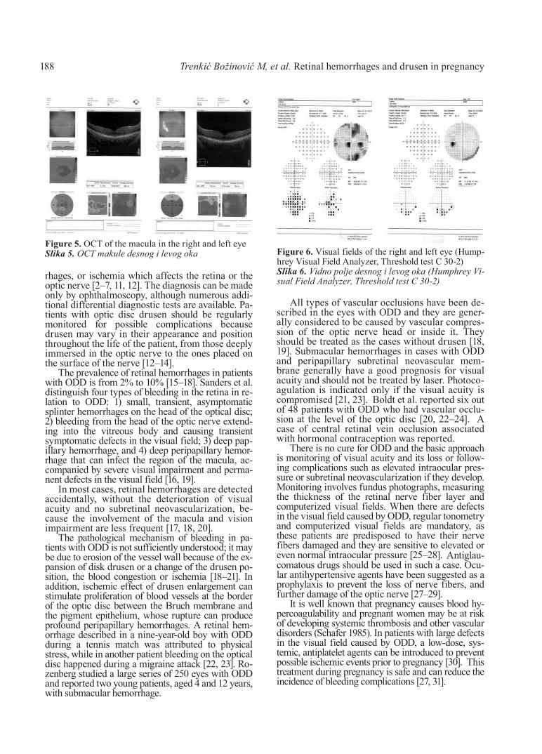
188
rhages, or ischemia which affects the retina or the optic nerve [2–7, 11, 12]. The diagnosis can be made only by ophthalmoscopy, although numerous addi-tional differential diagnostic tests are available. Pa-tients with optic disc drusen should be regularly monitored for possible complications because drusen may vary in their appearance and position throughout the life of the patient, from those deeply immersed in the optic nerve to the ones placed on the surface of the nerve [12–14].
The prevalence of retinal hemorrhages in patients with ODD is from 2% to 10% [15–18]. Sanders et al. distinguish four types of bleeding in the retina in re-lation to ODD: 1) small, transient, asymptomatic splinter hemorrhages on the head of the optical disc; 2) bleeding from the head of the optic nerve extend-ing into the vitreous body and causing transient symptomatic defects in the visual field; 3) deep pap-illary hemorrhage, and 4) deep peripapillary hemor-rhage that can infect the region of the macula, ac-companied by severe visual impairment and perma-nent defects in the visual field [16, 19].
In most cases, retinal hemorrhages are detected accidentally, without the deterioration of visual acuity and no subretinal neovascularization, be-cause the involvement of the macula and vision impairment are less frequent [17, 18, 20].
The pathological mechanism of bleeding in pa-tients with ODD is not sufficiently understood; it may be due to erosion of the vessel wall because of the ex-pansion of disk drusen or a change of the drusen po-sition, the blood congestion or ischemia [18–21]. In addition, ischemic effect of drusen enlargement can stimulate proliferation of blood vessels at the border of the optic disc between the Bruch membrane and the pigment epithelium, whose rupture can produce profound peripapillary hemorrhages. A retinal hem-orrhage described in a nine-year-old boy with ODD during a tennis match was attributed to physical stress, while in another patient bleeding on the optical disc happened during a migraine attack [22, 23]. Ro-zenberg studied a large series of 250 eyes with ODD and reported two young patients, aged 4 and 12 years, with submacular hemorrhage.
All types of vascular occlusions have been de-scribed in the eyes with ODD and they are gener-ally considered to be caused by vascular compres-sion of the optic nerve head or inside it. They should be treated as the cases without drusen [18, 19]. Submacular hemorrhages in cases with ODD and peripapillary subretinal neovascular mem-brane generally have a good prognosis for visual acuity and should not be treated by laser. Photoco-agulation is indicated only if the visual acuity is compromised [21, 23]. Boldt et al. reported six out of 48 patients with ODD who had vascular occlu-sion at the level of the optic disc [20, 22–24]. A case of central retinal vein occlusion associated with hormonal contraception was reported.
There is no cure for ODD and the basic approach is monitoring of visual acuity and its loss or follow-ing complications such as elevated intraocular pres-sure or subretinal neovascularization if they develop. Monitoring involves fundus photographs, measuring the thickness of the retinal nerve fiber layer and computerized visual fields. When there are defects in the visual field caused by ODD, regular tonometry and computerized visual fields are mandatory, as these patients are predisposed to have their nerve fibers damaged and they are sensitive to elevated or even normal intraocular pressure [25–28]. Antiglau-comatous drugs should be used in such a case. Ocu-lar antihypertensive agents have been suggested as a prophylaxis to prevent the loss of nerve fibers, and further damage of the optic nerve [27–29].
It is well known that pregnancy causes blood hy-percoagulability and pregnant women may be at risk of developing systemic thrombosis and other vascular disorders (Schafer 1985). In patients with large defects in the visual field caused by ODD, a low-dose, sys-temic, antiplatelet agents can be introduced to prevent possible ischemic events prior to pregnancy [30]. This treatment during pregnancy is safe and can reduce the incidence of bleeding complications [27, 31].
Trenkić Božinović M, et al. Retinal hemorrhages and drusen in pregnancy
Figure 5. OCT of the macula in the right and left eyeSlika 5. OCT makule desnog i levog oka Figure 6. Visual fields of the right and left eye (Hump-
hrey Visual Field Analyzer, Threshold test C 30-2)Slika 6. Vidno polje desnog i levog oka (Humphrey Vi-sual Field Analyzer, Threshold test C 30-2)

Med Pregl 2014; LXVII (5-6): 185-189. Novi Sad: maj-juni. 189
Conclusion
Hemodynamic changes during pregnancy are possible factors affecting the development of opti-cal disc and retinal hemorrhages. Although the lit-erature describes the radial neurotomy fiber optic
nerve decompression, there is no established ther-apeutic treatment for optic disk drusen. Knowing that the treatment of optic disc drusen is limited, the recognition of optic nerve drusen as a cause of hemorrhage during pregnancy prevents unneces-sary diagnostic and therapeutic interventions.
References1. Morris RW, Ellerbrock JM, Hamp AM, Joy JT, Roels
P, Davis Jr CN. Advanced visual field loss secondary to optic nerve head drusen: case report and literature review. Optome-try 2009;80(2):83-100.
2. Wilkins JM, Pomeranz HD. Visual manifestations of visible and buried optic disc drusen: review. J Neuroophthal-mol 2004;24(2):125-9.
3. Optic disc drusen (editorial). Review of optometry. New York: Jobson Publishing, LLC; 2013. p. 75-7.
4. Friedman AH, Beckerman B, Gold DH, Walsh JB, Gar-tner S. Druse of the optic disc: review. Surv Ophthalmol 1977;21(5):375-90.
5. Auw-Haedrich C, Staubach F, Witschel H. Optic disk drusen: review. Surv Ophthalmol 2002;47(6):515-31.
6. Katz BJ, Pomeranz HD. Visual field defects and retinal nerve fiber layer defects in eyes with buried optic nerve dru-sen. Am J Ophthalmol 2006;141:248-53.
7. Lee AG, Zimmerman MB. The rate of visual field loss in optic nerve head drusen. Am J Ophthalmol 2005;139:1062-6.
8. Gili P, Flores-Rodríguez P, Yangüela J, Ordu˜na-Azcona J, Martín-Ríos MD. Evaluation of optic disc size in patients with optic nerve head drusen using fundus photo-graphy. J Optom 2013;6:75-9.
9. Manzanaro PG, Rodilla JY, Caravaca GR, Font CC, Rodrigo JCR, Puent AA. Decreased visual acuity from optic disc drusen. Arch Soc Esp Oftalmol 2010;85(2):64-9.
10. Ogawa R, Usui T, Takagi M, Hasegawa S, Abe H, Na-nba K. Bilateral optic disc drusen with severe progressive vi-sual field defect. Neuroophthalmol 2001;2(4):229-32.
11. Brodrick JD. Drusen of the disc and retinal haemorr-hages. Br J Ophthalmol 1973;57:299-306.
12. Lee KM, Woo SJ, Hwang JM. Morphologic characteri-stics of optic nerve head drusen on spectral-domain optical cohe-rence tomography. Am J Ophthalmol 2013;155:1139-47.
13. Sato T, Mrejen S, Spaide RF. Multimodal imaging of optic disc drusen. Am J Ophthalmol 2013;156:275-82.
14. Floyd MS, Katz BJ, Digre KB. Measurement of the scle-ral canal using optical coherence tomography in patients with optic nerve drusen. Am J Ophthalmol 2005;139:664-9.
15. Rubinstein K, Ali M. Retinal complications of optic disc drusen. Br J Ophthalmol 1982;66:83-95.
16. Romero J, Sowka J, Shechtman D. Hemorrhagic com-plications of optic disc drusen and available treatment opti-ons. Optometry 2008;79:496-500.
17. Sanders TE, Gay AJ, Newman M. Hemorrhagic compli-cations of drusen of the optic disk. Am J Ophthalmol 1971;71: 204-17.
18. Neffendorf JE, Mulholland C, Quinlan M, Lyons CJ. Disc drusen complicated by optic disc hemorrhage in childho-od. Can J Ophthalmol 2010;45:537-8.
19. Abeysiri P, Sivaprasad S, Trew D. Optic disc drusen. Lancet 2003;362(9):468.
20. Silbert Aumiller M. Optic disc drusen: complications and management. Optometry 2007;78:10-6.
21. Nicholson B, Ahmad B, Sears JE. Congenital optic ner-ve malformations. Int Ophthalmol Clin 2011;51(1):49-76.
22. Zaidi FH, Corbett MC. Optic disc drusen associated with neovascularization of optic disc. Eye 2005;19:814-6.
23. Borruat FX, Sanders MD. Vascular papillary anomali-es and complications in optic disc drusen. Klin Monatsbl Au-genheilkd 1996;208(5):294-6.
24. Austin JK. Optic disc drusen and associated venous stasis retinopathy. J Am Optom Assoc 1995;66(2):91-5.
25. Flores-Rodriguez P, Gili P, Martin-Rios MD. Sensitivity and specificity of time-domain and spectral-domain optical cohe-rence tomography in differentiating optic nerve head drusen and optic disc oedema. Ophthalmic Physiol Opt 2012;32:213-21.
26. Yehoshua Z, Gregori G, Sadda SR, Penha FM, Goldhar-dt R, Nittala MG, et al. Comparison of drusen area detected by spectral domain optical coherence tomography and color fundus imaging. Invest Ophthalmol Vis Sci 2013;54(4):2429-34.
27. Mikel M, Chidambaram Y, Amadeo RR. Optic disc drusen and anterior ischaemic optic neuropathy in pregnancy. Neuroophthalmol 2012;36(1):23-5.
28. Rott D, Leibowitz D. Optic nerve head drusen mimic-king papilledema and malignant hypertension. Eur J Intern Med 2009;20:112-3.
29. Johnson LN, Diehl ML, Hamm CW, Sommerville DN, Petroski GF. Differentiating optic disc edema from optic ner-ve head drusen on optical coherence tomography. Arch Opht-halmol 2009;127(1):45-9.
30. Sullu Y, Yıldız L, Erkan D. Submacular surgery for choroidal neovascularization secondary to optic nerve drusen. Am J Ophthalmol 2003;136:367-70.
31. Mehta JS, Bates A, Chan J, Kuteesa W, Acheson JF. Pregnancy associated, optic disc drusen related visual loss. Acta Ophthalmol Scand 2005;271-3.
Rad je primljen 5. II 2014.Recenziran 4. III 2014.Prihvaćen za štampu 5. III 2014.BIBLID.0025-8105:(2014):LXVII:5-6:185-189.

Časopis objavljuje sledeće kategorije radova: 1. Uvodnici (editorijali) – do 5 stranica. Sadrže
mišljenje ili diskusiju o nekoj temi važnoj za Časo-pis. Uobičajeno ih piše jedan autor po pozivu.
2. Originalni naučni radovi – do 12 stranica. Sadr-že rezultate sopstvenih originalnih naučnih istraživanja i njihova tumačenja. Originalni naučni radovi treba da sadrže podatke koji omogućavaju proveru dobijenih re-zultata i reprodukovanje istraživačkog postupka.
3. Pregledni članci – do 10 strana. Predstavljaju sa-žet, celovit i kritički pregled nekog problema na osnovu već publikovanog materijala koji se analizira i rasprav-lja, ilustrujući trenutno stanje u jednoj oblasti istraživa-nja. Radovi ovog tipa biće prihvaćeni samo ukoliko au-tori navođenjem najmanje 5 autocitata potvrde da su eksperti u oblasti o kojoj pišu.
4. Prethodna saopštenja – do 4 stranice. Sadrže naučne rezultate čiji karakter zahteva hitno objavlji-vanje, ali ne mora da omogući i ponavljanje iznetih rezultata. Donosi nove naučne podatke bez detaljnijeg obrazlaganja metodologije i rezultata. Sadrži sve de-love originalnog naučnog rada u skraćenom obliku.
5. Stručni članci – do 10 stranica. Odnose se na proveru ili reprodukciju poznatih istraživanja i pred-stavljaju koristan materijal u širenju znanja i prilago-đavanja izvornih istraživanja potrebama nauke i prakse.
6. Prikazi slučajeva – do 6 stranica. Obrađuju retku kazuistiku iz prakse, važnu lekarima koji vode neposrednu brigu o bolesnicima i imaju karakter stručnih radova. Prikazi slučajeva ističu neuobičajene karakteristike i tok neke bolesti, neočekivane reakcije na terapiju, primenu novih dijagnostičkih postupaka ili opisuju retko ili novo oboljenje.
7. Istorija medicine – do 10 stranica. Pišu se na poziv uredništva Medicinskog pregleda i obrađuju podatke iz prošlosti sa ciljem održavanja kontinuite-ta medicinske i zdravstvene kulture, a imaju karak-ter stručnih radova.
8. Druge vrste publikacija (feljtoni, prikazi knji-ga, izvodi iz strane literature, izveštaji sa kongresa i stručnih sastanaka, saopštenja o radu pojedinih zdravstvenih ustanova, podružnica i sekcija, saop-štenja Uredništva, pisma Uredništvu, novine u me-dicini, pitanja i odgovori, stručne i staleške vesti i In memoriam).
Priprema rukopisaPropratno pismo– Mora da sadrži svedočanstvo autora da rad
predstavlja originalno delo, kao i da nije objavljivan u drugim časopisima, niti se razmatra za objavljiva-nje u drugim časopisima.
– Potvrditi da svi autori ispunjavaju kriterijume za autorstvo nad radom, da su potpuno saglasni sa tekstom rada, kao i da ne postoji sukob interesa.
– Navesti u koju kategoriju spada rad koji se šalje (originalni naučni rad, pregledni članak, prethodno saopštenje, stručni članak, prikaz slučaja, istorija medicine).
RukopisZa pisanje teksta koristiti Microsoft Word for Win-
dows. Tekst treba otkucati koristeći font Times New Roman, na stranici formata A4, proredom od 1,5 (i u tabelama), sa marginama od 2,5 cm i veličinom slova od 12 pt. Rukopis treba da sadrži sledeće elemente:
1. Naslovna strana. Naslovna strana treba da sa-drži kratak i jasan naslov rada, bez skraćenica, za-tim kratki naslov (do 40 karaktera), puna imena i prezimena autora (najviše 6 autora) indeksirana broj-kama koje odgovaraju onima kojim se u zaglavlju navode uz pun naziv i mesta ustanova u kojima au-tori rade. Na dnu ove stranice navesti titulu, punu adresu, e-mail i broj telefona ili faksa autora zaduže-nog za korespondenciju.
2. Sažetak. Sažetak treba da sadrži do 250 reči, bez skraćenica, sa preciznim prikazom problematike, cilje-va, metodologije, glavnih rezultata i zaključaka. Saže-tak treba da ima sledeću strukturu:
– originalni naučni radovi: uvod (sa ciljem rada), materijal i metode, rezultati i zaključak;
– prikaz slučaja: uvod, prikaz slučaja i zaključak;– pregled rada: uvod, odgovarajući podnaslovi koji
odgovaraju onima u tekstu rada i zaključak.U nastavku navesti do deset ključnih reči iz spi-
ska medicinskih predmetnih naziva (Medical Su-bjects Headings, MeSH) Američke nacionalne me-dicinske biblioteke.
3. Sažetak na engleskom jeziku. Sažetak na en-gleskom jeziku treba da bude prevod sažetka na srp-skom jeziku, da ima istu strukturu i da sadrži do 250 reči, bez upotrebe skraćenica.
4. Tekst rada– Tekst originalnih članaka mora da sadrži slede-
će celine:Uvod (sa jasno definisanim ciljem rada), Materi-
jal i metode, Rezultati, Diskusija, Zaključak, spisak skraćenica (ukoliko su korišćene u tekstu) i eventu-alna zahvalnost autora onima koji su pomogli u istraživanju i izradi rada.
– Tekst prikaza slučaja treba da sadrži sledeće ce-line: Uvod (sa jasno definisanim ciljem rada), Prikaz slučaja, Diskusija i Zaključak.
– Tekst treba da bude napisan u duhu srpskog je-zika, oslobođen suvišnih skraćenica, čija prva upo-treba zahteva navođenje punog naziva. Skraćenice ne upotrebljavati u naslovu, sažetku i zaključku. Ko-ristiti samo opšte prihvaćene skraćenice (npr. DNA, MRI, NMR, HIV,...). Spisak skraćenice koje se na-vode u radu, zajedno sa objašnjenjem njihovog zna-čenja, dostaviti na poslednjoj stranici rukopisa.
– Koristiti mere metričkog sistema prema Inter-nacionalnom sistemu mera (International System Units – SI). Temperaturu izražavati u Celzijusovim stepenima (°C), a pritisak u milimetrima živinog stuba (mmHg).
– Ne navoditi imena bolesnika, inicijale ili broje-ve istorija bolesti.
Uvod sadrži precizno definisan problem kojim se bavi studija (njegova priroda i značaj), uz navođenje relevantne literature i sa jasno definisanim ciljem istraživanja i hipotezom.
Materijal i metode treba da sadrže podatke o na-činu dizajniranja studije (prospektivna/retrospektiv-na, kriterijumi za uključivanje i isključivanje, traja-nje, demografski podaci, dužina praćenja). Statistič-ke metode koje se koriste treba da budu jasne i de-taljno opisane.
Rezultati predstavljaju detaljan prikaz podataka dobijenih tokom studije. Sve tabele, grafikoni, she-me i slike moraju da budu citirani u tekstu, a njihova
UPUTSTVO AUTORIMA
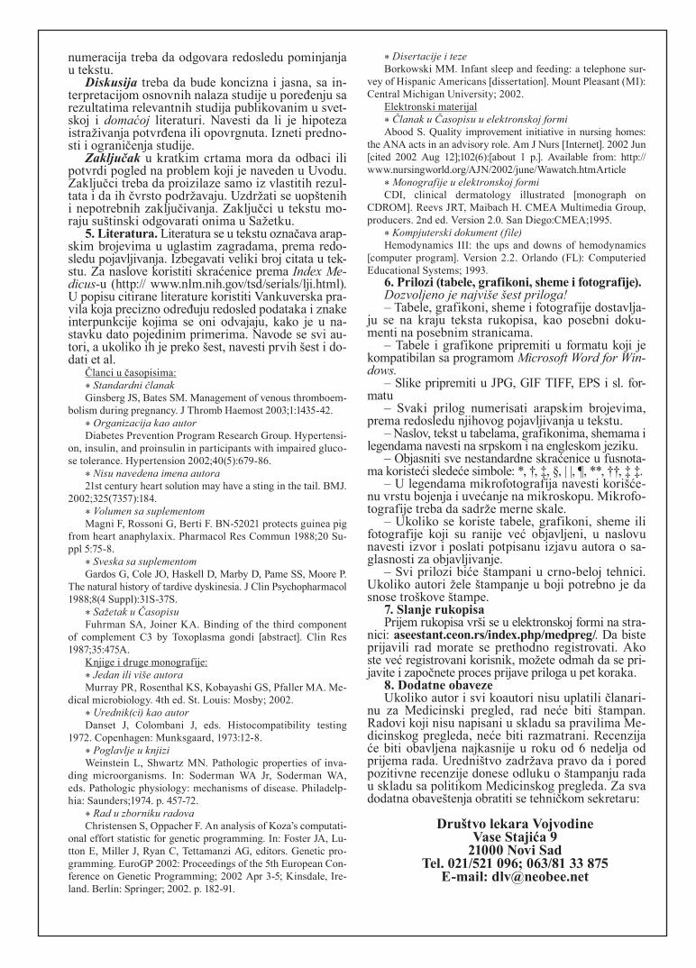
numeracija treba da odgovara redosledu pominjanja u tekstu.
Diskusija treba da bude koncizna i jasna, sa in-terpretacijom osnovnih nalaza studije u poređenju sa rezultatima relevantnih studija publikovanim u svet-skoj i domaćoj literaturi. Navesti da li je hipoteza istraživanja potvrđena ili opovrgnuta. Izneti predno-sti i ograničenja studije.
Zaključak u kratkim crtama mora da odbaci ili potvrdi pogled na problem koji je naveden u Uvodu. Zaključci treba da proizilaze samo iz vlastitih rezul-tata i da ih čvrsto podržavaju. Uzdržati se uopštenih i nepotrebnih zaključivanja. Zaključci u tekstu mo-raju suštinski odgovarati onima u Sažetku.
5. Literatura. Literatura se u tekstu označava arap-skim brojevima u uglastim zagradama, prema redo-sledu pojavljivanja. Izbegavati veliki broj citata u tek-stu. Za naslove koristiti skraćenice prema Index Me-dicus-u (http:// www.nlm.nih.gov/tsd/serials/lji.html). U popisu citirane literature koristiti Vankuverska pra-vila koja precizno određuju redosled podataka i znake interpunkcije kojima se oni odvajaju, kako je u na-stavku dato pojedinim primerima. Navode se svi au-tori, a ukoliko ih je preko šest, navesti prvih šest i do-dati et al.
Članci u časopisima:* Standardni članakGinsberg JS, Bates SM. Management of venous thromboem-
bolism during pregnancy. J Thromb Haemost 2003;1:1435-42.* Organizacija kao autorDiabetes Prevention Program Research Group. Hypertensi-
on, insulin, and proinsulin in participants with impaired gluco-se tolerance. Hypertension 2002;40(5):679-86.
* Nisu navedena imena autora21st century heart solution may have a sting in the tail. BMJ.
2002;325(7357):184.* Volumen sa suplementomMagni F, Rossoni G, Berti F. BN-52021 protects guinea pig
from heart anaphylaxix. Pharmacol Res Commun 1988;20 Su-ppl 5:75-8.
* Sveska sa suplementomGardos G, Cole JO, Haskell D, Marby D, Pame SS, Moore P.
The natural history of tardive dyskinesia. J Clin Psychopharmacol 1988;8(4 Suppl):31S-37S.
* Sažetak u ČasopisuFuhrman SA, Joiner KA. Binding of the third component
of complement C3 by Toxoplasma gondi [abstract]. Clin Res 1987;35:475A.
Knjige i druge monografije:* Jedan ili više autoraMurray PR, Rosenthal KS, Kobayashi GS, Pfaller MA. Me-
dical microbiology. 4th ed. St. Louis: Mosby; 2002.* Urednik(ci) kao autorDanset J, Colombani J, eds. Histocompatibility testing
1972. Copenhagen: Munksgaard, 1973:12-8.* Poglavlje u knjiziWeinstein L, Shwartz MN. Pathologic properties of inva-
ding microorganisms. In: Soderman WA Jr, Soderman WA, eds. Pathologic physiology: mechanisms of disease. Philadelp-hia: Saunders;1974. p. 457-72.
* Rad u zborniku radovaChristensen S, Oppacher F. An analysis of Koza’s computati-
onal effort statistic for genetic programming. In: Foster JA, Lu-tton E, Miller J, Ryan C, Tettamanzi AG, editors. Genetic pro-gramming. EuroGP 2002: Proceedings of the 5th European Con-ference on Genetic Programming; 2002 Apr 3-5; Kinsdale, Ire-land. Berlin: Springer; 2002. p. 182-91.
* Disertacije i tezeBorkowski MM. Infant sleep and feeding: a telephone sur-
vey of Hispanic Americans [dissertation]. Mount Pleasant (MI): Central Michigan University; 2002.
Elektronski materijal* Članak u Časopisu u elektronskoj formiAbood S. Quality improvement initiative in nursing homes:
the ANA acts in an advisory role. Am J Nurs [Internet]. 2002 Jun [cited 2002 Aug 12];102(6):[about 1 p.]. Available from: http://www.nursingworld.org/AJN/2002/june/Wawatch.htmArticle
* Monografije u elektronskoj formiCDI, clinical dermatology illustrated [monograph on
CDROM]. Reevs JRT, Maibach H. CMEA Multimedia Group, producers. 2nd ed. Version 2.0. San Diego:CMEA;1995.
* Kompjuterski dokument (file)Hemodynamics III: the ups and downs of hemodynamics
[computer program]. Version 2.2. Orlando (FL): Computeried Educational Systems; 1993.
6. Prilozi (tabele, grafikoni, sheme i fotografije).Dozvoljeno je najviše šest priloga!– Tabele, grafikoni, sheme i fotografije dostavlja-
ju se na kraju teksta rukopisa, kao posebni doku-menti na posebnim stranicama.
– Tabele i grafikone pripremiti u formatu koji je kompatibilan sa programom Microsoft Word for Win-dows.
– Slike pripremiti u JPG, GIF TIFF, EPS i sl. for-matu
– Svaki prilog numerisati arapskim brojevima, prema redosledu njihovog pojavljivanja u tekstu.
– Naslov, tekst u tabelama, grafikonima, shemama i legendama navesti na srpskom i na engleskom jeziku.
– Objasniti sve nestandardne skraćenice u fusnota-ma koristeći sledeće simbole: *, †, ‡, §, | |, ¶, **, ††, ‡ ‡.
– U legendama mikrofotografija navesti korišće-nu vrstu bojenja i uvećanje na mikroskopu. Mikrofo-tografije treba da sadrže merne skale.
– Ukoliko se koriste tabele, grafikoni, sheme ili fotografije koji su ranije već objavljeni, u naslovu navesti izvor i poslati potpisanu izjavu autora o sa-glasnosti za objavljivanje.
– Svi prilozi biće štampani u crno-beloj tehnici. Ukoliko autori žele štampanje u boji potrebno je da snose troškove štampe.
7. Slanje rukopisaPrijem rukopisa vrši se u elektronskoj formi na stra-
nici: aseestant.ceon.rs/index.php/medpreg/. Da biste prijavili rad morate se prethodno registrovati. Ako ste već registrovani korisnik, možete odmah da se pri-javite i započnete proces prijave priloga u pet koraka.
8. Dodatne obavezeUkoliko autor i svi koautori nisu uplatili članari-
nu za Medicinski pregled, rad neće biti štampan. Radovi koji nisu napisani u skladu sa pravilima Me-dicinskog pregleda, neće biti razmatrani. Recenzija će biti obavljena najkasnije u roku od 6 nedelja od prijema rada. Uredništvo zadržava pravo da i pored pozitivne recenzije donese odluku o štampanju rada u skladu sa politikom Medicinskog pregleda. Za sva dodatna obaveštenja obratiti se tehničkom sekretaru:
Društvo lekara VojvodineVase Stajića 9
21000 Novi SadTel. 021/521 096; 063/81 33 875
E-mail: [email protected]

Medical review publishes papers from various fields of biomedicine intended for broad circles of doc tors. The papers are published in Serbian language with an ex-panded summary in English language and contri butions both in Serbian and English language, and se lected pa-pers are published in English language at full length with the summary in Serbian language. Papers coming from non-Serbian speaking regions are pub lished in English language. The authors of the papers have to be Medical Review subscribers.
This journal publishes the following types of articles: editorials, original studies, preliminary reports, review articles, professional articles, case reports, articles from history of medicine and other types of publications.
1. Editorials – up to 5 pages – convey opinions or discussions on a subject relevant for the journal. Editori-als are commonly written by one author by invitation.
2. Original studies – up to 12 pages – present the authors’ own investigations and their interpreta tions. They should contain data which could be the basis to check the obtained results and reproduce the investi-gative procedure.
3. Review articles – up to 10 pages – provide a con-densed, comprehensive and critical review of a problem on the basis of the published material being analyzed and discussed, reflecting the current situation in one area of research. Papers of this type will be accepted for publica tion provided that the authors confirm their ex-pertise in the relevant area by citing at least 5 auto-cita-tions.
4. Preliminary reports – up to 4 pages – contain scientific results of significant importance requiring ur gent publishing; however, it need not provide detailed description for repeating the obtained results. It presents new scientific data without a detailed explana-tion of methods and results. It contains all parts of an original study in an abridged form.
5. Professional articles – up to 10 pages – examine or reproduce previous investigation and represent a valu able source of knowledge and adaption of original inves tigations for the needs of current science and prac-tise.
6. Case reports – up to 6 pages – deal with rare casu-istry from practise important for doctors in direct charge of patients and are similar to professional articles. They emphasize unusual characteristics and course of a disease, unexpected reactions to a therapy, application of new di-agnostic procedures and describe a rare or new disease.
7. History of medicine – up to 10 pages – deals with history in the aim of providing continuity of medi-cal and health care culture. They have the character of professional articles.
8. Other types of publications – The journal also publishes feuilletons, book reviews, extracts from for-eign literature, reports from congresses and profession-al meet ings, communications on activities of certain medical in stitutions, branches and sections, announce-ments of the Editorial Board, letters to the Editorial Board, novel ties in medicine, questions and answers, professional and vocational news and In memoriam.
Preparation of the manuscriptThe covering letter:– It must contain the proof given by the author that
the paper represents an original work, that it has neither been previously published in other journals nor is un-der consideration to be published in other journals.
– It must confirm that all the authors meet criteria set for the authorship of the paper, that they agree complete ly with the text and that there is no conflict of interest.
– It must state the type of the paper submitted (an original study, a review article, a preliminary report, a professional article, a case report, history of medicine)
The manuscript:Use Microsoft Word for Windows to type the text.
The text must be typed in font Times New Roman, page format A4, space 1.5 (for tables as well), borders of 2.5 cm and font size 12pt. The manuscript should contain the following elements:
1. The title page. The title page should contain a con-cise and clear title of the paper, without abbreviations, then a short title (up to 40 characters), full names and sur-names of the authors (not more than 6) indexed by num-bers corresponding to those given in the heading along with the full name and place of the institutions they work for. Contact information including the academic degree(s), full address, e-mail and number of phone or fax of the corresponding author (the author responsible for corre-spondence) are to be given at the bottom of this page.
2. Summary. The summary should contain up to 250 words, without abbreviations, with the precise re-view of problems, objectives, methods, important re-sults and conclusions. It should be structured into the paragraphs as follows:
– original and professional papers should have the introduction (with the objective of the paper), material and methods, results and conclusion
– case reports should have the introduction, case re port and conclusion
– review papers should have the introduction, sub-titles corresponding to those in the paper and conclu-sion. It is to be followed by up to 10 Key Words from the list of Medical Subject Headings, MeSH of the American National Medical Library.
3. The summary in Serbian language. The sum-mary in Serbian should be the translation of the sum-mary in English, it should be structured in the same way as the English summary, containing up to 250 words, without any abbreviations.
4. The text of the paper. The text of original stud-ies must contain the following: introduction (with the clearly defined objective of the study), material and methods, results, discussion, conclusion, list of abbre-viations (if used in the text) and not necessarily, the ac-knowledgment mentioning those who have helped in the investigation and preparation of the paper.
– The text of a case report should contain the fol-lowing: introduction (with clearly defined objective of the study), case report, discussion and conclusion.
– The text should be written in the spirit of Serbian language, without unnecessary abbreviations, whose first mentioning must be explained by the full term they stand for. Abbreviations should not be used in the title, summa-ry and conclusion. Only commonly ac cepted abbrevia-tions (such as DNA, MRI, NMR, HIV…) should be used. The list of abbreviations used in the text, together with the explanation of their mean ing, is to be submitted at the last page of the manuscript.
– All measurements should be reported in the met-ric system of the International System of Units – SI. Tem perature should be expressed in Celsius degrees (°C). and pressure in mmHg.
– No names, initials or case history numbers should be given.
Introduction contains clearly defined problem dealt with in the study (its nature and importance), with the relevant references and clearly defined objec-tive of the investigation and hypothesis.
Material and methods should contain data on de-sign of the study (prospective/retrospective, eligibility
INFORMATION FOR AUTHORS

and exclusion criteria, duration, demographic data, follow-up period). Statistical methods applied should be clear and described in details.
Results give a detailed review of data obtained dur-ing the study. All tables, graphs, schemes and figures must be cited in the text and numbered consecutively in the order of their first citation in the text.
Discussion should be concise and clear, interpreting the basic findings of the study in comparison with the results of relevant studies published in international and national literature. It should be stated whether the hypot-hesis has been confirmed or denied. Merits and demerits of the study should be mentioned.
Conclusion must deny or confirm the attitude to-wards the problem mentioned in the introduction. Con-clusions must be based solely on the author’s own results, corroborating them. Avoid generalised and unnecessary conclusions. Conclusions in the text must be in accord-ance with those given in the summary.
5. References. References are to be given in the text under Arabic numerals in parentheses consecu tively in the order of their first citation. Avoid a large number of ci-tations in the text. The title of journals should be abbrevi-ated according to the style used in In dex Medicus (http://www.nlm.nih.gov/tsd/serials/lji.html). Apply Vancouver Group’s Criteria, which define the order of data and punc-tuation marks separating them. Examples of correct forms of references are giv en below. List all authors, but if the number exceeds six, give the names of six authors fol-lowed by et ’al’.
Articles in journals* A standard articleGinsberg JS, Bates SM. Management of venous thromboembo-
lism during pregnancy. J Thromb Haemost 2003;1:1435-42.* An organisation as the author Diabetes Prevention Program Research Group. Hypertensi-
on, insulin, and proinsulin in participants with impaired gluco-se tolerance. Hypertension 2002;40(5):679-86.
* No author given 21st century heart solution may have a sting in the tail. BMJ.
2002;325(7357):184.* A volume with supplement Magni F, Rossoni G, Berti F. BN-52021 protects guinea pig from
heart anaphylaxix. Pharmacol Res Commun 1988;20 Suppl 5:75-8.* An issue with supplement Gardos G, Cole JO, Haskell D, Marby D, Pame SS, Moore
P. The natural history of tardive dyskinesia. J Clin Psychophar-macol 1988;8(4 Suppl):31S-37S.
* A summary in a journal Fuhrman SA, Joiner KA. Binding of the third component
of complement C3 by Toxoplasma gondi [abstract]. Clin Res 1987;35:475A.
Books and other monographs * One or more authors Murray PR, Rosenthal KS, Kobayashi GS, Pfaller MA. Me-
dical microbiology. 4th ed. St. Louis: Mosby; 2002.* Editor(s) as author(s) Danset J, Colombani J, eds. Histocompatibility testing 1972.
Copenhagen: Munksgaard, 1973:12-8.* A chapter in a book Weinstein L, Shwartz MN. Pathologic properties of invad-
ing microorganisms. In: Soderman WA Jr, Soderman WA, eds. Patho logic physiology: mechanisms of disease. Philadelphia: Saunders; 1974. p. 457-72.
* A conference paper Christensen S, Oppacher F. An analysis of Koza’s computa-
tional effort statistic for genetic programming. In: Foster JA, Lutton E, Miller J, Ryan C, Tettamanzi AG, editors. Genetic pro-gramming. EuroGP 2002: Proceedings of the 5th European Con-ference on Genetic Programming; 2002 Apr 3-5; Kinsdale, Ire-land. Berlin: Springer; 2002. p. 182-91.
* A dissertation and theses Borkowski MM. Infant sleep and feeding: a telephone sur-
vey of Hispanic Americans [dissertation]. Mount Pleasant (MI): Central Michigan University; 2002.
Electronic material * A journal article in electronic formatAbood S. Quality improvement initiative in nursing homes:
the ANA acts in an advisory role. Am J Nurs [Internet]. 2002 Jun [cited 2002 Aug 12];102(6):[about 1 p.]. Available from: http://www.nursingworld.org/AJN/2002/june/Wawatch.htmArticle
* Monographs in electronic formatCDI, clinical dermatology illustrated [monograph on CD-
ROM]. Reevs JRT, Maibach H. CMEA Multimedia Group, pro-ducers. 2nd ed. Version 2.0. San Diego:CMEA;1995.
* Acomputer fileHemodynamics III: the ups and downs of hemodynamics
[computer program]. Version 2.2. Orlando (FL): Computerized Educational Systems; 1993.
6. Attachments (tables, graphs, schemes and pho-tographs). The maximum number of attachments al-lowed is six!
– Tables, graphs, schemes and photographs are to be submitted at the end of the manuscript, on separate pages.
– Tables and graphs are to be prepared in the for-mat compatible with Microsoft Word for Windows progra mme. Photographs are to be prepared in JPG, GIF, TIFF, EPS or similar format.
– Each attachment must be numbered by Arabic numerals consecutively in the order of their appear-ance in the text
– The title, text in tables, graphs, schemes and legen-ds must be given in both Serbian and English language.
– Explain all non-standard abbreviations in footnotes using the following symbols *, †, ‡, §, | |, ¶, **, † †, ‡ ‡ .
– State the type of colour used and microscope ma-gnification in the legends of photomicrographs. Photo-micrographs should have internal scale markers.
– If a table, graph, scheme or figure has been previ-ously published, acknowledge the original source and submit written permission from the copyright holder to reproduce it.
– All attachments will be printed in black and whi-te. If the authors wish to have the attachments in colo-ur, they will have to pay additional cost.
7. Manuscript submissionThe manuscripts can be submited on the web-page:
aseestant.ceon.rs/index.php / medpreg/. The authors have to register with the journal prior to submitting their manuscript, or, if already registered, they can simply log in and begin the 5 step process.
8. Additional requirementsIf the author and all co-authors have failed to pay
the subscription for Medical Review, their paper will not be published.
Papers which have not met the criteria of Medical Review will not be taken into consideration. The Edito-rial review of the paper will be announced not later than six weeks after the submission of the paper. The Editori al Board reserves the right to make a decision regarding the publication of the paper according to the policy of Medical Review even if the review is positive. Contact the technical secretary for all additional infor-mation:
Društvo lekara VojvodineVase Stajića 9
21000 Novi SadTel. 021/521 096; 063/81 33 875
E-mail: [email protected]