hydrolyzable tannins of tamaricaceous plants. v. structures of monomeric–trimeric tannins and...
TRANSCRIPT
Hydrolyzable Tannins of Tamaricaceous Plants. V. Structures ofMonomeric−Trimeric Tannins and Cytotoxicity of Macrocyclic-TypeTannins Isolated from Tamarix nilotica1
Mohamed A. A. Orabi,† Shoko Taniguchi,‡ Hiroshi Sakagami,§ Morio Yoshimura,⊥ Takashi Yoshida,⊥
and Tsutomu Hatano*,‡
†Faculty of Pharmacy, Al-Azhar University, Assiut 71524, Egypt‡Graduate School of Medicine, Dentistry and Pharmaceutical Sciences, Okayama University, Tsushima, Okayama 700-8530, Japan§Division of Pharmacology, Department of Diagnostic and Therapeutic Sciences, School of Dentistry, Meikai University, Japan⊥College of Pharmaceutical Sciences, Matsuyama University, Bunkyo-cho, Matsuyama 790-8578, Japan
*S Supporting Information
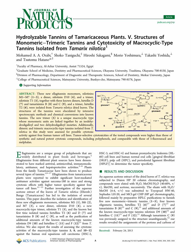
ABSTRACT: Three new ellagitannin monomers, nilotininsM5−M7 (1−3), a dimer, nilotinin D10 (4), and a trimer,nilotinin T1 (5), together with three known dimers, hirtellin D(7) and tamarixinins B (8) and C (9), and a trimer, hirtellinT2 (6), were isolated from Tamarix nilotica dried leaves. Thestructures of the tannins were elucidated by intensivespectroscopic methods and chemical conversions into knowntannins. The new trimer (5) is a unique macrocyclic typewhose monomeric units are linked together by an isodehy-drodigalloyl and two dehydrodigalloyl moieties. Additionally,dimeric and trimeric macrocyclic-type tannins isolated from T.nilotica in this study were assessed for possible cytotoxicactivity against four human tumor cell lines. Tumor-selective cytotoxicities of the tested compounds were higher than those ofsynthetic and natural potent cytotoxic compounds, including polyphenols, and comparable with those of 5-fluorouracil andmelphalan.
Ellagitannins are a unique group of polyphenols that arewidely distributed in plant foods and beverages.2
Ellagitannins from different plant sources have been demon-strated to have marked antiviral, antimicrobial, immunomodu-latory, antitumor, and hepatoprotective activities.2−8 Plantsfrom the family Tamaricaceae have been shown to produceseveral types of tannins.1,9−17 Ellagitannins from tamaricaceousplants were reported to exhibit significant host-mediatedantitumor activities against sarcoma-180 in mice and strongcytotoxic effects with higher tumor specificity against fourtumor cell lines.11−13 Further investigation of the aqueousacetone extract of the leaves of Tamarix nilotica (Ehrenb.)Bunge (Tamaricaceae) has led to the isolation of severaltannins. This paper describes the isolation and identification ofthree new ellagitannin monomers, nilotinins M5 (1), M6 (2),and M7 (3), a new dimer, nilotinin D10 (4), a newmacrocyclic-type trimer, nilotinin T1 (5), and the known butfirst time isolated tannins hirtellins T2 (6) and D (7) andtamarixinins B (8) and C (9), as well as the purification ofadditional amounts of the known macrocyclic-type tanninsnilotinin D9 (10) and hirtellins C (11) and F (12)11 from T.nilotica. We also report the results of assessing the cytotoxicactivities of the macrocyclic-type tannins 5, 8, and 10−12against the human oral squamous cell carcinoma (HSC-2,
HSC-3, and HSC-4) and human promyelocytic leukemia (HL-60) cell lines and human normal oral cells [gingival fibroblast(HGF), pulp cell (HPC), and periodontal ligament fibroblast(HPLF)] to determine the tumor specificity.
■ RESULTS AND DISCUSSION
An aqueous acetone extract of the dried leaves of T. nilotica wassubjected to Diaion HP 20 column chromatography, andcompounds were eluted with H2O, MeOH/H2O (40:60%, v/v), MeOH, and acetone, successively. The eluate with H2O/MeOH (6:4, v/v) was submitted to Toyopearl HW-40,Sephadex LH-20, and MCI-gel CHP-20P gel chromatography,followed mainly by preparative HPLC purifications to furnishfive new monomeric−trimeric tannins (1−5), four knownoligomeric tannins, hirtellins T2 (6)17 and D (7)13 andtamarixinins B (8)15 and C (9),15 and additional amounts ofthe known macrocyclic-type tannins nilotinin D9 (10)11 andhirtellins C (11)11 and F (12).11 Although tamarixinin C (9)was previously assigned to the structure unambiguously,15 ourresults revealed the assignments of the protons and carbons of
Received: February 24, 2013
Article
pubs.acs.org/jnp
© XXXX American Chemical Society andAmerican Society of Pharmacognosy A dx.doi.org/10.1021/np4001625 | J. Nat. Prod. XXXX, XXX, XXX−XXX

the sugar cores in the NMR spectra should be corrected asshown in Tables 1 and 2.Structural Elucidation of Monomeric Ellagitannins.
Nilotinin M5 (1) was isolated as an off-white, amorphouspowder. Its molecular formula was C55H38O36 from theprominent ion peak ([M + Na]+) at m/z 1297.10358 in the
HRESIMS spectrum. The 1H NMR spectrum of 1 displayedaromatic proton signals of two mutually coupled doublets [δH7.21 and 6.46 (each 1H, d, J = 1.8 Hz)] and a one-protonsinglet (δH 7.06), characteristic of the dehydrodigalloyl moiety(DHDG).9−11,17 The spectrum also showed a two-protonsinglet (δH 6.96) and one-proton singlet (δH 6.89), character-istic of an isodehydrodigalloyl moiety (isoDHDG),9−11 and asecond two-proton singlet (δH 6.93) and a pair of one-protonsinglets (δH 6.58 and 6.51) ascribable to galloyl andhexahydroxydiphenoyl (HHDP) units, respectively.18 Thealiphatic region of the spectrum showed seven sets of well-resolved signals (δH 5.50−3.80) assignable to the protons of afully O-acylated glucose core (Table 1).9−11 The large couplingconstants (J1,2 = 8.4 Hz, J2,3 = J3,4 = J4,5 = 9.6 Hz) of theseproton signals indicated the existence of the glucose core in thepyranose form with a 4C1 conformation and a β-oriented acylgroup at the anomeric carbon. The 13C NMR spectrum of 1showed the aliphatic, aromatic, and the carbonyl carbon peaks(Tables 2 and 3), which were assigned on the basis ofcorrelations in the HSQC and HMBC spectra, characteristic ofthe structural moieties of 1. A galloyl unit was placed at C-3 ofthe glucose core, as indicated from an HMBC correlationbetween a galloyl proton signal (δH 6.93, 2H, s) and a glucoseH-3 signal (δH 5.47, 1H, t) through a common carbonyl peak(δC 166.6). The acylation of the glucose core C-4/C-6 by anHHDP unit was suggested by the large chemical shift difference(ΔδH 1.45) between the geminal coupled C-6 methyleneprotons.19 Bridging of the HHDP unit at C-4/C-6 of theglucose core was further confirmed by HMBC correlationsamong the HHDP proton signals (δH 6.58 and 6.51) andsignals of H-6 (δH 5.25) and H-4 (δH 5.11) of the glucose corethrough the common carbonyl carbon peaks (δC 168.3 and167.7), respectively. In the 13C NMR spectrum of 1, the carbon
Table 1. 1H NMR Data δH (J in Hz) for the Glucose Protons of 1−5 and 9 (600 MHz, acetone-d6/D2O, 9:1)
1 2 3 9 4 5
glucose-11 5.50, d (8.4) 6.02, d (7.8) 6.02, d (7.8) 6.21, d (8.4) 5.24, d (7.8) 6.16 br s2 5.55, dd (8.4, 9.6) 5.65, dd (7.8, 9.6) 5.63, dd (7.8, 9.6) 5.63, dd (8.4, 9.6) 5.31, dd (7.8, 9.6) 5.56a
3 5.47, t (9.6) 5.72, t (9.6) 5.74, t (9.6) 5.81, t (9.6) 5.34, t (9.6) 5.70,b t (9.6)4 5.11, t (9.6) 5.19, t (9.6) 5.19, t (9.6) 5.22, t (9.6) 3.85, t (9.6) 5.12,c t (9.6)5 4.23, dd (6.6, 9.6) 4.43, dd (6.6, 9.6) 4.43, ddd (1.2, 6.6, 9.6) 4.54, dd (6.6, 9.6) ∼3.60d 4.25,e dd (6.6, 9.6)6 5.25, dd (6.6, 13.2) 5.28,dd (6.6, 13.2) 5.28,dd (6.6, 13.2) 5.33, dd (6.6, 12.6) 3.78, dd (6.6, 13.2) 5.26,f dd (6.6, 13.2)
3.80, br d (13.2) 3.83, d (13.2) 3.83, dd (1.2, 13.2) 3.92, d (12.6) 3.93, dd (2.4, 13.2) 3.84,g d (13.2)glucose-21 6.03, d (8.4) 5.44, d (7.8) 5.76 br s2 5.63, dd (8.4, 9.6) 5.51, dd (7.8, 9.6) 5.44, dd (7.8, 9.6)3 5.76, t (9.6) 5.59, t (9.6) 5.80, t (9.6)4 5.19, t (9.6) 5.10, t (9.6) 5.19, t (9.6)5 4.45, dd (6.6, 9.6) 4.24, dd (6.6, 9.6) 4.34e,h
6 5.30, dd (6.6, 13.2) 5.26, dd (6.6, 13.2) 5.23,f,i dd (6.6, 13.2)3.85, d (13.2) 3.81, br d (13.2) 3.76,g, d (13.2)
glucose-31 5.24i
2 5.56a
3 5.56a,b
4 5.07,c t (10.2)5 4.34e,h
6 5.36,f dd (6.6, 13.2)3.72,g d (13.2)
a,h,iProton signals with these chemical shifts overlapped, and thus their coupling constants were not measured. b,c,e−gInterchangeable. dOverlappedby solvent signal.
Table 2. 13C NMR Data for the Glucose Carbons of 1−5 and9 (151 MHz, acetone-d6/D2O, 9:1)
1 2 3 9 4 5
glucose-11 93.8 93.7 93.5 93.7 93.3 93.32 71.4 71.9 72.0 71.7 71.5 71.6a
3 73.6 73.5 73.4 73.4 76.4 73.4b
4 70.5 70.6 70.6 70.65 68.7 70.8c
5 72.8 72.8 72.7 72.9 78.0 72.9d
6 62.9 63.0 63.0 63.0 61.4 63.4e
glucose-21 93.5 93.6 93.32 71.9 71.6 72.53 73.3 73.5 73.44 70.58 70.6 70.55 72.8 72.8 72.7d
6 63.0 63.0 63.5e
glucose-31 93.82 71.2a
3 74.4b
4 70.7c
5 73.0.d
6 63.1e
a−eInterchangeable.
Journal of Natural Products Article
dx.doi.org/10.1021/np4001625 | J. Nat. Prod. XXXX, XXX, XXX−XXXB

peak at δC 150.3 was assigned to C-3/C-5 of an isoDHDGmoiety since it has been demonstrated that C-3/C-5 of theisoDHDG moiety resonate at lower field (δC ∼150) than C-3/C-5 of the usual galloyl unit (δC ∼145.7) (Table 3).9,10
Assignment of a two-proton singlet (δH 6.96) to H-2/H-6 of anisoDHDG moiety was confirmed by HMBC connectivity withthe characteristic carbon peak (δC 150.3) of C-3/C-5 of thesame moiety.9,10 The HMBC correlation of this two-protonsinglet with the glucose H-1 signal (δH 5.50) through the estercarbonyl carbon (δC 164.3) indicated the location of the
isoDHDG moiety at C-1 of the glucose core. A carboxyliccarbon peak (δC 170.3) was assigned to C-7′ of an isoDHDGunit, as indicated from its HMBC correlation with the H-6′signal (δH 6.89) of the same isoDHDG moiety. The absence ofa similar correlation of any of the glucose proton signals withthe latter carboxylic carbon peak satisfied orientation of theisoDHDG moiety as shown by the formula 1. Similarly,placement of the DHDG moiety at C-2 of the glucose core wassubstantiated from connectivity in the HMBC spectrumbetween the H-6′ singlet signal (δH 7.06) of the DHDG
Table 3. 13C NMR Data for the Aromatic Skeleton of the Tannins 1−5 (151 MHz, acetone-d6/D2O, 9:1)
1 2 3 4 5a
DHDG1 121.8 119.9, 122.2 119.3, 119.62 106.7 106.6, 107.1 107.2, 107.83 147.6 147.4, 147.5 147.7 (2C)4 138.8b 138.5,c 139.5c 139.43,d 139.7d
5 146.0 145.7, 145.8 145.6, 145.76 112.7 112.6, 112.8 112.2, 112.57 168.1 164.3, 168.9 163.4, 164.41′ 113.4 113.1, 113.5 112.3, 112.52′ 136.8 136.78, 136.84 135.8, 137.23′ 140.4b 140.3,c 140.59c 139.9,d 140.2d
4′ 140.8b 140.62,c 141.2c 140.1,d 140.9d
5′ 143.4 143.2, 143.8 142.6, 143.46′ 110.6 110.18, 110.23 109.6, 110.67′ 164.7 164.8 (2C) 164.0, 164.1isoDHDG1 124.6 124.9, 126.5 126.5 123.32/6 110.8 (2C) 110.5, 110.9 (2C each) 110.5 110.4 (2C)3/5 150.3 (2C) 149.5, 150.7 (2C each) 149.4 148.1(2C)4 139.3 138.4, 139.8 138.3 139.36d
7 164.3 164.8, 168.33 168.2 164.91′ 114.4 113.5 (2C) 113.5 114.52′ 139.5b 139.06, 139.14 139.2e 139.7d
3′ 139.6b 139.6, 140.8 140.6e 139.65d
4′ 139.9b 139.3, 140.1 140.1 139.9d
5′ 142.2 142.0, 142.3 142.1 142.46′ 108.3 108.2, 108.5 108.4 108.27′ 170.3 166.6, 170.7 166.8 166.3galloyl1 119.9 120.1 119.4, 120,0 119.1, 119.7, 120.5 120.4, 120.2, 120.02/6 110.16 (2C) 110.2 (2C) 110.18, 110.23 110.1, 110.18, 110.3 (2C each) 110.3 (2C), 110.4 (4C)3/5 145.7 (2C) 145.7 (2C) 145.7, 145.9 (2C each) 145.8 (4C), 145.7 (2C) 145.7, 145.8, 145.9 (2C each)4 139.0 139.3 139.3, 139.8 139.2, 139.3, 139.8 139.23, 139.26 139.67 166.6 166.8 165.2, 166.8 164.6, 166.8, 167.1 167.0 (2C), 167.4HHDP1 115.7 115.6 115.6 115.7 115.6, 115.8 (2C)1′ 115.8 115.9 115.9 115.8 115.86, 115.93, 116.22, 2′ 125.4, 126.0 125.5, 126.1 125.4, 126.1 125.4, 126.0 125.5, 125.6, 125.8, 126.1, 126.2 (2C)3 107.9 108.0 107.9 107.84 108.1, 108.2 (2C)3′ 107.8 107.8 107.8 107.78 107.9 (3C)4, 4′ 145.1, 145.2 145.16, 145.23 145.15, 145.2 145.1, 145.2 145.2 (6C)5 136.3 136.3 136.3 136.3 136.3, 136.4 (2C)5′ 136.5 136.6 136.6 136.5 136.5, 136.6, 136.86, 6′ 144.3 (2C) 144.33, 144.36 144.3 (2C) 144.3 (2C) 144.27 (2C), 144.33 (4C)7 168.3 168.3 168.3 168.3 168.3, 168.5 (2C)7′ 167.7 167.8 167.8 167.8 167.75, 167.87, 167.92
aThe assignments of the carbons of the DHDG and isoDHDG moieties in 5 were achieved on the basis of HSQC and comparison withspectroscopic data of the monomeric tannins 1−3 and those of the previously isolated analogous macrocyclic-type tannins 10−12.11 b−
eInterchangeable.
Journal of Natural Products Article
dx.doi.org/10.1021/np4001625 | J. Nat. Prod. XXXX, XXX, XXX−XXXC

moiety and the glucose H-2 signal (δH 5.55) through an estercarbonyl carbon peak (δC 164.7). The carboxylic carbon peak(δC 168.1) was assigned to C-7 of the DHDG moiety becauseof the HMBC correlation with the meta-coupled signals (δH7.21 and 6.46) of the same ring. Lack of HMBC correlations ofany of the glucose proton signals with this carboxylic carbonpeak satisfied the attachment mode of the DHDG moiety asshown by formula 1 (Figure 1). The chiral HHDP unit in 1 wasshown to have an S configuration, as deduced from a positiveCotton effect at 236 nm in the electronic circular dichroism(ECD) spectrum.20 On the basis of these findings, nilotinin M5was formulated as 1. It is the first monomeric hydrolyzabletannin including both DHDG and isoDHDG moieties.Nilotinin M6 (2) was isolated as an off-white, amorphous
powder. Its molecular formula, C55H38O36, the same as that of1, was determined by HRESIMS. The 1H NMR spectrumdisplayed two two-proton singlets (δH 7.06 and 6.99) and a pairof one-proton singlets (δH 6.87 and 6.84), characteristic of twoisoDHDG moieties.9−11,15 Proton signals due to a galloyl (δH6.97) and an HHDP (δH 6.60 and 6.49) unit were evident inthe aromatic region.18 The aliphatic proton signals comprisedseven spin systems of well-resolved signals (δH 6.02−3.83) withlarge coupling constants (Table 1) assignable to protons of afully O-acylated glucopyranose core with a 4C1 conformationincluding a β-oriented acyl group at the anomeric center. The13C NMR spectrum also showed the aliphatic (Table 2),aromatic, and carbonyl carbon peaks (Table 3) consistent withthe structural moieties of 2. Acylation of the C-4/C-6 of theglucose core by an HHDP unit was suggested because of thelarge chemical shift difference (ΔδH 1.45) between the gem-coupled proton signals (δH 5.28 and 3.83) of the C-6methylene protons.19 It was also evidenced by HMBCcorrelations between HHDP proton signals (δH 6.60 and6.49) and signals of H-6 (δH 5.28) and H-4 (δH 5.19) of theglucose core through common carbonyl carbon peaks (δC 168.3and 167.8), respectively (Figure 2). A galloyl unit was shown tobe at C-3 of the glucose core by HMBC correlation betweenthe galloyl proton signal (δH 6.97) and the glucose H-3 signal(δH 5.72) through a common carbonyl carbon peak (δC 166.8).Consequently, the isoDHDG moieties should be at C-1 and C-2 of the glucose core. A pair of two-proton singlets (δH 7.06and 6.99) was assigned to H-2/H-6 of the two isoDHDGmoieties, as indicated from two-bond HMBC connectivity withthe characteristic carbon peaks (δC 150.7 and 149.5) of C-3/C-5 of the same moieties, respectivly.9−11,16 The attachmentmode of the isoDHDG moieties in 2 (Figure 1) wassubstantiated from HMBC correlations of H-2/H-6 [δH 7.06(2H, s)] of an isoDHDG moiety and H-6′ [δH 6.84 (1H, s)] ofthe other isoDHDG moiety with the glucose H-1 signal [δH6.02 (1H, d)] and the glucose H-2 signal [δH 5.65 (1H, dd)]via the carbonyl carbon peaks (δC 164.8 and 166.6),respectively. On the other hand, the remaining H-6′ signal[δH 6.87 (1H, s)] of the isoDHDG moiety at C-1 of glucoseand the H-2/H-6 signal [δH 6.99 (2H, s) of the other oneshowed HMBC correlations with the carboxylic carbon peaks(δC 170.7 and 168.33), respectively. Lack of HMBCcorrelations from the aliphatic proton signals to thesecarboxylic carbon peaks satisfied orientations of the isoDHDGmoieties as shown by formula 2 (Figure 1). A positive Cottoneffect at 236 nm in the ECD spectrum indicated an Sconfiguration of the chiral HHDP unit.20 On the basis of thisspectroscopic evidence, nilotinin M6 was assigned the structure2 (Figure1), which is isomeric to nilotinin M5 (1), where the
DHDG moiety at C-2 of the glucose core in 1 was replacedwith an isoDHDG moiety in 2. It is the first monomerichydrolyzable tannin including two isoDHDG moieties.Nilotinin M7 (3) was isolated as an off-white, amorphous
powder. Its molecular formula, C48H34O31, the same as that ofremurin A,9 was determined from a prominent ion peak at m/z
Figure 1. Structures of the new tannins 1−3 and the known oneremurin A. The arrows (H→C) indicate important HMBCcorrelations.
Journal of Natural Products Article
dx.doi.org/10.1021/np4001625 | J. Nat. Prod. XXXX, XXX, XXX−XXXD

Figure 2. Structures of the new tannins 4 and 5 and the known tannin 6. The arrows (H→C) indicate important HMBC correlations.
Journal of Natural Products Article
dx.doi.org/10.1021/np4001625 | J. Nat. Prod. XXXX, XXX, XXX−XXXE

1129.09566 ([M + Na]+) in the HRESIMS spectrum. The 1HNMR spectrum indicated the presence of two galloyl units [δH7.07 and 6.965 (each 2H, s)], an HHDP unit [δH 6.60 and 6.48(each 1H, s)], an isoDHDG moiety [δH 6.968 (2H, s) and 6.81(1H, s)], and a fully O-acylated β-glucose core in the 4C1conformation (Table 1). The 13C NMR spectrum showedcarbon peaks (Tables 2 and 3) consistent in numbers andchemical shifts with the carbons in the structural componentsof 3. Locations of the HHDP and galloyl units were the same asthose of remurin A by close similarity of the respective glucosesignals in the 1H and 13C NMR spectra and also HMBCcorrelations as shown in Figure 1. As mentioned for 2, the two-proton singlet (δH 6.968) of H-2/H-6 of the isoDHDG moietywas differentiated from those of the galloyl (H-2/H-6) signals[δH 6.965 and 7.07 (each 2H, s)] on the basis of HMBCconnectivity of the former proton signal with the carbon peak(δC 149.4) diagnostic of C-3/C-5 of the isoDHDG moiety.9,10
One of the galloyl units was confirmed at C-1 of the glucosecore by HMBC correlations between the galloyl-A protonsignal [δH 7.07 (2H, s)] and the glucose H-1 signal [δH 6.02(1H, d)] through the carbonyl carbon peak (δC 165.2). It wasnoteworthy that the H-6′ signal [δH 6.81 (1H, s)] of theisoDHDG and the H-2/H-6 signal [δH 6.965(2H, s)] of thegalloyl-B moieties exhibited HMBC correlations with theglucose H-2 [δH 5.63 (1H, dd)] and H-3 [δH 5.74 (1H, t)]signals through two carbonyl carbons resonating at δC 166.8(2C). This did not permit exact placement of these structuralmoieties on the glucose core on the basis of spectroscopicevidence. Alternatively, since isomerization of the isoDHDGmoiety into DHDG through Smiles rearrangement in a weakalkaline solution (phosphate buffer, pH 7.4) was reported,9,10,16
this reaction was applied here, and the rearranged product from3 was identified as remurin A (Figure 1), to secure structure 3including the S configuration of the HHDP unit for nilotininM7.Nilotinins M5−M7 (1−3) are further examples of the unique
hydrolyzable tannins of T. nilotica. Among these tannins, theanomeric position, unless esterified with a galloyl unit, is alwaysesterified by the galloyl part bearing two hydrogens of theDHDG or the isoDHDG moiety, while C-2 esterification ofglucose was always by the galloyl part bearing an isolatedhydrogen of the DHDG or the isoDHDG moiety. The upfieldshift of H-1 (δH 5.50) of the glucose core in 1 relative to that(δH 6.02) of the glucose cores in 2 and 3 could be attributableto the anisotropic effect of the DHDG moiety at C-2 of theglucose core (Figure 1).9,13
Structural Elucidation of the Dimeric Ellagitannin.Nilotinin D10 (4) was isolated as an off-white, amorphouspowder. Its HRESIMS exhibited a prominent ion peak ([M +Na]+) at m/z 1763.17637, corresponding to the molecularformula C75H56O49. The 1H NMR spectrum exhibited 11signals due to three galloyl units [δH 7.03, 6.948, 6.92 (each 2H,s)], an HHDP unit [δH 6.58 and 6.51 (each 1H, s)], and twoDHDG moieties [δH 7.13, 6.95, 6.37, and 6.31 (each 1H, d, J =1.8 Hz), DHDG (H-2 and H-6) × 2, δH 7.06, 7.02 (each 1H, s),DHDG H-6′ × 2]. In the upper-field region of the spectrum,two seven-spin aliphatic proton systems with large couplingconstants (Table 1) were distinguished by the 1H−1H COSYexperiment, indicating the presence of two β-glucopyranosecores in the 4C1 conformation. The 13C NMR spectrumexhibited carbon peaks (Tables 2 and 3) consistent with thestructural units of 4 (Figure 2). An HHDP unit was located atC-4/C-6 of glucose-2, as indicated by the large chemical shift
difference (ΔδH 1.45) between the C-6 gem-proton signals (δH5.26 and 3.81). Conversely, the C-4 and C-6 hydroxy groups ofglucose-1 were unacylated because of the upfield shifts (δH3.78−3.93) of the glucose-1 H-4 and H-6. The HMBCspectrum showed correlations among the galloyl proton signals[δH 7.03, 6.948, 6.92 (each 2H, s)] and signals of H-3 (δH 5.34)of glucose-2 and H-1 (δH 5.44) and H-3 (δH 5.59) of glucose-1through the respective carbonyl carbon peaks (δC 167.1, 164.6and 166.8). The remaining hydroxy groups of the glucose cores(at C-1 and C-2 of glucose-2 and C-1 of glucose-1) were alsoO-acylated because of the downfield shifts of the correspondingproton signals (Table 1). Consequently, three galloyl parts ofthe two DHDG moieties should be placed at these hydroxygroups. The HMBC correlations among two DHDG H-6′signals [δH 7.05 and 7.02 (each 1H, s)] with the glucose-1 H-2signal (δH 5.31) and the glucose-2 H-2 signal (δH 5.51) via twoequivalent carbonyl carbon peaks (δC 164.8, 2C) substantiatedattachment of the galloyl parts bearing an isolated hydrogen(G1 and G3) of the DHDG moieties to C-2 of both glucosecores. The orientation of the galloyl portion (G4) of theDHDG moiety was confirmed as shown in Figure 2 on thebasis of the HMBC correlations of the meta-coupled protonsignals due to the DHDG H-2 (δH 6.37) and H-6 (δH 7.13)with the carboxylic carbon peak (δC 168.9, DHDG C-7). Inspite of the overlapping of the glucose-1 H-1 signal (δH 5.24)with the glucose-2 H-6 signal (δH 5.26) and that of the DHDGH-6 signal (δH 6.95) with a galloyl (H-2/H-6) signal (δH 6.948)in the 1H NMR spectrum, and also the lack of HMBC cross-peaks for these proton signals, the galloyl part (G2) bearingtwo meta-coupled proton signals (δH 6.31 and 6.95) of theother DHDG moiety should necessarily be attached to C-1 ofglucose-1. A positive Cotton effect at 238 nm in the ECDspectrum of 4 indicated an S configuration for the HHDPunit.20 On the basis of the above spectroscopic findings,nilotinin D10 was assigned the structure 4 (Figure 2).
Structural Elucidation of the Trimeric Ellagitannin.Nilotinin T1 (5) was indicated to be a trimeric ellagitanninpossessing the molecular formula C123H84O78, as revealed bythe ion peak ([M +Na]+) at m/z 2831.24916 in the HRESIMSspectrum and the spectroscopic data shown below. The 1HNMR spectrum showed proton signals [δH 6.97 (2H, br s) and6.76 (1H, br s)] diagnostic for an isoDHDG moiety. Two pairsof mutually coupled broad signals [δH 6.81 and 6.44 (each 1H,br s), and δH 7.04 and 6.48 (each 1H, br s)] and a pair of one-proton singlets (δH 7.15 and 6.99) indicated the presence oftwo DHDG moieties.9−11,16 Proton signals attributable to threeHHDP units [δH 6.51, 6.52, 6.53, 6.58, 6.60, and 6.62 (each 1H,s)] and three galloyl units [δH 7.09, 6.972, and 6.968 (each 2H,s)] were also evident in the aromatic region of the spectrum.The 1H NMR spectrum also showed three seven-spin systemsassignable to protons of three glucose cores (Table 1). Amongthese signals, three broad signals (δH 6.16, 5.76, and 5.24) wereassigned to anomeric protons of the glucose-1, 2, and 3moieties on the basis of HSQC correlations with anomericcarbon peaks [δC 93.3 (2C) and 93.8], respectively. Despite thebroadening of these anomeric proton signals, the 4C1conformations of the glucose cores were implied from thelarge coupling constants of the remaining proton signals (Table1). Chemical shifts of the glucose proton signals (Table 1)indicated acylation of all of the hydroxy groups on theglucopyranose rings. The 13C NMR spectrum of 5 showedcarbon peaks (Tables 2 and 3) consistent with the presence ofthese acyl moieties and the glucose cores. The locations of the
Journal of Natural Products Article
dx.doi.org/10.1021/np4001625 | J. Nat. Prod. XXXX, XXX, XXX−XXXF

galloyl units were assigned to C-3 of each of the glucose cores,as determined from the HMBC correlations of the galloylproton signals (δH 7.09, 6.972, and 6.968) with the H-3 signals(δH 5.80, 5.70, and 5.56) of the three glucose cores throughcommon carbonyl carbon peaks [δC 167.4 and 167.0 (2C)].Bridging of an HHDP unit at C-4/C-6 of each glucose core wasevidenced from the large chemical shift difference (ΔδH 1.42−1.64) between the C-6 methylene proton signals (δH 5.26/3.84,5.23/3.76, and 5.36/3.72) of the glucosyl moieties. Thelocation of the HHDP units was further confirmed byHMBC correlations among the HHDP proton signals (δH6.51, 6.52, 6.53, 6.59, 6.60, and 6.63) and signals of H-4 (δH5.07, 5.12, and 5.19) and H-6 (δH 5.23, 5.26, and 5.36) of theglucose cores through common ester carbonyl carbon peaks [δC167.75, 167.87, 167.92, 168.3, and 168.5 (2C)]. Broadening ofthe ester carbonyl carbon peaks of the DHDG (δC 163.4, 164.0,164.1, and 164.4) and the isoDHDG (δC 164.9 and 166.3)moieties suggested a macrocyclic structure for nilotinin T1,since analogous features were also observed in the 13C NMRspectra of macrocyclic-type tannins with related structures suchas nilotinin D9 (10) and hirtellins C (11) and F (12).11
Consequently, the two DHDG and the isoDHDG moieties in 5were presumed to be bridged between C-1 of a glucose coreand the C-2 of another to form a macrocyclic structure. Linkingmodes of the DHDG and isoDHDG moieties to the glucoseresidues could not be established by HMBC due to thebroadening of their proton signals. The structure of nilotininT1 was subsequently formulated as 5 by its chemicalconversion into hirtellin T2 (6) (Figure 2), a known tanninwith the same molecular formula, through Smiles rearrange-ment of the less stable isoDHDG into the more stable DHDGunit under mild conditions. It is noteworthy that despite thelack of HMBC cross-peaks among the proton signals of theDHDG and isoDHDG moieties with their associated carbons,HSQC correlations of these proton signals with thecorresponding carbons were clearly detected. Thus, assign-ments of the carbon resonances of 5 (Tables 2 and 3) weredetermined on the basis of HSQC data and comparison withthe corresponding signals of the monomeric nilotinins M5−M7and with signals of the analogous macrocyclic dimers nilotininD9 (10) and hirtellins C (11) and F (12) (Figure 3).Cytotoxic Activity of Tannins. Studies conducted with
tumor cell lines have shown that several monomeric−oligomeric ellagitannins exhibit potent cytotoxicity againstcarcinoma cell lines and lower cytotoxicity to normalcells.21,22 In our preceding paper,11 we presented the effectsof some monomeric and dimeric tannins on oral squamous cellcarcinoma (HSC-2, HSC-3, and HSC-4) and promyelocyticleukemia (HL-60) cell lines compared with their effect onhuman oral normal cells (HGF, HPC, and HPLF). In ourefforts to find candidates for developing into efficient antitumoragents, five macrocyclic-type tannins, nilotinin T1 (5, Figure 2),tamarixinin B (8), nilotinin D9 (10), and hirtellins C (11) andF (12) (Figure 3), were also investigated against these cell linesfollowing the procedures shown in the Experimental Section.Compounds examined in the present study showed
significant cytotoxic effects with medium to high tumor-specificity (TS) indices against all of the tested tumor cell lines,as shown in Table 4. Although the effects of these macrocyclic-type tannins (5, 8, and 10−12) (TS = 2.2−2.9) are relativelylower than the linear and hellinoyl-type tannins isolated fromthe same plant (TS = 2.1−7.1),11 their potencies are still higherthan synthetic ketones (TS = 1.2−1.8), natural potent cytotoxic
polyphenols (flavones, flavonols, and prenylated flavonoids)(TS = 1.2−2.3), terpenes (triterpene aglycones and triterpeneglycosides) (TS = 1.2−1.6),23 and coumarins (TS = 2.4) and
Figure 3. Structures of the known tannins 7−12.
Journal of Natural Products Article
dx.doi.org/10.1021/np4001625 | J. Nat. Prod. XXXX, XXX, XXX−XXXG

comparable with those of 5-fluorouracil (5-FU) (TS > 3.35)and melphalan (TS = 4.09).24
The ellagitannins, hirtellins A and B and tamarixinin A, fromTamarix plants exhibited strong antitumor activity upon singleintraperitoneal injection to mice four days before intra-peritoneal inoculation of the sarcoma-180 cells,25 which agreeswith our results of the cytotoxic activity study of the same andother tannins11 and with the results of the cytotoxicinvestigation shown in this paper. These results support thepossibility of developing antitumor ellagitannins with lowtoxicity from tamaricaceous plants.T. nilotica has been known since pharaonic times and has
been mentioned in medical papyri to relieve headache and as anantipyretic, an anti-inflammatory, and an aphrodisiac.6 In ourpreceding paper on T. nilotica we reported several ellagitanninswith strong cytotoxic effects.11 We report here the isolation andstructural elucidation of five new monomeric−trimericellagitannins, together with four known ones, from T. nilotica.Cytotoxic effects of macrocyclic-type tannins are also discussed.The mode of action of these compounds remains to beestablished.
■ EXPERIMENTAL SECTIONGeneral Experimental Procedures. Optical rotations were
recorded on a JASCO DIP-1000 digital polarimeter. UV spectrawere measured on a JASCO V-530 spectrophotometer, and ECDspectra on a JASCO J-720W spectrophotometer. ESIMS wereperformed using a Micromass Auto Spec OA-Tof spectrophotometerin positive ion mode. The solvent used was 50% MeOH + 0.1%NH4OAc, and the flow rate was set at 20 μL/min. HRESIMS wasperformed using a micrOTOF-Q (Bruker Daltonics, USA) massspectrometer. The 1H and 13C NMR spectra were recorded on aVarian INOVA AS 600 instrument (600 MHz for 1H and 151 MHz for13C NMR). Chemical shifts are given in δ (ppm) values relative tothose of the solvent signal [acetone-d6 (δH 2.04; δC 29.8)] on thetetramethylsilane scale. The standard pulse sequences programmedinto the instrument (Varian INOVA AS600) were used for each 2Dmeasurement. The JCH value was set at 5 Hz in the HMBC spectra.Normal-phase HPLC was conducted on a YMC-Pack SIL A-003(YMC, Kyoto, Japan) column (4.6 i.d. × 250 mm) developed with n-hexane/MeOH/THF/formic acid (47:39:13:1) containing oxalic acid(450 mg/L) (flow rate, 1.5 mL/min; 280 nm UV detection) at roomtemperature. Analytical reversed-phase HPLC was performed on aYMC-Pack ODS-A A-303 column (4.6 i.d. × 250 mm) eluted with0.01 M H3PO4/0.01 M KH2PO4/MeOH (2:2:1) (flow rate, 1 mL/min; 280 nm UV detection) at 40 °C. Preparative reversed-phaseHPLC was performed at 40 °C on a YMC-Pack ODS-A A-324 column(10 i.d. × 300 mm) using 0.01 M H3PO4/0.01 M KH2PO4/MeOH[either 43:43:16 (solvent I), 40:40:20 (solvent II), 41:41:18 (solventIII), 41.5:41.5:17 (solvent IV), or 37.5:37.5:25 (solvent V), v/v], at aflow rate of 2 mL/min with detection at 280 nm UV. The tumor celllines were obtained from Riken Bioresource Center, Tsukuba, Ibaraki,
Japan. The normal cells were prepared from periodontal tissues,according to the guidelines of the intramural board of MeikaiUniversity Ethics Committee (no. A0808) after obtaining informedconsent from the patients. Because HGF, HPC, and HPLF cells have alimited lifespan due to in vitro senescence, these cells were used at apopulation doubling level of 5−8.
Plant Material. Leaves of T. nilotica were collected at Al-Wadi Al-Assiuty, 20 km northeast of Assiut, Egypt, in October 2006, andidentified by Prof. Mo’men Mostafa Zareh, Department of Botany,Faculty of Science, Assiut University. A voucher specimen (No. 1024)is deposited in the same department.
Extraction and Isolation. Air-dried, powdered leaves (1 kg) werehomogenized in H2O/acetone (30:70, v/v, 22 L) at room temper-ature. The filtered homogenate was concentrated in vacuo to 1.5 L attemperatures not exceeding 40 °C. The concentrated homogenate wasapplied to a Diaion HP-20 (5 i.d. × 50 cm) column and eluted withH2O, MeOH/H2O (40:60, v/v), MeOH, and acetone, successively, togive the corresponding H2O (116 g), MeOH/H2O (40:60) (52 g),MeOH (12.4 g), and acetone (0.629 g) fractions, respectively. Therespective fractions were fractionated by monitoring normal- andreversed-phase HPLC. The MeOH/H2O (40:60, v/v) fraction (52 g)was divided into two equal portions (26 g each). Each portion wasseparately subjected to a Toyopearl HW-40 (coarse) column (2.2 i.d.× 65 cm) and eluted with EtOH/H2O (50:50 → 70:30, v/v), EtOH/H2O (70:30, v/v)/acetone/H2O (70:30, v/v) (90:10 → 80:20 →70:30 → 50:50, v/v), and acetone/H2O (70:30, v/v), successively,collecting 500 mL fractions. The corresponding fractions from bothToyopearl columns were combined as seven Toyopearl (T1) fractions(T1−7).
The T1 fraction 3 (6.12 g), eluted with EtOH/H2O (70:30, v/v)/acetone/H2O (70:30, v/v) (90:10, v/v), was subjected to the sameToyopearl (coarse) column and eluted with EtOH/H2O (70:30, v/v),EtOH/H2O (70:30, v/v)/acetone/H2O (70:30, v/v) (90:10), andacetone/H2O (70:30, v/v), successively, collecting 900 drop fractions,to give 700 Toyopearl (T2) fractions. The T2 fractions 158−183 (322mg), eluted with EtOH/H2O (70:30, v/v), were subjected to an MCI-gel CHP-20P column (1.1 i.d. × 35 cm) with H2O and H2O/MeOH(85:15→ 80:20→ 75:25→ 70:30 and 0:100, v/v). The H2O−MeOH(80:20, v/v) eluate (31 mg) was rechromatographed over an MCI-gelCHP-20P column (1.1 i.d. × 21 cm) with H2O and H2O/MeOH(85:15 → 80:20 and 0:100, v/v). A preparative HPLC purificationwith solvent I of the late eluate with H2O/MeOH (85:15, v/v) (7 mg)yielded a pure sample of nilotinin M5 (1) (2.1 mg). Rechromatog-raphy of the H2O/MeOH (80:20, v/v) eluate (63 mg) over the sameMCI-gel CHP-20P column and by the same elution pattern afforded aH2O/MeOH (75:25, v/v) eluate (19.8 mg), which upon preparativeHPLC purification with solvent II gave nilotinin D10 (4) (4.5 mg).The T2 fractions 184−210 (297 mg), eluted with EtOH/H2O (70:30,v/v), were subjected to an MCI-gel CHP-20P column (1.1 i.d. × 35cm) with H2O and H2O/MeOH (85:15 → 80:20 → 75:25 → 70:30and 0:100, v/v). The early eluate with H2O/MeOH (80:20, v/v) (33.7mg) was rechromatographed over an MCI-gel CHP-20P column (1.1i.d. × 21 cm) isocratically with H2O/MeOH (85:15, v/v), followed bypreparative HPLC purification of the eluate (21 mg) with solvent II,affording an additional pure sample (8.2 mg) of nilotinin M5 (1). The
Table 4. Cytotoxicity of Tannins 5, 8, and 10−12 against Human Normal and Tumor Cells
CC50 (μM)a
human oral squamous cell carcinoma cells promyelocytic leukemia cells human normal oral cells
tannin MW HSC-2 HSC-3 HSC-4 HL-60 HGF HPC HPLF TSb,c
nilotinin T1 (5) 2808 22.3 ± 2.0 34.2 ± 1.1 42.9 ± 0.31 30.5 ± 0.23 96.6 ± 2.5 90.7 ± 2.1 >100 2.9tamarixinin B (8) 1872 23.0 ± 1.8 47.8 ± 1.9 43.0 ± 0.78 55.9 ± 15.7 93.6 ± 3.2 88.3 ± 1.5 >100 >2.2nilotinin D9 (10) 1570 18.5 ± 0.30 21.9 ± 0.98 24.5 ± 4.6 70.8 ± 17.0 >100 81.0 ± 1.7 >100 >2.8hirtellin C (11) 1872 19.9 ± 1.2 24.3 ± 2.3 28.7 ± 2.6 54.2 ± 8.3 95.0 ± 1.6 84.3 ± 2.1 >100 >2.9hirtellin F (12) 1570 20.2 ± 0.75 20.8 ± 0.90 26.6 ± 1.6 62.0 ± 0.48 >100 80.3 ± 2.1 >100 >2.9
aEach value represents the mean of at least three independent experiments. bTS, tumor specificity. cTS = {[CC50(HGF) + CC50(HPC) +CC50(HPLF)]/[CC50(HSC-2) + CC50(HSC-3)] + CC50(HSC-4) + CC50(HL-60) ]} × (4/3).
Journal of Natural Products Article
dx.doi.org/10.1021/np4001625 | J. Nat. Prod. XXXX, XXX, XXX−XXXH

late eluate (30 mg) with H2O/MeOH (80:20, v/v) was alsorechromatographed over an MCI-gel CHP-20P column (1.1 i.d. ×21 cm) with H2O and H2O/MeOH (90:10 → 85:15 → 80:20 →75:25 and 0:100, v/v), and preparative HPLC purification of the H2O/MeOH (75:25, v/v) eluate (55 mg) with solvent II yielded nilotininM7 (3) (9.0 mg). The T2 fractions 211−264 (555 mg), eluted withEtOH/H2O (70:30, v/v), were subjected to an MCI-gel CHP-20Pcolumn (1.1 i.d. × 37 cm) with H2O and H2O/MeOH (90:10 →85:15 → 80:20 → 75:25 →70:30 and 0:100, v/v). The late eluate withH2O/MeOH (80:20, v/v) (43 mg) with preparative HPLCpurification with solvent II yielded nilotinin M6 (2) (7.2 mg). TheT2 fractions 265−318 (535 mg), early eluted with EtOH/H2O (70:30,v/v)/acetone/H2O (70:30, v/v) (90:10, v/v), were subjected to anMCI-gel CHP-20P column (1.1 i.d. × 37 cm) with H2O and H2O/MeOH (90:10 → 85:15 → 80:20 → 75:25 →70:30 and 0:100, v/v),and the late eluate with H2O/MeOH (75:25, v/v) (59 mg) wasrechromatographed over the same column with H2O/MeOH (80:20→ 75:25 → 70:30 and 0:100, v/v). The eluate (42.0 mg) with H2O/MeOH (75:25 and 70:30, v/v) was subjected to preparative HPLCpurification with solvent III to yield nilotinin D9 (10) (5.4 mg) andhirtellin F (6.3 mg), respectively.A part (3.6 g) of the T1 fraction 5 (4.6 g), eluted with EtOH−H2O
(70:30, v/v)/acetone/H2O (70:30, v/v) (70:30, v/v), was rechroma-tographed on a Toyopearl HW-40 (coarse) column (2.2 i.d. × 45 cm)with EtOH/H2O (70:30, v/v)/acetone/H2O (70:30, v/v) (90:10 →80:20 → 70:30, v/v), successively. The eluate (1.4g) with EtOH/H2O(70:30, v/v)/acetone/H2O (70:30, v/v) (80:20) was subjected to anMCI-gel CHP-20P column (1.1 i.d. × 36 cm) with H2O and H2O/MeOH (95:5 → 90:10 → 85:15 → 80:20 → 75:25 → 70:30 → 60:40and 0:100, v/v), yielding 600 MCI fractions (MCIfrs, 600 drops each).The MCIfrs 11−60 (159 mg) and the MCIfrs 61−195 (235 mg),eluted with H2O/MeOH (85:15, v/v), were separately rechromato-graphed over a Sephadex LH-20 column (2.2 i.d. × 21 cm) withEtOH/H2O (70:30, v/v), EtOH/H2O (70:30, v/v)/acetone/H2O(70:30, v/v) (90:10, v/v), and acetone/H2O (70:30, v/v), and thecrude tannins (69 mg) in the eluate with EtOH/H2O (70:30, v/v)/acetone/H2O (70:30, v/v) (90:10, v/v) from these Sephadex columnsyielded hirtellin T2 (6) (15.2 mg) upon preparative HPLC purificationwith solvents I and II, respectively. The MCIfrs 196−300 (268 mg),eluted with H2O/MeOH (80:20, v/v), were subjected to an MCI-gelCHP-20P column (1.1 i.d. × 37 cm) with H2O and H2O/MeOH(95:5 → 90:10 → 85:15 → 80:20 → 75:25 → 70:30 → 60:40 and0:100, v/v), and the early eluate (57 mg) with H2O/MeOH (70:30, v/v) was purified by preparative HPLC with solvent IV to afford hirtellinT2 (6) (7.55 mg) and nilotinin T1 (5) (16.6 mg). The MCIfrs 301−471 (496 mg), eluted with H2O/MeOH (75:25, v/v), were subjectedto a Sephadex LH-20 column (1.1 i.d. × 37 cm) with EtOH/H2O(70:30, v/v), EtOH−H2O (70:30, v/v)/acetone/H2O (70:30, v/v)(90:10 → 80:20, v/v), and acetone/H2O (70:30, v/v), and the crudetannins (32 mg) in the EtOH/H2O (70:30, v/v) eluate yieldedhirtellin C (11) (7 mg) upon preparative HPLC purification withsolvent II. The early eluate with EtOH/H2O (70:30, v/v)/acetone/H2O (70:30, v/v) (90:10, v/v) (88.7 mg) from the Sephadex columnwas dissolved in MeOH. Tamarixinin B (8) (29 mg) was obtained asan MeOH-insoluble fraction, whereas the MeOH-soluble fraction waspurified by preparative HPLC with solvent IV and yielded additionalcrops of tamarixinin B (8) (10 mg) and nilotinin T1 (5) (9.3 mg). TheMCIfrs 523−572 (72 mg), eluted with H2O/MeOH (60:40, v/v),were rechromatographed over an MCI-gel CHP-20P column (1.1 i.d.× 21 cm) with H2O and H2O/MeOH (70:30 → 60:40 and 0:100, v/v). Preparative HPLC purification of the eluate with H2O/MeOH(60:40, v/v) (17 mg) with solvent V afforded tamarixinin C (9) (5.7mg).Nilotinin M5 (1): off-white, amorphous powder; [α]26D +23.6 (c 1.0,
MeOH); UV (MeOH) λmax (log ε) 218.5 (5.05), 270 (4.7); CD(MeOH) [θ] (nm) [θ] +0.75 × 10⁵ (236), −1.8 × 10⁴ (265), +1.3 ×10⁴ (285); 1H NMR (acetone-d6/D2O, 9:1, 600 MHz) δH 7.21 (1H, d,J = 1.8 Hz, DHDG H-6), 7.06 (1H, s, DHDG H-6′), 6.96 [2H, s,isoDHDG (H-2/H-6)], 6.93 [2H, s, galloyl (H-2/H-6)], 6.89 (1H, s,isoDHDG H-6′), 6.58 (1H, s, HHDP H-3), 6.51 (1H, s, HHDP H-3′),
6.46 (1H, d, J = 1.8 Hz, DHDG H-2), and glucose protons (Table 1);13C NMR assignments, see Tables 2 and 3; FABMS m/z 1297 [M +Na]+; HRESIMS m/z 1297.10358 [M + Na]+ (calcd for C55H38O36Na, 1297.10350).
Nilotinin M6 (2): off-white, amorphous powder; [α]18D +20.5 (c 1.0,MeOH); UV (MeOH) λmax (log ε) 218.5 (5.43), 264 (5.04); CD(MeOH) [θ] (nm) +1.5 × 10⁵ (236), −7.6 × 10⁴ (262), +4.8 × 10⁴
(286); 1H NMR (acetone-d6/D2O, 9:1) δH 7.06, 6.99 [each 2H, s,isoDHDG (H-2/H-6) × 2], 6.97 [2H, s, galloyl (H-2/H-6)], 6.87,6.84 [each 1H, s, isoDHDG (H-6′) × 2], 6.60 (1H, s, HHDP H-3),6.49 (1H, s, HHDP H-3′), and glucose protons (Table 1); 13C NMRassignments, see Tables 2 and 3; HRESIMS m/z 1297.10145 [M +Na]+ (calcd for C55H38O36Na, 1297.10351).
Nilotinin M7 (3): off-white, amorphous powder; [α]25D +21.4 (c 1.0,MeOH); UV (MeOH) λmax (log ε) 217.5 (5.11), 274 (4.74); CD(MeOH) [θ] (nm) +1.1 × 10⁵ (237), −3.3 × 10⁴ (262), +2.3 × 10⁴
(283); 1H NMR (acetone-d6/D2O, 9:1) δH 7.07 [2H, s, galloyl-A (H-2/H-6)], 6.968, [2H, s, isoDHDG (H-2/H-6)], 6.965 [2H, s, galloyl-B(H-2/H-6)], 6.81 (1H, s, isoDHDG H-6′), 6.60 (1H, s, HHDP H-3),6.48 (1H, s, HHDP H-3′), and glucose protons (Table 1); 13C NMRassignments, see Tables 2 and 3; FABMS m/z 1229 [M + Na]+;HRESIMS m/z 1129.09566 [M + Na]+ (calcd for C48H34O31Na,1129.09763).
Nilotinin D10 (4): off-white, amorphous powder; [α]27D +53.1 (c1.0, MeOH); UV (MeOH) λmax (log ε) 218.5 (5.43), 277 (5.08); CD(MeOH) [θ] (nm) +0.9 × 10⁵ (238), −1.6 × 10⁴ (266), +2.6 × 10⁴
(291); 1H NMR (acetone-d6/D2O, 9:1) δH 7.13, 6.95 [each 1H, d, J =1.8 Hz, (DHDG H-6) × 2], 7.06, 7.02 [each 1H, s, (DHDG H-6′) ×2], 7.03, 6.948, 6.92 [each 2H, s, galloyl (H-2/H-6) × 3], 6.58 (1H, s,HHDP H-3), 6.51 (1H, s, HHDP H-3′), 6.37, 6.31 [each 1H, d, J =1.8 Hz, (DHDG H-2) × 2], and glucose protons (Table 1); 13C NMRassignments, see Tables 2 and 3; FABMS m/z 1763 [M + Na]+;HRESIMS m/z 1763.17637 [M + Na]+ (calcd for C75H56O49 Na,1763.17824).
Nilotinin T1 (5): off-white, amorphous powder; [α]31D −5.8 (c 1.0,MeOH); UV (MeOH) λmax (log ε) 218 (5.63), 274.5 (5.28); CD(MeOH) [θ] (nm) +4.2 × 10⁵ (235), −1.9 × 10⁵ (262), +1.2 × 10⁵
(286); 1H NMR (acetone-d6/D2O, 9:1) δH 7.15, 6.99 [each 1H, s,(DHDG H-6′) × 2], 7.04, 6.81 [each 1H, br s, (DHDG H-6) × 2],6.48 and 6.44 [each 1H, br s, (DHDG H-2) × 2], 6.97 (2H, br s,isoDHDG H-2/H-6), 6.76 (1H, br s, isoDHDG H-6′), 7.09, 6.972,and 6.968 [each 2H, s, galloyl (H-2/H-6) × 3], 6.62, 6.60, and 6.58[each 1H, s, HHDP (H-3′) × 3], 6.53, 6.52, 6.51 [each 1H, s, HHDP(H-3) × 3], and glucose protons (Table 1); 13C NMR assignments,see Tables 2 and 3; HRESIMS m/z 2831.24916 [M + Na]+ (calcd forC123H84O78Na, 2831.24989).
Chemical Conversion of Nilotinin M7 (3) into Remurin A. Asolution of 3 (2 mg) in 200 μL of phosphate buffer (pH 7.4)containing 1 drop of acetone was warmed at 37 °C for 1 h. Thereaction mixture was acidified with 1 N HCl and poured into a Sep-Pak C18 cartridge. Inorganic solutes were washed out from thecartridge with H2O, while adsorbed tannin was recovered (1.9 mg)with MeOH. Conversion of 3 into remurin A was monitored by co-chromatographic analyses with standard samples isolated from theplant on normal-phase (tR 6.48 min, remurin A and 3) and reversed-phase HPLC (tR 7.02 min for remurin A and tR 9.70 min for 3) andwas confirmed by the 1H NMR data comparison.
Chemical Conversion of Nilotinin T1 (5) into Hirtellin T2 (6). Asolution of 5 (2 mg) in 200 μL of phosphate buffer (pH 7.4)containing 1 drop of acetone was incubated at 37 °C for 2 h. Theexperiment was completed as mentioned above. A conversion of 5 into6 was detected by co-chromatographic analyses with standard samplesisolated from the plant on normal-phase (tR 15.56 min, 5 and 6) andreversed-phase HPLC (tR 5.76 min for 1 and tR 5.28 min for 6) andthe 1H NMR data comparison.
Cytotoxic Activity Assay. HL-60 cells (Riken, Tsukuba, Japan)were cultured at 37 °C in RPMI1640 supplemented with 10% heat-inactivated fetal bovine serum (FBS) (Sigma Chemical Corp., St.Louis, MO, USA), under a humidified 5% CO2 atmosphere. Humanoral squamous cell carcinoma cell lines (HSC-2, HSC-3, HSC-4)
Journal of Natural Products Article
dx.doi.org/10.1021/np4001625 | J. Nat. Prod. XXXX, XXX, XXX−XXXI

(kindly provided by Professor Nagumo, Showa University, Japan)were cultured in Dulbecco’s modified Eagle medium (Gibco BRL,Grand Island, NY, USA) supplemented with 10% heat-inactivatedFBS. Normal human oral cells, HGF, HPC, and HPLF were preparedfrom periodontal tissues, as previously reported,24 and used at 8−15population doubling levels. The cells (other than HL-60) wereinoculated at 5 × 10³ cells/well in 96-microwell plates (BectonDickinson, Franklin Lakes, NJ, USA) unless otherwise stated. After 48h, the medium was removed by suction with an aspirator and replacedwith 0.1 mL of fresh medium containing different test compoundconcentrations. Each test compound was dissolved in DMSO at aconcentration of 20 mM. The first well contained 100 μM of the testcompound and was sequentially diluted 2-fold, with three replicatewells for each concentration. The cells were incubated for anadditional 48 h, and the relative viable cell number was determinedby the 3-(4,5-dimethylthiazol-2-yl)-2,5-diphenyltetrazolium bromide(MTT) method. In brief, the cells were washed with phosphate-buffered saline without calcium and magnesium, which was replacedwith fresh culture medium containing 0.2 mg/mL MTT, and the cellswere incubated for another 4 h. The cells were lysed with 0.1 mL ofDMSO, and the absorbance of the cell lysate at 540 nm (A540) wasdetermined using a microplate reader (Biochromatic Labsystem,Helsinki, Finland).26 The A540 of the control cells was usually in therange from 0.40 to 0.90. The HL-60 cells were inoculated at 3.0 × 10⁴
cells/0.1 mL in 96-microwell plates, and different concentrations oftest compounds were added. After a 48 h incubation, the viable cellnumber was determined with a hemocytometer under a lightmicroscope after trypan blue staining. The CC50 was determinedfrom the dose−response curve.
■ ASSOCIATED CONTENT*S Supporting Information1H and 13C NMR spectra for 1−5 are available free of chargevia the Internet at http://pubs.acs.org.
■ AUTHOR INFORMATIONCorresponding Author*Tel: +81-86-251-7936. Fax: +81-86-251-7926. E-mail:[email protected] authors declare no competing financial interest.
■ ACKNOWLEDGMENTSThe authors thank the SC-NMR Laboratory of OkayamaUniversity for experiments using the NMR instrument. Thiswork was supported in part by a Grant-in-Aid for ScientificResearch from the Japan Society for the Promotion of Science.
■ REFERENCES(1) Part IV: Orabi, M. A. A.; Taniguchi, S.; Terabayashi, S.; Hatano,T. Phytochemistry 2011, 72, 1978−1989.(2) Yoshida, T.; Hatano, T.; Ito, H.; Okuda, T. In Chemistry andBiology of Ellagitannins: An Underestimated Class of Bioactive PlantPolyphenols; Quideau, S., Ed.; World Scientific Publishing: Singapore,2009; Chapter 2, pp 55−93.(3) Fukuchi, K.; Sakagami, H.; Okuda, T.; Hatano, T.; Tanuma, S.;Kitajima, K.; Inoue, Y.; Inoue, S.; Ichikawa, S.; Nonoyama, M.; Konno,K. Antiviral Res. 1989, 11, 285−297.(4) Nakashima, H.; Murakami, T.; Yamamoto, N.; Sakagami, H.;Tanuma, S.; Hatano, T.; Yoshida, T.; Okuda, T. Antiviral Res. 1992,18, 91−103.(5) Sakagami, H.; Asano, K.; Tanuma, S.-I.; Hatano, T.; Yoshida, T.;Okuda, T. Anticancer Res. 1992, 12, 377−388.(6) Abouzid, S.; Sleem, A. Pharm. Biol. 2011, 49, 392−395.(7) Yoshida, T.; Hatano, T.; Ito, H.; Okuda, T. In Bioactive NaturalProducts; Atta-ur-Rahman, Ed.; Studies in Natural Products Chemistry:Elsevier Science B.V., 2000; Vol. 23, Part D, Chapter 9, pp 395−453.
(8) Okuda, T.; Yoshida, T.; Hatano, T. In Antioxidant Effects ofTannins and Related Polyphenols; Huang, M.-T.; Ho, C.-T.; Lee, C.,Eds.; Phenolic Compounds in Food and Their Effects on Health II:Antioxidants and Cancer Prevention; ACS Symposium Series, 1992;Vol. 507, Chapter 7, pp 87−97.(9) Orabi, M. A. A.; Taniguchi, S.; Hatano, T. Phytochemistry 2009,70, 1286−1293.(10) Orabi, M. A. A.; Taniguchi, S.; Yoshimura, M.; Yoshida, T.;Hatano, T. Heterocycles 2010, 1, 463−475.(11) Orabi, M. A. A.; Taniguchi, S.; Yoshimura, M.; Yoshida, T.;Kishino, K.; Sakagami, H.; Hatano, T. J. Nat. Prod. 2010, 75, 870−879.(12) Yoshida, T.; Hatano, T.; Ahmed, A. F.; Okonogi, A.; Okuda, T.Tetrahedron 1991, 47, 3575−3584.(13) Yoshida, T.; Ahmed, A. F.; Memon, M. U.; Okuda, T. Chem.Pharm. Bull. 1991, 39, 2849−2854.(14) Yoshida, T.; Ahmed, A. F.; Okuda, T. Chem. Pharm. Bull. 1993,41, 672−679.(15) Yoshida, T.; Ahmed, A. F.; Memon, M. U.; Okuda, T.Phytochemistry 1993, 33, 197−202.(16) Ahmed, A. F.; Yoshida, T.; Okuda, T. Chem. Pharm. Bull. 1994,42, 246−253.(17) Ahmed, A. F.; Yoshida, T.; Memon, M. U.; Okuda, T. Chem.Pharm. Bull. 1994, 42, 254−264.(18) Okuda, T.; Yoshida, T.; Hatano, T. J. Nat. Prod. 1989, 52, 1−31.(19) (a) Jochim, J. C.; Taigel, G.; Schmdit, O. Th.. Justus Liebigs Ann.Chem. 1968, 717, 169−185. (b) Wilkins, C. K.; Bohm, B. A.Phytochemistry 1976, 15, 211−214.(20) Okuda, T.; Yoshida, T.; Hatano, T.; Koga, T.; Toh, N.;Kuriyama, K. Tetrahedron Lett. 1982, 23, 3937−3940.(21) Ito, H.; Kobayashi, E.; Takamatsu, Y.; Li, S.-H.; Hatano, T.;Sakagami, H.; Kusama, K.; Satoh, K.; Ugita, D.; Shimura, S.; Itoh, Y.;Yoshida, T. Chem. Pharm. Bull. 2000, 48, 687−693.(22) (a) Sakagami, H.; Jiang, Y.; Kusama, K.; Atsumi, T.; Ueha, T.;Toguchi, M.; Iwakura, I.; Satoh, K.; Ito, H.; Hatano, T.; Yoshida, T.Phytomedicine 2000, 7, 39−47. (b) Miyamoto, K.; Murayama, T.;Yoshida, T.; Hatano, T.; Okuda, T. In Antinutrients and Phytochemicalsin Food; Shahidi, F., Ed.; ACS Series 662, American Chemical Society:Washington, DC, 1997; Chapter 14, pp 245−259. (c) Zunino, F.;Capranico, G. In Cancer Therapeutics: Experimental and Clinical Agents;Teicher, B. A., Ed.; Cancer Drug Discovery and Development;Humana Press: Totowa, NJ, 1997; Chapter 9, pp 195−214. (d) Wang,C. C.; Chen, L. G.; Yang, L. L. Cancer Lett. 1999, 140, 195−200.(e) Yang, L. L.; Lee, C. Y.; Yen, K. Y. Cancer Lett. 2000, 157, 65−75.(23) Sakagami, H.; Kobayashi, M.; Chien, C.-H.; Kanegae, H.;Kawase, M. In Vivo 2007, 21, 311−320.(24) Koh, T.; Machino, M.; Murakami, Y.; Umemura, N.; Sakagami,H. In Vivo 2013, 27, 85−96.(25) Miyamoto, K.; Nomura, M.; Murayama, T.; Furukawa, T.;Hatano, T.; Yoshida, T.; Koshiura, R.; Okuda, T. Biol. Pharm. Bull.1993, 16, 379−387.(26) Takano, A.; Hashimoto, K.; Ogawa, M.; Koyanagi, J.; Kurihara,T.; Wakabayashi, H.; Kikuchi, H.; Nakamura, Y.; Motohashi, N.;Sakagami, H.; Yamamoto, K.; Tanaka, A. Anticancer Res. 2009, 29,455−464.
Journal of Natural Products Article
dx.doi.org/10.1021/np4001625 | J. Nat. Prod. XXXX, XXX, XXX−XXXJ